Vitamin D: current status and perspectives.
8
0
0
Texte intégral
(2) Article in press - uncorrected proof Cavalier et al.: Vitamin D: current status and perspectives 121. 25VTD by itself plays important physiological roles. Indeed, it can directly enhance the intestinal absorption of calcium, 200–400 times less potently, but plasma concentration of 25VTD is much higher than of 125VTD (500–1000 times) (11).. Vitamin D deficiency and ‘‘desirable’’ serum values. Figure 1 Chemical structure of calciferol and ergosterol.. Figure 2 Emergence of publications on vitamin D in the last decade, compared to parathormone (PTH).. its implications on non-skeletal systems, as well as on its insufficiency in the general population rose dramatically (Figure 2).. Metabolism of vitamin D Vitamin D is thus produced by the skin or absorbed through the intestine. It enters the bloodstream where it is bound to the vitamin D binding protein. It is then hydroxylated in the liver on the carbon in position 25. The production of the 25-hydroxyvitamin D (25VTD) is not regulated and increases with the amount of vitamin D available. In the kidneys, the 1-a hydroxylase adds a second hydroxyle group on the carbon in position 1 to finally give the 1,25-(OH)2 vitamin D (125VTD) or calcitriol, the active form of vitamin D. This hydroxylation is regulated, mainly by parathyroid hormone (PTH), hypophosphatemia, and low dietary calcium (10). The half-life of 125VTD is much shorter than of 25VTD (4 h vs. 2–3 weeks) (11). 125VTD acts by binding to the cytosol receptor VDR, found in many tissues. If 125VTD is the active form,. It is now consensual that the status of vitamin D must be evaluated by the measurement of the 25VTD and not by the measurement of the 125VTD. Indeed, in vitamin D insufficiency, the level of 125VTD in serum can be normal, high, or low (12). The measurement of 125VTD is thus not adapted to evaluate the vitamin D status of the patient and should be kept for the explorations of some specific diseases, such as granulomatous diseases, pseudovitamin D-deficiency rickets, hereditary vitamin-D resistant rickets, hypophosphatemic vitamin-D resistant rickets, and Xlinked hypophosphatemic rickets. It is the 25VTD, representing the reserve of vitamin D of the body, which must be assayed to identify if a patient has or has not a vitamin D insufficiency. However, many experts have claimed that the reference values of 25VTD are unsuitable (too low) and that the supplementations recommended are insufficient (13–16). This brought about a new approach for the establishment of these reference values. Indeed, in our laboratories, when we establish the reference values for a biological parameter, we generally determine this parameter in a large number of ‘‘healthy’’ subjects, representative of the reference population, and we calculate a reference range, corresponding, e.g., to 95% of the population. If we did the same for 25VTD, and even if we suppose that the same assay technique is used everywhere, the reference range will depend on the studied population and in particular on the season during which the blood samples were collected, of the latitude and the altitude of the place of residence of the subjects, as well as their age and skin pigmentation. The lower limit obtained would thus vary from 10 nmol/L (4 ng/mL), e.g., for black subjects in winter living at latitudes higher than 408, to 50 or even 75 nmol/L (20 or 30 ng/mL) for white people who were exposed to intense sunshine for a long period. Even if it is interesting to know these ‘‘usual’’ values, one should not forget that the physician who prescribes a 25VTD determination generally wants to know the level of vitamin D of his patient in order to prescribe it if necessary. For that purpose, the reliability of the reference range of 25VTD was questioned by Heaney who, in 2000, wrote in an editorial (13): ‘‘When ordering and interpreting serum 25VTD concentration, the physician needs, in virtually all cases, to ignore the laboratory’s published reference range« ’’. Lips (17) proposed to define vitamin D insufficiency as the concentrations of 25VTD for which there is no deleterious effects for health and particularly for the bone. However, this concept is unusual for the Clinical Biologists, and the value.
(3) Article in press - uncorrected proof 122. Cavalier et al.: Vitamin D: current status and perspectives. threshold defining the insufficiency in vitamin D is not easy to determine. Let us also remember that this value (if it exists) depends on the assay used and should be given for all the assays available on the market (there is no standardization of these kits to date) (18–20). Moreover, these assays should also be able to recognize both 25-OH vitamin D2 and 25-OH vitamin D3. Indeed, although skin exposure to UVB produces vitamin D3 and food sources of vitamin D contain mainly vitamin D3, supplementation is often made with vitamin D2, especially in the USA. As long as vitamin D2 is available, it is mandatory to measure 25VTD with a method that recognizes both 25VTD2 and 25VTD3 in order to avoid underestimation of the vitamin D status and thus potentially lead to overtreatment, and to expensive and stressful explorations (21). The different approaches used to define the levels of 25VTD associated with an optimal vitamin D status, and consequently to define the insufficiency in vitamin D, can be separated into several categories and are listed as follows: 1) Study of the relationship between the serum concentrations of 25VTD and PTH in populations (apparently in healthy populations). • Dawson-Hughes et al. observed seasonal variations of PTH conversely related to the variations of 25VTD in subjects older than 65 years (182 men and 209 women) in the area of Boston, MA, USA. They observed that PTH could increase as soon as the serum 25VTD decreased below 44 ng/mL (110 nmol/L) (22). In this study, the average calcium food content was approximately 730 mg/day. • Malabanan et al. gave vitamin D2 (50,000 IU/week during 8 weeks, thus 400,000 U over a period of 2 months) and calcium (1000–1500 mg/day) to 35 patients aged 49 to 83 years old, whose basal serum 25VTD was comprised between 10 and 25 ng/mL (25–62.5 nmol/L) (23). After these 8 weeks, they observed a significant PTH decrease in the subjects whose basal 25VTD was -20 ng/mL but not in those (ns7) whose 25VTD was higher than 20 ng/mL. The authors concluded that a serum 25VTD )20 ng/mL at least was necessary to obtain an optimal concentration of PTH. • Many studies evaluated the relation between the concentrations of 25VTD and the concentrations of PTH in populations apparently in good health while seeking to define the concentration of 25VTD below which the PTH can increase. Most of these studies observed a negative statistical relation between these two parameters (the concentration of PTH drops when the level of 25VTD increases). However, this relation was generally not linear: PTH decreases until the 25VTD reaches a value threshold above which the PTH plateaus. These studies were recently listed and commented by Aloia et al. (24). The reported thresholds varied coarsely between 16 and 44 ng/mL (40 and. 110 nmol/L) according to the methodology used to evaluate the relation between 25VTD and PTH but also according to the studied population. The average calcium intake of these populations seemed to be an important parameter. Approximately, the value threshold of 25VTD was around 20 ng/mL (50 nmol/L) when the average calcium intake was approximately 1200–1500 mg/day, and 30–32 ng/mL (75–80 nmol/L) with calcium intake of approximately 700–1000 mg/day. It should be noted that the relation between the concentrations of PTH and the concentrations of 25VTD is probably dependant on the magnesium status of the subjects tested. Indeed, a relative hypomagnesemia is able to explain why PTH does not rise, even if the patient is clearly vitamin D insufficient (25). 2) Evaluation of the concentrations of 25VTD for which the intestinal absorption of calcium is optimal. The evaluation of the intestinal absorption of calcium is not easy and very few studies have been published on this topic. The conclusions of these studies suggest that the intestinal absorption of calcium increases when the 25VTD increases from 30 to 80–85 nmol/L (12 to 32–34 ng/mL) approximately; beyond this point, it seems that there is no significant modification any longer (26). 3) Study of the relation between the concentrations of 25VTD and the frequency of certain diseases. If it can be generally agreed to consider that neither rickets nor osteomalacia occur when 25VTD is )12–25 nmol/L (5–10 ng/mL), many observational studies suggest a relation between low concentration of 25VTD and the frequency of many diseases (cancers, diabetes, multiple sclerosis, rheumatoid polyarthritis, tuberculosis, hypertension, cardiovascular ‘‘events’’«) or abnormalities (sarcopenia«). Generally, subjects in the highest quantile of 25VTD (concentrations generally )75 nmol/L or 30 ng/mL) have a lower relative risk than those in the lowest quantile (27–30). It should be noted, however, that these studies remain observational, and even if their methodology is irreproachable, they are not as well quoted as interventional studies in the hierarchy of evidence-based medicine. 4) Study of the mean concentration of 25VTD reached in interventional studies showing positive effects of vitamin D on: • the reduction of fracture risk (75–100 nmol/L or 30–40 ng/mL) (31), • the reduction of risk of falls (61–90 nmol/L or 24.4–36 ng/mL) (32), • the reduction of cancer risk wbased on a study where the mean 25VTD level reached was 96 nmol/L (38.4 ng/mL), starting from a basal value of 71 nmol/L (28.4 ng/mL)x (33), • the reduction of risk of tooth loss wstill, only one study where the concentrations of 25VTD raised from 71 to 110 nmol/L, or 28.4 to 44 ng/mL (34)x, • the reduction of blood pressure after 3 months of UVB, where 25VTD raised from 58 to 151 nmol/L.
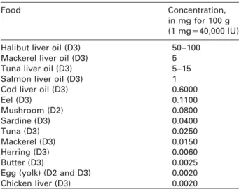
(4) Article in press - uncorrected proof Cavalier et al.: Vitamin D: current status and perspectives 123. (23.2 to 60.4 ng/mL) (35), or after 1200 mg of calcium and 800 IU/day of vitamin D3 for 2 months during which 25VTD increased from 25.7 to 64.8 nmol/L (10.3–25.9 ng/mL) (36). All the results of these different approaches, although quite variable, are coherent with the proposal made in 2005 by five of six experts assembled in a roundtable to define the insufficiency in vitamin D as a concentration of 25VTD -75 nmol/L (30 ng/mL) (‘‘«it is important to ensure that the serum 25VTD level obtained after vitamin D supplementation in individual patient reaches this new threshold.’’) (37). Based on such criterion, vitamin D deficiency becomes very common. It has been estimated that 1 billion people worldwide suffer from vitamin D deficiency (12). As vitamin D is not essentially present in our alimentation (Table 1), the main source remains as the cutaneous generation under UV exposure (38). However, this will be limited by multiple factors, which can be split into two categories: environmental (latitude, season, time of the day, ozone amount, cloud amount, aerosol and reflectivity of the surface) and personal (skin type and pigmentation, age, clothing, cultural habits, and use of sunscreen) (39). In some parts of the world, vitamin D synthesis will not occur during a significant part of the year. Generally, we can consider that dark-skinned migrants who have significantly moved to the north or south are at high risk of vitamin D deficiency because of melanin UVB absorption. On the other hand, fair-skinned individuals moving towards highly sunny areas will be at high risk of erythema and skin cancer and thus will particularly need to protect themselves.. Oral supplementation and ‘‘toxicity’’ of vitamin D Currently, the recommendations from the US Institute of Medicine are 200 IU/day from birth through to age 50 years, 400 IU for individuals aged 51–70 years, and 600 IU for those )70 years, with a tolerable upper Table 1 Vitamin D concentration found in some foodstuff. Food. Halibut liver oil (D3) Mackerel liver oil (D3) Tuna liver oil (D3) Salmon liver oil (D3) Cod liver oil (D3) Eel (D3) Mushroom (D2) Sardine (D3) Tuna (D3) Mackerel (D3) Herring (D3) Butter (D3) Egg (yolk) (D2 and D3) Chicken liver (D3). Concentration, in mg for 100 g (1 mgs40,000 IU) 50–100 5 5–15 1 0.6000 0.1100 0.0800 0.0400 0.0250 0.0150 0.0060 0.0025 0.0020 0.0020. input level of 2000 IU/day (40). Actually, these ‘‘old’’ recommendations are inadequate, misleading, potentially harmful, and should not be used any longer in clinical practice. Re-examination of the requirements for vitamin D is urgently required and may likely reveal the need for vitamin D intake exceeding 2000 IU/day for adults (14). On the other hand, textbooks have revealed that vitamin D, similar to vitamin A, is ‘‘toxic’’ as it accumulates in fat tissue and could induce severe hypercalcemia, nephrocalcinosis, or cardiovascular disorders. Some cases have been published in the literature, especially iatrogenic intoxications in children. However, the amounts of ingested vitamin D needed to obtain clinical signs of toxicity are enormous. Recently, Chambellan-Tison et al. have listed some cases of intoxications in children ranging from 3 months to 7 years (41). The lowest and highest cumulated doses ranged from 1,200,000 IU to 6,000,000 IU during 4 and 30 days, respectively. Moreover, no complications were observed with 300,000 IU/day for 10 days in a 6-month-old girl, 600,000 IU/day for 4 days in a 4-year-old boy and 300,000 IU/day for 15 days in a 7-year-old boy. These data do not minimize vitamin D toxicity, but these cases of iatrogenic accidents have also been published with other drugs or common daily used products. However, what is striking is the security margin between the levels needed to observe a clinical sign of intoxication and the levels needed to raise or maintain serum 25VTD over 30 ng/mL. Vitamin D is thus not to be considered as particularly ‘‘toxic’’. Moreover, the ‘‘intoxication’’ level of 25VTD has never been clearly determined, and in all cases of intoxications 25VTD levels were clearly over 100 ng/mL.. Skeletal action of vitamin D The role played by vitamin D in bone health is important and well known (42). Indeed, the curvilinear relationship between net calcium absorption and calcium intakes reflects the sum of two absorptive mechanisms (43). The first one is a passive, diffusional, and paracellular absorption driven by transepithelial electrochemical gradients. It accounts only for 10%–15% of dietary calcium and 60% of phosphorous. The second one is cell-mediated, saturable, and active. This active transport of calcium from the lumen of the intestine to the circulation is regulated by 125VTD. If the transport mechanisms are still unclear (44), they include calbindin-D9k, a calcium-binding protein induced by 125VTD which facilitates the diffusion of calcium through the cell interior toward the basolateral membrane, and TRPV6, a membrane calcium channel responsible for the first step in calcium absorption in the intestine. TRPV6 is colocalized with calbindin-D9k and induced by 125VTD (45). 125VTD also stimulates the intestinal expression of NPT2B, which activates absorption of phosphate. The precise result is that with adequate vitamin D, adults absorb.
(5) Article in press - uncorrected proof 124. Cavalier et al.: Vitamin D: current status and perspectives. 30%–40% of dietary calcium and 70%–80% of phosphorus. The main function of 125VTD for bone mineralization is to maintain a supersaturated Ca-P product in the circulation. This will allow the passive mineralization of the collagen bone matrix. A major deficit in vitamin D will thus lead to bone pathologies characterized by a defect of mineralization as a consequence: rickets in children and osteomalacia in adults. This is particularly frequent when this deficit is associated with a malabsorption (46). When the deficit in vitamin D is less important, there are no rickets or osteomalacia, but the reduction in the intestinal absorption of calcium and the trend to a decrease in ionized calcium which follows will induce a rise in the concentration of PTH. This secondary hyperparathyroidism will stimulate the 1-a hydroxylase, increasing the serum concentration of 125VTD. PTH also stimulates the expression of a protein, RANKL (receptor activator for nuclear factor kB ligand), a member of the tumor necrosis factor (TNF) family of proteins, on the cell surface of osteoblasts (47). On the other hand, pre-osteoclasts express RANK, a member of the TNF receptor family on their membrane. The interaction of RANKL of the osteoblast on the RANK of the pre-osteoclast allows the differentiation of the latter into mature osteoclasts. These mature osteoclasts produce hydrochloric acid to dissolve bone mineral and release calcium into the extracellular fluid. More information on the modulation of osteoclast differentiation and function by the members of the TNF receptor and ligand families has been published previously (48). 125VTD also has other effects on osteoblasts: it increases the expression of bone alkaline phosphatase, osteocalcin, osteonectin, osteoprotegerin, and a variety of cytokines. Finally, 125VTD exerts negative feedback on the secretion of PTH by the parathyroid, thus limiting its hyperplasia in the event of hyperparathyroidism. Osteoporosis is a major concern in our societies as it affects approximately 33% of women 60–70 years of age and 66% of those 80 years or older (12). Calcium and vitamin D are still essential components in osteoporosis management. It is not clear, however, whether calcium should be added to vitamin D supplementation or vitamin D to calcium supplementation (49). Nevertheless, it has been demonstrated that vitamin D by itself increases bone density (50). Next to the bone action of vitamin D by itself, it also plays an important role on muscle strength and prevents falls in elderly patients (32). Current recommendations for vitamin D supplementation in preventing bone loss and fracture in osteoporosis are in excess of 700–800 IU daily (51).. Non-skeletal actions of vitamin D If skeletal activity of vitamin D is well known, the knowledge of its action on other tissues is generally poorly shared in the medical community. However,. brain, prostate, and colon tissues, amongst others, have a vitamin D receptor and respond to 125VTD (12). Observational studies, and more and more interventional studies, are raising the importance of a significant vitamin D supplementation for not-only skeletal benefits. A systematic review of these studies would be tedious and thus we will only discuss some of them in the following sections. Vitamin D and diabetes Several studies have suggested that vitamin D supplementation in children reduced the risk of type 1 diabetes. Moreover, living in northern latitude has been associated with an increase of the risk of suffering from this disease. A cohort study, where Finn children were given 2000 IU/day during their first year of life, showed an 80% reduction of the risk of developing type 1 diabetes at 31 years (52). Vitamin D also plays a role in type 2 diabetes. Indeed, vitamin D deficiency is associated with the metabolic syndrome, and a combined therapy of calcium/vitamin D (1200 mg/800 IU) was shown to lower the risk of type 2 diabetes by 33% vs. 600 mg/400 IU (53). Autoimmune diseases, immune regulation, and infections In 1903, Finsen was awarded the Nobel Prize for demonstrating that sun exposure was beneficial for patients suffering from lupus vulgaris. It has also been known for decades that sun exposure is beneficial to patients suffering from tuberculosis. This fact was explained later, with the role of cathelicidin, a peptide produced by the monocytes and the macrophages. Indeed, these cells present on their surface Toll-like receptors, which trigger direct antimicrobial activity against intracellular bacteria. The activation of these receptors upregulates the expression of the vitamin D receptor and the vitamin D-1-a-hydroxylase genes. This leads to the induction of the antimicrobial peptide cathelicidin and kills intracellular Mycobacterium tuberculosis. The production of cathelicidin stops when extracellular 25VTD levels fall below 20 ng/mL (54). Vitamin D is also known to modulate autoimmunity. Ecological studies (relation between sunshine or latitude with the apparition of the diseases) and observational studies have shown its importance in various diseases, such as multiple sclerosis (27, 55) or rheumatoid arthritis (56). Vitamin D and cancer More than 20 years ago, different ecological studies hypothesized that sunshine and thus (?) vitamin D was associated with a reduced risk of different cancers, such as breast (57), ovarian (58), colorectal (59), and prostate cancers (60). Since then, many different epidemiological studies have confirmed this initial hypothesis (61). The basis for antitumoral effect exists: many cell types – including tumoral cells –.
(6) Article in press - uncorrected proof Cavalier et al.: Vitamin D: current status and perspectives 125. contain vitamin D receptors and when these receptors are activated, they induce differentiation of the cells and inhibit proliferation of the tumoral cells, invasiveness, angiogenesis, and metastatic potential (62). The first randomized controlled study that involved vitamin D intervention sufficient to raise serum 25VTD over 30 ng/mL and reporting favorable cancer outcome in postmenopausal women was recently published (33). It showed that, after 4 years, the relative risk of suffering from a cancer was reduced by 35% for every 10 ng/mL increase in serum 25VTD. Vitamin D and cardiovascular diseases Implications of vitamin D in the cardiovascular system are also very important. This has been suggested by different epidemiological studies (63, 64) and finds its roots in the fact that vitamin D receptors are found in many cells of the cardiovascular system. Moreover, there is evidence that 125VTD suppresses the renin gene expression (65) and regulates the growth and proliferation of vascular smooth cells and cardiomyocytes (66). Clinical studies have associated low levels of vitamin D with blood pressure (67, 68), coronary artery calcification (69), and cardiovascular diseases, such as myocardial infarction (70), acute stroke (71), congestive heart failure (72). Recently, in the Framingham Offspring Study, vitamin D deficiency has also been associated with incident cardiovascular diseases (64). If the field seems promising, interventional studies are, however, still lacking. Vitamin D, pregnancy, and lactation Vitamin D insufficiency is common in healthy pregnant women (73). There are many reasons for this observation, and one of these is certainly the low concentration of vitamin D in vitamin supplements given during pregnancy (74). However, it has been shown that reduced concentrations of 25VTD in mothers during pregnancy was associated with reduced bonemineral content in children at the age of 9 years (75), as well as other bone disturbances (76, 77). Moreover, maternal vitamin D deficiency at -22 weeks gestation was also found to be a strong and independent risk factor for preeclampsia (78). The benefits of vitamin D supplementation during pregnancy have been alleged to decrease the risk of recurrent wheeze in early childhood (79) and many hypotheses have emerged to associate maternal vitamin D deficiency with the asthma epidemic (80), schizophrenia (81, 82), and autism (83). Nowadays, expert recommendations for vitamin D supplementation during pregnancy and lactation are actually as high as 6000 IU/day (84).. Conclusions Vitamin D deficiency is common in our population. Indeed, this prohormone is essential for bone health, but its positive role has recently been demonstrated in many other fields. However, vitamin D is not ‘‘mag-. ic’’ and there should be more scientific investigations, such as controlled trials vs. placebo to understand the ecological or epidemiological studies. In these trials, one should clearly reveal serum levels of vitamin D necessary to achieve the goals, which is sometimes lacking. Nevertheless, the cost/benefit ratio and some recently published studies are now in favor of a controlled and efficient vitamin D supplementation in patients presenting a 25VTD level -30 ng/mL. More attention should also be focused on pregnant and lactating women, as well as children and adolescents. Next to the medical aspects of vitamin D, some brave and interesting evolutionary theories have emerged to associate the evolution of skin color, human migration from East Africa (the cradle of humanity), rickets and vitamin D (14, 85–89). In these theories, our African ancestries, certainly very darkly pigmented, have migrated to populate more northern or southern latitudes. This migration would have led to a gradual adaptation of the skin pigmentation, linked to a necessary synthesis of vitamin D. Indeed, this evolution was necessary for the survival of the human species, as rickets is not compatible with childbirth due to the misshapen pelvis of women suffering from this disease. Moreover, these darkly pigmented people living in the north would probably rapidly become vitamin D depleted, with resulting mobility and reproductive problems associated with this deficiency. In the same direction, we are not supposed to live covered with clothes inside our buildings. The ‘‘return to nature’’ would certainly fit with a rise in serum 25VTD concentrations. Hopefully, for those who would not dare, vitamin D supplementation remains a solution!. References 1. Ide M. The bios of Wildiers and the cultivation of yeast. J Biol Chem 1921;XLVI(3):521–3. 2. Funk C. The etiology of deficiency disease. J State Med 1912;XX:341–68. 3. Funk C, Dubin HE. The vitamins of yeast and their role in animal nutrition. Proc Soc Exp Biol 1921;19:15. 4. Heaton TB. On the Vitamin D. Biochem J 1922;16:800–8. 5. Hess AF. The prevention and cure of rickets by sunlight. Am J Public Health (NY) 1922;12:104–7. 6. Hess AF, Weinstock M, Heelman FD. The antirachitic value of irradiated phytosterol and cholesterol. J Biol Chem 1925;63:305–9. 7. Rosenheim O, Webster TA. Further observations on the photo-chemical formation of vitamin D. J Soc Chem Ind 1926;45:932. 8. Rosenheim O, Webster TA. The parent substance of vitamin D. Biochem J 1927;21:389–97. ¨ ber das Provitamin aus dem Sterin 9. Windaus A, Bock F. U der Schweineschwarte. Z Physiol Chem 1937;245:168– 70. 10. Houillier P, Nicolet-Barousse L, Maruani G, Paillard M. What keeps serum calcium levels stable? Joint Bone Spine 2003;70:407–13. 11. Souberbielle JC, Friedlander G, Kahan A, Cormier C. Evaluating vitamin D status. Implications for preventing.
(7) Article in press - uncorrected proof 126. 12. 13. 14.. 15. 16. 17.. 18.. 19.. 20.. 21.. 22.. 23. 24.. 25.. 26.. 27.. 28.. 29.. 30.. 31.. 32.. Cavalier et al.: Vitamin D: current status and perspectives. and managing osteoporosis and other chronic diseases. Joint Bone Spine 2006;73:249–53. Holick MF. Vitamin D deficiency. N Engl J Med 2007; 357:266–81. Heaney RP. Vitamin D: how much do we need, and how much is too much? Osteoporos Int 2000;11:553–5. Hollis BW. Circulating 25-hydroxyvitamin D levels indicative of vitamin D sufficiency: implications for establishing a new effective dietary intake recommendation for vitamin D. J Nutr 2005;135:317–22. Zittermann A. Vitamin D in preventive medicine: are we ignoring the evidence? Br J Nutr 2003;89:552–72. Holick MF. Too little vitamin D in premenopausal women: why should we care? Am J Clin Nutr 2002;76:3–4. Lips P. Vitamin D deficiency and secondary hyperparathyroidism in the elderly: consequences for bone loss and fractures and therapeutic implications. Endocr Rev 2001;22:477–501. Lips P, Chapuy MC, Dawson-Hughes B, Pols HA, Holick MF. An international comparison of serum 25-hydroxyvitamin D measurements. Osteoporos Int 1999;9:394–7. Binkley N, Krueger D, Cowgill CS, Plum L, Lake E, Hansen KE, et al. Assay variation confounds the diagnosis of hypovitaminosis D: a call for standardization. J Clin Endocrinol Metab 2004;89:3152–7. Hollis BW. The determination of circulating 25-hydroxyvitamin D: no easy task weditorialx. J Clin Endocrinol Metab 2004;89:3149–51. Cavalier E, Wallace AM, Knox S, Mistretta VI, Cormier C, Souberbielle JC. Serum vitamin D measurement may not reflect what you give to your patients. J Bone Miner Res 2008;23:1864–5. Dawson-Hughes B, Harris SS, Dallal GE. Plasma calcidiol, season, and serum parathyroid hormone concentrations in healthy elderly men and women. Am J Clin Nutr 1997;65:67–71. Malabanan A, Veronikis IE, Holick MF. Redefining vitamin D insufficiency. Lancet 1998;351:805–6. Aloia JF, Talwar SA, Pollack S, Feuerman M, Yeh JK. Optimal vitamin D status and serum parathyroid hormone concentrations in African American women. Am J Clin Nutr 2006;84:602–9. Sahota O, Mundey MK, San P, Godber IM, Hosking DJ. Vitamin D insufficiency and the blunted PTH response in established osteoporosis: the role of magnesium deficiency. Osteoporos Int 2006;17:1013–21. Heaney RP, Dowell MS, Hale CA, Bendich A. Calcium absorption varies within the reference range for serum 25-hydroxyvitamin D. J Am Coll Nutr 2003;22:142–6. Munger KL, Levin LI, Hollis BW, Howard NS, Ascherio A. Serum 25-hydroxyvitamin D levels and risk of multiple sclerosis. J Am Med Assoc 2006;296:2832–8. Garland CF, Gorham ED, Mohr SB, Grant WB, Giovannucci EL, Lipkin M, et al. Vitamin D and prevention of breast cancer: pooled analysis. J Steroid Biochem Mol Biol 2007;103:708–11. Gorham ED, Garland CF, Garland FC, Grant WB, Mohr SB, Lipkin M, et al. Optimal vitamin D status for colorectal cancer prevention: a quantitative meta analysis. Am J Prev Med 2007;32:210–6. Forman JP, Giovannucci E, Holmes MD, Bischoff-Ferrari HA, Tworoger SS, Willet WC, et al. Plasma 25-hydroxyvitamin D levels and risk of incident hypertension. Hypertension 2007;49:1063–9. Bischoff-Ferrari HA, Willett WC, Wong JB, Giovannucci E, Dietrich T, Dawson-Hughes B. Fracture prevention with vitamin D supplementation: a meta-analysis of randomized controlled trials. J Am Med Assoc 2005; 293:2257–64. Bischoff-Ferrari HA, Dawson-Hughes B, Willett WC, Staehelin HB, Bazemore MG, Zee RY et al. Effect of vitamin. 33.. 34.. 35.. 36.. 37.. 38.. 39.. 40.. 41.. 42.. 43.. 44.. 45.. 46.. 47.. 48.. 49. 50.. 51.. D on falls: a meta-analysis. J Am Med Assoc 2004;291:1999–2006. Lappe JM, Travers-Gustafson D, Davies KM, Recker RR, Heaney RP. Vitamin D and calcium supplementation reduces cancer risk: results of a randomized trial. Am J Clin Nutr 2007;85:1586–91. Krall EA, Wehler C, Garcia RI, Harris SS, Dawson-Hughes B. Calcium and vitamin D supplements reduce tooth loss in the elderly. Am J Med 2001;111:452–6. Baynes KC, Boucher BJ, Feskens EJ, Kromhout D. Vitamin D, glucose tolerance and insulinaemia in elderly men. Diabetologia 1997;40:344–7. Pfeifer M, Begerow B, Minne HW, Nachtigall D, Hansen C. Effects of a short-term vitamin D(3) and calcium supplementation on blood pressure and parathyroid hormone levels in elderly women. J Clin Endocrinol Metab 2001;86:1633–7. Dawson-Hughes B, Heaney RP, Holick MF, Lips P, Meunier PJ, Vieth R. Estimates of optimal vitamin D status. Osteoporos Int 2005;16:713–6. Holick MF. Sunlight and vitamin D for bone health and prevention of autoimmune diseases, cancers, and cardiovascular disease. Am J Clin Nutr 2004;80:1678S–88S. Webb AR. Who, what, where and when – influences on cutaneous vitamin D synthesis. Prog Biophys Mol Biol 2006;92:17–25. Institute of Medicine. Dietary reference intakes: calcium, phosphorus, magnesium, vitamin D, fluoride. Washington DC, USA: National Academy Press, 1997. Chambellan-Tison C, Horen B, Plat-Wilson G, Moulin P, Claudet I. wSevere hypercalcemia due to vitamin D intoxicationx. Arch Pediatr 2007;14:1328–32 win French with English abstractx. Reginster JY. The high prevalence of inadequate serum vitamin D levels and implications for bone health. Curr Med Res Opin 2005;21:579–86. Favus MJ, Bushinsky DA, Leman J Jr. Regulation of calcium, magnesium, and phosphate metabolism. In: Primer on the metabolic bone diseases and disorders of mineral metabolism, 6th ed. Washington DC, USA: The American Society of Bone and Mineral Research, 2006: 76–83. Walters JR, Balesaria S, Khair U, Sangha S, Banks L, Berry JL. The effects of vitamin D metabolites on expression of genes for calcium transporters in human duodenum. J Steroid Biochem Mol Biol 2007;103:509–12. Benn BS, Ajibade D, Porta A, Dhawan P, Hediger M, Peng JB, et al. Active intestinal calcium transport in the absence of TRPV6 and calbindin-D9k. Endocrinology 2008;149:3196–205. Basha B, Rao DS, Han ZH, Parfitt AM. Osteomalacia due to vitamin D depletion: a neglected consequence of intestinal malabsorption. Am J Med 2000;108:296–300. Takahashi N, Udagawa N, Takami M, Suda T. Cells of bone: osteoclast generation. In: Bilezikian JP, Raisz LG, Rodan GA, editors. Principles of bone biology. New York, NY, USA: Academic Press, 2005:109–26. Suda T, Takahashi N, Udagawa N, Jimi E, Gillespie MT, Martin TJ. Modulation of osteoclast differentiation and function by the new members of the tumor necrosis factor receptor and ligand families. Endocr Rev 1999;20: 345–57. Reginster JY. Calcium and vitamin D for osteoporotic fracture risk. Lancet 2007;370:632–4. Bischoff-Ferrari HA, Dietrich T, Orav EJ, Dawson-Hughes B. Positive association between 25-hydroxy vitamin D levels and bone mineral density: a population-based study of younger and older adults. Am J Med 2004; 116:634–9. Rizzoli R, Boonen S, Brandi ML, Burlet N, Delmas P, Reginster JY. The role of calcium and vitamin D in the management of osteoporosis. Bone 2008;42:246–9..
(8) Article in press - uncorrected proof Cavalier et al.: Vitamin D: current status and perspectives 127. 52. Hypponen E, Laara E, Reunanen A, Jarvelin MR, Virtanen SM. Intake of vitamin D and risk of type 1 diabetes: a birth-cohort study. Lancet 2001;358:1500–3. 53. Pittas AG, Dawson-Hughes B, Li T, Van Dam RM, Willett WC, Manson JE, et al. Vitamin D and calcium intake in relation to type 2 diabetes in women. Diabetes Care 2006;29:650–6. 54. Liu PT, Stenger S, Li H, Wenzel L, Tan BH, Krutzik SR, et al. Toll-like receptor triggering of a vitamin D-mediated human antimicrobial response. Science 2006;311:1770– 3. 55. Smolders J, Damoiseaux J, Menheere P, Hupperts R. Vitamin D as an immune modulator in multiple sclerosis, a review. J Neuroimmunol 2008;194:7–17. 56. Merlino LA, Curtis J, Mikuls TR, Cerhan JR, Criswell LA, Saag KG. Vitamin D intake is inversely associated with rheumatoid arthritis: results from the Iowa Women’s Health Study. Arthritis Rheum 2004;50:72–7. 57. Garland FC, Garland CF, Gorham ED, Young JF. Geographic variation in breast cancer mortality in the United States: a hypothesis involving exposure to solar radiation. Prev Med 1990;19:614–22. 58. Lefkowitz ES, Garland CF. Sunlight, vitamin D, and ovarian cancer mortality rates in US women. Int J Epidemiol 1994;23:1133–6. 59. Garland CF, Garland FC. Do sunlight and vitamin D reduce the likelihood of colon cancer? Int J Epidemiol 1980;9:227–31. 60. Schwartz GG, Hulka BS. Is vitamin D deficiency a risk factor for prostate cancer? (hypothesis). Anticancer Res 1990;10:1307–11. 61. Giovannucci E. Vitamin D and cancer incidence in the Harvard cohorts. Ann Epidemiol 2008;Feb 19. wEpub ahead of printx. 62. Giovannucci E. The epidemiology of vitamin D and cancer incidence and mortality: a review (United States). Cancer Causes Control 2005;16:83–95. 63. Martins D, Wolf M, Pan D, Zadshir A, Tareen N, Thadhani R, et al. Prevalence of cardiovascular risk factors and the serum levels of 25-hydroxyvitamin D in the United States: data from the Third National Health and Nutrition Examination Survey. Arch Intern Med 2007;167:1159–65. 64. Wang TJ, Pencina MJ, Booth SL, Jacques PF, Ingelsson E, Lanier K, et al. Vitamin D deficiency and risk of cardiovascular disease. Circulation 2008;117:503–11. 65. Li YC, Kong J, Wei M, Chen ZF, Liu SQ, Cao LP. 1,25Dihydroxyvitamin D(3) is a negative endocrine regulator of the renin-angiotensin system. J Clin Invest 2002; 110:229–38. 66. O’Connell TD, Berry JE, Jarvis AK, Somerman MJ, Simpson RU. 1,25-Dihydroxyvitamin D3 regulation of cardiac myocyte proliferation and hypertrophy. Am J Physiol 1997;272:H1751–8. 67. Kristal-Boneh E, Froom P, Harari G, Ribak J. Association of calcitriol and blood pressure in normotensive men. Hypertension 1997;30:1289–94. 68. Lind L, Hanni A, Lithell H, Hvarfner A, Sorensen OH, Ljunghall S. Vitamin D is related to blood pressure and other cardiovascular risk factors in middle-aged men. Am J Hypertens 1995;8:894–901. 69. Watson KE, Abrolat ML, Malone LL, Hoeg JM, Doherty T, Detrano R, et al. Active serum vitamin D levels are inversely correlated with coronary calcification. Circulation 1997;96:1755–60.. 70. Scragg R, Jackson R, Holdaway IM, Lim T, Beaglehole R. Myocardial infarction is inversely associated with plasma 25-hydroxyvitamin D3 levels: a community-based study. Int J Epidemiol 1990;19:559–63. 71. Poole KE, Loveridge N, Barker PJ, Halsall DJ, Rose C, Reeve J, et al. Reduced vitamin D in acute stroke. Stroke 2006;37:243–5. 72. Zittermann A, Schleithoff SS, Tenderich G, Berthold HK, Korfer R, Stehle P. Low vitamin D status: a contributing factor in the pathogenesis of congestive heart failure? J Am Coll Cardiol 2003;41:105–12. 73. Dawodu A, Agarwal M, Hossain M, Kochiyil J, Zayed R. Hypovitaminosis D and vitamin D deficiency in exclusively breast-feeding infants and their mothers in summer: a justification for vitamin D supplementation of breast-feeding infants. J Pediatr 2003;142:169–73. 74. Cavalier E, Delanaye P, Morreale A, Carlisi A, Mourad I, Chapelle JP et al. La carence en vitamine D chez les femmes enceintes en re´gion lie´geoise: un proble`me me´connu. Rev Med Liege 2008;74:87–91. 75. Javaid MK, Crozier SR, Harvey NC, Gale CR, Dennison EM, Boucher BJ, et al. Maternal vitamin D status during pregnancy and childhood bone mass at age 9 years: a longitudinal study. Lancet 2006;367:36–43. 76. Dijkstra SH, van Beek A, Janssen JW, de Vleeschouwer LH, Huysman WA, van den Akker EL. High prevalence of vitamin D deficiency in newborn infants of high-risk mothers. Arch Dis Child 2007;92:750–3. 77. Pawley N, Bishop NJ. Prenatal and infant predictors of bone health: the influence of vitamin D. Am J Clin Nutr 2004;80:1748S–51S. 78. Bodnar LM, Catov JM, Simhan HN, Holick MF, Powers RW, Roberts JM. Maternal vitamin D deficiency increases the risk of preeclampsia. J Clin Endocrinol Metab 2007;92:3517–22. 79. Camargo CA Jr, Rifas-Shiman SL, Litonjua AA, RichEdwards JW, Weiss ST, Gold DR, et al. Maternal intake of vitamin D during pregnancy and risk of recurrent wheeze in children at 3 y of age. Am J Clin Nutr 2007;85:788–95. 80. Litonjua AA, Weiss ST. Is vitamin D deficiency to blame for the asthma epidemic? J Allergy Clin Immunol 2007; 120:1031–5. 81. McGrath JJ, Welham JL. Season of birth and schizophrenia: a systematic review and meta-analysis of data from the Southern Hemisphere. Schizophr Res 1999; 35:237–42. 82. Altschuler EL. Low maternal vitamin D and schizophrenia in offspring. Lancet 2001;358:1464. 83. Cannell JJ. Autism and vitamin D. Med Hypotheses 2008;70:750–9. 84. Hollis BW. Vitamin D requirement during pregnancy and lactation. J Bone Miner Res 2007;22(Suppl 2):V39–V44. 85. Diamond J. Evolutionary biology: geography and skin colour. Nature 2005;435:283–4. 86. Jablonski NG, Chaplin G. The evolution of human skin coloration. J Hum Evol 2000;39:57–106. 87. Jablonski NG, Chaplin G. Skin deep. Sci Am 2002; 287:74–81. 88. Relethford JH. Hemispheric difference in human skin color. Am J Phys Anthropol 1997;104:449–57. 89. Vieth R. What is the optimal vitamin D status for health? Prog Biophys Mol Biol 2006;92:26–32..
(9)
Figure

Documents relatifs
As the input monomer vapor is carried across the surface of a substrate, the monomer reacts with the initiator free radical making an active site on the monomer
La conviction de l’installation d’une maladie grave oblige à maintenir une attention permanente sur le soma dans la conti- nuité, comme pour rétablir un lien entre la psyché et
Les êtres fantomatiques seraient-ils fortement présents quand la vie n’est plus traversée par la possibilité de rencontre et d’accordage émotionnels avec un autre ? La nostalgie
Il va donc être nécessaire de construire un modèle BPMN générique des processus pouvant être trouvés dans une PUI, un diagramme générique des classes UML des entités
Proposition 2 When labor market integration generates a bad equilibrium with child labor in the country that featured higher wage rates without migration, this equilibrium may be
An initial VB12 intramuscular boost followed by an oral VB12 dose of 1000 µg daily is invariably sufficient.. Consequently, there should be little need for continuing
The objective of this study, therefore, was to determine the relationship between age, renal function (serum creatinine and estimated creatinine clearance) and BMI and parameters
The overall mean level of 25(OH) D was ~ 20 ng/mL Deficiency was found among all age groups, in both sexes and in both local and non-local populations: overall 85.4% were vitamin
