VIABILITY OF CLONED BOVINE EMBRYOS AFTER ONE OR TWO CYCLES OF NUCLEAR TRANSFER AND IN VITRO CULTURE
F.J. Ectors,1 A. Delval,1 L.C. Smith,2 K. Touati,1 B. Remy,1 J-F. Beckers1 and F. Ectors1
1Department of Animal Endocrinology and Reproduction Faculty of Veterinary Medicine University of Liege
B-4000 Liege, Belgium
2Centre de Recherche en Reproduction Animale, Faculté de Médecine Vétérinaire Université de Montréal, Saint-Hyacinthe, Québec, Canada
Received for publication:
Accepted:
ABSTRACT
We described an exclusively in vitro procedure for cloning and recloning bovine embryos. Embryos obtained by IVM/IVF/IVC developed to the morula stage were used as blastomere donors in cunjunction with IVM recipient oocytes. Reconstructed embryos were developed in vitro in co-culture using bovine oviductal epithelial cells. The resulting morulae were used as donors for recloning under the same experimental conditions. No significant difference was observed between cloning and recloning in terms of development (rates of blastocysts: 12.9 versus 14.9%), in the number of nuclei per blastocyst (63.8 versus 49.1), or in pregnancy rates (35.7 versus 33.3%). The high variability observed between replicates and the correlation between results in first and second cycle nuclear transfer may suggest an inherant potential of individual donor embryos to support development by cloning.
Key words: nuclear transfer, bovine, embryo, in vitro culture, in vitro fertilization INTRODUCTION
Development of reproducible cloning methods in mammals is of considerable scientific and economic interest. Due to the relatively small number of offspring obtained after a single cycle of nuclear transfer, some authors have suggested the use of viable reconstructed embryos as donors for second or additional cycles of nuclear transfer. Recloning of mammalian embryos has been reported by Willadsen et al (9), Westhusin et al (8) and Stice and Keefer (7). The latter authors have described the attainment of live calves after 3 generations of nuclear transfer.
Acknowledgements
Reports on the potential of preimplantation development of nuclear transplanted bovine embryos have varied considerably within and between laboratories. Two main factors might account for this variability: donor and recipient cell cycle compatibility (1) and/or partial ability of the recipient cytoplasm to undifferentiate the donor nucleus. In addition, Stice and Keefer (7) have postulated that some donor embryos may have an intrinsic potential to support proper development after multiple nuclear transfers.
We described a nuclear transfer procedure in conjunction with in vitro embryo culture. Donor embryos were produced by IVM, IVF and in vitro culture. Development of reconstituted embryos was performed in vitro in co-culture with bovine oviductal epithelial cells. Some of these reconstituted embryos were used as donors for recloning. Afterwards, a limited number of embryos was evaluated in vitro by counting nuclei. Relatively large numbers of first and second cycle blastocysts obtained by nuclear transfer were implanted into synchronous recipient heifers in order to evaluate their ability to induce ongoing pregnancies.
MATERIALS AND METHODS
Two experiments were conducted: in Experiment 1, first cycle nuclear transfer embryos were produced, while in Experiment 2 the first cycle reconstructed morulae were used as donor embryos for recloning.
Micromanipulations
In vitro maturation of oocytes and in vitro fertilization of donor embryos have been previously described (2). Micromanipulations were carried out on an inverted Nikon Diaphot microscope(x100 magnification) equipped with Leitz mechanical micromanipulators. All the micromanipulation procedures were performed at 25 to 30°C, in 250-µl doplets under oil (light mineral oil, Sigma Chemical, St. Louis MO; M-3516).
Oocyte enucleation. After 24 h of IVM, expanded ovocyte-cumulus complexes were placed in calcium- and magnesium-free PBS medium supplemented with hyaluronidase (300 IU/ml; Sigma: H-3506) and vigorously shaken for 5 min (vortexed) to remove the cumulus cells. Denuded oocytes with a homogeneous cytoplasm and a first polar body were selected as recipient cytoplasms.
These oocytes were placed in PBS added with 10% fetal calf serum (FCS: Sebak: ref.: 30.01.21) containing cytochalasin B (7.5 µg/ml; Sigma: C-6762) 10 min before starting the micromanipulations. For enucleation of oocytes, a small amount (≈1/3) of cytoplasm adjacent to the first polar body and the first polar body itself was removed by aspiration. Immediately after enucleation, the micromanipulated oocytes were incubated at room temperature in PBS containing 10% FCS and Hœchst 33342 (5 µg/ml; bisbenzimide trihydrochloride, Sigma: B-2261) for 10 min. After staining, the micromanipulated oocytes were rinced in fresh PBS added with 10% FCS and then examined individually at x100 magnification with a Nikon TMD Diaphot inverted microscope equipped with an epifluorescence detector. A 100-watt mercury lamp (Osram, Germany) and an ultraviolet filter block (365 nm excitation and 400 nm emission, UV-1A, Nikon) were used to irradiate the oocytes. Exposition of oocytes to UV light lasted no more than 5 sec. Only oocytes with no observable chromatin were
considered as correctly enucleated, and were used as recipient cytoplasm for an isolated blastomere. The enucleated oocytes were then returned to maturation medium at 39°C until 44 to 46 h post-onset of IVM.
Donor embryos. In Experiments 1 and 2, donor embryos were used on Day 5 after IVF (Day 0 = IVF); in Experiment 2, morulae were used on Day 6 after first cycle nuclear transfer for recloning (Day 0 = Day of enucleation). After selection, they were incubated in calcium- and magnesium-free PBS + 10% FCS containing cytochalasin B (7.5 µg/ml) for 10 min. Each donor was placed individually in a 25-µl droplet under oil. The embryonic cell mass was mechanically removed from its zona pellucida with a glass needle mounted on a micromanipulator. The blastomeres were disaggregated by gentle pipetting throughout a calibrated holding pipette (inner diameter: ≈30 µm).
Injection and fusion. Immediately after the disaggregation of the donor morula, an appropriate number of recipient cytoplasms was added to the droplet containing the isolated blastomeres. Single blastomeres isolated from approximately 32-cell embryos were inserted into the perivitelline space of enucleated oocytes. Injection was performed via the hole remaining in the zona pellucida from the day before. Recipient oocyte-blastomere pairs were transferred to the electrofusion medium: 0.275 M mannitol (Sigma, ref.: M-1902) added with 0.1 mM MgSO4.7H2O (Sigma, ref.: M-7774) and 0.05 mM CaCl2 (Sigma, ref.: C-7902). The pairs were gently placed in a home-made fusion chamber with 2 platinium parallel electrodes 240 µm apart. A direct current pulse of 2.7 kVolts/cm was applied for 50 microsec. An electric fusion processor was used as pulse generator (Jouan, CHT 1287; Saint-Herblain, France). Stimulated pairs were immediatly incubated in co-culture after the electrofusion procedure.
Embryo Development
The bovine oviducts were recovered at a local slaughterhouse from normally cycling heifers presenting a corpus luteum. They were carried to the laboratory in saline at 0°C within 1 h. After dissection, the fallopian tubes were washed by immersion in ethanol for a few seconds and then rinsed 3 times in saline. The scratching of the oviductal mucosae was performed using a glass microscope slide. After washing the cells 5 times in PBS + 10% FCS, they were cultured for 24 h in TCM 199 supplemented with 20% FCS and antibiotics/antimicotic preparation (penicillin: 100 IU/ml, streptomycin: 100 µg/ml and amphotericin B: 1 µg/ml - Life Technologies, Inc., Grand Island NY; USA; ref.: 600-5245AE). Before use for embryo culture, they were washed twice in PBS + 10% FCS to remove the dead cells. After washing, the tissue pellet was resuspended in Menezo B2 medium (tissue/medium ratio of 1/50) supplemented with 20% heat-treated proestrus cow serum (Day 20), pyruvate (30 µg/ml), antibiotics (penicillin: 100 IU/ml, streptomycin: 100 µg/ml). The zygotes or reconstructed embryos were cultured in 50-µl droplets of tissue suspension under oil.
Embryos were classified on Day 3 as cleaved (II-cell, IV-cell and VIII-cell stages) and as VIII-cell stages, on Day 5 or 6 as morula or on Day 7 as blastocyst.
Blastocyst quality obtained from first and second cycle nuclear transfer was assessed in vitro by counting the nuclei or in vivo by their ability to induce gestation in synchronized heifers.
Blastocyst fixing and staining. On Day 7, a portion of the blastocysts was fixed in ethanol/2.3% NaCitrate (1/3: v/v) to determine the cell number based on the nuclear counts after staining with Hœchst 33342 (10 µg/ml).
Embryo transfer. Red Holstein heifers were synchronized by intramuscular injection of 750 µg (3 ml) of cloprostenol (Estrumate, Pitman-Moore) about 60 h before the onset of desired estrus. Blastocysts obtained in co-culture after nuclear transfer were transferred non surgically to recipient heifers 7 d after estrus. Two embryos of comparable quality from the same blastomere donor were transferred together into the recipient's uterine horn ipsilateral to the corpus luteum. Pregnancies were diagnosed on Day 35 by pregnancy-associated glycoprotein (PAG) determination (11). All pregnant recipients were examined after 90 d of gestation by palpation per rectum in order to control the ongoing pregnancy.
Statistical Analysis
Data were analyzed by Chi-square (Tables 1 and 2) and of Student's t-test (Tables 3 and 4). Correlations between development after first and second cycle nuclear transfer were also assessed.
RESULTS Experiment 1: First Cycle Nuclear Transfer
On average, 19.1 (248/13) reconstructed embryos were obtained per donor. The development potential of first cycle nuclear transfer embryos was compared to that of embryos produced by IVF (Table 1). Although similar cleavage rates were obtained in both groups, first cycle reconstructed embryos had significantly lower development rates to 8-cell and later development stages (P<0.01). Moreover, substantially higher standard errors and wider ranges were obtained in the reconstructed embryos, indicating a large variability in this group, compared with that of IVF embryos.
Table 1. Percentage of embryos developing after IVF and first cycle nuclear transfer
Mean % ±SEM Range Number Replicates IVF:
Cleaved (Day 3) 76.0 ± 4 60 to 86 (511/672) 6 8-cell stage (Day 3) 49.1 ± 4 34 to 59 (330/672) 6 Morula (Day 5) 21.4 ± 4 20 to 43 (144/672) 6 First cycle nuclear transfer:
Cleaved (Day 3) 68.7 ± 15 35 to 83 (170/248) 13 8-cell stage (Day 3) 21.8 ± 16 0 to 41 (54/248) 13 Blastocysts (Day 7) 12.9 ± 10 0 to 27 (32/248) 13
Experiment 2: Second Cycle Nuclear Transfer
The mean number of reconstructed embryos obtained per donor was similar in first (228/11) and second (303/15) cycle nuclear transfer (20.7 versus 20.2; P>0.05). As shown in Experiment 1, cleavage rate in IVF was similar to that in first and second cycle nuclear transfer; whereas the development to 8-cell and later stages was significantly lower in both reconstructed groups (Table 2).
No significant difference was observed at any stage between first and second cycle nuclear transfer embryos. Standard errors and ranges observed in the reconstructed embryos were also higher than in the IVF group. No difference in these parameters was observed between first and second cycle nuclear transfer groups, indicating that increased variability observed after cloning was not influenced by a recloning procedure.
To evaluate a possible donor effect, first cycle nuclear transfer embryos obtained in Experiment 2 were classified according to the development rate (Table 3). In Table 3, when the first cycle nuclear transfer embryos obtained in Experiment 2 were classified according to the development rate, there was evidence of a possible donor effect, but the correlation between the first and second cycle was weak. Table 2. Percentage of embryos developing after IVF, first and second cycle nuclear
transfer
Mean % ±SEM Range Number Replicates IVF:
Cleaved (Day 3) 76.5 ± 10 60 - 86 (485/634) 5 8-cell (Day 3) 48.4 ± 11 34 - 59 (307/634) 5 Morula (Day 5) 23.8 ± 6 20 - 35 (151/634) 5 First cycle nuclear transfer:
Cleaved (Day 3) 86.8 ± 17 41 to 92 (198/228) 11 8-cell (Day 3) 22.8 ± 10 8 to 36 (52/228) 11
Morula (Day 6) 14.5 ± 12 0 to 30 (33/228) 11
Second cycle nuclear transfer:
Cleaved (Day 3) 78.2 ± 7 66 to 89 (237/303) 8 8-cell (Day 3) 28.7 ± 9 18 to 43 (87/303) 8
Blastocysts (Day 7) 14.9 ± 12 0 to 41 (45/303) 8
Two tests were performed to evaluate the quality of first and second cycle nuclear transfer blastocysts obtained in the Experiments 1 and 2 described above. Some embryos were fixed and stained in order to determine their nuclear number (Table
4), while others were non-surgically transferred to recipient heifers (Table 5).
A reduced number of nuclei was observed in blastocysts from nuclear transfer embryos when compared to IVF embryos (P<0.0005). Moreover, the number of nuclei observed in recloned blastocysts was also reduced when compared with those of first cycle nuclear transfer, but the results were not significantly different (P>0.05). The absence of a quality difference between cloned and recloned blastocysts was confirmed by the similar rates of pregnancies obtained in the 2 groups (Table 5).
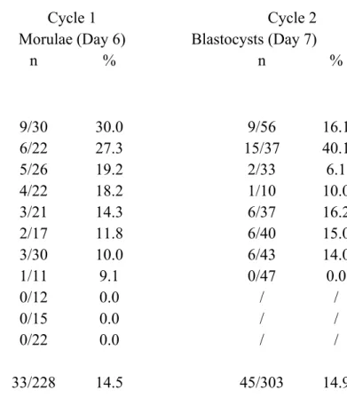
Table 3. Developmental potential of cloned embryos over 2 cycles
Donors Cycle 1 Cycle 2
Morulae (Day 6) Blastocysts (Day 7)
n % n % 1 9/30 30.0 9/56 16.1 2 6/22 27.3 15/37 40.1 3 5/26 19.2 2/33 6.1 4 4/22 18.2 1/10 10.0 5 3/21 14.3 6/37 16.2 6 2/17 11.8 6/40 15.0 7 3/30 10.0 6/43 14.0 8 1/11 9.1 0/47 0.0 9 0/12 0.0 / / 10 0/15 0.0 / / 11 0/22 0.0 / / Totals 33/228 14.5 45/303 14.9 Parent donor embryos were classified by function of their developmental rate after the first cycle nuclear transfer .
Correlation between Cycle 1 and 2 nuclear transfers: r2 = 0.58; P=0.13.
A total of 11 calves were obtained in the first and the second experiments. All of them were morphologically normal and presented normal karyotypes. In the second cycle nuclear transfer group, we obtained the clones of male twins and of female triplets.
Table 4. Number of nuclei in IVF, Cycle 1 and 2 nuclear transfer Day 7 blastocysts
Mean n of nuclei Range No. of fixed
(± SEM) blastocysts
IVF 105.2 ± 3.21 69-148 48
Experiment 1 (Cycle 1 nuclear transfer) 63.8 ± 4.9 35-135 25 Experiment 2 (Cycle 2 nuclear transfer) 49.1 ± 5.0 30-83 11 Table 5. Percentage of pregnancies following transfer of 2 blastocysts from Cycle 1
and 2 nuclear transfers a.
Day 35 Day 90 Term Newbornb
Experiment 1 (Cycle 1 nuclear transfer)
% 50.0 50.0 35.7 21.4
n 7/14 7/14 5/14 6/28
Experiment 2 (Cycle 2 nuclear transfer)
% 50.0 33.3 33.3 20.8
n 6/12 4/12 4/12 5/24
a: All recipients heifers received two embryos from the same clone.
b: No. calves / No. of transferred embryos.
DISCUSSION
Our results demonstrate that viable multiple generation nuclear transfer embryos can be obtained through an exclusively in vitro procedure for cloning and recloning in the bovine species. Previous reports on multiple generation nuclear transfer have shown that 2 or more generations of nuclear transfer embryos can be produced when using in vivo donor embryos and by culturing the nuclear transfer embryos in the oviducts of intermediate hosts, either sheep or rabbit (7,8). Developmental rates to the blastocyst stage similar to those reported using in vivo procedures have been obtained in vitro (3), suggesting that temporary passage in the reproductive tract of nuclear transfer embryos is not necessary. In vitro derived donor embryos have previously been shown to support the development of single generation nuclear transfer embryos with a potential to produce live offspring (3,5,10). Although some authors have reported reduced development from in vitro donor embryos (10), others have shown similar development rates of nuclear transfer embryos derived from in vitro and in vivo donor embryos (2,3).
Significantly lower developmental rates and lower nuclei counts in blastocysts were observed in both first and second cycle nuclear transfer derived embryos when compared with these of IVF embryos. This loss in potential may, however, not always be present, since others have shown similar rates of development between first cycle nuclear transfer and IVF embryos (3). Similar to the findings of Westhusin et al (6) and Stice et al (5), our experiments show no change in development potential between first and second generation clones. All 3 viability parameters studied, (i.e., blastocyst development rates, nuclei number in Day 7 blastocysts, and pregnancy rates) showed a remarkable similarity between both nuclear transfer groups, indicating that viability is not affected by a second round of nuclear transfer. These findings are in contrast with those reported by Stice and Keefer (5) and Westhusin et al (7), who showed decreased development in second cycle nuclear transfer embryos. It is somewhat surprising that an embryo, or its genome, can remain viable after being cultured entirely in vitro for up to 18 d (Cycle 2). In addition to the prolonged in vitro culture period, Cycle 2 embryos undergo 2 cycles of micromanipulation comprised of exposure to conditions such as low temperature, UV irradiation, microsurgery, cytoskeletal inhibitors and electric pulses. The cytoplasm and plasma membrane, however, are renewed at each cycle of nuclear transfer, bringing, apart from these non genetic components, fresh (viable) post-transcriptional machinery wich enables several cleavage divisions at limited or no expense to the transplanted nucleus. On the contrary, repeated exposure of the chromatin to reprogramming factors in the cytoplasm of oocytes may even enhance developmental potential, as, is indicated in the amphibian (6). In support of this notion, Stice and Keefer (5) reported a rebound in development rates after third and fourth cycles of bovine nuclear transfer (5). The latter, however, appear to produce lower pregnancy rates and fewer live offspring.
Like Westhusin et al (6) and Stice and Keefer (5), we observed a large variability in the percentages of blastocysts between the first and second cycle of nuclear transfer. In our study, the between replicate variability (expressed as the coefficient of variation: CV) for in vitro blastocyst development was low in the IVF program (19 and 25%). In contrast, after cloning it increased greatly but was not amplified in the second cycle when compared with that of the first cycle (80.5 versus 77.5%). As noted by others (7), the origin of such inter-replicate variability remains unclear. A nuclear donor effect is most likely a cause of inter-replicate variation since only a small number of donor embryos is used in any micromanipulation session. As suggested by Stice and Keefer (5), nuclei from different embryos may have variable intrinsic potential to support development after nuclear transfer. Such intrinsic potential of donor embryos should be transmitted through generations of multiple nuclear transfers, leading to a clonal family effect. In our study, a positive correlation (r2=0.58) but not a significant (P=0.13) one was observed between the development of first and second cycles of nuclear transfer embryos. However, since clonal families that produce no embryos in first nuclear transfer cycle provide no information for the second generation, it is possible that this correlation value is biased. If we assume that the donor embryos which are not able to support development at the first cycle nuclear transfer are also unable to do so in the second cycle, then the resulting correlation value is higher (r2=0.74) and significant (P<0.01). Moreover, clustering of our clonal families into high (21 to 30%; Donors 1 and 2), average (11 to 20%; Donors 3 to 7) and low (0 to 10%; Donors 8 to 11) groups at the first cycle nuclear transfer indicates that, with a few exceptions, the development of most second cycle nuclear transfer embryos remained in the same group or fell to the group just below (Table 3). Further
investigations are needed to determine whether or not a clonal family effect is present in nuclear transfer, and if these donor embryos can be identified prior to the cloning procedure. Screening of good donor embryos would allow for the production of larger numbers of genetically identical embryos and would lead to a substantial improvement in the efficiency of the embryo cloning procedure in cattle.
REFERENCES
1. Collas P, Balise JJ, Robl JM. Influence of cell cycle stage of the donor nucleus on development of nuclear transplant rabbit embryos. Biol Reprod 1992; 46:492-500.
2. Ectors FJ, Thonon F, Delval A, Fontes RS, Touati K, Beckers J-F. Comparison between culture of bovine embryos in vitro versus development in rabbit oviducts and in vivo. Liv Prod Sci 1993; 36:29-34.
3. Heyman Y, Chesné P, Lebourhis D, Peynot N, Renard JP. Developmental ability of bovine embryos after nuclear transfer based on the nuclear source: in vivo versus in vitro. Theriogenology 1994; 42:695-702.
4. Keefer CL, Stice SL. In vitro culture of bovine nucleus transfer embryos. Biol Reprod 1992; 46:166.
5. Kinis A, Vergos E, Gallagher M, Gordon I. Factors affecting nuclear transfer in cattle using oocytes and embryos produced by in vitro culture. J Reprod Fertil 1991; 43 (Suppl): 261-262.
6. Orr NH, DiBerardino MA, McKinnel RG. The genome of frog erythrocytes displays centuplicates replications. Proc Natl Acad Sci 1986; 83:1369-1373.
7. Stice SL, Keefer CL. Multiple generational bovine embryo cloning. Biol Reprod 1993; 48: 715-719.
8. Westhusin ME, Pryor JH, Bondioli KR. Nuclear transplantation in the bovine embryo: a comparison of 5-day, 6-day, frozen thawed and nuclear transfer donor embryos. Mol Reprod Dev 1991; 28:119-123.
9. Willadsen SM. Cloning sheep and cow embryos. Genome 1989; 31:956-962.
10. Yang X, Jiang S, Farrell P, Foote RH, McGrath AB. Nuclear transfer in cattle: effect of nuclear donor cells, cytoplast age, co-culture and embryo transfer. Mol Reprod Dev 1993; 35:29-36.
11. Zoli A, Guilbault LA, Delahaut P, Ortiz WB, Beckers J-F. Radioimmunoassay of a bovine pregnancy-associated glycoprotein in serum: its application for pregnancy diagnosis. Biol Reprod 1992; 46:83-92.