HAL Id: dumas-03211444
https://dumas.ccsd.cnrs.fr/dumas-03211444
Submitted on 28 Apr 2021
HAL is a multi-disciplinary open access
archive for the deposit and dissemination of sci-entific research documents, whether they are pub-lished or not. The documents may come from teaching and research institutions in France or abroad, or from public or private research centers.
L’archive ouverte pluridisciplinaire HAL, est destinée au dépôt et à la diffusion de documents scientifiques de niveau recherche, publiés ou non, émanant des établissements d’enseignement et de recherche français ou étrangers, des laboratoires publics ou privés.
Concordance between echocardiographic assessment
performed in the initial phase of decompensated
cirrhosis with edema and ascites by an expert intensive
care physician and a non-expert gastroenterologist: the
echocirrho study
Adrien Ardavan Prost
To cite this version:
Adrien Ardavan Prost. Concordance between echocardiographic assessment performed in the initial phase of decompensated cirrhosis with edema and ascites by an expert intensive care physician and a non-expert gastroenterologist: the echocirrho study. Human health and pathology. 2018. �dumas-03211444�
UNIVERSITE DE MONTPELLIER FACULTE DE
MEDECINE MONTPELLIER-NIMES
THESE
Pour obtenir le titre de
DOCTEUR EN MEDECINE
Présentée et soutenue publiquement
Par Adrien Ardavan PROST Le 26 Octobre 2018.
Concordance Between Echocardiographic
Assessment Performed in the Initial Phase of
Decompensated Cirrhosis with Edema and Ascites by
an Expert Intensive Care Physician and a Non-expert
Gastroenterologist.
The EchoCirrho study.
Directeur de thèse : M. Le Docteur Jean-François BOURGAUX
JURY :
M. le Professeur Philippe POUDEROUX, Président
M. le Professeur Georges-Philippe PAGEAUX, Assesseur
M. le Professeur Jean-Yves LEFRANT, Assesseur
ANNEE UNIVERSITAIRE 2017 - 2018 PERSONNEL ENSEIGNANT Professeurs Honoraires ALLIEU Yves ALRIC Robert ARNAUD Bernard ASTRUC Jacques AUSSILLOUX Charles AVEROUS Michel AYRAL Guy BAILLAT Xavier BALDET Pierre BALDY-MOULINIER Michel BALMES Jean-Louis BALMES Pierre BANSARD Nicole BAYLET René BILLIARD Michel BLARD Jean-Marie BLAYAC Jean Pierre BLOTMAN Francis BONNEL François BOUDET Charles
BOURGEOIS Jean-Marie BRUEL Jean Michel BUREAU Jean-Paul BRUNEL Michel CALLIS Albert CANAUD Bernard CASTELNAU Didier CHAPTAL Paul-André CIURANA Albert-Jean CLOT Jacques D’ATHIS Françoise DEMAILLE Jacques DESCOMPS Bernard DIMEGLIO Alain DU CAILAR Jacques DUBOIS Jean Bernard DUMAS Robert DUMAZER Romain ECHENNE Bernard FABRE Serge
FREREBEAU Philippe GALIFER René Benoît GODLEWSKI Guilhem GRASSET Daniel GROLLEAU-RAOUX Robert GUILHOU Jean-Jacques HERTAULT Jean HUMEAU Claude JAFFIOL Claude JANBON Charles JANBON François JARRY Daniel JOYEUX Henri LAFFARGUE François LALLEMANT Jean Gabriel LAMARQUE Jean-Louis LAPEYRIE Henri LESBROS Daniel LOPEZ François Michel LORIOT Jean LOUBATIERES Marie Madeleine MAGNAN DE BORNIER Bernard MARY Henri MATHIEU-DAUDE Pierre MEYNADIER Jean MICHEL François-Bernard MICHEL Henri MION Charles MION Henri MIRO Luis NAVARRO Maurice NAVRATIL Henri OTHONIEL Jacques PAGES Michel PEGURET Claude POUGET Régis PUECH Paul PUJOL Henri PUJOL Rémy RABISCHONG Pierre RAMUZ Michel RIEU Daniel RIOUX Jean-Antoine ROCHEFORT Henri
ROUANET DE VIGNE LAVIT Jean Pierre
SAINT AUBERT Bernard SANCHO-GARNIER Hélène SANY Jacques SENAC Jean-Paul SERRE Arlette SIMON Lucien SOLASSOL Claude THEVENET André VIDAL Jacques VISIER Jean Pierre
Professeurs Émérites ARTUS Jean-Claude BLANC François BOULENGER Jean-Philippe BOURREL Gérard BRINGER Jacques CLAUSTRES Mireille DAURES Jean-Pierre DAUZAT Michel DEDET Jean-Pierre ELEDJAM Jean-Jacques GUERRIER Bernard JOURDAN Jacques MAURY Michèle MARES Pierre MONNIER Louis PRAT Dominique PRATLONG Francine PREFAUT Christian PUJOL Rémy ROSSI Michel SULTAN Charles TOUCHON Jacques VOISIN Michel ZANCA Michel
Professeurs des Universités - Praticiens Hospitaliers PU-PH de classe exceptionnelle
ALBAT Bernard - Chirurgie thoracique et cardiovasculaire
ALRIC Pierre - Chirurgie vasculaire ; médecine vasculaire (option chirurgie vasculaire) BACCINO Eric - Médecine légale et droit de la santé
BASTIEN Patrick - Parasitologie et mycologie BONAFE Alain - Radiologie et imagerie médicale CAPDEVILA Xavier - Anesthésiologie-réanimation COMBE Bernard - Rhumatologie
COSTA Pierre - Urologie
COTTALORDA Jérôme - Chirurgie infantile COUBES Philippe - Neurochirurgie
CRAMPETTE Louis - Oto-rhino-laryngologie
CRISTOL Jean Paul - Biochimie et biologie moléculaire DAVY Jean Marc - Cardiologie
DE LA COUSSAYE Jean Emmanuel - Anesthésiologie-réanimation DELAPORTE Eric - Maladies infectieuses ; maladies tropicales
DE WAZIERES Benoît - Médecine interne ; gériatrie et biologie du vieillissement, médecine générale, addictologie
DOMERGUE Jacques - Chirurgie générale DUFFAU Hugues - Neurochirurgie
DUJOLS Pierre - Biostatistiques, informatique médicale et technologies de la communication ELIAOU Jean François - Immunologie
FABRE Jean Michel - Chirurgie générale GUILLOT Bernard - Dermato-vénéréologie
HAMAMAH Samir-Biologie et Médecine du développement et de la reproduction ; gynécologie médicale HEDON Bernard-Gynécologie-obstétrique ; gynécologie médicale
HERISSON Christian-Médecine physique et de réadaptation JABER Samir-Anesthésiologie-réanimation
JEANDEL Claude-Médecine interne ; gériatrie et biologie du vieillissement, médecine générale, addictologie
JONQUET Olivier-Réanimation ; médecine d’urgence
JORGENSEN Christian-Thérapeutique ; médecine d’urgence ; addictologie KOTZKI Pierre Olivier-Biophysique et médecine nucléaire
LANDAIS Paul-Epidémiologie, Economie de la santé et Prévention LARREY Dominique-Gastroentérologie ; hépatologie ; addictologie LEFRANT Jean-Yves-Anesthésiologie-réanimation
LE QUELLEC Alain-Médecine interne ; gériatrie et biologie du vieillissement, médecine générale, addictologie
MARTY-ANE Charles - Chirurgie thoracique et cardiovasculaire MAUDELONDE Thierry - Biologie cellulaire
MERCIER Jacques - Physiologie MESSNER Patrick - Cardiologie MOURAD Georges-Néphrologie
PELISSIER Jacques-Médecine physique et de réadaptation
RENARD Eric-Endocrinologie, diabète et maladies métaboliques ; gynécologie médicale REYNES Jacques-Maladies infectieuses, maladies tropicales
RIBSTEIN Jean-Médecine interne ; gériatrie et biologie du vieillissement, médecine générale, addictologie
RIPART Jacques-Anesthésiologie-réanimation ROUANET Philippe-Cancérologie ; radiothérapie SCHVED Jean François-Hématologie; Transfusion TAOUREL Patrice-Radiologie et imagerie médicale
PU-PH de 1re classe
AGUILAR MARTINEZ Patricia-Hématologie ; transfusion AVIGNON Antoine-Nutrition
AZRIA David -Cancérologie ; radiothérapie
BAGHDADLI Amaria-Pédopsychiatrie ; addictologie BEREGI Jean-Paul-Radiologie et imagerie médicale
BLAIN Hubert-Médecine interne ; gériatrie et biologie du vieillissement, médecine générale, addictologie
BLANC Pierre-Gastroentérologie ; hépatologie ; addictologie BORIE Frédéric-Chirurgie digestive
BOULOT Pierre-Gynécologie-obstétrique ; gynécologie médicale CAMBONIE Gilles -Pédiatrie
CAMU William-Neurologie CANOVAS François-Anatomie
CARTRON Guillaume-Hématologie ; transfusion
CHAMMAS Michel-Chirurgie orthopédique et traumatologique COLSON Pascal-Anesthésiologie-réanimation
CORBEAU Pierre-Immunologie
COSTES Valérie-Anatomie et cytologie pathologiques COURTET Philippe-Psychiatrie d’adultes ; addictologie CYTEVAL Catherine-Radiologie et imagerie médicale DADURE Christophe-Anesthésiologie-réanimation DAUVILLIERS Yves-Physiologie
DE TAYRAC Renaud-Gynécologie-obstétrique, gynécologie médicale DEMARIA Roland-Chirurgie thoracique et cardio-vasculaire
DEMOLY Pascal-Pneumologie ; addictologie DEREURE Olivier-Dermatologie - vénéréologie DROUPY Stéphane -Urologie
DUCROS Anne-Neurologie -
FRAPIER Jean-Marc-Chirurgie thoracique et cardiovasculaire KLOUCHE Kada-Réanimation ; médecine d’urgence
KOENIG Michel-Génétique moléculaire LABAUGE Pierre- Neurologie
LAFFONT Isabelle-Médecine physique et de réadaptation LAVABRE-BERTRAND Thierry-Cytologie et histologie LECLERCQ Florence-Cardiologie
LEHMANN Sylvain-Biochimie et biologie moléculaire LUMBROSO Serge-Biochimie et Biologie moléculaire
MARIANO-GOULART Denis-Biophysique et médecine nucléaire MATECKI Stéfan -Physiologie
MEUNIER Laurent-Dermato-vénéréologie MONDAIN Michel-Oto-rhino-laryngologie MORIN Denis-Pédiatrie
NAVARRO Francis-Chirurgie générale
PAGEAUX Georges-Philippe-Gastroentérologie ; hépatologie ; addictologie PETIT Pierre-Pharmacologie fondamentale ; pharmacologie clinique ; addictologie
PERNEY Pascal-Médecine interne ; gériatrie et biologie du vieillissement, médecine générale, addictologie
PUJOL Jean Louis-Pneumologie ; addictologie PUJOL Pascal-Biologie cellulaire
PURPER-OUAKIL Diane-Pédopsychiatrie ; addictologie
QUERE Isabelle-Chirurgie vasculaire ; médecine vasculaire (option médecine vasculaire) SOTTO Albert-Maladies infectieuses ; maladies tropicales
TOUITOU Isabelle-Génétique TRAN Tu-Anh-Pédiatrie
VERNHET Hélène-Radiologie et imagerie médicale
PU-PH de 2ème classe
CANAUD Ludovic-Chirurgie vasculaire ; Médecine Vasculaire CAPDEVIELLE Delphine-Psychiatrie d'Adultes ; addictologie CAPTIER Guillaume-Anatomie
CAYLA Guillaume-Cardiologie
CHANQUES Gérald-Anesthésiologie-réanimation
COLOMBO Pierre-Emmanuel-Cancérologie ; radiothérapie COSTALAT Vincent-Radiologie et imagerie médicale
COULET Bertrand-Chirurgie orthopédique et traumatologique CUVILLON Philippe-Anesthésiologie-réanimation
DAIEN Vincent-Ophtalmologie
DE VOS John-Cytologie et histologie DORANDEU Anne-Médecine légale -
DUPEYRON Arnaud-Médecine physique et de réadaptation
FESLER Pierre-Médecine interne ; gériatrie et biologie du vieillissement, médecine générale, addictologie
GARREL Renaud -Oto-rhino-laryngologie GAUJOUX Viala Cécile-Rhumatologie GENEVIEVE David-Génétique
GODREUIL Sylvain-Bactériologie-virologie ; hygiène hospitalière GUILLAUME Sébastien-Urgences et Post urgences psychiatriques -
GUILPAIN Philippe-Médecine Interne, gériatrie et biologie du vieillissement; addictologie GUIU Boris-Radiologie et imagerie médicale
HAYOT Maurice-Physiologie
HOUEDE Nadine-Cancérologie ; radiothérapie JACOT William-Cancérologie ; Radiothérapie JUNG Boris-Réanimation ; médecine d'urgence KALFA Nicolas-Chirurgie infantile
KOUYOUMDJIAN Pascal-Chirurgie orthopédique et traumatologique LACHAUD Laurence-Parasitologie et mycologie
LALLEMANT Benjamin-Oto-rhino-laryngologie
LAVIGNE Jean-Philippe-Bactériologie-virologie ; hygiène hospitalière LE MOING Vincent-Maladies infectieuses ; maladies tropicales LETOUZEY Vincent-Gynécologie-obstétrique ; gynécologie médicale LOPEZ CASTROMAN Jorge-Psychiatrie d'Adultes ; addictologie LUKAS Cédric-Rhumatologie
MAURY Philippe-Chirurgie orthopédique et traumatologique MILLET Ingrid-Radiologie et imagerie médicale
MORANNE Olvier-Néphrologie MOREL Jacques -Rhumatologie
NAGOT Nicolas-Biostatistiques, informatique médicale et technologies de la communication NOCCA David-Chirurgie digestive
PANARO Fabrizio-Chirurgie générale
PARIS Françoise-Biologie et médecine du développement et de la reproduction ; gynécologie médicale PASQUIE Jean-Luc-Cardiologie
PEREZ MARTIN Antonia-Physiologie
POUDEROUX Philippe-Gastroentérologie ; hépatologie ; addictologie PRUDHOMME Michel-Anatomie
RIGAU Valérie-Anatomie et cytologie pathologiques RIVIER François-Pédiatrie
ROGER Pascal-Anatomie et cytologie pathologiques ROSSI Jean François-Hématologie ; transfusion ROUBILLE François-Cardiologie
SEBBANE Mustapha-Anesthésiologie-réanimation SEGNARBIEUX François-Neurochirurgie
SIRVENT Nicolas-Pédiatrie
VINCENT Denis -Médecine interne ; gériatrie et biologie du vieillissement, médecine générale, addictologie
VINCENT Thierry-Immunologie
WOJTUSCISZYN Anne-Endocrinologie-diabétologie-nutrition
PROFESSEURS DES UNIVERSITES
1re classe :
COLINGE Jacques - Cancérologie, Signalisation cellulaire et systèmes complexes 2ème classe :
LAOUDJ CHENIVESSE Dalila - Biochimie et biologie moléculaire VISIER Laurent - Sociologie, démographie
PROFESSEURS DES UNIVERSITES - Médecine générale
1re classe :
LAMBERT Philippe 2ème classe :
AMOUYAL Michel
PROFESSEURS ASSOCIES - Médecine Générale
DAVID Michel RAMBAUD Jacques
PROFESSEUR ASSOCIE - Médecine
BESSIS Didier - Dermato-vénéréologie)
PERRIGAULT Pierre-François - Anesthésiologie-réanimation ; médecine d'urgence ROUBERTIE Agathe – Pédiatrie
Maîtres de Conférences des Universités - Praticiens Hospitaliers MCU-PH Hors classe
CACHEUX-RATABOUL Valère-Génétique
CARRIERE Christian-Bactériologie-virologie ; hygiène hospitalière CHARACHON Sylvie-Bactériologie-virologie ; hygiène hospitalière
FABBRO-PERAY Pascale-Epidémiologie, économie de la santé et prévention
HILLAIRE-BUYS Dominique-Pharmacologie fondamentale ; pharmacologie clinique ; addictologie PELLESTOR Franck-Cytologie et histologie
PUJOL Joseph-Anatomie
RAMOS Jeanne-Anatomie et cytologie pathologiques RICHARD Bruno-Thérapeutique ; addictologie RISPAIL Philippe-Parasitologie et mycologie
SEGONDY Michel-Bactériologie-virologie ; hygiène hospitalière STOEBNER Pierre -Dermato-vénéréologie
MCU-PH de 1re classe
ALLARDET-SERVENT Annick-Bactériologie-virologie ; hygiène hospitalière BADIOU Stéphanie-Biochimie et biologie moléculaire
BOUDOUSQ Vincent-Biophysique et médecine nucléaire BOULLE Nathalie-Biologie cellulaire
BOURGIER Céline-Cancérologie ; Radiothérapie BRET Caroline -Hématologie biologique COSSEE Mireille-Génétique Moléculaire GABELLE DELOUSTAL Audrey-Neurologie
GIANSILY-BLAIZOT Muriel-Hématologie ; transfusion GIRARDET-BESSIS Anne-Biochimie et biologie moléculaire LAVIGNE Géraldine-Hématologie ; transfusion
MENJOT de CHAMPFLEUR Nicolas-Neuroradiologie MOUZAT Kévin-Biochimie et biologie moléculaire PANABIERES Catherine-Biologie cellulaire
PHILIBERT Pascal-Biologie et médecine du développement et de la reproduction RAVEL Christophe - Parasitologie et mycologie
SCHUSTER-BECK Iris-Physiologie
STERKERS Yvon-Parasitologie et mycologie
TUAILLON Edouard-Bactériologie-virologie ; hygiène hospitalière YACHOUH Jacques-Chirurgie maxillo-faciale et stomatologie
MCU-PH de 2éme classe
BERTRAND Martin-Anatomie
BRUN Michel-Bactériologie-virologie ; hygiène hospitalière DU THANH Aurélie-Dermato-vénéréologie
GALANAUD Jean Philippe-Médecine Vasculaire GOUZI Farès-Physiologie
JEZIORSKI Éric-Pédiatrie
KUSTER Nils-Biochimie et biologie moléculaire
LESAGE François-Xavier-Médecine et Santé au Travail MAKINSON Alain-Maladies infectieuses, Maladies tropicales
MURA Thibault-Biostatistiques, informatique médicale et technologies de la communication OLIE Emilie-Psychiatrie d'adultes ; addictologie
THEVENIN-RENE Céline-Immunologie
MAITRES DE CONFERENCES DES UNIVERSITES - Médecine Générale
COSTA David
FOLCO-LOGNOS Béatrice
MAITRES DE CONFERENCES ASSOCIES - Médecine Générale
CLARY Bernard GARCIA Marc MILLION Elodie PAVAGEAU Sylvain REBOUL Marie-Catherine SEGURET Pierre
MAITRES DE CONFERENCES DES UNIVERSITES Maîtres de Conférences hors classe
BADIA Eric - Sciences biologiques fondamentales et cliniques
Maîtres de Conférences de classe normale
BECAMEL Carine - Neurosciences BERNEX Florence - Physiologie
CHAUMONT-DUBEL Séverine - Sciences du médicament et des autres produits de santé CHAZAL Nathalie - Biologie cellulaire
DELABY Constance - Biochimie et biologie moléculaire
GUGLIELMI Laurence - Sciences biologiques fondamentales et cliniques HENRY Laurent - Sciences biologiques fondamentales et cliniques
LADRET Véronique - Mathématiques appliquées et applications des mathématiques LAINE Sébastien - Sciences du Médicament et autres produits de santé
LE GALLIC Lionel - Sciences du médicament et autres produits de santé
LOZZA Catherine - Sciences physico-chimiques et technologies pharmaceutiques MAIMOUN Laurent - Sciences physico-chimiques et ingénierie appliquée à la santé MOREAUX Jérôme - Science biologiques, fondamentales et cliniques
PRATICIENS HOSPITALIERS UNIVERSITAIRES
CLAIRE DAIEN-Rhumatologie
BASTIDE Sophie-Epidémiologie, économie de la santé et prévention FAILLIE Jean-Luc-
Pharmacologie fondamentale ; pharmacologie clinique ; addictologie GATINOIS Vincent-Histologie, embryologie et cytogénétique
HERLIN Christian -Chirurgie plastique ; reconstructrice et esthétique ; brûlologie HERRERO Astrid-Chirurgie générale
PANTEL Alix-Bactériologie-virologie ; hygiène hospitalière
PERS Yves-Marie-Thérapeutique, médecine d’urgence ; addictologie
PINETON DE CHAMBRUN Guillaume-Gastroentérologie ; hépatologie ; addictologie TORRE Antoine-Gynécologie-obstétrique ; gynécologie médicale
Aux membres de mon jury,
Au Pr Philippe POUDEROUX, merci de m’avoir fait l’honneur de présider cette thèse. Merci de m'avoir accueilli, accompagné dans le service et de me faire confiance pour le clinicat.
Au Pr Jean-Yves LEFRANT, merci d’avoir accepté, sans me connaitre de faire partie de mon jury de thèse et d’apporter votre expertise à ce travail.
Au Pr Georges-Philippe PAGEAUX, merci de me faire l’honneur de faire partie de mon jury de thèse. Merci pour votre pédagogie, votre disponibilité et vos conseils toujours précieux.
Au Dr Claire ROGER, merci de nous avoir aidé Jean-François et moi tout au long de ce projet, de sa création jusqu’à la rédaction, merci pour tout le temps que tu nous as consacré et encore merci de me faire l’honneur de faire partie de mon jury de thèse. Au Dr Jean-François BOURGAUX, merci de m’avoir proposé ce sujet et d’avoir été mon directeur de thèse, c’est un honneur. Merci pour ton implication et ta gentillesse, pas uniquement pour ma thèse mais depuis mon tout premier semestre. Tu as su petit à petit me laisser endoscoper sans reprendre le tube toutes les 30 secondes et ça aussi c’est ma fierté.
A ma famille,
Maman et Papa, merci pour votre soutien et votre amour, pour l’éducation que vous
m’avez apportée et si j’en suis là aujourd’hui c’est grâce à vous. J’espère que vous êtes aussi fiers de moi aujourd’hui que je le suis d’avoir des parents comme vous. Je sais que je n’appelle pas aussi souvent que vous le voudriez et que je ne le dis pas assez mais je vous aime.
Dorian, encore une fois je ne le dis pas assez mais je t’aime et je suis fier de toi. Tu es
quelqu’un de brillant, tu as su trouver ta voie et tu la trace avec intelligence et créativité. Alors continue de faire ce qui te plait, c’est le plus important et continue de me faire rêver à travers tes photos et tes voyages.
Khaleh Kathy, merci pour tout l’amour que tu nous portes, pour être la confidente et la
tante la plus cool du monde. On dirait que votre sorcellerie à maman et toi a plutôt bien fonctionné pour le moment. Pour toutes ces vacances en Iran qui reste parmi mes plus beaux souvenirs. Je reviendrais bientôt te voir en Iran je te le promets (et tant pis pour les États Unis).
Aux équipes médicales,
Au service de gastro de Nîmes, merci aux infirmier(e)s, aides- soignant(e)s, ASH, secrétaires médicales, de m’avoir accompagné, soutenu et aussi supporté de mon premier à mon dernier semestre. En tout cas moi c’est avec plaisir que je reviens pour 2 ans, mon mégaphone est prêt.
A Ludo, merci pour tout ce que tu m’as appris et que m’apprendras encore, c’est un réel plaisir de bosser avec toi et ce malgré tes blagues et tes goûts musicaux parfois douteux. A Valérie, tu as essayé de me faire aimer l’oncologie digestive mais je crois que c’est peine perdue, tu as quand même réussi à m’apprendre quelques bases et ça c’est très fort. A Camille, merci pour ta bienveillance et toute l’aide que tu m’as apporté. Ne m’en veux pas si parfois j’oublie volontairement le tableau nutritionnel dans les
comptes rendus. Maintenant que la thèse est passée, promis je vais reprendre du poids. A Antho, mon premier chef, merci d’avoir soutenu et encouragé un interne de premier semestre un peu à la masse. Merci pour ta patience et aussi pour toutes ces pauses clopes. A Béné, tu une chef géniale. Merci pour ta bonne humeur et ta disponibilité. Et aussi pour tous les conseils que tu m'as donné en vue du clinicat. J'ai hâte d'être co-chef avec toi. A Stéphane et M. Ribard, pour vos conseils et votre accueil dans le service. Au MAD, merci aux infirmier(e)s, aides- soignant(e)s, ASH, secrétaires médicales, de m’avoir accompagné et soutenu durant mon passage dans le service.
A Romain, merci pour toutes ces visites aussi divertissantes qu'instructives, pour ta disponibilité, tes encouragements en toutes circonstances et ces macdo qui rendent les gardent vachement plus sympas. Toi aussi tu es le meilleur. Marie, je ne t'ai pas eu longtemps comme chef mais ce fut un réel plaisir de travailler avec toi, merci pour ta gentillesse. Magda, merci pour ta gentillesse et ta patience, j'ai adoré travailler avec toi. A Stéphanie, Jean Christophe, Michael et Guillaume, merci pour votre accueil et votre aide dans le service.
Au service de gastro de Béziers, merci à toute l'équipe, à Mohammed, Yann,
Marie-Pierre et Ali, pour votre accueil et votre gentillesse. J'ai passé un semestre génial dans
Au service de radiologie du CHU St Eloi,
A Julien, merci d'avoir été mon directeur de mémoire et de m'avoir énormément appris en radiologie. J'espère ne pas tout oublier. A Ali, mon sosie, et dire qu'il aura fallu 4 mois à certains pour réussir à nous différencier. En tous cas j'ai adoré travailler avec toi, ta pédagogie, ta patience et tes goûts musicaux y sont pour beaucoup. A Laure, merci pour ta gentillesse tout au long du semestre, ça a été un réel plaisir de bosser avec toi. A
M. Guiu, Marie-Ange, Julie, Valentina, Christophe et Carole, pour leur accueil dans le
service.
Au service de nutrition de l'ICM Val d'Aurelle,
A Pierre et Nicolas, merci pour votre accueil, votre bonne humeur (sauf certains lundi)
et votre pédagogie tout au long du semestre. Merci à toute l'équipe des diététiciennes pour leur accueil dans l’unité.
Un remerciement tout particulier aux médecins des services de réanimation
chirurgicale, réanimation médicale et RUSC du CHU Caremeau. Sans vous, ce travail n’aurait pas pu voir le jour. Merci aux Dr Aurélien Daurat, Pierre Barbaste, Aurelle
Buzancais, Stéphanie Bulyez et Martin Mahul, pour votre implication et votre
disponibilité. Merci à Sophie et aux autres ARC du service de réanimation et à M.
Lyamin Bendjeddou. Et un immense merci aux Dr Laurent Muller et Benjamin Louart, merci pour votre bienveillance, vos conseils précieux et votre expertise dans la
A tous les Gastropotes,
José, avoir été confondu avec toi autant de fois est flatteur et j'espère atteindre ton
niveau un jour, en attendant je me contenterai de mon imitation de toi qui n'est pas si mal tu l’avoueras. Lucile, merci pour toute l'aide que tu m'as apporté, à la fois comme co-interne et chef et pour ta bonne humeur en toutes circonstances. Gaspard, j'ai eu la chance de t'avoir comme co-interne et chef, merci d'avoir toujours été là quand j'avais besoin de toi avec tout le calme et la patience qui te caractérise. Roman, que ce soit à Béziers ou Montpellier t'avoir comme chef est un régal, autant pour tout ce que tu m'as appris en endoscopie que pour les blagues à faire aux infirmier(e)s, même si je suis très loin de ton niveau. Lucy, « El general », j’emploierai ta méthode pour mater les externes nîmois et ils ont intérêt à bien se tenir. Avoir était ton co-interne fut un plaisir, merci pour ta gentillesse, tes encouragements et tes conseils avisés. Barbara, ma première co-interne, merci de m’avoir soutenu lors de notre premier semestre. J’ai adoré nos 3 semestre passés ensemble. Et grâce à toi je sais changer une roue de voiture sur une borne d’arrêt d’urgence. Thomas, j’ai adoré être ton co-interne. Non seulement parce que tu es quelqu’un de brillant mais aussi pour ta fausse susceptibilité et ta capacité à t’énerver tout seul après un téléphone (Jules en témoignera). J’espère que tu vas revenir à Nîmes, promis je serai gentil. Anne, Claire et Sarah, vous êtes des co-internes en or. J’ai adoré travailler avec vous. Merci de m’avoir supporté quand je râlais (surement plus souvent que je veux bien l’admettre mais surement moins que ce que vous
prétendez !). Florence, t’avoir comme un chef fut un bonheur. Merci pour ton calme et ta patience mais aussi pour tous ces staff bières dans votre bureau. Ça rend le MAD tellement plus agréable. Maxime, tu m’as régalé pour mon dernier semestre. J’ai hâte que tu reviennes à Nîmes (tant que j’y suis encore s’il te plait). Héloïse, la gentillesse incarnée. Tu as fait preuve de beaucoup de patience pour nous écouter râler Barbara, Thomas et moi. On t’a vu t’affirmer petit à petit entre ton premier et ton deuxième semestre et j’espère que tu reviendras nous voir à Nîmes. Caroline, Émilie, Nicolas, merci d’avoir permis les premières inclusions pour ma thèse. Je vous dois toujours un verre, je n’oublie pas. Victoria, Antoine, Mélanie, Jules, Frantz et ceux que j’aurais oublié, merci pour tous les bons moments passés en soirées, congrès et journée de DES.
A mes amis,
A la Moutre : Rémy, quand je repense à cette première soirée de P2, heureusement que je ne suis pas susceptible et j’ai bien fait parce que ce soir-là j’ai trouvé l’un de mes meilleurs amis. Merci pour ces moments en soirée et en voyage aux quatre coins du monde. J’ai failli cette année et mais on repart l’année prochaine ! Léo, heureusement que tu es là pour me trouver des surnoms. Merci pour tes encouragements et tout le renforcement positif, tu n’imagines pas comme ça a pu compter pour moi. Et merci pour la petite place que tu me fais dans ton palace parisien à chaque fois je que viens squatter. Ralex, le MAS, que des bons moments avec toi, surtout en voyage. J’espère qu’on remettra ça très vite. Vous suivre à Montpellier Célia, Rémy et toi a été la
meilleure décision de ma vie (après avoir choisi gastro bien sûr). Tu es le plus mauvais gagnant que je connaisse mais je te pardonne (parce qu’heureusement tu ne gagnes pas souvent). Célia, tu as toujours été là pour me soutenir, dans les bons et les mauvais moments. Tu es surement l’une de mes plus fidèles compagnonnes de soirée. Et j’espère qu’il y en aura encore beaucoup d’autres. Thomas, mon premier coloc. La distance n’a pas entamé notre amitié et j’en suis fier. Il nous reste un concert de Kendrick à voir ensemble n’oublie pas. Merci pour toutes ces fessées que je t’ai mises à NBA2K. Si un jour tu acceptes de rejouer sans t’énerver, je suis ton homme. Manon, tu es restée fidèle à Nice autant que tu es restée fidèle en amitié. Je sais que je ne suis pas très bavard au téléphone mais à chaque fois que tu appelles, ça n’est que du bonheur. Laulau, tu remarqueras que je suis resté très (mais alors très) soft sur ton surnom (dieu sait que tu en as des tellement plus rigolos). Ils ont bien de la chance de t’avoir récupéré dans le nord. Et nous on est vraiment chanceux de t’avoir comme amie. Guigui, les meilleurs moments de mon externat, je les ai passé avec toi. Ta porte a toujours été ouvertes pour nous (de même que tes bières) et la mienne le sera toujours pour toi.
Aux Montpelliérains : Chris, mi ricain, mi breton et adopté niçois que tu le veuille ou non. J’ai trouvé avec toi un ami en or. Merci pour ta bonne humeur permanente et ton enthousiasme. Comme pour Rémy, j’ai hâte de repartir voyager avec toi. Et puis avec qui je partagerais toute mes bd et mes découvertes musicales si tu n’étais pas là (merci à FIP et au Tiny Desk). Brot, Ouaoua, Mama, Dub, Camille, Gaou, Suzy et Steph, vous
comme on en rêve mais surtout tu es devenu un très bon ami. Ben et Max, merci pour ces 6 mois de coloc, c’était vraiment cool quand on arrivait à tous sortir à 14h de stage.
Charlotte, j’ai passé un super semestre avec toi à Béziers. Et merci d’avoir rattrap mes
errances capillaires, j’ai retenu la leçon. Et à tous les autres internes de Béziers : Marie,
Edwige, Mika, Aurélie, Aude, Guillaume, François, Alex merci pour ce semestre
génial.
Laura, j’ai adoré être ton co-interne le temps d’un semestre. Je te passerais des clopes
quand Lucy aura le dos tourné, t’inquiètes pas.
A tous mes copains niçois : Vincent, toujours là pour moi. Merci pour tous ces moments mémorables, pour le soutien et les encouragements en sous colles aussi, ça a
énormément compté et ça a payé. FX, c’est toujours un bonheur de te voir, je ne compte plus tous les bons moments qu’on a passé en soirée tous ensembles. Benja, tu vas voir le Madison sans moi, je te pardonne mais ne t’avise pas d’aller au Garden sans moi ! J’attends toujours que le blanc bec que tu es me dunk dessus. Greg, Charles et Rémi, faire du kayak au milieu des orques reste une mauvaise idée, mais pour tout le reste je vous suis les yeux fermés. Chloé, Pierre (dit l’homme le plus caisse du monde), Célia,
Zoé, Camille, j’aurais pu (j’aurais dû selon Charles) être dans votre promo mais c’est
tout comme. Merci pour toutes ces soirées et les bons moments (sauf pour la fois où vous m’avez transformé en chanteur de Tokyo hotel). Camille, Zuzu, Bensad, Ricou,
Gautier, et tous les autres, si j’ai autant aimé l’externat, c’est aussi grâce à vous, je
recommencerai bien. Céline, merci pour ton soutien infaillible et ton amitié.
A Maeva et Manu, il va falloir que je me remette en forme si je veux pouvoir continuer à vous suivre tous les 2.
Aux Zinzins : Loulou, Pierre, Florence, Fab, Thibault, Gaëlle, Marion, Vincent,
Estelle, John, Nina, Kéké et Nehrnehr, merci de m’avoir accepté dans la bande (mais
A Iléana, au début tu t’es accroché (certains diraient même que tu as galéré) et grâce à toi je suis l’homme le plus heureux du monde. Ces deux dernières années à tes cotés sont les plus belles de ma vie et elles en appellent beaucoup d’autres. Merci de m’avoir soutenue, encouragé, secoué, fait rire, parfois engueulé, motivé, aimé et même fait danser. Tu me rends meilleur un peu plus chaque jour. J’ai hâte de continuer à
découvrir le monde avec toi (à l’exception des pays où il fait froid je l’ai bien compris). Je t’aime.
SUMMARY
Abstract
19
Introduction
20
Patients & Methods
21
Patients 21 Data 21 Echography Data 22 Statistical analysis 23
Results
24
Population characteristics 24 Echography 24Discussion
25
Conclusion
27
Bibliography
28
Tables
31
Figures
36
ABSTRACT
Introduction: Ascites is a frequent complication of cirrhosis and a factor of poor
prognosis. There are no recommendations for a systematic evaluation of the hemodynamic state assessed by echocardiography at the initial phase of the decompensation. The aim of our study is to evaluate the concordance of the
echocardiographic assessment made at the initial phase of the decompensated cirrhosis with edema and ascites by intensivists considered as experts and non-expert
gastroenterologists after a short training.
Patients and methods: All patients admitted to the gastroenterology unit for
decompensated cirrhosis defined by edema of the lower limbs, and/or ascites, have been prospectively included. Patient admitted for another type of cirrhosis worsening that may affect the hemodynamic state were not included. We performed 2
echocardiography exams for each patient, one by a gastroenterologist and one by an intensivist. The gastroenterologists participating in the study were all trained in the same way: 2 hours of training course and 2 days of hands-on training in intensive care unit. The primary endpoint is the agreement between gastroenterologist and ICU physicians for 3 echocardiography parameters: visual LVEF (Left Ventricular Ejection Fraction (%)), sub aortic VTI (velocity time integral, cm) and E wave velocity (m.s-1).
Results: From March 2018 to August 2018, 27 patients were included. The median age
was 62 years, 81,5 % were men. Alcohol was the main etiology of cirrhosis (21 patients, 78%). There was mostly advanced liver disease with 40.7% Child C, 55.6% Child B. The median MELD score was 13.3. All Patients had an estimated LVEF > 40%, estimated by both intensivist and gastroenterologists (perfect agreement). The median VTI measured by intensivists was 18.8 versus 17 cm by gastroenterologists (ICC (Intraclass Correlation Coefficient) = 0.89, good agreement). The median E wave velocity was 0.68 by
intensivists versus 0.8 m/s by gastroenterologists (ICC = 0.30, poor agreement).
Conclusion: This preliminary study shows that, after a short training period,
Concordance Between Echocardiographic
Assessment Performed in the Initial Phase of
Decompensated Cirrhosis with Edema and
Ascites by an Expert Intensive Care Physician
and a Non-expert Gastroenterologist
The EchoCirrho study.
INTRODUCTION:
Ascites is a frequent complication of cirrhosis. About 5% to 10% of patients with compensated cirrhosis per year develop this complication (1). Ascites impairs the cirrhotic patients quality of life, leading to repeated hospitalizations, identification of chronic medication (2) and is also responsible for complications like spontaneous bacterial peritonitis (SBP)(3). Ascites is also a factor of poor prognosis: the five-year survival drops from about 80% in compensated patients to about 30% in patients with decompensated cirrhosis and ascites (2).
During cirrhosis, there are some hemodynamic variations (4) resulting in a peripheral arterial splanchnic vasodilation and an hyperdynamic circulation, leading to ascites and edema worsening.
To date, there are no recommendations for a systematic evaluation of the hemodynamic state at the initial phase of decompensated cirrhosis for patients hospitalized in hepatology ward. For ward patients, hemodynamic assessment needs to be non-invasive. In ICU, even though invasive hemodynamic monitoring is usually performed, transthoracic echocardiography (TTE) is widely used as a non-invasive tool for hemodynamic assessment (5). Evaluation of ascites and pleural effusion is also of importance. Therefore, coupling TTE with pleural and ascites assessment is useful in ICU and called POCUS (Point Of Care Ultrasound) (6).
Several studies have shown that a short training in echocardiography in residents without previous knowledge in echography appears feasible and efficient to allows a simple and reliable assessment of cardiovascular structure (7,8) and hemodynamic statu of patients (9,10).
ascites by intensivists considered as experts and non-expert gastroenterologists after a short training. The secondary aims were: the agreement between hepatologist and ICU physicians for evaluation of ascites and pleural effusion; to describe hemodynamics profiles observed in hepatology ward patients with decompensated cirrhosis and to assess mortality at D28.
PATIENTS & METHODS
Patients:
A prospective study in the gastroenterology department of Nîmes University Hospital (France) was conducted from March 30, 2018 to August 13, 2018. All patients admitted to the gastroenterology ward for decompensated cirrhosis defined by edema of the lower limbs, and/or ascites, associated or not with weight gain, have been included.
Other inclusion criteria were: patients > 18 years old and a known or suspected cirrhosis diagnosed by biopsy or based on recognized clinical/biological/radiological criteria. Non-inclusion criteria were cirrhotic patients admitted for another episode of decompensated cirrhosis that may affect the hemodynamic state (active gastrointestinal bleeding or sepsis), patient already included in the present study, refusal to participate in the study, patient under guardianship, pregnant or breastfeeding woman.
Exclusion criteria were poor echogenicity, withdrawal of consent during the study, ultrasound diagnosis of a severe cardiac pathology requiring specialized treatment in ICU or cardiology (severe cardiac failure, massive acute pulmonary edema, endocarditis, severe valvulopathy, cardiac tamponade, intracardiac thrombus).
Questionnaires and data were collected using the REDCap software to secure the collection (11).
The protocol was approved by the local ethics committee (Personal Protection Committee - Ile de France VI n° 89-17, approval date on 02/21/2018) of the Nimes University hospital. All patients signed a written consent form.
MELD score (13), comorbidities, treatment that can influence cardiovascular status (beta blocker, diuretics, antihypertensive agents), clinical examination at D0.
The following biological data were collected at D0: prothrombin ratio (PR), International Normalized Ratio (INR), serum alanin aminotransferase (ALAT), serum aspartate aminotransferase (ASAT), serum bilirubin, serum creatinine, serum urea, platelet count, serum hemoglobin level, serum natremia, serum kaliemia, serum albuminemia, reactive C-Protein level, serum gamma glutamyl transferase (GGT), serum alkaline phosphatase (PAL).
As the end of follow up at day 28, we collected treatments received by each patient during their stay in gastroenterology ward as well as his outcome.
Echocardiography data:
We performed 2 POCUS exams for each patient, one by a gastroenterologist considered as non-expert and one by an intensivist considered as expert in echocardiography. Ultrasound exams were performed with a SAMSUNG MEDISON M70 model echo device using an US probe PE 2-4 Hz (Samsung, Seoul, Korea).
Gastroenterologist physicians participating in the study were all trained according to the same training protocol: 2 hours of training course and 2 days of hands-on training in intensive care unit, supervised by an intensivist.
ICU physicians were all well-trained physicians (level 2 or 3, (14))
- Data collected by both expert and non-expert operators were: a semi quantitative (≥40% or <40%) visual estimation of the Left Ventricular Ejection Fraction (LVEF); mitral E wave velocity (m/s), sub aortic velocity time integral (VTI) (cm), pleural effusion and ascites evaluation. The amount of pleural effusion was estimated according to the formula: interpleural distance (mm) x 20, as shown in the Balik & al. study (15). For ascites we used a semi quantitative assessment (low, medium, high abundance)
- Data collected by intensivists only were: the S wave and E 'wave velocities (cm/s) at the lateral mitral ring, inferior vena cava collapsibility index (%), mitral A wave velocity (m/s), the presence of intrapulmonary shunt, the presence of a valvulopathy, left atrium surface area (cm2), left ventricle diameter (cm), septal wall thickness (mm), posterior wall thickness (mm), tricuspid annular plane systolic excursion (TAPSE) (mm), tricuspid annular plane systolic velocity (TAPSV) (cm/s), the presence of cardiac chamber dilation, a pulmonary arterial hypertension and a pericardial effusion.
Statistical analysis:
The primary endpoint was the agreement of 3 echocardiographic measurements between gastroenterologists and ICU physicians: visual LVEF, sub aortic VTI and E wave velocity. Secondary endpoints were the presence of pleural effusion and ascites assessed by intensivists and gastroenterologists, mortality at D28 and the description of the hemodynamic profiles observed in decompensated cirrhotic patients.
We assessed that 120 patients were required for a presumed intra-class correlation coefficient (ICC) rho of 0.85 with an accuracy of 0.1 (+/- 10 %). Here we present the preliminary results.
Descriptive statistics were reported using median and interquartile range (25th to 75th
percentile) for continuous variables. For categorical variables, frequencies and proportions were given. To study the inter-rater reliability between expert and non-expert we computed for continuous variables (mitral E wave, sub-aortic VTI) the intra-class correlation coefficient (ICC). To estimate the agreement for continuous variables we used the Bland and Altman method (16) while the agreement for the LVEF (above or below 40%) was tested with a Cohen's kappa coefficient (κ)(17). ICC estimates were calculated with a single-rating, absolute-agreement, 2-way mixed-effects model (a fixed set of judges rate each patient)(18). We also computed a 95% bias-corrected and accelerated bootstrap confidence interval of the ICC based on 10 000 resampling with replacement (19). Reliability value (ICC) ranges between 0 and 1, with values closer to 1 representing stronger reliability. Based on the 95% confident interval of the ICC estimate, values less than 0.5, between 0.5 and 0.75, between 0.75 and 0.9, and greater than 0.90 are indicative of poor, moderate, good, and excellent reliability, respectively (18). The Bland and Altman method consists in plotting the difference between the paired measurements (expert and non-expert assessment) against their mean value. The agreement is evaluated according to the mean difference (bias), its 95% confidence interval, limits of agreement defined as mean difference ± 1.96 standard deviation (SD) and outliers defined as
of κ ranges from −1 (total disagreement) to 1 (perfect agreement) and 0 indicates no agreement than chance. We used the Landis and Koch convention to characterize κ values: <0 indicating no agreement, from 0 to 0.20 a slight agreement, from 0.21 to 0.40 a fair agreement, from 0.41 to 0.60 a moderate agreement, from 0.61 to 0.80 a substantial agreement and from 0.81 to 1 an almost perfect agreement (20). All statistical analysis were performed using R software (21) (version 3.3.2).
RESULTS
Population characteristics
In the preliminary study, 27 patients were included. Study population characteristics at admission are shown in Table 1, the characteristics of liver disease are shown in Table 2. The causes of decompensated cirrhosis were : infectious episodes (1 otitis and 1 urinary tract infection); surgery in 2 patients; the occurrence of hepatocellular carcinoma (HCC) in 2 patients, associated with tumor portal thrombosis in 2 other patients; the occurrence of a portal thrombosis alone in 4 patients; acute renal failure (1 patient); acute alcoholic hepatitis (1 patient); and an inflammatory syndrome of undetermined etiology without evidence for an infection (1 patient). In 3 cases, the etiology was a lack of compliance to diuretic treatment. In third of cases, no cause of decompensation was found.
Echocardiography
Echocardiographic Characteristics evaluated by Experts and Non-Experts are shown in
Table 3. All Patients had an estimated LVEF > 40%, estimated by both intensivists and
gastroenterologists (perfect agreement) (Table 3). Data were not exploitable for VTI in 2 patients. The median sub aortic VTI measured by intensivists was 18.8 versus 17.0 cm by gastroenterologists. The median E wave velocity was 0.68 by intensivists versus 0.80 m/s by gastroenterologists. ICC for sub aortic VTI was 0.89 indicating good confidence and 0.30 for E-wave velocity indicating a poor confidence.
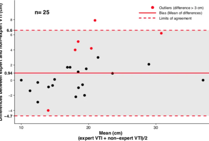
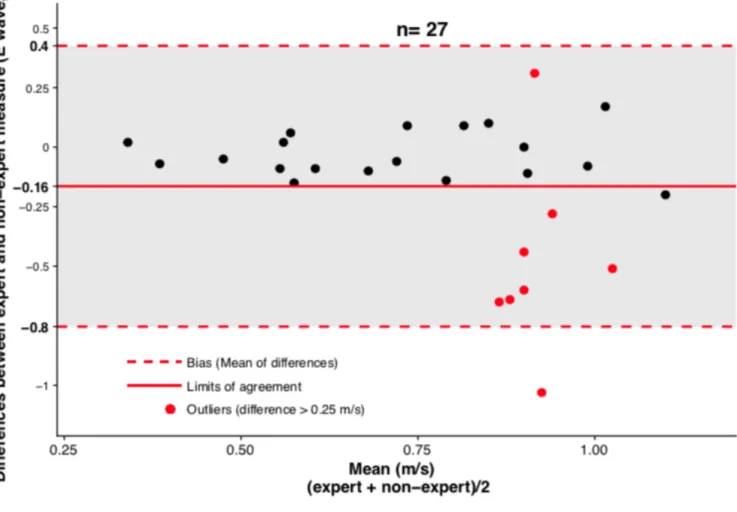
The Bland and Altman plots are shown in Figures 1 and 2. For Sub-aortic VTI, 6 patients were considered as outliers (difference > 3 cm) and 8 patients were considered as outliers for E wave velocity (difference > 0.25 m/s).
The presence of pleural effusion was diagnosed in 10 patients by intensivists and in 9 patients by gastroenterologists (Cohen’s Kappa Coefficient = 0.76). The ultrasound diagnosis of ascites was made in 25 patients by intensivists and 27 patients by gastroenterologists.
Echocardiographic Characteristics Evaluated by Experts alone are shown in Table 4.
DISCUSSION
Key findings
Our preliminary results demonstrate that echocardiography can be performed by previously trained gastroenterologists. In our study, the LVEF estimation by gastroenterologists was well correlated to intensivist’s estimation. We report a good agreement between gastroenterologists and ICU physicians for sub aortic VTI assessment while a poor agreement was observed for E wave velocity assessment. These results can be explained by the limited number of echography performed by each operator (between 3 and 6) over the study period. It could be postulated that the accuracy will increase with experience. This means that, for non-trained physicians, both LVEF (visual assessment) and stroke volume (indirectly assessed by sub aortic VTI) are easier to assess as compared to static left ventricle filling pressure (assessed by E wave velocity). For sub aortic VTI, the difference in measurement between expert and non-expert was less than the outlier (3 cm) in 76% of cases. This means that there was no clinically relevant difference in the assessment of cardiac output for 19 out of 27 patients. In the same way, there was no clinically relevant difference in the evaluation of left ventricle filing pressure for 19 patients.
The agreement for evaluation of pleural effusion and ascites was also good between gastroenterologists and intensivists with a good correlation as the Cohen’s Kappa coefficient was 0.76 for pleural effusion. We couldn't calculate a Cohen’s kappa coefficient for the evaluation of ascites, due to the lack of a diagnosis of absence of ascites
Relationship with previous papers
No study has yet evaluated the place of systematic echocardiographic evaluation of patients admitted for decompensated cirrhosis. In all studies evaluating the hemodynamics and cardiac function of cirrhotic patients, echocardiography is always performed by an expert physician (cardiologist or intensivist) and never by a hepatologist or gastroenterologist limiting its routine use in daily clinical practice (22–24)
Available data report that velocity time integral reflecting stroke volume can be accurately assessed by previously trained non-expert physicians after 25 US exams (25). LVEF assessment using a qualitative approach has been shown to adequately evaluate systolic function in septic shock (26). Similarly, in the present study, the semi-quantitative assessment of LEVF performed by gastroenterologists was well correlated to expert evaluation emphasizing the feasibility and usefulness of such approach.
During cirrhosis, there are some hemodynamic variations (4) : increase in total blood volume and splanchnic blood volume; decrease in central blood volume, arterial blood volume and systemic vascular resistance; increase of cardiac frequency, cardiac output and arterial compliance. This results in a peripheral arterial splanchnic vasodilation and a hyperdynamic circulation. However, to date, the description of typical hemodynamic profiles of cirrhotic patients has not been reported. Due to the preliminary nature of our results and the limited number of patients included, we can’t present these profiles yet. One of the pathophysiological mechanisms for the formation of ascites and edema is the peripheral arterial splanchnic vasodilatation, responsible for effective hypovolemia, which will activate arterial and cardiopulmonary volume receptors. This results in an activation of the renin angiotensin aldosterone system (RAAS) and the sympathetic nervous system, responsible for sodium and salt retention (27). This emphasizes the importance of blood volume assessment during cirrhosis decompensation. Stroke volume assessment by TTE is one of the key issue for blood volume evaluation (5). The ability of gastroenterologists to accurately perform stroke volume assessment as reported in the present study could be of great value for the management of decompensated cirrhosis and fluid optimization.
The role of TTE in the management of decompensated cirrhosis with acute kidney injury has been studied in one case report (22). Echocardiography was used to assess the hemodynamic status of the patient and guide the timing of PLA, to increase venous
determine the best location for ascites paracentesis (28) and for the diagnosis of hepatopulmonary syndrome by performing a bubble test, especially in patients waiting for transplantation (29). This is also true in hepatology ward for ascites paracentesis. Quantification of ascites and pleural effusion is also of importance for evaluating fluid retention. In ICU patients, POCUS influences patient management in 85% of cases as demonstrated in a large one-day observational trial. (30). It helps improving diagnosis performance where clinical evaluation failed (31).
Therefore, we could hypothesize that there is a potential utility of POCUS (coupling TTE with pleural and abdominal ultrasound) to optimize the hemodynamic state in patients with decompensated cirrhosis with ascites.
Clinical implication of study findings
The use of POCUS for patients with decompensated cirrhosis could therefore: guide the treatment of ascites (fluid infusion or diuretics), allow an early identification of patients requiring vasopressor treatment motivating a transfer to the ICU and prevent the occurrence of complications such as hepato-renal syndrome. A prospective interventional study based on the results of the present study seems crucial to further investigate the potential benefit of echocardiographic management of decompensated cirrhotic patients.
Our study has several limitations: the limited number of patients included at the time of the analysis and so the limited number of echography performed by each operator, responsible for a lack of statistical power. The choice of a semi-quantitative estimate of the LVEF, with a 40% cutoff can lead to selection bias. Indeed cirrhotic patients most often have preserved systolic function(32).
CONCLUSION:
This preliminary study shows that, after a short training period, echocardiography can be performed by gastroenterologists in order to assess both LVEF and cardiac output in patient with decompensated cirrhosis. Evaluation of left ventricle filing pressure appears
REFERENCES
1. Ginés P, Quintero E, Arroyo V, Terés J, Bruguera M, Rimola A, et al.
Compensated cirrhosis: natural history and prognostic factors. Hepatol. Baltim. Md. 1987;7:122–128.
2. D’Amico G, Garcia-Tsao G, Pagliaro L. Natural history and prognostic indicators of survival in cirrhosis: a systematic review of 118 studies. J. Hepatol. 2006;44:217–231. 3. Tandon P, Garcia-Tsao G. Bacterial infections, sepsis, and multiorgan failure in cirrhosis. Semin. Liver Dis. 2008;28:26–42.
4. Møller S, Henriksen JH, Bendtsen F. Extrahepatic complications to cirrhosis and portal hypertension: haemodynamic and homeostatic aspects. World J. Gastroenterol. 2014;20:15499–15517.
5. Cecconi M, De Backer D, Antonelli M, Beale R, Bakker J, Hofer C, et al. Consensus on circulatory shock and hemodynamic monitoring. Task force of the European Society of Intensive Care Medicine. Intensive Care Med. 2014;40:1795–1815. 6. Moore CL, Copel JA. Point-of-care ultrasonography. N. Engl. J. Med.
2011;364:749–757.
7. Perez-Avraham G, Kobal SL, Etzion O, Novack V, Wolak T, Liel-Cohen N, et al. Left ventricular geometric abnormality screening in hypertensive patients using a hand-carried ultrasound device. J. Clin. Hypertens. Greenwich Conn. 2010;12:181–186.
8. Mjølstad OC, Andersen GN, Dalen H, Graven T, Skjetne K, Kleinau JO, et al. Feasibility and reliability of point-of-care pocket-size echocardiography performed by medical residents. Eur. Heart J. Cardiovasc. Imaging. 2013;14:1195–1202.
9. Vignon P, Dugard A, Abraham J, Belcour D, Gondran G, Pepino F, et al. Focused training for goal-oriented hand-held echocardiography performed by noncardiologist residents in the intensive care unit. Intensive Care Med. 2007;33:1795–1799.
10. Nguyen VTQ, Ho JE, Ho CY, Givertz MM, Stevenson LW. Handheld echocardiography offers rapid assessment of clinical volume status. Am. Heart J. 2008;156:537–542.
11. Harris PA, Taylor R, Thielke R, Payne J, Gonzalez N, Conde JG. Research electronic data capture (REDCap)—A metadata-driven methodology and workflow process for providing translational research informatics support. J. Biomed. Inform. 2009;42:377–381.
et al. A model to predict survival in patients with end-stage liver disease. Hepatol. Baltim. Md. 2001;33:464–470.
14. Mayo PH, Beaulieu Y, Doelken P, Feller-Kopman D, Harrod C, Kaplan A, et al. American College of Chest Physicians/La Société de Réanimation de Langue Française statement on competence in critical care ultrasonography. Chest. 2009;135:1050–1060. 15. Balik M, Plasil P, Waldauf P, Pazout J, Fric M, Otahal M, et al. Ultrasound
estimation of volume of pleural fluid in mechanically ventilated patients. Intensive Care Med. 2006;32:318.
16. Bland JM, Altman DG. Statistical methods for assessing agreement between two methods of clinical measurement. Lancet Lond. Engl. 1986;1:307–310.
17. Cohen J. A Coefficient of Agreement for Nominal Scales. Educ. Psychol. Meas. 1960;20:37–46.
18. Koo TK, Li MY. A Guideline of Selecting and Reporting Intraclass Correlation Coefficients for Reliability Research. J. Chiropr. Med. 2016;15:155–163.
19. DiCiccio T, Efron B. Bootstrap confidence intervals. Stat. Sci. 1996;11:189–228. 20. Landis JR, Koch GG. The measurement of observer agreement for categorical data. Biometrics. 1977;33:159–174.
21. R Core Team. A language and environment for statistical computing. R Found. Stat. Comput. Vienna Austria [Internet]. 2016;Available from:
https://www.R-project.org/
22. Huggins JT, Doelken P, Walters C, Rockey DC. Point-of-Care Echocardiography Improves Assessment of Volume Status in Cirrhosis and Hepatorenal Syndrome. Am. J. Med. Sci. 2016;351:550–553.
23. Karagiannakis DS, Vlachogiannakos J, Anastasiadis G, Vafiadis-Zouboulis I, Ladas SD. Diastolic cardiac dysfunction is a predictor of dismal prognosis in patients with liver cirrhosis. Hepatol. Int. 2014;8:588–594.
24. Dadhich S, Goswami A, Jain VK, Gahlot A, Kulamarva G, Bhargava N. Cardiac dysfunction in cirrhotic portal hypertension with or without ascites. Ann.
Gastroenterol. 2014;27:244–249.
25. Charron C, Prat G, Caille V, Belliard G, Lefèvre M, Aegerter P, et al. Validation of a skills assessment scoring system for transesophageal echocardiographic monitoring of hemodynamics. Intensive Care Med. 2007;33:1712–1718.
arterial vasodilation hypothesis: a proposal for the initiation of renal sodium and water retention in cirrhosis. Hepatol. Baltim. Md. 1988;8:1151–1157.
28. Frankel HL, Kirkpatrick AW, Elbarbary M, Blaivas M, Desai H, Evans D, et al. Guidelines for the Appropriate Use of Bedside General and Cardiac Ultrasonography in the Evaluation of Critically Ill Patients-Part I: General Ultrasonography. Crit. Care Med. 2015;43:2479–2502.
29. Levitov A, Frankel HL, Blaivas M, Kirkpatrick AW, Su E, Evans D, et al.
Guidelines for the Appropriate Use of Bedside General and Cardiac Ultrasonography in the Evaluation of Critically Ill Patients-Part II: Cardiac Ultrasonography. Crit. Care Med. 2016;44:1206–1227.
30. Zieleskiewicz L, Muller L, Lakhal K, Meresse Z, Arbelot C, Bertrand P-M, et al. Point-of-care ultrasound in intensive care units: assessment of 1073 procedures in a multicentric, prospective, observational study. Intensive Care Med. 2015;41:1638–1647. 31. Bossone E, DiGiovine B, Watts S, Marcovitz PA, Carey L, Watts C, et al. Range and prevalence of cardiac abnormalities in patients hospitalized in a medical ICU. Chest. 2002;122:1370–1376.
32. Møller S, Henriksen JH. Cardiovascular complications of cirrhosis. Gut. 2008;57:268–278.
TABLES
Table 1. Population characteristics (n=27)
Age (years) 62 [55; 72.5]
Male gender 22 (81.5%)
Weight (kg) 79 [65 ; 86]
BMI 26.58 [23.27 ; 30.68]
Preexisting conditions
Chronic arterial hypertension 9 (33.3 %)
Chronic heart disease 3 (11.1 %)
Ischemic heart disease 5 (18.5 %)
COPD 2 (7.4 %)
Chronic kidney disease 1 (3.7 %)
Diabetes 10 (37 %) Obesity 6 (22.2 %) HCC 7 (25.9 %) Home medication Diuretics 19 (70.4 %) ACE inhibitors 5 (18.5 %) Sartans 4 (14.8 %)
Calcium channel blockers 2 (7.4 %) Cardioselective beta blockers 6 (22.2 %) Non cardioselective beta blockers 7 (25.9 %) Mean Arterial Pressure (mmHg) 90 [82; 97] Systolic Arterial Pressure (mmHg) 125 [110.5; 134.0] Diastolic Arterial Pressure (mmHg) 71 [68; 81] Heart rate (beats.min-1) 86 [79.0; 94.5]
For categorical variables, results are given as number of patients and percentages while for continuous variables as median and interquartile range (25th;75th percentiles).
COPD= Chronic Obstructive Pulmonary Disease, ACE= Angiotensin Converting Enzyme. HCC= hepatocellular carcinoma
Table 2. Characteristics, management of the decompensated liver disease and status at day 28 Etiology Alcohol 21 (77.8 %) Viral hepatitis B 2 (7.4 %) Viral hepatitis C 5 (18.5 %) Nonalcoholic steatohepatitis, 7 (25.9 %) Unknown 1 (3.7 %) Precipitating event Non-compliance 3 (11.1 %) Dietary errors 0 Sepsis 2 (7.4 %) Other 13 (48.1 %) Unknown 9 (33.3 %) Encephalopathy 3 (11.1 %) Ascites 26 (96.3 %)
Lower limb edema 14 (51.9 %)
Biological data
Plasma Sodium (mmol.L-) 136 [133.5 ; 139.5]
Plasma Creatinine ("mol.L-) 93 [72 ; 109]
Plasma Urea (mmol.L-) 5.8 [4.0 ; 8.7]
Albumin (g.L-1) 28.4 [24.35 ; 31.85] Bilirubin (µmol.L-) 32 [19 ; 54] Platelets (count/mm3) 129000 [10000 ; 188500] Prothrombin activity (%) 62 [49.5 ; 74.5] Child-Pugh Score 9 [8.0 ; 10.5] A 1 (3.7 %) B 15 (55.6 %) C 11 (40.7 %) MELD score 13.3 [11.15 ; 17.05] Management Albumin infusion (n) 23 (85.2 %)
Fluid amount when used (mL) 10250 [6500 ; 12500] Furosemide (n) 20 (74.1 %) Spironolactone (n) 19 (70.4 %) Thiazide diuretics (n) 2 (7.4 %) Vasoactive drug 0 Ascites removal (n) 22 (81.5 %) Status at day 28 Discharge at home 7 (25.9 %) Non-survivor 4 (14.8 %)
Readmitted after discharge 11 (40.7 %) Discharge to a rehabilitation unit 1 (3.7 %)
Admitted to the ICU 3 (11.1 %)
Still in the unit 1 (3.7 %)
Results are given as median and interquartile range (25th-75th percentiles)
and as number of patients and percentages for categorical variables.
Table 3. Echocardiographic characteristics assessed by experts and non-experts.
LVEF above 40%
Expert 27 (100 %)
Non-expert 27 (100 %)
Peak mitral E-wave velocity (m.s-1) ICC = 0.30 [-0.16 ; 0.63]
Expert 0.68 [0.55 ; 0.86]
Non-expert 0.80 [0.65 ; 1.10]
Sub Aortic VTI (cm) ICC = 0.89 [0.74 ; 0.96]
Expert 18.8 [14.7 ; 22.7] Non-expert 17.0 [14.6 ; 19.7] Pleural effusion Expert 10 (37 %) Non-expert 9 (33.3 %) Ascites Expert 25 (92.6 %) Non-expert 27 (100 %)
Results are given as median and interquartile range (25th-75th percentiles) for
continuous variables and as number of patients and percentages for categorical variables.
LVEF = Left Ventricular Ejection Fraction, VTI = velocity time-integral. ICC = intra-class correlation coefficient.
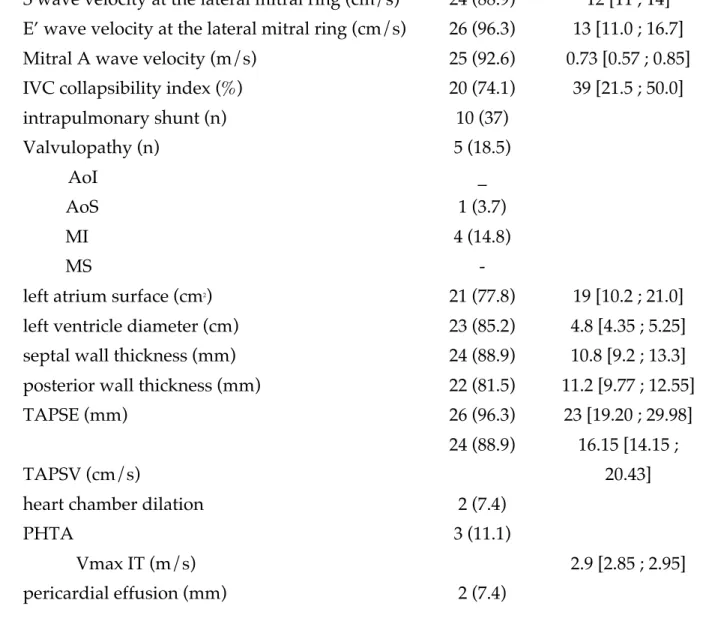
Table 4. Echocardiographic Characteristics Evaluated by Experts alone
n (%)
S wave velocity at the lateral mitral ring (cm/s) 24 (88.9) 12 [11 ; 14] E’ wave velocity at the lateral mitral ring (cm/s) 26 (96.3) 13 [11.0 ; 16.7] Mitral A wave velocity (m/s) 25 (92.6) 0.73 [0.57 ; 0.85] IVC collapsibility index (%) 20 (74.1) 39 [21.5 ; 50.0]
intrapulmonary shunt (n) 10 (37) Valvulopathy (n) 5 (18.5) AoI _ AoS 1 (3.7) MI 4 (14.8) MS -
left atrium surface (cm2) 21 (77.8) 19 [10.2 ; 21.0]
left ventricle diameter (cm) 23 (85.2) 4.8 [4.35 ; 5.25] septal wall thickness (mm) 24 (88.9) 10.8 [9.2 ; 13.3] posterior wall thickness (mm) 22 (81.5) 11.2 [9.77 ; 12.55]
TAPSE (mm) 26 (96.3) 23 [19.20 ; 29.98]
TAPSV (cm/s)
24 (88.9) 16.15 [14.15 ; 20.43]
heart chamber dilation 2 (7.4)
PHTA 3 (11.1)
Vmax IT (m/s) 2.9 [2.85 ; 2.95]
pericardial effusion (mm) 2 (7.4)
Results are given as median and interquartile range (25th-75th percentiles) and as
number of patients and percentages for categorical variables.
IVC: Inferior Vena cava; AoI: Aortic insufficiency; AoS : Aortic stenosis; MI : Mitral insufficiency; MS : Mitral Stenosis; TAPSE : tricuspid annular plane systolic excursion; TAPSV: tricuspid annular plane systolic velocity; PHTA: Pulmonary arterial hypertension.
FIGURES
SERMENT
Ø
En présence des Maîtres de cette école, de mes chers condisciples et
devant l’effigie d’Hippocrate, je promets et je jure, au nom de l’Etre
suprême, d’être fidèle aux lois de l’honneur et de la probité dans l’exercice
de la médecine.
Ø
Je donnerai mes soins gratuits à l’indigent et n’exigerai jamais un salaire
au-dessus de mon travail.
Ø
Admis (e) dans l’intérieur des maisons, mes yeux ne verront pas ce qui s’y
passe, ma langue taira les secrets qui me seront confiés, et mon état ne
servira pas à corrompre les mœurs, ni à favoriser le crime.
Ø
Respectueux (se) et reconnaissant (e) envers mes Maîtres, je rendrai à
leurs enfants l’instruction que j’ai reçue de leurs pères.
Ø
Que les hommes m’accordent leur estime si je suis fidèle à mes promesses.
Que je sois couvert (e) d’opprobre et méprisé (e) de mes confrères si j’y
manque.
Introduction : L'ascite est une complication fréquente de la cirrhose et un facteur de
mauvais pronostic. Il n'existe aucune recommandation pour une évaluation
systématique de l'état hémodynamique des patients par échographie cardiaque, à la phase initiale d’une décompensation de la maladie. Le but de notre étude est d'évaluer la concordance de l'évaluation écho-cardiographique faite lors de la phase initiale de la décompensation œdémato-ascitique par des médecins réanimateurs considérés comme experts et des gastro-entérologues non experts, après une courte formation.
Patients et méthodes : Tous les patients admis dans notre service de gastroentérologie
pour une décompensation œdémato-ascitique de cirrhose ont été inclus de façon prospective. Les patients admis pour une autre complication de la cirrhose pouvant affecter l'état hémodynamique n'ont pas été inclus. Nous avons effectué 2 examens échographiques pour chaque patient, un par un gastro-entérologue et un par un
médecin réanimateur. Les gastro-entérologues participant à l'étude ont tous été formés de la même façon : 2 heures de cours théorique et 2 journées de mise en pratique en service de réanimation. Le critère de jugement principal est la concordance entre les gastro-entérologues et les médecins réanimateurs pour 3 paramètres échographiques : la FEVG visuelle (Fraction d'éjection ventriculaire gauche (%)), l’ITV sub-aortique (intégrale temps-vitesse, (cm)) et la vitesse de l’onde E (m.s-1).
Résultats : De mars à août 2018, 27 patients ont été inclus. L'âge médian était de 62 ans,
81,5 % étaient des hommes. L'alcool était la principale étiologie de la cirrhose (21 patients, 78 %). La maladie hépatique était le plus souvent avancée avec 40,7 % CHILD C et 55,6 % CHILD B. Le score de MELD médian était de 13,3. Tous les patients
présentaient une FEVG > 40 %, estimée à la fois par les réanimateurs et les
gastro-entérologues (concordance parfaite). L’ITV médian mesuré par les réanimateurs était de 18,8 contre 17 cm par les gastroentérologues (CCI (Coefficient de corrélation intraclasse) = 0,89, bonne concordance). La vitesse médiane mesurée de l'onde E était de 0,68 par les réanimateurs contre 0,8 m/s par les gastroentérologues (CCI = 0,30, faible
concordance).
Conclusion : Cette étude préliminaire montre qu'après une courte formation,
l'échographie cardiaque peut être réalisée par des gastro-entérologues afin d'évaluer la FEVG et le débit cardiaque chez des patients avec une cirrhose décompensée.
L'évaluation de la pression de remplissage du ventricule gauche semble plus difficile. D'autres études sont nécessaires pour préciser la place de l'échographie cardiaque afin