Chemistry an Asian Journal
12- to 22-Membered bridged -lactams as potential Penicillin Binding Proteins inhibitors. Aline Sliwa,[a] Georges Dive,*[b] and Jacqueline Marchand-Brynaert*[a]
[a] Prof. Dr. J. Marchand-Brynaert, Ir. A. Sliwa
Institute of Condensed Matter and Nanosciences (IMCN) Molecules, Solids and Reactivity (MOST)
Université catholique de Louvain
Bâtiment Lavoisier, Place Louis Pasteur L4.01.02 1348 Louvain-la-Neuve, Belgium
Phone : +32 (0)10 47 27 40 Fax : +32 (0)10 47 41 68
E-mail : jacqueline.marchand@uclouvain.be
[b] Dr. G. Dive
Centre d’ingénierie des Protéines (CIP) Université de Liège
Bâtiment B6, Allée de la Chimie 4000 Sart-Tilman (Liège), Belgium
Keywords
Ab initio calculations, -lactams, Macrocycles, Ring Closing Metathesis, Serine-enzyme inhibition Abstract
As potential inhibitors of Penicillin Binding Proteins (PBPs), we focused our research on the synthesis of non-traditional 1,3-bridged -lactam embedded into macrocycles. 12- to 22-Membered bicyclic -lactams were synthesized by RCM reaction of bis--alkenyl-3(S)-amino-azetidinone precursors. The reactivity of 1,3-bridged -lactams was estimated by the determination of the energy barrier of a concerted nucleophilic attack and lactam ring-opening process, using ab initio calculations. The results predict that 16-membered cycles should be the more reactive. Biochemical evaluations against R39 DD-peptidase and two resistant PBPs, namely PBP2a and PBP5fm, revealed indeed the inhibition effect of compound 4d featuring a 16-membered bridge and the N-Boc chain at C(3) position of -lactam ring. Surprisingly, the corresponding bicycle 12d with the V side-chain at C(3) was inactive. Very elaborate reactivity models of the R39 active site have been built that allowed to explain these results.
Introduction
The introduction of penicillins to the therapeutic arsenal, in the early 40’s, is the starting point of the antibiotic era which allowed to save millions men from potentially fatal infectious diseases. However, since
the initial use of penicillins as chemotherapeutic agents, phenomena of bacterial resistance were reported.[1]
Due to the antibiotic pressure, the occurrence of resistant bacteria increases, leading to a worrisome situation about antibiotics efficiency, so worrying that the World Health Organization decided to dedicate the World Health Day 2011 to microbial resistance.
Despite the discovery of several other classes of antibiotics, the so-called -lactam class (exemplified nowadays with cephalosporins, carbapenems and penems)[2] still remain the most prescribed one. Initially
the activity of -lactam antibiotics (i.e. penicillins) has been attributed to the strain of the four-membered ring and the twisted amide bond of the 2-azetidinone function. Therefore the search of novel -lactam antibiotics has been extensively focused on strained 1,4-fused bicyclic structures, intended to improve the so-called "acylating power" of the -lactam ring versus the target serine-proteases (i.e. bacterial DD-peptidases).[3] Tebipenem, tomopenem or razupenem, (Figure 1) three carbapenems in phase II clinical
studies, represent examples of this strategy.[4]
N O CO2H H OH H SR Tomopenem R = N O N HN O HN NH NH2 Tebipenem R = N S N Razupenem R = S N H N
Figure 1. Examples of Carbapenems in development.
However this traditional model of reactivity seems to be over-evaluated, as there is still no clear relationship between structural characteristics and biological activity. An alternative model of reactivity has been proposed by our group some years ago, based on 1,3-bridged, planar -lactam motifs embedded into macrocycles. The aim was to possibly decrease the activation barrier of the 2-azetidinone N–C(O) bond cleavage by increasing the conformational adaptability that would be involved in the atoms reorganization
during the formation of an acyl-enzyme intermediate.[5] A first series of compounds A (Figure 2), structurally
related to carbapenems, has been previously reported and evaluated against bacterial enzymes with a mitigated success attributed to an inadequate configuration of the C(3) chiral center. To further explore our hypothesis, we decided to synthesize the series of compounds B (Figure 2) structurally related to cephalosporins and penicillins, with an amino substituent and inversion of configuration at C(3). Since the nucleophilic attack by serine enzymes occurs normally on the α-face of the -lactam, the macrocycle of B surrounding the -face would not hamper the serine enzyme processing.
N O O X Y 1 23 4 R = H, n-Pr, OAc R1= H, Me, COR 2 X = Y = O ; X = Y = H,H ; X = O and Y = H,H R A n n N O N X Y R1 1 2 3 4 n n B
Figure 2. 1,3-bridged bicyclic -lactam compounds.
N O N Y n R X n N O N X Y n n R NH O N R1 N OH O OH X = Y = O ; R = H, Me, Boc ; (R1, R2) = (Boc, H), (Boc, Me)
X = Y = H,H ; R = Boc, PhOCH2CO ; (R1, R2) = (Boc, H), (PhOCH 2CO, H) R2 1 2 3 4 B C D R1 R2
Scheme 1. Retrosynthetic strategy.
The RCM (Ring Closing Metathesis) reaction was chosen as the key step for the formation of the bridging macrocycles. The precursors are chiral azetidin-2-ones C with -alkenoyl or -alkenyl chains on the positions N(1) and C(3)-N. The starting chirons D, derivatives of (S)-3-aminoazetidin-2-one, come from L-serine.
First we tried to synthesize the bicyclic family B with bis-acylated chains (X = Y = O). But unexpectedly, when applying the RCM reaction to the bis-acylated precursors C (X = Y = O, Scheme 1), we recovered exclusively cyclodimers, except in one case (R = Boc; n = 2).[6] Due to the presence of amide and
imide functions in the precursors C, the conformers leading to the desired cyclizations are strongly disfavoured. This should not be the case for the bis-alkylated precursors (X = Y = H,H). Therefore, we kept the same retrosynthetic strategy to synthesize the 1,3-bridged bicyclic -lactam compounds B with bis-alkylated chains (X = Y = H,H).
In this article we describe the successful synthesis of target molecules B belonging to the bis-alkylated family (X = Y = H,H). All these compounds were investigated from a theoretical point of view, and their reactivity into a model of Penicillin Binding Protein (PBP) cavity was studied. Theoretical predictions could be experimentally confirmed by in vitro evaluations against R39 D,D-peptidase,[7] the
commonly used model of bacterial enzymes, and against two resistant PBPs, namely PBP2a from Staphylococcus aureus[8] and PBP5fm from Enterococcus faecium.[9]
Results and Discussion Synthesis
The first series of bis-alkylated bicycles B synthesized was the Boc family (X = Y = H,H; R = Boc). The starting material is the known (S)-3-(tertbutyloxycarbonyl)amino-2-azetidinone 1, readily prepared from the commercially available Boc-L-serine, as previously described.[6]
NH O BocHN N O BocN 1 N O n n BocN N O n n BocN n n a b c 2a, n = 1 (50%) 2b, n = 3 (41%) 2c, n = 4 (43%) 2d, n = 5 (39%) 2e, n = 6 (46%) 2f, n = 8 (35%) 3b, n = 3 (66%) 3c, n = 4 (64%) 3d, n = 5 (90%) 3e, n = 6 (76%) 3f, n = 8 (72%) 4b, n = 3 (84%) 4c, n = 4 (83%) 4d, n = 5 (98%) 4e, n = 6 (67%) 4f, n = 8 (60%)
Scheme 2. Synthesis of bis-alkylated azetidinones 2 and RCM reaction: a) NaH, Br-(CH2)n-CH=CH2, DMF,
0 °C to 20 °C, 12 h; b) Grubbs II catalyst (2 x 5 mol%), DCM, 40 °C, 12 h; c) H2, Pd-C catalyst, MeOH, 20
-Alkenyl bromides of various lengths were used to access various sizes of bicycles. All the -alkenyl bromides used are commercially available reagents. The bis-alkyl derivatives 2a-f were synthesized in one step, with moderate yields, by using 2 equivalents of NaH and 2.2 equivalents of -alkenyl bromides (step (a) of Scheme 2). Other strong bases were tested (namely LiHMDS, KOH in presence of Bu4NHSO4 and
NaI), without improvement of yields. The use of an excess of base led to racemisation of the bis-alkyl derivatives 2. The bis-alkyl derivative 2 with n = 2 was not obtained, probably because HBr elimination occurred from 1-butenyl bromide. With these precursors in hands, we could validate the RCM strategy: using second generation Grubbs’ catalyst, the macrocycles 3b-f were readily formed and isolated in good yields after chromatographic purification (step (b) of Scheme 2) leading to 12- to 22-membered rings. The bis-alkyl derivative 2a did not cyclise, the expected 8-membered ring bicycle being too small. Catalytic hydrogenation led to the corresponding saturated macrocycles 4b-f with high yields (step (c) of Scheme 2). Since the N-unprotected derivatives B (X = Y = H,H ; R = H) were desirable for biological evaluation and/or further derivatization with penicillin’s side-chain, we attempted to remove the Boc group from the precursors 2, and bicycles 3 and 4. Several conditions were tested (TFA in DCM, HCl 2 M solution, CAN in refluxing ACN,[10] TMSCl in presence of NaI[11]) without success; in all cases the -lactam degradation was
observed (IR, NMR analyses).
We tried to replace the Boc protecting group by the penicillin V side-chain (V = PhOCH2CO) in situ, by
direct acylation (with phenoxyacetyl chloride and TEA or DIEA) of the crude mixtures resulting from the treatment of 2, 3 and 4 under the above mentioned deprotection conditions. Here again, the results were disappointing with the recovery of untractable mixtures. Hence to access the bis-alkylated bicycles B with the V side-chain (X = Y = H,H ; R = PhOCH2CO) we re-started the total synthesis from L-serine (Scheme
3). OH O VHN OH N H O VHN OH OBn N O VHN OBn O NH VHN b c d 87% 6 7 8 9 64% 100% a 71% 5 OH O H2N OH
Scheme 3. Synthesis of chiron 9: a) phenoxyacetyl chloride, saturated NaHCO3, CH3CN, rt, 12 h; b) DCC,
NH2OBn, THF, 0 °C to rt, 12 h; c) PPh3, CCl4, TEA, CH3CN, 0 °C to rt, 12 h; d) H2, Raney-Ni,
L-serine 5 was acylated under Schotten-Baumann conditions into (S)-3-hydroxy-2-(2-phenoxyacetamido)propanoic acid 6 in 71% yield. This compound was converted to the corresponding hydroxamate 7, using O-benzylhydroxylamine and DCC, in 87% yield. Intramolecular cyclization via the method proposed by Miller et al.,[12] afforded the -lactam 8 in 64% yield. Subsequent hydrogenation in the
presence of Raney nickelgave the desired chiron 9 in quantitative yield.
NH O VHN N O VN 9 N O n n VN N O n n VN n n a b c 10a, n = 1 (55%) 10b, n = 3 (22%) 10c, n = 4 (43%) 10d, n = 5 (30%) 11b, n = 3 (59%) 11c, n = 4 (47%) 11d, n = 5 (67%) 12b, n = 3 (90%) 12c, n = 4 (91%) 12d, n = 5 (93%)
Scheme 4. Synthesis of bis-alkylated azetidinones 10 and RCM reaction: a) NaH, Br-(CH2)n-CH=CH2,
DMF, 0 °C to 20 °C, 12 h; b) Grubbs II catalyst (2 x 5 mol%), DCM, 40 °C, 12 h; c) H2, Pd-C catalyst,
MeOH, 20 °C, 3 h.
Similarly to the Boc family, the bis-alkyl derivatives 10 were synthesized in one step, from 9 in moderate yields. Under conditions of RCM reaction, the precursors 10b-d (but not 10a) cyclized into bicycles 11b-d in good yields, affording 12- to 16-membered rings. Catalytic hydrogenation led to the saturated bicycles 12b-d. (Scheme 4)
All precursors (2, 10) and bicycles (3-4, 11-12) were characterized by IR, NMR and MS (see Experimental). In particular, MS was useful to detect the possible occurrence of side-products issued from (cyclo)oligomerizations and/or double bond migrations (in precursors) leading in fine to cyclic products with the formal extrusion of one CH2 unit. Only the 18- and 22-membered ring bicycles, i.e. compounds 3e-f, and
consequently 4e-f, were contaminated with a small amount of lower homologues (respectively 17- and 21-membered ring bicycles).
The NMR patterns confirmed our hypothesis that 1,3-bridged -lactams embedded into large rings are endowed with a certain conformational adaptability, as discussed in the next section.
A lot of conformers can exist for the monocyclic molecules 2 and 10 and also for the bicyclic molecules 3, 4, 11 and 12. In solution, the coexistence of conformers for these compounds was experimentally detected by NMR spectroscopy. For example, in Figure 3 are presented the 13C NMR spectra of compound 12d
recorded in 1,1,2,2-tetrachloroethane-d2 at different temperatures. At 30 °C, a lot of signals are visible
because some carbons give rise to signal splitting into several peaks. Rising the temperature to 120 °C leads to signals coalescence and thus allows the structural assignment. The quaternary carbons (Cq) at 167.8 (C=O), 165.3 (C=O) and 158.0 (PhO) ppm at 120 °C appeared as multiple signals at 30 °C. The methylene peak of the V side-chain at 67.9 ppm at 120 °C presented two distinct signals at 30 °C. The C(3) carbon of the -lactam ring at 62.2 ppm at 120 °C gave two very spaced signals at 30 °C. Similar NMR studies recorded for compounds 10d and 11d can be found in supporting information.
Figure 3. 13C NMR spectra of compound 12d recorded at 125 MHz in 1,1,2,2-tetrachloroethane-d 2.
The geometry of all the molecules has been fully optimized at the RHF level using the minimal basis set MINI-1’.[13]
In the case of bis-alkylated precursors 2, 10 (X = Y = H,H), the molecules are conformationally less constrained than the bis-acylated compounds (X = Y = O),[6] as the carbonyls are replaced by methylene
groups which can accommodate more conformations. Depending on the nature of the N-substituent (Boc or V), a great number of local minima could be trapped.
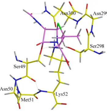
Two conformations « i » and « ii » of the bridging cycle (of compounds 3, 11 and 4, 12) have been located with respect to the -lactam ring for all the studied compounds. The conformation « i » expands the cycle to the right upper corner of the -lactam C(4); in the conformation « ii », the cycle is more orientated to the carbonyl C(2) of the -lactam. For each conformation, the carbonyl of the side-chain (Boc or V) can rise above the 4-membered ring (« a » conformation) or below (« b » conformation) (Figure 4). For all the unsaturated compounds 3, 11, the trans configuration of the substituted ethylene has been considered.
Figure 4. Conformers ib and iia of compound 4d.
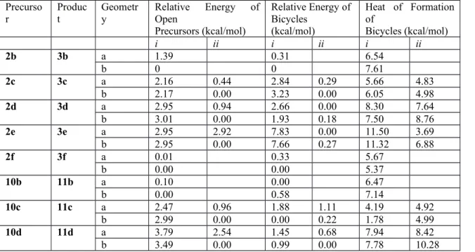
The heat of formation has been computed with respect to the open precursor with the same conformation as the one of the corresponding cyclized molecule. For Boc as well for V side-chains, the 4 conformations lie in the same range of stability, the relative energies being less than 8 kcal/mole in the case of ii conformations often more stable than the i ones (Table 1). The size of the cycle has a significant impact on the conformations for the smallest (12-membered cycle) and the largest ones (22-membered cycle). For the
compounds 3b and 11b, only the conformation i can be trapped. In the 3f molecule, the ring is so large that it expands on both sides of the -lactam in a pseudo i conformation only.
Since the bis-alkylated precursors 2 and 10 are highly flexible, their cyclization can easily lead to the desired compounds 3 and 11. Similar reaction was not possible with the bis-acylated precursors.[6]
Table 1. Relative energies of the precursors/bicycles in the selected conformations and respective heat of formation resulting from the cyclization.
Precurso
r Product Geometry Relative Energy ofOpen Precursors (kcal/mol) Relative Energy of Bicycles (kcal/mol) Heat of Formation of Bicycles (kcal/mol) i ii i ii i ii 2b 3b a 1.39 0.31 6.54 b 0 0 7.61 2c 3c a 2.16 0.44 2.84 0.29 5.66 4.83 b 2.17 0.00 3.23 0.00 6.05 4.98 2d 3d a 2.95 0.94 2.66 0.00 8.30 7.64 b 3.01 0.00 1.93 0.18 7.50 8.76 2e 3e a 2.95 2.92 7.83 0.00 11.50 3.69 b 2.95 0.00 7.66 0.27 11.32 6.88 2f 3f a 0.01 0.33 5.67 b 0.00 0.00 5.37 10b 11b a 0.10 0.00 6.47 b 0.00 0.58 7.14 10c 11c a 2.47 0.96 1.88 1.11 4.19 4.92 b 2.99 0.00 0.00 0.22 1.78 4.99 10d 11d a 3.79 2.54 1.45 0.68 7.94 8.42 b 3.49 0.00 0.99 0.00 7.78 10.28
Reactivity versus serine enzyme models
The reactivity of the bridged molecules has been studied using a simple model of PBP cavity (Figure 5) at the RHF/MINI-1’ level. (Table 2)
N Me O H NH N O H H O O H Me H
In this model, the -lactam ring opening occurs via a concerted process: the nucleophilic serine is mimicked by 2-(formyl)amino-1-ethanol, in interaction with methylamine working as a proton relay to methanol which, in fine, transfers the proton to the -lactam nitrogen.[14] The formamide moiety mimicks the oxyanion
hole stabilization. At the transition state (TS), this pseudo 8-membered ring is described by the reaction coordinate associated to the negative curvature of the energy second derivative matrix.
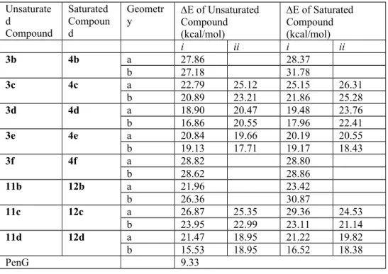
Table 2. Activation energy of concerted nucleophilic attack (MINI-1’).
Unsaturate d Compound Saturated Compoun d Geometr
y E of UnsaturatedCompound (kcal/mol) E of Saturated Compound (kcal/mol) i ii i ii 3b 4b a 27.86 28.37 b 27.18 31.78 3c 4c a 22.79 25.12 25.15 26.31 b 20.89 23.21 21.86 25.28 3d 4d a 18.90 20.47 19.48 23.76 b 16.86 20.55 17.96 22.41 3e 4e a 20.84 19.66 20.19 20.55 b 19.13 17.71 19.17 18.43 3f 4f a 28.82 28.80 b 28.62 28.86 11b 12b a 21.96 23.42 b 26.36 30.87 11c 12c a 26.87 25.35 29.36 24.53 b 23.95 22.99 23.11 21.14 11d 12d a 21.47 18.95 21.22 19.82 b 15.53 18.95 16.52 18.38 PenG 9.33
The results obtained with Boc and V side-chains present some common features concerning the lowest energy barriers: the optimum size of the cycle is a 16-membered ring (i.e. 3d-4d and 11d-12d) while the shorter and the bigger ones enhance the energy barrier calculated with the model. In most of the cases, the activation energy is also higher for the saturated cycle with respect to the corresponding unsaturated one (with the trans configuration at the C=C bond).
Inhibition of R39, PBP2a and PBP5fm
All products were evaluated for their potential inhibition effect on bacterial serine enzymes. R39 from Actinomadura is a model serine-enzyme of low molecular weight D,D-peptidases, usually considered for a
preliminary screening of penicillin-like compounds. R39 and the tested -lactams (100 M) were incubated (1 h, 25 °C). Then the enzyme residual activity (RA) was determined by observing the hydrolysis of the thioester S2d substrate[15] in the presence of DTNB for labeling the formed thiol, and reading at 412 nm.
The results are given in Table 3 as percentages (%) of initial activity. The activity in the absence of inhibitors is set at 100% and therefore low values indicate very active compounds since the bacterial enzyme has been inhibited by the tested compound and consequently cannot hydrolyze its substrate. A tested compound is considered as a “hit” (i.e. potential inhibitor) for a RA < 80%. All the compounds were also evaluated against two high-molecular-weight D,D-peptidases responsible for bacterial resistance to β-lactam antibiotics: PBP2a from methicillin-resistant S. aureus and PBP5fm from E. faecium. The tested -lactams (1 mM) were incubated with the PBPs (4h, 30 °C), then fluorescein-labelled ampicillin was added to detect the residual activity. This reagent is an inhibitor forming a stable acyl-enzyme intermediate. After denaturation, and SDS-PAGE separation of the acylated enzyme from the reagent band, fluorescence was measured. The fluorescence intensity is proportional to the residual active protein, i.e. protein non acylated by the tested compound.
Table 3. Evaluation of bis-alkylated azetidinones against R39 D,D-peptidase, PBP5f and PBP2a Entr y compoun d n R39 RA [%] PBP5f RA [%] PBP2a RA [%] 1 2a 1 101 4 81 96 2 2b 3 >100 94 71 3 2c 4 97 11 69 68 4 2d 5 80 4 80 59 5 2e 6 97 2 100 95 6 2f 8 101 ± 1 100 100 7 10a 1 103 8 100 96 8 10b 3 97 ± 3 90 95 9 10c 4 101 ± 1 100 98 10 10d 5 100 ± 4 96 91 11 3b 3 >100 68 53 12 3c 4 103 9 78 96 13 3d 5 83 4 74 58 14 3e 6 98 1 99 96 15 3f 8 97 ± 3 72 69 16 11b 3 102 ± 4 95 89 17 11c 4 101 ± 3 100 97
18 11d 5 104 ± 4 100 93 19 4b 3 >100 96 98 20 4c 4 96 11 89 88 21 4d 5 52 3 61 61 22 4e 6 95 ± 1 100 89 23 4f 8 96 ± 1 86 90 24 12b 3 101 ± 3 100 97 25 12c 4 97 ± 4 100 100 26 12d 5 105 ± 9 99 100
Interestingly, the lowest residual activities occur for Boc molecules with the saturated (4d, entry 21) and unsaturated (3d, entry 13) 16-membered cycles, and their open precursor (2d, entry 4). Some activities on PBP2a are also observed for other compounds of the Boc family (entries 2, 3, 11, 15). By opposite, none of the V side-chain molecules has a significant activity on the R39 DD-peptidase, nor on PBP2a and PBP5f. Only the saturated 16-membered cycle of the Boc family (4d) has a high activity on the R39 DD-peptidase. In order to understand this phenomenon, more elaborate reactivity models of the active site have been built.
Building of the models
The R39 active site is constituted by the three conserved motifs found in PBP and -lactamases, as highlighted from the X-ray data.[7] The first motif connects Ser49 (nucleophilic serine) and Lys52 by Asn50
and Met51. Remarkably, the conformation of the backbone is stabilized by a hydrogen bond between the carbonyl of Ser49 backbone and the NH of Lys52 allowing the lysine residue extension in such a way that the amino group N lies in the vicinity of O of Ser49. The second motif is formed by Ser298, Asn299 and Asn300. Ser298 is in connection with the amino group of Lys52 residue. Due to the turn in the conformations of both Asn299 and Asn300, the NH of Asn300 interacts with the ligand carbonyl side-chain. The third motif is formed by Lys410, Thr411, Gly412 and Thr413. Several interactions stabilize its conformation. The carbonyl backbone of Thr413 makes a hydrogen bond with the NH ligand side-chain. The NH backbone of Thr413 interacts with the oxyanion of the ligand, while the amino group of Lys410 side-chain and OH group of Thr411 stabilize the carboxylic group of the penicillin-type antibiotics. Gly416 starts the 4 sheet with Val417 and Ser418 parallel to the 3 one. The bottom of the cavity is delimited by Gly348, Leu349, Ser350 and Arg351 on one hand and by Ala146, Tyr147 and Ser148 on the other hand. As
depicted on the 2D drawing (Figure 6), side-chains of Arg351, Leu349 and Tyr147 could interact with the side-chain of the ligand. They could also give rise to a steric hindrance with some part of the ligand.
Ser350 Arg351 Leu349 Gly348 N N N C N O O Tyr147 Ser148 Ala146 HO-Phe Ser415 Met414 Thr413 Gly412 Thr411 Lys410 Gly416 Val417 Ser418
Figure 6. Bottom of R39 cavity. The fragment in red is a part of the ligand bearing a side-chain on C(3)
Three models have been built by increase of their complexity in order to locate the transition structure with penicillin bearing a side-chain limited to a formamide group (referred to as Pen). The first one contains the 49 to 52 amino acids of the first motif and methanol mimicking Ser298 (82 atoms, 250 basis functions) as in the simple model (Figure 7). In the second model, the second motif has been added with the 298 to 300 amino acids (110 atoms, 342 basis functions) (Figure 8). Last, the inclusion of the 410 to 413 amino acids of the third motif constitutes the third model formed by 168 atoms and 508 basis functions (Figure 9, hydrogen atoms have been deleted for clarity, model 3 with hydrogen atoms and hydrogen bonds is presented in supporting information. For sight of clarity also, the point of view is rotated around the Y axis showing that the motif 3 lies above the -lactam ring). The aim of these calculations is not, at the present stage, to determine an energy barrier which could be representative of the energy involved in the enzymatic reaction with the complete protein but to analyze the geometrical constraints due to the models. At a geometric point of view, the position of the thiazolidine ring of penicillin or the tetrahydrothiazine ring of cephalosporin (results not shown) lies at the entrance of the cavity. This feature could be related to the fact that many DD-peptidases can easily accommodate large -lactam antibiotics such as the tricyclic carbapenems.[16] A second
important geometry constraint is related to the conformation of the third motif which defines the accessible volume above the -lactam ring.
Figure 7. Pen in the first model. Legend of figures 7, 8, 9, 10: C of Pen or 4d are purple, others in yellow, H are in grey, O in red, N in blue, hydrogen bond are in thin stick
Figure 9. Pen in the third model. See legend figure 7.
The three TS structures have been located as first order saddle points for which the imaginary frequency well describes the motion of the hydrogen between Ser49 and Lys52, Lys52 and Ser298 and Ser298 to the nitrogen of the -lactam ring. Remarkably, the three equilibrium structures are nearly superimposable, as well as the simple model of Figure 5, when looking at the 8-membered ring formed by the proton shuttle.
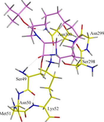
Additional calculations with this 4d molecule have thus been performed and the transition state structures located for the second and third models, using ib and iib conformations as starting geometry (143 atoms, 407 basis functions). It appears that conformation iib has an important steric hindrance with the third motif and that only conformation ib could accommodate the geometry of the active site model (Figure 10). The goodness of fit between this structure and the one obtained with Pen could partially explain the 52% of residual activity observed with 4d on R39.
Figure 10. Compound 4d in conformation ib in the second model. See legend figure 7.
One question remains about the lack of activity of 12d. This molecule can adopt two conformations of the phenoxy side-chain with the ib conformation of the bridged cycle differing only by 1.52 kcal. Both of them have been superimposed on the conformation ib of 4d (Figure 11). In one conformation, the phenoxy group has a steric hindrance with Tyr147 phenol; on the other, the phenoxy lies in too short contact with Leu349 and Arg351 side-chains. This geometric feature occurring with both conformations could be related to the biological results as 12d cannot accommodate the active site model geometry while 4d can do it.
Figure 11. Compounds 4d (black) and 12d (orange) in conformation ib.
Conclusion
In the bis-acylated family previously reported (X = Y = O, Scheme 1) cyclodimers were obtained when applying the RCM reaction as key-step for the macrocyclization. In the present work, this synthetic strategy was successful to obtain cyclomonomers with the bis-alkylated family (X = Y = H,H) due to the high conformational adaptability of the bis-alkylated precursors. 12- to 22-Membered 1,3-bridged -lactams for the Boc series and 12- to 16-membered bicyclic -lactams for the V side-chain series were synthesized. The reactivity of the 1,3-bridged -lactams in serine enzyme active site highly depends on the ligand conformation explaining why the compounds bearing a V side-chain do not fit with the geometry of the catalytic cavity. This feature has been analyzed using different models of the R39 active site including the three conserved motifs of this enzyme family. Within the synthesized bicycles, 4d is a good inhibitor of the R39 enzyme (as theoretically predicted), and some compounds also exhibit a significant activity against resistant PBP2a from MRSA. The activity could be related to the flexibility of the bicycles which can accommodate their conformation into the enzyme active site.
The activity of the 1,3-bridged macrocycles deriving from bis--alkenyl-3(S)-amino-azetidinone precursors suggests a way to design novel lactam antibiotics with a planar amide bond, and devoid of carboxylic group. Indeed, this acid function only plays a role by stabilizing the conformation of penicillin-like molecules by H-bonds with the residues of the third motif.
Experimental Section Synthesis
Experiments were performed under argon atmosphere in flame-dried glassware. All solvents, including anhydrous solvents, and reagents were purchased from Acros Organics, Alfa Aesar, Fluka, Sigma-Aldrich or VWR, and used without any further purification. TLC analyses were performed on aluminum plates coated with silica gel 60F254 (Merck) and visualized with a KMnO4 solution and UV (254 nm) detection, and
column chromatographies were performed on silica gel (40-63 or 63-200 m) purchased from Rocc. Melting points (mp) were determined on a Büchi B-540 apparatus calibrated with caffeine, vanillin, and phenacetin. [α]D was measured on Perkin-Elmer 241 MC polarimeter, at 20 °C. Concentrations are given in g/100 mL.
Nuclear magnetic resonance (1H and 13C) spectra were recorded at 300 MHz for proton and 75 MHz for
carbon (Bruker Avance 300) or 500 MHz for proton and 125 MHz for carbon (Bruker Avance 500). Chemical shifts are reported in parts per million relative to residual solvent peak from the deuterated solvent or relative to peak of TMS. NMR coupling constants (J) are reported in hertz. Infrared (IR) spectra were recorded using FTIR-8400S Shimadzu apparatus. Products were analyzed as thin films deposited on an Se-Zn crystal by evaporation from CH2Cl2 solutions. Low resolution mass spectra were obtained using a
ThermoFinnigan LCQ Quantum spectrometer or using a FinniganMat TSQ7000. High Resolution Mass Spectrometry (HRMS) analyses were performed at the University College London (UK). The Supporting Information of this paper includes structural data for compounds 2a-c, 2e-f, 3b-c, 3e-f, 4b-c, 4e-f, 10a-c, 11b-c, 12b-c, 13C NMR spectra of all new compounds.
tert-butyl N-(hept-6-en-1-yl)-N-[(3S)-1-(hept-6-en-1-yl)-2-oxoazetidin-3-yl]carbamate 2d: NaH (60% dispersion in oil, 279 mg, 6.98 mmol) was added to a stirred solution of 1 (0.65 g, 3.49 mmol) in dry DMF (41 mL) at 0 °C. Alkyl bromide (1.17 mL, 7.68 mmol) was added after 30 min and the mixture was then slowly warmed to r.t. and stirred overnight. The mixture was quenched with water (30 mL) at 0 °C and then the medium diluted with brine (50 mL). The resulting mixture was extracted with EtOAc (3x30 mL) and the organic layers were washed with brine (70 mL). After drying over MgSO4 and removing the solvent under
reduced pressure, the residue was purified by flash column chromatography (hexane/EtOAc 4:1) to provide 2d as a pale-yellow oil (515 mg, 39%). Rf=0.63 (hexane/EtOAc 3:2); [α]20D =+3.0 (c=1.7 in CH3OH); 1H
NMR (300 MHz, CDCl3, 20 °C): δ=5.84-5.71 (m, 2H), 5.01-4.90 (m, 4H + 0.4H, rotamer), 4.52 (br s, 0.6H,
rotamer), 3.46-3.43 (m, 1H), 3.26-3.08 (m, 5H), 2.07-2.00 (m, 4H), 1.57-1.24 ppm (m, 21H); 13C (125 MHz,
CDCl3, 20 °C): δ=167.1, 154.7, 138.9, 138.7, 114.7, 114.5, 80.9, 80.5, 63.2, 62.9, 47.9, 47.1, 46.7, 41.7,
33.8, 33.7, 29.6, 29.0, 28.7, 28.5, 27.5, 26.5, 26.4 ppm; IR: =2974-2856, 1753, 1693, 1639, 1456 cm-1; MS
(ESI): m/z (%): 779 (12) [2M + Na]+, 401 (100) [M + Na]+; HRMS (ESI): m/z calcd for
C22H38N2O3Na: 401.2780 [M + Na]+; found: 401.2778.
tert-butyl 16-oxo-1,14-diazabicyclo[13.1.1]heptadec-7-ene-14-carboxylate 3d: Grubbs catalyst (second generation) (56 mg, 66.09 mol) was added to a stirred solution of 2d (0.50 g, 1.32 mmol) in dry CH2Cl2
(264 mL) and the solution was stirred at reflux under argon for 4 h. Then a second addition of Grubbs catalyst (56 mg, 66.09 mol) was made and the mixture was additionally stirred at reflux for 20 h. Then the solvent was removed under reduced pressure and the crude product was purified thrice by column chromatography (hexane/EtOAc 4:1), to provide 3d as a pale-brown solid (416 mg, 90%). Rf=0.43
(hexane/EtOAc 3:2); m.p. 90.1-90.9 °C; [α]20D =-24.4 (c=1.2 in CHCl3); 1H NMR (500 MHz, CDCl3, 20 °C): δ=5.51 (br s, 0.6H, rotamer), 5.34-5.18 (m, 2H + 0.4H, rotamer), 3.73-3.67 (m, 1H), 3.42-3.35 (m, 1H), 3.25-2.93 (m, 3H), 2.81-2.76 (m, 1H), 2.13-1.94 (m, 4H), 1.69-1.17 ppm (m, 21H); 13C (125 MHz, CDCl 3, 20 °C): δ=167.6, 167.4, 155.1, 154.3, 131.9, 131.7, 130.4, 130.2, 80.6, 80.3, 62.5, 61.9, 46.9, 45.4, 45.1, 41.2, 41.0, 40.8, 32.2, 30.5, 28.8, 28.4, 28.1, 27.9, 27.2, 27.1, 26.9, 26.7, 26.2, 26.1, 25.1, 25.0, 24.8, 24.5 ppm; IR: =2924-2852, 1755, 1697, 1454 cm-1; MS (ESI): m/z (%): 373 (100) [M + Na]+, 317 (23) [M + Na
– C4H8]+, 295 (25) [M + H – C4H8]+; HRMS (ESI): m/z calcd for C20H34N2O3Na: 373.2467 [M + Na]+; found:
tert-butyl 16-oxo-1,14-diazabicyclo[13.1.1]heptadecane-14-carboxylate 4d: To a stirred solution of 3d (110 mg, 0.31 mmol) in methanol (10 mL) was added 10% Pd/C (10 mg). After being stirred under hydrogen atmosphere (P = 1 atm) for 3 h at room temperature, the mixture was filtered through a short pad of Celite and concentrated under reduced pressure. The residue was purified by column chromatography (hexane/EtOAc 4:1), to provide 4d as a white solid (108 mg, 98%). Rf=0.48 (hexane/EtOAc 3:2); m.p.
55.4-56.2 °C; [α]20D =-24.9 (c=1.0 in CHCl3); 1H NMR (500 MHz, CDCl3, 20 °C): δ=5.44 (br s, 0.6H, rotamer), 5.18 (br s, 0.4H, rotamer), 3.68 (br s, 1H), 3.43 (br s, 1H), 3.26-2.99 (m, 3H), 2.84-2.79 (m, 1H), 1.75-1.69 (m, 1H), 1.59-1.20 ppm (m, 28H); 13C (125 MHz, CDCl 3, 20 °C): δ=167.4, 167.1, 155.2, 155.1, 80.5, 62.6, 62.1, 48.0, 47.7, 45.1, 41.9, 29.1, 28.5, 28.2, 26.9, 26.8, 26.6, 26.5, 26.0, 25.4 ppm; IR: =2924-2854, 1757, 1697, 1458 cm-1; MS (ESI): m/z (%): 375 (100) [M + Na]+, 319 (10) [M + Na – C 4H8]+, 297 (26) [M + H –
C4H8]+; HRMS (ESI): m/z calcd for C20H36N2O3Na: 375.2624 [M + Na]+; found: 375.2621.
(2S)-3-hydroxy-2-(2-phenoxyacetamido)propanoic acid 6: Phenoxyacetyl chloride (6.57 mL, 47.58 mmol) was added to a solution of L-serine (5.00 g, 47.58 mmol) in saturated aqueous NaHCO3 (200 mL) and
MeCN (40 mL). The reaction mixture was stirred vigorously overnight at room temperature and the aqueous phase was extracted with diethyl ether (2 x 200 mL). The aqueous solution was acidified to pH 2-3 with HCl 36% and extracted with EtOAc (4 x 200 mL). The organic layer was dried over MgSO4 and
concentrated under reduced pressure. The crude product was solubilized in a minimum of EtOAc and hexane was added until the apparition of a precipitate. After 5-6h the suspension was filtered, washed with hexane and dried under reduced pressure to provide 6 as a white solid (8.08 g, 71%). m.p. 130.1-130.9 °C;
[α]D 20 =+30.4 (c=1.1 in (CH3)2CO); 1H NMR (300 MHz, (CD3)2CO, 20 °C): δ=7.62-7.60 (br d, J = 6.5 Hz, 1H), 7.35-7.30 (m, 2H), 7.04-6.98 (m, 3H), 4.65-4.60 (m, 1H), 4.55 (s, 2H), 4.06-4.00 (dd, J = 4.1, 11.1 Hz, 1H), 3.95-3.90 ppm (dd, J = 3.6, 11.1 Hz, 1H); 13C (75 MHz, (CD 3)2CO, 20 °C): δ=171.7, 168.6, 158.7, 130.5 (2C), 122.5, 115.7 (2C), 68.0, 62.7, 55.0 ppm; IR: =3393-3060, 2754-2469, 1731, 1643, 1539, 1495 cm-1; MS (ESI): m/z (%): 262 (100) [M + Na]+, 240 (38) [M + H]+, 222 (52) [M + H – H 2O]+; HRMS (CI):
m/z calcd for C11H14NO5: 240.08720 [M + H]+; found: 240.08649.
(2S)-N-(benzyloxy)-3-hydroxy-2-(2-phenoxyacetamido)propanamide 7: A solution of N,N-dicyclohexylcarbodiimide (DCC) (2.72 g, 13.17 mmol) in THF (10 mL) was added dropwise, at 0 °C, into a well-stirred solution of 6 (3.00 g, 12.54 mmol) and O-(phenylmethyl)hydroxylamine (1.46 mL, 12.54 mmol) in THF (120 mL). The reaction mixture was stirred for 1 h at 0 °C and overnight at room temperature. The obtained white precipitate (DCU) was separated by filtration and the resulting clear reaction mixture was concentrated under reduced pressure. After addition of Et2O (100 mL), the precipitate was filtered, washed
with Et2O and dried under reduced pressure to provide 7 as a white solid (3.76 g, 87%). Rf=0.30
(CH2Cl2/MeOH 95:5); m.p. 145.0-145.7 °C; [α]D 20 =-1.5 (c=1.0 in (CH3)2SO); 1H NMR (300 MHz, (CD3)2SO, 20 °C): δ=11.33 (br s, 1H), 8.01 (br d, J = 7.9 Hz, 1H), 7.39-7.28 (m, 7H), 6.99-6.95 (m, 3H), 5.08 (br s, 1H), 4.78 (s, 2H), 4.55 (s, 2H), 4.23-4.29 (m, 1H), 3.59 ppm (br d, J = 5.5 Hz, 2H); 13C (75 MHz, (CD3)2SO, 20 °C): δ=167.6, 166.7, 152.7, 135.8, 129.5 (2C), 128.9 (2C), 128.3 (3C), 121.2, 114.7 (2C), 76.9, 66.6, 61.4, 52.8 ppm; IR: =3313-2999, 1650, 1556, 1499 cm-1; MS (ESI): m/z (%): 367 (100) [M + Na]+, 345 (30) [M + H]+, 222 (12) [M + H – NH
2OBn]+; HRMS (CI): m/z calcd for C18H21N2O5: 345.14505
[M + H]+; found: 345.14649.
N-[(3S)-1-(benzyloxy)-2-oxoazetidin-3-yl]-2-phenoxyacetamide 8: A solution of PPh3 (2.09 g, 7.98 mmol)
in dry MeCN (35 mL) was added dropwise, at 0 °C and under Ar atmosphere, to a stirred solution of 7 (2.50 g, 7.26 mmol) in dry CH3CN (20 mL), containing CCl4 (0.77 mL, 7.98 mmol) and anhydrous triethylamine
(1.52 mL, 10.89 mmol). The resultant reaction mixture was stirred for 2 h at 0 °C and overnight at room temperature. After completion of the reaction, the obtained white precipitate (OPPh3) was separated by
filtration and the resulting clear reaction mixture concentrated under reduced pressure. The residue was dissolved with EtOAc (50 mL), washed with a saturated NH4Cl solution (2x40 mL) and brine (40 mL). After
drying over MgSO4 and removing the solvent under reduced pressure, the residue was purified by flash
(hexane/EtOAc 4:6); m.p. 138.8-139.4 °C; [α]D 20 =-6.2 (c=1.0 in (CH3)2CO); 1H NMR (300 MHz, (CD3)2CO, 20 °C): δ=8.19 (br d, J = 7.0 Hz, 1H), 7.49-7.28 (m, 7H), 7.01-6.96 (m, 3H), 4.98 (s, 2H), 4.91-4.85 (m, 1H), 4.53 (br d, J = 1.4 Hz, 2H), 3.72-3.69 (m, 1H), 3.53 ppm (dd, J = 2.4, 4.3 Hz, 1H); 13C (75 MHz, (CD3)2CO, 20 °C): δ 169.1, 163.4, 158.7, 136.5, 130.4 (2C), 130.0 (2C), 129.5, 129.3 (2C), 122.4, 115.6 (2C), 77.9, 67.9, 52.6, 52.4 ppm; IR: =3363-3193, 1776, 1677, 1598, 1531, 1495 cm-1; MS (ESI):
m/z (%): 365 (8) [M + K]+, 349 (100) [M + Na]+, 327 (10) [M + H]+; HRMS (ESI): m/z calcd for
C18H18N2O4Na: 349.1164 [M + Na]+; found: 349.1151.
N-[(3S)-2-oxoazetidin-3-yl]-2-phenoxyacetamide 9: Compound 8 (1.00 g, 3.06 mmol) dissolved in methanol (18 mL) and EtOAc (18 mL) was placed under H2 (1 atm) at rt in the presence of Raney-Ni (50%
in water) catalyst for 12 h. Then the mixture was filtered through a pad of Celite and concentrated under reduced pressure to provide 9 as a white solid (0.67 g, 100%), which was used without further purification. Rf=0.42 (EtOAc/MeOH 95:5); m.p. 155.7-156.6 °C; [α]D 20 =-19.1 (c=1.0 in CH3OH); 1H NMR (500 MHz, CD3OD, 20 °C): δ=7.32-7.28 (m, 2H), 7.01-6.97 (m, 3H), 5.04 (dd, J = 2.7, 5.4 Hz, 1H), 4.54 (br d, J = 1.2 Hz, 2H), 3.58-3.56 (m, 1H), 3.39 ppm (dd, J = 2.6, 5.5 Hz, 1H); 13C (125 MHz, CD 3OD, 20 °C): δ=171.5, 170.8, 159.2, 130.6 (2C), 122.9, 115.9 (2C), 68.1, 57.8, 44.1 ppm; IR: =3281-3020, 1776, 1703, 1664, 1628, 1556, 1498 cm-1; MS (ESI): m/z (%): 243 (100) [M + Na]+; HRMS (CI): m/z calcd for C
11H13N2O3:
221.09262 [M + H]+, found: 221.09253.
N-(hept-6-en-1-yl)-N-[(3S)-1-(hept-6-en-1-yl)-2-oxoazetidin-3-yl]-2-phenoxyacetamide 10d: Prepared by the same procedure as described for 2d. Yield: 30%; Rf=0.47 (hexane/EtOAc 1:1); [α]D
20 =+3.1 (c=1.0 in CHCl3); 1H NMR (500 MHz, C2D2Cl4, 20 °C): δ=7.33 (t, J = 7.7 Hz, 2H), 7.03 (t, J = 7.3 Hz, 1H), 6.93 (br d, J = 8.0 Hz, 2H), 5.84-5.75 (m, 2H), 5.12-4.95 (m, 4H + 0.3H, rotamer), 4.76-4.62 (m, 2H + 0.7H, rotamer), 3.53-3.10 (m, 6H), 2.07-2.03 (m, 4H), 1.71-1.52 (m, 4H), 1.44-1.25 ppm (m, 8H); 13C (125 MHz, C2D2Cl4, 90 °C): δ=167.9, 165.1, 157.9, 138.3, 129.4, 121.7, 114.8, 114.4, 67.6, 62.9, 46.8, 41.9, 33.2, 28.3, 28.2, 27.1, 26.3, 26.1 ppm; IR: =3003-2854, 1748, 1668, 1599, 1587, 1497 cm-1; MS (ESI): m/z (%): 435
(100) [M + Na]+, 413 (6) [M + H]+, 288 (10); HRMS (ESI): m/z calcd for C
25H36N2O3Na: 435.2624 [M + Na] +; found: 435.2625.
(1S)-14-(2-phenoxyacetyl)-1,14-diazabicyclo[13.1.1]heptadec-7-en-16-one 11d: Prepared by the same procedure as described for 3d. Yield: 67%; Rf=0.42 (hexane/EtOAc 3:7); m.p. 86.7-87.3 °C; [α]20D =-2.8
(c=1.9 in CHCl3); 1H NMR (500 MHz, C2D2Cl4, 20 °C): δ=7.34-7.30 (m, 2H), 7.02 (t, J = 7.3 Hz, 1H), 6.93-6.91 (m, 2H), 5.84-5.74 (m, 0.4H, rotamer), 5.37-5.16 (m, 2H + 0.6H, rotamer), 4.79-4.62 (m, 2H), 3.72-3.66 (m, 1H), 3.55-3.08 (m, 4H), 2.88-2.66 (m, 1H), 2.16-1.85 (m, 4H), 1.73-1.19 ppm (m, 12H); 13C (125 MHz, C2D2Cl4, 120 °C): δ=167.8, 165.5, 158.0, 131.8, 131.1, 130.3, 130.1, 129.4, 121.7, 115.0, 67.9, 62.3, 49.1, 47.1, 46.7, 45.2, 44.2, 41.8, 33.7, 31.7, 31.5, 31.3, 30.5, 28.4, 28.2, 28.0, 27.8, 27.4, 27.2, 27.1, 26.5, 26.1, 25.5, 25.2, 24.6 ppm; IR: =3003-2853, 1740, 1664, 1497 cm-1; MS (ESI): m/z (%): 407 (100)
[M + Na]+, 385 (89) [M + H]+, 357 (21) [M + H - CO]+; HRMS (ESI): m/z calcd for C
23H32N2O3Na:
407.2311 [M + Na]+; found: 407.2322.
(15S)-14-(2-phenoxyacetyl)-1,14-diazabicyclo[13.1.1]heptadecan-16-one 12d: Prepared by the same procedure as described for 4d. Yield: 93%; Rf=0.33 (hexane/EtOAc 1:1); m.p. 85.2-85.7 °C; [α]D
20 =-1.0 (c=1.7 in CHCl3); 1H NMR (500 MHz, C2D2Cl4, 120 °C): δ=7.40-7.36 (m, 2H), 7.10-7.03 (m, 3H), 5.46 (br s, 1H), 4.83-4.74 (m, 2H), 4.05 (br s, 0.4H, rotamer), 3.81-3.75 (m, 1H), 3.60-3.50 (m, 3H + 0.6H, rotamer), 3.38-3.36 (m, 1H), 2.93-2.89 (m, 1H), 2.03-1.47 ppm (m, 19H); 13C (125 MHz, C 2D2Cl4, 120 °C): δ=167.8, 165.3, 158.0, 129.3, 121.7, 115.0, 67.9, 62.2, 49.1, 47.7, 45.0, 42.1, 33.7, 28.8, 27.7, 27.0, 26.9, 26.8, 26.3, 26.2, 26.1, 25.6, 25.5, 24.6 ppm; IR: =3028-2856, 1740, 1662, 1437 cm-1; MS (ESI): m/z (%): 409 (100)
[M + Na]+, 387 (25) [M + H]+, 359 (7) [M + H - CO]+; HRMS (ESI): m/z calcd for C
23H34N2O3Na: 409.2467
Computational chemistry
All the calculations have been performed with the Gaussian 03 suite of programs.[17] The geometry is
optimized by analytical gradient energy minimization. The nature of the located extrema is defined by the inertia of the second derivative energy (Hessian matrix). For the minima, all the eigenvalues are positive; in the case of a first order saddle point as the TS structures, the first eigenvalue is negative and is associated to the imaginary frequency. In the studied models, the related eigenvector components are the geometric variables involved in the H transfer in the pseudo 8 membered ring.
All absolute energies of the precursors, unsaturated bicycles, saturated bicycles, and all the absolute energies of the transition state structures in the simple model, figure 5, computed for the unsaturated bicycles and saturated bicycles in the selected conformations can be found in supporting information.
Biochemical evaluation Assay with resistant PBPs
Purified PBP5f from Enterococcus faecium D63r and PBP2a from Meticillin-Resistant Staphylococcus aureus ATCC 43300 were used as target proteins to test inhibitory activity of synthesized -lactams. Each of the purified PBPs (2.5 µM) were first incubated with 1 mM potential inhibitor in 100 mM phosphate buffer, 0.01% Triton X-100,[18] pH 7, for 4 h at 30 °C. Then, 25 µM fluorescein-labeled ampicillin[19] was
added to detect the residual penicillin binding activity (RA). The samples were further incubated for 30 min at 37 °C in a total volume of 20 µL. Denaturation buffer was added (0.1 M Tris/HCl, pH 6.8, containing 25% glycerol, 2% SDS, 20% β-mercaptoethanol and 0.02% bromophenol blue) and the samples were heated to 100 °C for 1 min. The samples were then loaded onto a 10% SDS-acrylamide gel (10 x 7 cm) and electrophoresis was performed for 45 min at 180 V (12 mA). Detection and quantification of the RAs were done with Molecular Image FX equipment and Quantity One software (BioRad, Hercules, CA, USA). Three independent experiments were carried out for each inhibitor.
Assay with R39
All assays with R39 have been done in microtiter plates 96-wells (Brand, Wertheim, Germany). 20 mM of the tested compounds have been solved in DMF. Finally 7.5 µL of the solution have been used in the assay. The final concentration of the compounds in the assays was 100 µM. The final concentration of DMF in the assays was 0.25%. R39 (3.5 nM) was incubated in the presence of the potential inhibitors in 10 mM sodium phosphate buffer (pH 7.2) with 100 mM NaCl, 100 mM D-alanine, 0.01 mg/ml BSA and 0.01% Triton for 60 min at 25 °C. This preincubation was realized, in order to detect also slow binding inhibitors. After the preincubation the residual activity RA of R39 was determined by observing the hydrolysis of the thioester S2d substrate, in the presence of DTNB, catalyzed by the non-inhibited enzyme. The initial rate of hydrolysis of 1 mM S2d in the presence of 1 mM DTNB was determined by monitoring the increase of absorbance at 412 nm (DTNB: [] = 13600 M-1 s-1) using a microplate absorbance reader (Power Wave
X, Biotek Instruments, Winooski, U.S.A.). The rate of spontaneous hydrolysis of S2d in the presence of the inhibitors was also determined in absence of R39. All assays have been done three times. The determination of RA of R39 in absence of inhibitors has been done six times on each plate. In order to detect false positives which could be slow binding non-competitive promiscuous inhibitors, the assays have been done in the presence of 0.01% Triton-X-100.[20]
Acknowledgements
This work was supported by the Interuniversity Attraction Pole (IAP P6/19 PROFUSA), F.R.S.-FNRS, UCL and ULg (computational facilities). G. D. and J. M.-B. are senior research associates of the F.R.S.-FNRS (Belgium). Dr. Astrid Zervosen is acknowledged for the R39 testing and Dr. Ana Amoroso and Olivier Verlaine for the PBP2a and PBP5f testing. Ir. Raoul Rozenberg and Dr. Cécile Le Duff have contributed to the structural analysis.
[1] a) E. P. Abraham, E. Chain, Nature 1940, 146, 837; b) C. Walsh, Nature 2000, 406, 775-781; c) D. M. Livermore, J. Antimicrob. Chemother. 2009, 64 (suppl 1), i29-i36; d) G. L. French, Int. J. Antimicrob. Agents 2010, 36S3, S3-S7.
[2] C. Hubschwerlen in Comprehensive Medicinal Chemistry, 2nd ed. (Eds: J. B. Taylor, D. J. Triggle),
Elsevier, Oxford, UK, 2007, chapter 7.17, pp. 479–517.
[3] a) J. L. Strominger, Antibiotics 1967, 1, 705-713; b) R. B. Woodward in The Chemistry of Penicillin (Eds. H. T. Clarke, J. R. Johnson et R. Robinson), Princeton University Press, Princeton, New Jersey, 1949, pp. 440–454; c) B. Alcaide, C. Aragoncillo, P. Almendros in Comprehensive Heterocyclic
Chemistry III (Ed.: C. Stevens), Elsevier, Oxford, 2008, vol. 2, chapter 2.02, pp. 111–171; d) J.
Marchand-Brynaert, C. Brulé in Comprehensive Heterocyclic Chemistry III (Ed.: C. Stevens), Elsevier, Oxford, 2008, vol. 2, chapter 2.03, pp. 173–237.
[4] a) M. Bassetti, L. Nicolini, S. Esposito, E. Righi, C. Viscoli, Curr. Med. Chem. 2009, 16, 564-575; b) L. I. Llarrull, S. A. Testero, J. F. Fisher, S. Mobashery, Curr. Opin. Microbiol. 2010, 13, 551-557; c) M. Bassetti, F. Ginocchio, M. Mikulska, Critical Care 2011, 15, 215.
[5] a) A. Urbach, G. Dive, J. Marchand-Brynaert, Eur. J. Org. Chem. 2009, 11, 1757-1770; b) A. Urbach, G. Dive, B. Tinant, V. Duval, J. Marchand-Brynaert, Eur. J. Med. Chem. 2009, 44, 2071-2080.
[6] a) A. Sliwa, G. Dive, J.-L. Habib Jiwan, J. Marchand-Brynaert, Tetrahedron 2010, 66, 9519-9527 and references therein; b) A. Sliwa, J. Marchand-Brynaert, M. Luhmer, Magn. Reson. Chem. 2011, in press.
[7] a) E. Sauvage, R. Herman, S. Petrella, C. Duez, F. Bouillenne, J.M. Frère, P. Charlier, J. Biol. Chem. 2005, 280, 31249-31256; b) E. Sauvage, A. J. Powell, J. Heilemann, H. R. Josephine, P. Charlier, C. Davies, R. F. Pratt, J. Mol. Biol. 2008, 381, 383–393.
[8] D. Lim, N. C. Strynadka, Nat. Struct. Biol. 2002, 9, 870-876.
[9] E. Sauvage, F. Kerff, E. Fonzé , R. Herman, B. Schoot, J.-P. Marquette, Y. Taburet, D. Prevost, J. Dumas, G. Leonard, P. Stefanic, J. Coyette, P. Charlier, Cell. Mol. Life Sci. 2002, 59, 1223-1232. [10] J. R. Hwu, M. L. Jain, S.-C. Tsay, G. H. Hakimelahi, Tet. Lett. 1996, 37,2035-2038.
[11] G. A. Olah, S. C. Narang, B. G. B. Gupta, R. Malhotra, J. Org. Chem. 1979, 44, 1247-1251.
[12] M. J. Miller, P. G. Mattingly, M. A. Morrison, J. F. Kerwin Jr., J. Am. Chem. Soc. 1980, 102, 7026-7032.
[13] a) H. Tatewaki, S. Huzinaga, J. Comp. Chem. 1980, 1, 205-228; b) G. Dive, D. Dehareng, J. M. Ghuysen, Theor. Chim. Acta 1993, 85, 409-421.
[14] G. Dive, D. Dehareng, Int. J. Quant. Chem. 1999, 73, 161-174. [15] N-benzoyl-D-alanyl-thioglycolate (S2d)
a) R. Schwyzer, C. Hurlimann, C. Coenzyle, Helv. Chim. Acta 1954, 37, 155–166;b) M. Adam, C. Damblon, M. Jamin, W. Zorzi, V. Dusart, M. Galleni, A. el Kharroubi, G. Piras, B. G. Spratt, W. Keck, J. Coyette, J.-M. Ghuysen, M. Nguyen-Disteche, J.-M. Frère, Biochem. J. 1991, 279, 601-604. [16] H. S. Sader, A. C. Gales, Drugs 2001, 61, 553-564.
[17] Gaussian 03, Revision D.02, M. J. Frisch, G. W. Trucks, H. B. Schlegel, G. E. Scuseria, M. A. Robb, J. R. Cheeseman, J. A. Montgomery, Jr., T. Vreven, K. N. Kudin, J. C. Burant, J. M. Millam, S. S. Iyengar, J. Tomasi, V. Barone, B. Mennucci, M. Cossi, G. Scalmani, N. Rega, G. A. Petersson, H. Nakatsuji, M. Hada, M. Ehara, K. Toyota, R. Fukuda, J. Hasegawa, M. Ishida, T. Nakajima, Y. Honda, O. Kitao, H. Nakai, M. Klene, X. Li, J. E. Knox, H. P. Hratchian, J. B. Cross, C. Adamo, J. Jaramillo, R. Gomperts, R. E. Stratmann, O. Yazyev, A. J. Austin, R. Cammi, C. Pomelli, J. W. Ochterski, P. Y. Ayala, K. Morokuma, G. A. Voth, P. Salvador, J. J. Dannenberg, V. G. Zakrzewski, S. Dapprich, A. D. Daniels, M. C. Strain, O. Farkas, D. K. Malick, A. D. Rabuck, K. Raghavachari, J. B. Foresman, J. V. Ortiz, Q. Cui, A. G. Baboul, S. Clifford, J. Cioslowski, B. B. Stefanov, G. Liu, A. Liashenko, P. Piskorz, I. Komaromi, R. L. Martin, D. J. Fox, T. Keith, M. A. Al-Laham, C. Y. Peng, A. Nanayakkara, M. Challacombe, P. M. W. Gill, B. Johnson, W. Chen, M. W. Wong, C. Gonzalez, and J. A. Pople, Gaussian, Inc., Wallingford CT, 2004.
[18] B. Y. Feng, B. K. Shoichet, Nat. Protoc. 2006, 1, 550-553.
[19] B. Lakaye, C. Damblon, M. Jamin, M. Galleni, S. Lepage, B. Joris, J. Marchand-Brynaert, C. Frydrych, J.-M. Frère, Biochem. J. 1994, 300, 141–145.
[20] A. Zervosen, W.-P. Lu, Z. Chen, R. E. White, T. P. Jr. Demuth, J.-M. Frère, Antimicrob. Agents Chemother. 2004, 48, 961-969.


![Table 3. Evaluation of bis-alkylated azetidinones against R39 D,D-peptidase, PBP5f and PBP2a Entr y compound n R39RA [%] PBP5fRA[%] PBP2aRA[%] 1 2a 1 101 4 81 96 2 2b 3 >100 94 71 3 2c 4 97 11 69 68 4 2d 5 80 4 80 59 5 2e 6 97 2 100 95 6 2f 8 10](https://thumb-eu.123doks.com/thumbv2/123doknet/5430947.127262/11.892.253.645.716.1156/table-evaluation-alkylated-azetidinones-peptidase-pbp-entr-compound.webp)