I
NCREASED
E
XPRESSION OF
“P
ERIPHERAL
-T
YPE
”
B
ENZODIAZEPINE
R
ECEPTORS IN
H
UMAN
T
EMPORAL
L
OBE
E
PILEPSY
:
I
MPLICATIONS FOR
PET
I
MAGING OF
H
IPPOCAMPAL
S
CLEROSIS
Anny Sauvageau,
1Paul Desjardins,
1Violina Lozeva,
1Christopher
Rose,
1Alan S. Hazell,
1Alain Bouthillier,
1;2and Roger F.
Butterworth
1;3Neuroscience Research Unit, CHUM (Campus Saint-Luc), Montreal, Quebec, Canada. Department of Surgery, CHUM (Campus Notre-Dame), Montreal, Quebec, Canada. To whom correspondence should be addressed at Neuroscience Research Unit, CHUM (Campus Saint-Luc), 1058 St-Denis Street, Montreal, Quebec, Canada H2X 3J4. E-mail: butterwr@medclin.umontreal.ca
ABSTRACT
Increased binding sites for “peripheral-type” benzodiazepine receptor (PTBR) ligands have been described in a wide range of neurological disorders including both human and exper-imental epilepsy. This study was undertaken to assess PTBR expression in relation to the presence of hippocampal sclerosis in human temporal lobe epilepsy (TLE). For this pur-pose, hippocampal CA1 subfields were dissected from surgical samples from patients with therapy-refractive TLE with (n D 5) or without (n D 2) hippocampal sclerosis and from age-matched nonepileptic postmortem controls (n D 5). PTBR expression was assessed by immunohistochemistry and reverse-transcription polymerase chain reaction. Receptor sites were evaluated using an in vitro binding assay and the selective PTBR ligand [3H]PK11195. Epileptic patients with hippocampal sclerosis showed increases in
PTBR binding sites, im-munoreactivity, and mRNA expression compared to both nonsclerotic TLE patients and postmortem nonepileptic controls. Induction of PTBR expression and binding sites were directly correlated with the presence of hippocampal sclerosis and the accompanying reac-tive gliosis.
Key words:
Temporal lobe epilepsy; hippocampus; peripheral-type benzodiazepine receptors; position emission tomography; astrocytes.INTRODUCTION
The “peripheral-type” benzodiazepine receptor (PTBR) is a multimeric complex found mainly on the outer mitochondrial membrane of astrocytes (Anholt et al., 1986; Basile and Skolnick, 1986). PTBR may also be colocalized with activated microglia in injured brain tissue (Kuhlmann and Guilarte, 2000). PTBR is composed of three subunits, an 18 KDa iso-quinoline carboxamide-binding protein (IBP), a 34 KDa voltage-dependent anion channel, and a 30 KDa adenine nucleotide carrier. PTBR has been implicated in various
functions, such as steroidogenesis, respiration, cell growth, and differentiation, and in response to stress. PTBR expression is increased following brain injury and this increase in PTBR has been used as an indication of neuronal damage or loss in several
neurodegenerative disorders, such as Alzheimer’s disease (Diorio et al., 1991), Huntington’s disease (Messmer and Reynolds, 1998), Wernicke’s encephalopathy (Desjardins et al., 1999), multiple sclerosis (Banati et al., 2000), and stroke-induced brain injury (Raghavendra Rao et al., 2000; Stephenson et al., 1995). Increased PTBR
expression in these conditions has been associated with both microglial activation and reactive astrogliosis.
An association between alterations of PTBR and epilepsy has been proposed for some time. Epileptic patients manifest increased binding sites for PTBR ligands in blood mononuclear cells (Caldiroli et al., 1997; Ferrarese et al., 1996; Larkin et al., 1993) as well as in brain (Beaurain et al., 1994; Johnson et al., 1992). In addition, both EL (epileptic mice; Nakamoto et al., 1996), and kainate-induced epileptic rats (Benavides et al., 1987) show increased brain densities of PTBR sites. It was suggested that these increases in PTBR sites were either the consequence of the epileptogenic process per se or resulted from the gliosis accompanying neuronal cell loss in hippocampus of epileptic animals. The aim of this study was to measure PTBR expression in surgically excised samples of hippocampus from patients with temporal lobe epilepsy (TLE) with or without
hippocampal sclerosis. PTBR binding sites were assessed by an in vitro binding technique utilizing the highly selective ligand [3H]PK11195. Increased PTBR expression was
visualized using immunohistochemistry and PTBR gene expression was measured by semiquantitative reverse-transcription polymerase chain reaction (RT-PCR).
MATERIALS
AND
METHODS
Patients
Patients with intractable epilepsy (n = 5) suffered from complex partial seizures and the epileptic focus was localized to the temporal lobe in all patients as revealed by physical examination, magnetic resonance imaging, and long-term video monitoring. The hippocampus was removed surgically from these patients in order to achieve seizure control. Informed consent was obtained for all procedures. Hippocampal samples were rapidly frozen for molecular studies or immersion-fixed in phosphate-buffered saline (PBS) for neuropathological evaluation and subsequent correlative analysis. Epileptic patients with nonsclerotic hippocampus (n = 2), as well as postmortem hippocampus from neuropathologically con-firmed normal patients ( n = 5), were used as nonepileptic controls. CA1 region was microdissected from frozen samples according to the atlas of Paxinos and Watson (1986) and frozen at °80°C until RNA extraction.
[
3H]PK11195 Binding Assay
Membrane suspensions were prepared as described previously (Rao and Butterworth, 1997) with slight modifications. Briefly, each individual sample was homogenized in 100
volumes of ice-cold 50 mM Tris-HCl buffer (pH 7.5), with 5 up-and-down strokes of the glass-Teflon pestle. The homogenates were centrifuged at 40,000 g for 20 min and the pellet was resuspended in the initial volume of buffer and stored as aliquots at °80°C. Membrane protein concentrations were determined by the methods of Lowry et al. (1951) using bovine serum albumin (Sigma) as standard. Experiments were performed in duplicate, membranes from postmortem controls and epileptic patients being used in parallel sets. To determine Bmax and Kd values, saturation binding experiments were
performed. [3H]PK11195 was used at eight concentrations be-tween 0.1 and 7 nM. To examine the possible changes in receptor density, samples were incubated with 2 nM of [3H]PK11195. Aliquots of membrane suspension (400 L, 0.3– 0.6 mg/mL of protein) were incubated for 60 min at 4°C with [3H]PK11195 (specific activity 86 Ci/mmol) in a final volume of 500 L. Nonspecific binding was defined in the presence of 1 M unlabeled PK11195 (RBI). The incubation was stopped by dilution with ice-cold buffer, the samples were filtered under vacuum through Whatman GF/B filters using a Millipore harvester, and the filters were washed twice with 4 ml of ice-cold buffer. Filter-bound activity was extracted into 4.5 ml of Ultima-Gold (Packard), and then determined by liquid
scintillation counting at 40% efficiency. Saturation binding data were analyzed by nonlinear curve fitting, using the program GraphPad Prism (GraphPad Software, Inc., San Diego, CA). With this program, values for Kd and Bmax are obtained by fitting curves to the
data with the use of the equation Specific Binding D Bmax £ L=Kd C L , where Bmax is the
maximum specific binding, L is the concentration of [3H]PK11195, and Kd is the
dissociation constant.
Immunohistochemistry
Frozen sections (6 M) were fixed with methanol for 2 min at °20±C. The cell membranes were then permeabilized in PBS containing 1% Triton X-100 for 30 min at room
temperature. The sections were rinsed with PBS and incubated with 0.3% H2O2 for 5 min to inactivate endogenous peroxidase. Nonspecific sites were blocked in PBS containing 0.1% Triton X-100 (PBST) and 1.5% heat-inactivated goat serum for 30 min. Sections were incubated overnight at 4±C, with a mouse monoclonal antibody (1 g/mL) directed against human IBP (Sanofi Recherche, Montpellier, France) or HLA-DR, a human class II antigen of the major histocompatibility complex (PharMingen, NJ). After washing with PBST, sections were incubated with biotinylated goat antibody against mouse IgGs (Vector laboratory, CA) at a 1:200 dilution for 30 min at room temperature. Sections were washed with PBST and incubated with an avidin–biotin-peroxidase complex (Vectastain, Vector laboratory, CA) at a 1:500 dilution for 30 min. After washing with PBST, bound immunoglobulins were detected by incubation with 0.1 M Tris-HCl (pH 7.4) containing 0.5 mg/mL 3,3-diaminobenzidine–HCl and 0.01% hydrogen peroxide.
RNA Extraction
Total RNA was extracted using TRI Reagent (MRC, Inc., Ohio) according to the
RNase-free DNase I per 50 mg of total RNA at 37°C for 1 h. Purified RNA was then extracted with phenol, precipitated with ethanol, and resuspended in
diethylpyrocarbonate-treated water. RNA samples were kept at °70°C until use.
RT-PCR Analysis
Actin was used as an internal standard to monitor loading variations. Total RNA (0.5 mg) was mixed with 10 mM Tris-HCl (pH 8.3), 1.0 mM MgCl2, 50 mM KCl, 0.01% (w/v) bovine serum albumin, 100 mM dNTPs, primers at 1 mM each, AMV reverse transcriptase (80 U/mL), Taq DNA polymerase (20 U/mL), and 50 mCi/mL [32P]dCTP (3000 Ci/mmol), for
a total reaction volume of 50 mL. The reactions were initially car-ried out at 50°C for 15 min, followed by PCR at 95°C for 30 s, at 62°C for 45 s, and at 72°C for 1 min.
Amplification efficiency conditions were determined after a kinetic study, to ensure all experiments were performed within the exponential phase of amplification where PCR product remains proportional to initial template concentration (data not shown). Actin and PTBR (IBP subunit) were amplified for 18 and 33 cycles, respectively. After
amplification, the samples were electrophoresed onto 9% polyacrylamide gels, dried, and autoradiographed at °70°C with an intensifying screen. Each band was excised and Cerenkov radiation was quantitated using a b-counter. Oligonucleotide primers were de-signed using the PRIME program (Genetic Computer Group, Wisconsin) and synthesized by the Sheldon Biotechnology Center (McGill University, Quebec) based on the following GeneBank accession numbers: X00351 (b-Actin; Ponte et al., 1984) and L21951-L21954 (PTBR; Lin et al., 1993). The forward and reverse primer sequences were as follows: GACCTGACTGACTACCTCAT and AGACAGCACTGTGTTGGCGT (b-actin, 350 bp); and GCCCTTCCCGGAGCGTGCC and CGGCGTACCAGCGGAGAC (PTBR, 164 bp). The specificity of the oligonucleotide primers was verified using the program BLASTN (National Center for Biotechnology Information, Bethesda, MD).
Statistical Analysis
Data are expressed as means ±SEM. A probability of less than 5% ( p < 0:05) was considered to indicate statistical significance. All statistical analyses were performed using the computer software GraphPad Prism. Statistical significance of differences between the groups was determined with the unpaired, two-tailed Student’s t test.
RESULTS
Saturation binding curves for the binding of the PTBR ligand [3H]PK11195 to crude
synaptic membrane preparations from controls and from TLE patients obtained at sites was observed in TLE samples. Bmax and Kd values for TLE patients were 694 ± 61:7
fmol/mg protein and 2:15 ± 0:5 nM, respectively. In comparison, Bmax and Kd values for
controls were 282 ± 25:0 fmol/mg protein and 0:94 ± 0:29 nM, respectively.
Hematoxylin-eosin staining of sections from TLE patients showed, in five of seven cases, an almost complete loss of neurons from the CA1 subfield (Fig. 2). In all five cases PTBR
immunolabeling studies revealed increased immunoreactivity confined to astrocytes in these sections (Fig. 2, panel d).
Figure 1. Increased density of binding sites for the PTBR ligand [3 H]PK11195 in TLE. (A) Non-linear regression curve. (B) The corresponding Rosenthal–Scatchard plot of
3[H]PK11195 binding to the CA1 subfield of hippocampus in postmortem controls (o) and epileptic patients with hippocampal sclerosis ( ).
Figure 2. Loss of CA1 hippocampal neurons results in astrocytic proliferation and increased PTBR immunoreactivity. Panel a: HCE staining of CA1 subfield from a normal control; Panel b: H CE staining of CA1 subfield from a patient with TLE and
hipppocampal sclerosis. Note the neuronal cell loss throughout this field. Panels c and d: PTBR immunostaining. Note the lack of staining in control (Panel c) and the intense immunostaining in TLE patient (Panel d).
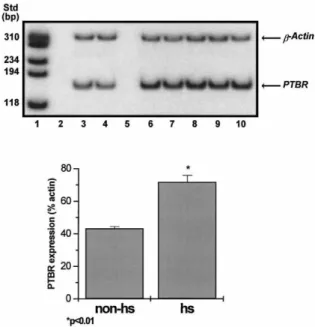
Figure 3. Upper panel: Expression of the PTBR mRNA in TLE patients. Total RNA was extracted from the hippocampal CA1 subfield of patients with (lanes 6–10) or without (lanes 3–4) hippocampal sclerosis. β-Actin (351 bp) and PTBR (164 bp) were amplified by RT-PCR for 18 and 33 cycles, respectively. AMV reverse transcriptase was omitted (as a negative control) in lanes 2 and 5. Lower panel: PTBR expression normalized to percentage of actin expression in patients with TLE with hippocampal sclerosis (hs) versus patients without hippocampal sclerosis (non-hs). PTBR was significantly increased in the hs group compared to the non-hs group ( p < 0:01 by Student’s t test).
At the mRNA level, TLE patients with hippocampal sclerosis showed significantly increased (by 66%, p < 0:01) PTBR expression compared to two patients with TLE who did not manifest hippocampal sclerosis (Fig. 3).
There was no evidence for the appearance of activated microglia/macrophages in the TLE patient samples used in this study as visualized using HLA-DR immunoreactivity (data not shown).
DISCUSSION
Results of the present study reveal significant increases in densities of binding sites for the PTBR ligand [3H]PK11195 in dissected hippocampal CA1 subfields from five patients
with TLE. In all cases, increased PTBR sites were associated with severe neuronal loss (hippocampal sclerosis) and reactive gliosis. These findings confirm and extend those of previous studies which observed increased PTBR sites in blood mononuclear cells (Caldiroli et al., 1997; Ferrarese et al., 1996; Laikin et al., 1993) and brain (Beaurain et al.,
1994; Johnson et al., 1992). Novel findings in the present study are those of increased PTBR sites in material from TLE patients with hippocampal sclerosis compared to similar material from patients with no pathological evidence of hippocampal sclerosis.
Immunohistochemical studies revealed increased PTBR immunolabeling in CA1 subfield of TLE patients with hippocampal sclerosis. Increased immunolabeling was particularly localized on astrocytic elements suggesting that the increased PTBR signal was the consequence of astrogliosis which accompanied neuronal cell loss in TLE. Similar findings of increased PTBR sites coincident with astrocytic proliferation in the presence of
neuronal loss were previously reported in Alzheimer’s disease (Diorio et al., 1991) and Wernicke’s encephalopathy (Desjardins et al., 1999; Leong et al., 1996). On the other hand, other disorders, such as multiple sclerosis (Banati et al., 2000) and stroke-induced brain injury (Stephenson et al., 1995), have been de-scribed in which increased PTBR sites coincided with the appearance of activated microglia rather than astrocytes. As previously reported by others (Beaurain et al., 1994), there was no evidence for a role of activated microglia activation in the samples used in the present study, suggesting that the hippocampal sclerosis in these patients was relatively longstanding.
Although unlikely (given the relatively low levels of expression of PTBR in nonscle-rotic TLE patients observed in the present study), there is conceivably a role for PTBR activation in the pathogenesis of the epileptic process per se. PTBR mediates the trans-port of cholesterol across the mitochondrial membrane and subsequently plays a role in the synthesis of the so-called neurosteroids, some of which have agonistic properties at GABA-A and NMDA receptors (Compagnone and Mellon, 2000). Activation of PTBR could therefore result in an imbalance between excitatory and inhibitory systems in the CNS (Majewska et al., 1987; Wu et al., 1991). Alternatively, increased PTBR in TLE may represent an adaptive phenomenon in hippocampal fields aimed at increasing neuroinhi-bition via the synthesis of epalons, a class of neurosteroids with anticonvulsant
properties shown previously to allosterically potentiate the effect of GABA on chloride conductance by the GABA-A receptor complex (Gee et al., 1995).
Findings in the present study of a correlation between the increased expression of PTBR sites in vulnerable CA1 subfields of TLE patients with the presence of hippocampal sclerosis suggest that the Positron Emission Tomography (PET) ligand [11C]PK11195 may
be useful in imaging the degree of hippocampal damage in TLE patients similar to that previously proposed for imaging of the extent of brain lesions in Wernicke’s
encephalopathy (Leong et al., 1996) and multiple sclerosis (Banati et al., 2000). Other PET ligands such as [11C]flumazenil ([ 11C]FMZ) have been used extensively to
evaluate the extent of neuronal loss in patients with refractory TLE. [11C]FMZ binds to the
GABA-related central benzodiazepine receptor and can be used in vivo to detect remaining neurons in sclerotic hippocampi. In many cases, however, [11C]FMZ showed
poor spatial correlation with neuronal cell loss and therefore has not been proven consistently helpful in localizing the epileptic foci (Koepp et al., 2000). Autoradiographic studies in experimental model of Wernicke’s encephalopathy revealed that increases in [3H]PK11195 binding sites paralleled the topographic distribution of neuronal cell loss
sites (Leong et al., 1996), suggesting that [11C]PK11195 represents a more reliable PET
ligand to evaluate neuronal cell loss. Further studies are clearly warranted in order to evaluate this possibility.
ACKNOWLEDGMENTS
The studies described were funded by the Savoy Foundation. P. Desjardins is the recipient of a fellowship from Claude Bertrand Foundation (University of Montreal). PTBR antibody (IBP subunit) was provided by Sanofi Recherche (Montpellier, France).
REFERENCES
Anholt, R.R.H., Pederson, P.L., DeSouza, E.B., and Snyder, S.H. (1986). The peripheral-type benzodiazepine receptor: Localization to the mitochondrial outer membrane. J. Biol. Chem. 261:576–583.
Banati, R.B., Newcombe, J., Gunn, R.N., Cagnin, A., Turkheimer, F., Heppner, F., Price, G., Wegner, F., Giovannonni, G., Miller, D.H., Perkin, G.D., Smith, T., Hewson, A.K., Bydder, G., Kreutzber, G.W., Jones, T., Cuzner, M.L., and Myers, R. (2000). The peripheral-type benzodiazepine binding site in the brain in multiple sclerosis: Quantitative in vivo imaging of microglia as a measure of disease activity.
Brain 123:2321–2337.
Basile, A.S. and Skolnick, P. (1986). Subcellular localization of “peripheral-type” binding sites for benzodiazepines in rat brain. J. Neurochem. 46:305–308.
Beaurain, J., Clemenceau, S., Duyckaerts, C., Benavides, J., Beaulac, M., Hauw, J.J., and Philippon, J. (1994). Etude morphom´etrique et radio-autographique de la perte neuronale et de la gliose dans la sclerose de l’hippocampe associ´ee a` l’´epilepsie du lobe temporal. Chirurgie 120:486–493. Benavides, J., Fage, D., Carter, C., and Scatton, B. (1987). “Peripheral type” benzodiazepine binding sites are a sensitive indirect index of neuronal damage. Brain Res. 421:167–172.
Caldiroli, E., De Ponti, F., Cosentino, F., Marino, F., Fietta, A.M., Taddei, M., Tartara, A., Zibetti, A., Mazzone, A., Lecchini, S., and Frigo, G.M. (1997). Carbamazepine affects neurophil function through an action on peripheral benzodiazepine receptors. Immunopharmacol. Immunotoxicol. 19:367–382. Compagnone, N.A. and Mellon, S.H. (2000). Neurosteroids: Biosynthesis and function of these novel neuromod-ulators. Front. Neuroendocrinol. 21:1–56.
Desjardins, P., Todd, K.G., Hazell, H.A., and Butterworth, R.F. (1999). Increased “peripheral-type” benzodiazepine receptor sites and mRNA in thalamus of thiamine-deficient rats. Neurochem. Int. 35:363–369.
Diorio, D., Welner, S.A., Butterworth, R.F., Meaney, M.J., and Suranyi-Cadotte, B.E. (1991). Peripheral benzodi-azepine binding sites in Alzheimer’s disease frontal and temporal cortex. Neurobiol. Aging 12:255–258.
Ferrarese, C., Perego, M., Marzorati, C., Bianchi, G., Frigo, M., Pecora, N., Riva, R., Moretti, G., and Frattola, L. (1996). Modifications of diazepam binding inhibitor and peripheral benzodiazepine receptors in the lymphocytes of epileptic patients. Ital. J. Neurol. Sci. 17:141–145.
Gee, K.W., McCauley, L.D., and Lan, N.C. (1995). A putative receptor for neurosteroids on the GABAA receptor complex: The pharmacological properties and therapeutic potential of epalons. Crit. Rev.
Neurobiol. 9:207–227.
Johnson, E.W., de Lanerolle, N.C., Kim, J.H., Sundaresan, S., Spencer, D.D., Mattson, R.H., Zoghbi, S.S., Baldwin, R.M., Hoffer, P.B., and Seibyl, J.P. (1992). “Central” and “peripheral” benzodiazepine receptors: Opposite changes in human epileptogenic tissue. Neurology 42:811–815. Koepp, M.J., Hammers, A., Labbe, C., Woerman, F.G., Brooks, D.J., and Duncan, J.S. (2000). 11C-Flumazenil PET in patients with refractory temporal lobe epilepsy and normal MRI. Neurology 54:332–339.
Kuhlmann, A.C. and Guilarte, T.R. (2000). Cellular and subcellular localization of peripheral benzodiazepine receptors after trimethyltin neurotoxicity. J. Neurochem. 74:1694–1704. Larkin, J.G., McKee, P.J., Thompson, G.G., and Brodie, M.J. (1993). Peripheral benzodiazepine receptors in platelets of epileptic patients. Bri. J. Clin. Pharmacol. 36:71–74.
Leong, D.K. and Butterworth, R.F. (1996). Neuronal cell death in Wernicke’s encephalopathy: Pathophysiologic mechanism and implications for PET imaging. Metab. Brain Dis. 11:71–79. Lin, D., Chang, Y.J., Strauss, J.F., III, and Miller, W.L. (1993). The human peripheral benzodiazepine receptor gene: Cloning and characterization of alternative splicing in normal tissues and in a patient with congenital lipoid adrenal hyperplasia. Genomics 18:643–650.
Lowry, O.H., Rosebrough, N.J., Farr, A.L., and Randall, R.J. (1951). Protein measurement with Folin phenol reagent. J. Biol. Chem. 193:265–275.
Majewska, M.D. and Schwartz, R.D. (1987). Pregnenolonesulfate: An endogenous antagonist of the -aminobutyric acid receptor complex in brain? Brain Res. 404:355–360.
Messmer, K. and Reynolds, G.P. (1998). Increased peripheral benzodiazepine binding sites in the brain of patients with Huntington’s disease. Neurosci. Lett. 241:53–56.
Nakamoto, Y., Watabe, S., Shiotani, T., and Yoshii, M. (1996). Peripheral-type benzodiazepine receptors in association with epileptic seizures in EL mice. Brain Res. 717:91–98.
Paxinos, G. and Watson, C. (1986). The Rat Brain in Stereotaxic Coordinates, Academic Press, New york. Ponte, P., Ng, S.Y., Engel, J., Gunning, P., and Kedes, L. (1984). Evolutionary conservation in the untranslatedregions of actin mRNAs: DNA sequence of a human beta-actin cDNA. Nucleic Acids Res. 12:1687–1696.
Raghavendra Rao, V.L., Dogan, A., Bowen, K.K., and Dempsey, R.J. (2000). Traumatic brain injury leads to increased expression of peripheral-type benzodiazepine receptors, neuronal death, and activation of astrocytes and microglia in rat thalamus. Exp. Neurol. 161:102–114.
Rao, V.L. and Butterworth, R.F. (1997). Characterization of binding sites for the omega3 receptor ligands [3 H]PK11195 and [3 H]RO5-4864 in human brain. Eur. J. Pharmacol. 340:89–99.
Stephenson, D.T., Schober, D.A., Smaltig, E.B., Mincy, E., Gehlert, D.R., and Clemens, J.A. (1995). Peripheral benzodiazepine receptors are colocalized with activated microglia following transient global forebrain is-chemia in the rat. J. Neurosci. 15:5263–5274.
Wu, F.-S., Gibbs, T.T., and Farb, D.H. (1991). Pregnenolone sulfate: A positive allosteric modulator at the N-Methyl-D-aspartate receptor. Mol. Pharmacol. 40:333–336.
![Figure 1. Increased density of binding sites for the PTBR ligand [ 3 H]PK11195 in TLE](https://thumb-eu.123doks.com/thumbv2/123doknet/11312024.282108/5.745.132.610.144.384/figure-increased-density-binding-sites-ptbr-ligand-tle.webp)