ii
Université de Montréal
Nanovecteurs pour cibler Pseudomonas aeruginosa dans la Fibrose Kystique
par
Juliana Campos Del’ Orto
Sciences pharmaceutiques Faculté de pharmacie
Mémoire présenté à la Faculté des études supérieures en vue de l’obtention du grade de Maitre en sciences (M.Sc.) en sciences pharmaceutiques option technologie pharmaceutique
Juin 2014
iii
Université de Montréal
Faculté des Études Supérieures et Postdoctorales
Ce mémoire intilulé:
Nanovecteurs pour cibler Pseudomonas aeruginosa dans la Fibrose Kystique
Présenté par: Juliana Campos Del’ Orto
A été évalué par les membres du jury :
Patrice Hildgen, directeur de recherche Jeanne Leblond- Chain, co-directrice de recherche
Valérie Gaëlle Roullin, présidente- rapporteur Valéry Waters, examinatrice externe
iv
RÉSUMÉ
La production excessive de mucus visqueaux dans les poumons des patients atteints de la fibrose kystique (FK) gêne la diffusion des médicaments et entraîne des infections bactériennes. En effet, l’infection pulmonaire par Pseudomonas aeruginosa (PA) est la principale cause de mortalité. Les travaux effectués dans cette thèse avaient pour but de développer des nouvelles formulations de nanoparticules (NP) et de liposomes (LP) chargées avec des antibiotiques pour erradiquer le PA chez les patients atteints de KF. Tout d’abord, les polymères PEG-g-PLA et PLA-OH ont été synthétisés et caractérisés. Ensuite, l'efficacité d'encapsulation (EE) de la tobramycine, du sulfate de colistine et de la lévofloxacine (lévo) a été testée dans des NP de PEG-g-PLA et / ou PLA-OH. Les premiers essais d'optimisation ont montré que les NP chargées avec la lévo présentaient une augmentation de l’EE. La lévo reste alors le médicament de choix. Cependant, la meilleure charge de médicament obtenue était de 0,02% m/m. Pour cette raison, nous avons décidé d'évaluer l'encapsulation de la lévo dans les LP. En fait, des LP chargés de lévo ont présenté une EE d’environ 8% m/m. De plus, la taille et la charge de ces LP étaient appropriées pour la pénétration du vecteur dans le mucus. Le test de biofilm n'est pas reproductible, mais le test standard a montré que la souche mucoïde de PA était susceptible à la lévo. Ainsi, nous avons comparé les activités des LP fraîchement préparées (vides et chargés ) et de la lévo libre sous la forme planctonique de PA. Les résultats ont montré que des LP vides ne gênent pas la croissance bactérienne. Pour la souche mucoïde (Susceptible à la lévo) les LP chargés et le médicament libre ont présenté la même concentration minimale inhibitrice (CMI). Toutefois, les souches non mucoïdes (résistant à la lévo) ont présenté une CMI deux fois plus faible que celle pour le médicament libre. Finalement, les LP se sont avérés plus appropriés pour encapsuler des médicaments hydrophiles que les NP de PEG-g-PLA. En outre, les LP semblent améliorer le traitement contre la souche résistante de PA. Toutefois, des études complémentaires doivent être effectuées afin d'assurer la capacité des liposomes èa traiter la fibrose kystique.
Mots-clés : antibiotique , nanoparticules polymèriques, PEG-g-PLA, liposomes, Pseudomonas aeruginosa, biofilm, Fibrose Kystique.
v
ABSTRACT
The increased production of viscoid mucus in the lungs of cystic fibrosis (CF) patients hinders the diffusion of therapeutics and favor bacterial infections. Indeed, lung infection by Pseudomonas aeruginosa (PA) relates with increased mortality in CF patients. This work is aimed at developing new antibiotic loaded nanoparticles and liposomes formulations to eradicate PA in CF. Firstly, PEG-g-PLA and PLA-OH polymers were synthesized and characterized. Afterwards, the loading efficiency (LE) of tobramycin, colistin sulfate and levofloxacin was evaluated in PEG-g-PLA and/or PLA-OH nanoparticles (NP). Early stage of optimization showed that levofloxacin NP exhibited increased LE thus this drug was selected for further optimization. However, the highest levofloxacin LE accomplished was 0.02% w/w. Thus, we decided to evaluate the levofloxacin LE into liposomes (LP). In fact, levofloxacin LP exhibited drug loading of 8% w/w with a size and charge suitable for mucus penetration. Preliminary evaluation of free tobramycin, colistin sulfate and levofloxacin against PA showed that the biofilm test was not reproducible. However, the traditional test in the planktonic form of PA showed that the mucoid strain was susceptible to levofloxacin. Thus, we evaluated fresh LP (blank and loaded) and free levofloxacin formulations against the planktonic form of PA (mucoid and non-mucoid strains). Results showed that blank LP did not interfere with the bacterial growth. Loaded LP presented similar minimal inhibitory concentration (MIC) for the susceptible mucoid strain and half MIC for the resistant non-mucoid strain when compared to the free drug. To conclude, LP seemed more appropriate to encapsulate hydrophilic drugs than polymeric PEG-g-PLA nanoparticles. Also, levofloxacin loaded LP seemed to improve the treatment against resistant strain of PA when compared to the free drug. However, further studies need to be performed to conclude whether levofloxacin LP are a promising option for the treatment of CF.
Keywords: antibiotics, polymeric nanoparticles, PEG-g-PLA, liposomes, Pseudomonas aeruginosa, biofilm, Cystic Fibrosis.
vi
TABLE OF CONTENTS
CHAPTER 1: INTRODUCTION 1 1.1. Cystic Fibrosis 2 1.1.1. Historical background 2 1.1.2. Symptoms 41.2. Lung Phase disease in Cystic Fibrosis 4
1.2.1. Pulmonary dysfunction due to CFTR mutations 4 1.2.2. Pulmonary Infection by Pseudomonas aeruginosa 6
1.2.3. Biofilm formation 8
1.2.4. Treatments 9
1.2.4.1. Treatment of the lung disease 9
1.2.4.2. Treatment of lung infection caused by Pseudomonas aeruginosa 10 1.2.4.3. Chronic Pseudomonas aeruginosa pulmonary infection 11
1.2.4.4. Recently approved therapies 12
vii
1.2.5.1. Patient compliance 13
1.2.5.2. Pseudomonas aeruginosa resistance to antibiotics 14 1.2.5.3. Antibiotic diffusion in the mucus 16
1.2.5.4. Biofilm formation 16
1.3. Nanotechnology for pulmonary delivery 17
1.3.1. Nanocarrier for drug delycery in the lungs 17
1.3.2. Polymeric nanoparticles 20
1.3.3. Engineered polymeric nanoparticles for drug delivery in the lungs 21
1.3.3.1. Mucus penetration particles 21
1.3.3.2. Nanoparticles size and charge 23
1.3.4. PEG-g-PLA nanoparticles as a model vector for pulmonary administration 24 1.3.5. Recent drug carriers and other formulations under development 26
CHAPTER 2: HYPOTHESIS AND OBJECTIVES 27
viii
2.2. Objectives 28
CHAPTER 3: EXPERIMENTAL 31
3.1. Synthesis and characterization of PLA-OH and PEG-g-PLA polymers 32
i. Materials 32
ii. Methodology 32
3.1.1. Synthesis of PLA and PEG-g-PLA 32
3.1.1.1. Synthesis of PLA-BGE 32
3.1.2. Synthesis of PLA-OH 33
3.1.3. Characterization of PLA-OH and PEG-g-PLA 34
iii. Results and discussion 35
3.2. Synthesis and characterization antibiotic loaded nanocarriers 39
3.2.1. Synthesis and characterization of tobramycin, colistin sulfate and levofloxacin
nanoparticles 40
i. Materials 40
ii. Methodology 40
ix
3.2.2.1. Development of quantitative method to dose tobramycin 40 3.2.2.2. Development of quantitative method to dose colistin sulfate 41 3.2.2.3. Development of a quantitative method to dose levofloxacin 41 3.2.3. Organic solvent analysis for drug extraction from nanoparticles 42 3.2.4. Synthesis and characterization of nanoparticles 43 3.2.4.1. Synthesis of tobramycin, colistin sulfate and levofloxacin nanoparticles
43 3.2.4.2. Characterization of nanoparticles 45
a) Nanoparticle size 44
b) Nanoparticle charge 44
c) Drug contend inside nanoparticles 44
iii. Results and discussion 46
3.3. Development of methodologies of analysis to quantify antibiotics 46 3.3.1. Development of quantitative method to dose tobramycin 46 3.3.2. Development of quantitative method to dose colistin sulfate 47
x
3.3.3. Development of quantitative method to dose levofloxacin 50 3.3.4. Organic solvent analysis for the drug extraction from nanoparticles 50 3.3.5. Synthesis and characterization of nanoparticles 52 3.3.5.1. Process of nanoparticle production 52 3.3.5.2. Synthesis and characterization of tobramycin loaded
nanoparticles 53
3.3.5.3. Synthesis and characterization of colistin sulfate and levofloxacin
nanoparticles 56
a) Polymer composition 59
b) Surfactants composition 61
c) Solvents and co-solvents in ESE by single emulsion 61
d) Nanoparticles formation method 62
e) pH 63
f) Co-solvents in NPP 63
g) Additives in NPP 64
xi
3.4. Synthesis and characterization of levofloxacin loaded liposomes 65
i. Materials 65 ii. Methodology 65 3.4.1. Preparation of liposomes 65 3.4.2. Characterization of liposomes 66 a) Liposomes size 66 b) Liposomes charge 67 c) Lipid quantification 67
d) Drug content inside liposomes 67
iii. Results 68
3.4.3. Preparation of liposomes 68
3.4.4. Characterization of liposomes 70
3.5. Assessment of free antibiotic versus antibiotic loaded nanocarriers efficiency in eradicating planktonic and biofilm forms of PA in antimicrobial susceptibility
testing 72
xii
ii. Methodology 73
3.5.1. Validation of antimicrobial susceptibility testing with free antibiotic against
PA 73
3.5.1.1. Bacterial Strains and culture conditions 73 3.5.1.1.1. Bacterial strains and culture conditions 73
3.5.1.1.2. Antibiotic formulations 73
3.5.2. Planktonic antimicrobial susceptibility testing against Pseudomonas
aeruginosa 74
3.5.2.1. Preparation of inoculums 75
3.5.2.2. Inoculum checks 75
3.5.2.3. Incubation and additionof resazurin 75 3.5.3. Antimicrobial susceptibility testing of the biofilm form of Pseudomonas
aeruginosa 76
3.5.3.1. Preparation of the inoculums and biofilm formation 77
3.5.3.2. Inoculum checks 78
xiii
3.5.3.4. Biofilm incubation in the recovery plate 79
3.5.3.5. Addition of resazurin 80
3.5.4. Broth Microbiological Test of liposomes against Pseudomonas aeruginosa 80
3.5.4.1. Antibiotic formulations 80
3.5.5. Liposomes Broth Microdilution Test in the planktonic form of Pseudomonas
aeruginosa 81
iii. Results and discussion 81
3.5.6. Validation of antimicrobial susceptibility testing with free antibiotic against PA
81
3.5.7. Planktonic antimicrobial susceptibility testing of liposomes against
Pseudomonas aeruginosa 83
CHAPTER IV: CONCLUSION AND PERSPECTIVES 84
xiv
LIST OF TABLES
Table I: Chronic medications for maintenance of lung health. 10 Table II: Aerosolized aminoglycosides for the chronic suppressive therapy of P.
aeruginosas.
11
Table III: Recently approved aerosolized antibiotics for the treatment of cystic fibrosis.
12
Table IV: Main mechanisms of resistance of P. aeruginosa to certain classes of antibiotics.
15
Table V: Properties of the main nanocarriers produced for drug delivery to the lungs.
18
Table VI: Recent formulations under development for CF treatment. 26
Table VII: Plan of work. 30
Table VIII: Polymers’ batches characterization. 38 Table IX: HPLC parameters used for the analysis of colistin sulfate. 41 Table X: HPLC parameters used for the analysis of levofloxacin. 42 Table XI: Applied methodologies for the production of tobramycin
nanoparticles.
xv
Table XII: Analysis of organic solvents for drug extraction of levofloxacin and colistin sulfate from nanoparticles.
51
Table XIII: Composition and characterization of tobramycin nanoparticles batches.
53
Table XIV: Composition of colistin sulfate and levofloxacin nanoparticle formulations.
57
Table XV: Characterization of colistin sulfate and levofloxacin nanoparticle formulations.
58
Table XVI: Characterization of blank and levofloxacin loaded liposomes. 70 Table XVII: Pseudomonas aeruginosa susceptibility to tobramycin, colistin
sulfate and levofloxacin.
82
xvi
LIST OF FIGURES
Figure 1. The cystic fibrosis (CF) transmembrane conductance regulator (CFTR) gene and its encoded polypeptide.
3
Figure 2: Normal mucus clearance mechanisms and failure to adequately hydrate CF airway surfaces.
6
Figure 3: Prevalence of respiratory infections, 2007-2011. 7
Figure 4: Age-specific prevalence of respiratory infection in CF patients, 2011. 7
Figure 5: A model of the stages of bacterial biofilm development. 9
Figure 6: Four hypothesized biofilm resistance mechanisms. 17
Figure 7: Schematic illustration of four nanoparticle platforms for antimicrobial drug delivery.
18
Figure 8: Summary schematic illustrating the fate of mucus-penetrating particles (MPP) and conventional mucoadhesive particles (CP) administered to a mucosal surface.
23
Figure 9: Scheme to represent the structure of PEG-g-PLA nanoparticles. 24
xvii
PEG-g-PLA.
Figure 11: 1H NMR spectra of PLA-BGE. 36
Figure 12: 1H NMR spectra of PLA-OH. 37
Figure 13: 1H NMR spectra of PEG-g-PLA-2. 37
Figure 14: Derivation of tobramycin with fluorescamine for spectrophotometric analysis.
47
Figure 15: Chemical structure of colistin and colistimethate sodium. 48
Figure 16: Colistin sulfate peaks at 6.6 and 6.9 minutes from the HPLC-ELSD analysis.
49
Figure 17: Chemical structure of levofloxacin . 50
Figure 18: Levofloxacin peak at 2.7 minutes from the HPLC-UV analysis. 50
Figure 19. Ammonium sulfate gradient for weak bases. 69
Figure 20: General scheme to illustrate the composition of plates for the regular microbiological test.
xviii
Figure 21: General scheme to illustrate the composition of the biofilm inoculators 96 well plates for biofilm formation.
77
Figure 22: Scheme to illustrate the biofilm rinsing in water to remove loosen planktonic cells and exposure to the challenge plate.
78
Figure 23: General scheme to illustrate the composition of the challenge plate.
79
Figure 24: Scheme to illustrate the biofilm rinsing in water to remove loosen planktonic cells and exposure to the Recovery Plate.
xix
ABREVIATIONS
AML Adherent Mucus Layer
ASL Airway Surface Liquid
ATCC American Type Culture Collection
ATP Adenosine tri-phosphate
CAMBH Cation Adjusted Mueller Hinton Broth
CF Cystic Fibrosis
CFTR Cystic Fibrosis Transmembrane Conductance Regulator
CFU Colony Forming Units
CLSI Clinical and Laboratory Standards Institute
DCM Dichloromethane
DE Double Emulsion
DLS Dynamic Light Scattering
DMF Dimethylformamide
DNA Deoxyribonucleic acid
DNFB 2,4 - Dinitrofluorobenzene
DPI Dry Powder for Inhalation
DSPC 1,2-Distearoyl-sn-glycero-3-phosphocholine
DSPE-PEG 1,2-distearoyl-sn-glycero-3-phosphoethanolamine-N-[biotinyl(polyethylene-glycol)-2000]
EE Encapsulation Efficiency
ELSD Evaporative Light Scattering Detection EPS Extracellular polysaccharide
ESE Emulsification Solvent Evaporation
GC Growth Control
GPC Gel Permeation Chromatography
HILIC Hydrophilic Interaction Column
xx
HPV Human Papilloma Virus
I Intermediate
IV Intra venous
LE Loading Efficiency
LML Luminal Mucus Layer
LOD Limit of Detection
LOQ Limit of Quantification
LPS Lipopolysaccharide
MBIC Minimal Biofilm Inhibitory Concentration
MHA Mueller Hinton Agar
MIC Minimal Inhibitory Concentration
mM Mili mol
Mn Number average molecular weight;
MPP Mucus Penetrating Particle
MPS Mononuclear Phagocytic System
MRSA Methicilin-Resistent Staphylococcus aureous
MS Mass Spectrometry
mV Milivolts
Mw Weight average molecular weight
MWCO Molecular Weight Cut off
nm nanometers NP Nanoparticles NPP Nanoprecipitation OD Optical Density PA Pseudomonas aeruginosa PCA Poly(cyanoacrylate)
PDI Polydispersity index
xxi
PEG-g-PLA Poly(ethylene glycol)-grafted-poly(lactic acid)
pH Potential of Hydrogen
PLA Poly(lactic acid)
PLA-OH Poly(lactic acid) polymer altered during the synthesis of PEG-g-PLA (intermediate product).
PLC Poly(ε-carprolactone)
PLGA Poly(glycolic acid)
PMMA- MA Poly(methyl methacrylate-co-methacrylic acid
PNP Polymeric Nanoparticle
ppm Parts per milion
PVA Polyvinyl alcohol
R Resistant
R2 Correlation coefficient
S Susceptible
RNA Ribonucleic acid
SA Sodium Alginate SB Stable bound SC Sterility Control SD Standard Deviation SE Single Emulsion TB Tobramycin Tg Transition temperature
TSB Tryptic Soy Broth
USP- NF United States Phamacopeia and National Formulary
UV Ultraviolet
xxii
ACKNOLEDGEMENTS
I thank God for always being by my side guiding my steps and making my dreams come true. You have provided me infinitely more then I would ever imagine.
I thank my beloved husband Marcelo Zanini for his support and cooperation. This accomplishment is also yours because I could never have made it without your incentive. I love you!
Also I thank my parents Pedro Del’ Orto and Terezinha Del’Orto who have taught me the best values in life. Even being all the way in Brazil I can still feel your love here in Canada.
I am thankful for the opportunity that Professor Patrice Hildgen gave to me to study at Université de Montréal and for his friendship and guidance. Thanks for trusting in me!
I am also enormously thankful to Professor Jeanne Leblond-Chain’s support and co-orientation. It was a great opportunity to work with you. Thanks for your dedication, expertise and friendship!
Similarly, I thank Professor Valérie Gaëlle Roullin that has recently jointed our university and already added so much to our team. Thanks for your precious cooperation and help. I am very lucky to have so many experts to count on.
I also thank the students and friends from Patrice’s and Jeanne’s lab : Jean-Michel Rabannel, Valérie Aoun, Igor Elkin, Mohamad Mokhtar, Mirza Hossain, Kevin Plourde, Warren Viricel, Amira Mbarek and Soudez Fakhari Tehrani (from Gaëlle’s laboratory). I would have not finished this work without your help.
Friends such as professor Grégoire Leclair and professor Xavier Banquy, Marie-Éve Lèclair, Martin Jutras, Alexandre Melkoumov, Sarra Zaraa, Amandine Chefson and Mihaela Friciu also participated in my project providing kindness help whenever necessary. Thanks for your cooperation and friendship.
xxiii
Finally, I thank Dr. Valérie Waters from the Hospital of Sick Kids in Toronto for kindly welcoming me and training me in the biofilm assay. I really enjoyed my time in Toronto. For sure you have a great team there.
1
CHAPTER 1:
2
1.1. Cystic Fibrosis
1.1.1 Historical background
Cystic fibrosis (FC) was first pathologically distinguished from celiac disease in 1938. Autopsy studies from malnourished children characterized a newborn disease of mucus plugging of the glandular ducts named “Cystic Fibrosis of the pancreas”. This disease was characterized by pancreatic damage and lack of pancreatic enzymes secretion resulting in nutritional failure which was assumed to be related with increased vulnerability to lung infection. At this time, the life expectancy was 6 months and death normally occurred as a result of lung inflammation. The disease was also designed as “Mucoviscidosis” and later was referred as “generalized exocrinopathy” since many exocrine glands were affected. At this time, CF was already considered an autosomal recessive disease. Later, better elucidation of the disease was possible due to the advents of the sweat electrolyte defect discovered in 1953. They found that the sweat of CF patients possessed an increased concentration of salts. Afterwards, the development of the standardized sweat test in 1959 allowed the identification of mild cases. Thus, the disease was no longer considered only a disorder of mucus production (1). In 1983, the chloride transport was identified as the major defect in CF accompanied by increased sodium reabsorption. Although CF has been diagnosed since 1938, it is only in 1989 that the mechanism was discovered, thanks to the discovery of the CFTR gene (2). Located in the long arms of chromosome 7, the CFTR gene encodes a protein that functions as an anion channel: the phosphorylated-dependent epithelial chloride channel (3) (Figure 1).
3
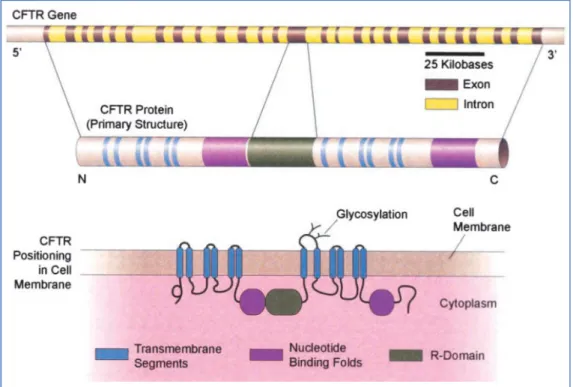
Figure 1. The cystic fibrosis (CF) transmembrane conductance regulator (CFTR) gene and its encoded polypeptide.The human CFTR gene (top) is located on the long arm of chromosome 7 and consists of 27 exon regions that encode the 1,480 amino acid CFTR proteins (middle). The mature protein after proper folding, glycosylation, and insertion into the cell membrane is shown at the bottom. The CFTR protein is a member of the ATP-binding cassette (ABC) family of transporters. It contains two nucleotide-binding domains that bind and hydrolyze ATP, two dual sets of membrane-spanning segments that form the channel, and a central regulatory (R) domain. The R domain, unique to CFTR, is highly charged with numerous phosphorylation sites for protein kinases A or C.(Reprinted by permission from Reference 490).
Taken from reference (3).
According to its primary structure, the CFTR channel is classified as a family member of the transport proteins class called ATP-biding cassette (ABC) transporter. These transporters utilize the energy of ATP hydrolysis to actively transport molecules across cell membranes. The CFTR channel is located in the apical membrane of epithelial tissues and is responsible for the regulation of chloride flow across epithelia cells. Thus, it has a crucial role in the control of transepithelial salt transport, fluid flow and ion concentration (4).
To date, more than 1900 mutations in the CFTR gene have been reported (5). In Canada, the most prevalent mutation (91.5%) is a deletion of a three-base pair which results in the loss of a phenylalanine residue at position 508 of the CFTR protein sequence (F508del) (6). Depending on the kind of mutation, the ion channel can work partially or completely fail
4
characterizing different symptoms and severity of the disease. However, the development of symptoms cannot be predicted based solely on DNA analysis, since some symptoms are also determined by phenotypic aspects, such as in the case of the pulmonary disease. However, the prediction of development of pancreatic disease is more genetically based.
1.1.2 Symptoms
As a variety of organs are lined by epithelial cells such as sweet ducts, airways, pancreatic ducts, biliary tree, intestines and vas deferens, cystic fibrosis leads to be a multi-systemic disease. The manifestations includes elevated sweat chloride concentration, lung disease, intestinal obstruction, pancreatic insufficiency with diabetes and impaired absorption, the latter due to inadequate secretion of digestive enzymes, biliary cirrhosis and congenital collateral absence of the vas deferens, often in combination (1, 7) .
Despite being a multi-systemic disease, approximately fifty percent of all patients are harmed with bacterial infection in the lungs (6). In addition, according to the Canadian Cystic Fibrosis Registry (2011), the main cause of death is pulmonary-related (6). Thus, given the impact of the pulmonary disease in the survival of CF patients, there is an unmet need for the development of new treatments for the chronic lung infections.
1.2. Lung Disease Phase in Cystic Fibrosis
1.2.1. Pulmonary dysfunctions due to CFTR mutations
The airways epithelium is formed by ciliated epithelial cells responsible to reabsorb electrolyte and goblet cells (glandular simple columnar epithelial cells). The latter are responsible for secreting mucins (the major component of mucus) and to generate the
5
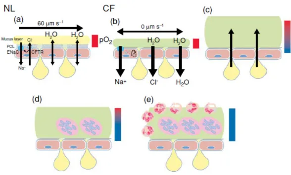
airway surface liquid (ASL). As shown in figure 2- scheme a, the normal airways are covered by the periciliary liquid layer which is composed of the cilium and a mucus layer. The function of the periciliary layer to provide a low-viscosity solution for ciliary beat which allows the mucus transport, therefore its volume and ion concentration are tightly regulated. The mucus layer is formed by high molecular-weight mucins whose properties are altered by electrolyte concentration, water content and pH. Ultimately, the mucin structure consists of numerous diversified carbohydrate side chains which are suitable for binding a wide variety of particles, as a mechanism to clear the airway through the mucus transport. Most importantly, it is widely accepted that the sensible adjustment in the electrolyte transport by the airway epithelium and submucosal glands controls the volume and composition of the ASL. Thus, as shown in the figure 2- schemes b and c, loss or dysfunction of CFTR leads to failure or decrease in the chloride secretion. However, as the sodium absorption still takes place, it leads to chloride paracellular absorption and water influx generating dehydration of ASL and build up of high viscous mucus. As a consequence, the cilium beat is impeded and the mucus transport does not take place. All these impairments together favors infection by bacteria and limits the pulmonary host defense due to loss of mucociliary clearance of bacteria(8) (Figure 2- schemes d and e).
6
Figure 2: Normal mucus clearance mechanisms and failure to adequately hydrate CF airway surfaces. (a) Normal airways coordinate rates of Na+ absorption and Cl- secretion to hydrate airway surfaces. (b) In CF, the absence of CFTR protein/function in the apical membrane leads to unregulated Na+(Cl- follows assively via paracellular path—not shown) and water absorption. The pathophysiologic sequence that follows CF dehydration is depicted in diagrams c–e. (c) Mucin secretion into adherent mucus plaque is depicted as emanating from goblet cells/glands. (d–e) Bacteria within mucus plaques/plugs are depicted as acrocolonies. Bars depict O2 tension in ASL (red, oxygenated; blue, hypoxic). NL=normal lung; CF= Lung of patients with CF.
Taken from reference (8).
1.2.2. Pulmonary Infection by Pseudomonas aeruginosa
Numerous microorganisms have been isolated from the lungs of CF patients such as Aspergillus fumigatus species, Haemophilus influenzae, Stenotrophomonas maltophilia(9), Burkholderia cepacia complex, Alcaligenes species, atypical mycobacteria and methicilin-resistent Staphylococcus aureous (MRSA) (6) (figure 3). In addition, viral infections also play an important role in the development of the CF disorder(10). However, the most prevalent bacteria are Pseudomonas aeruginosa and Staphylococcus aureus depending on age (6). Pseudomonas aeruginosa is a gram-negative bacillus, non-encapsulated and non-spore formers, which infects predominantly the lower respiratory tract.
7
Figure 3: Prevalence of respiratory infections, 2007-2011. Taken from reference (6).
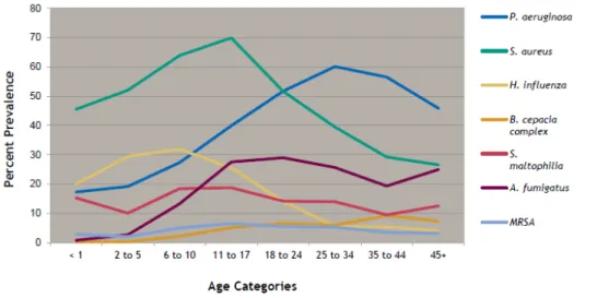
Studies have shown that early infection in the airways of CF patients is most frequently caused by S. aureus and H. influenzae. In the other hand, the most prevalent pathogen isolated in adults is Pseudomonas aeruginosa (Figure 4).
Figure 4: Age-specific prevalence of respiratory infection in CF patients, 2011. Taken from reference (6).
8
Indeed, pulmonary infection by Pseudomonas aeruginosa is closely related to an increase in mortality and morbidity of CF patients (6, 11) . However, it should be mentioned that other pathogens, such as Burkholderia ssp. (12)., Achromobacter ssp. (13) and Stenotrophomonas maltophilia (9) also contribute to morbidity and / or mortality .
Thus, these data support the importance of the development of new therapeutic strategies in order to prevent pulmonary exacerbations caused by these microorganisms. Pulmonary exacerbations are defined as the increased manifestation of respiratory symptoms such as cough and sputum production, often accompanied by systemic symptoms such as anorexia and malaise (14) . The occurrence of these symptoms defines the necessity of treatment adjustment in order to preserve pulmonary capacity and assure increased lifespan.
1.2.3. Biofilm formation
Numerous researchers have studied the underlying reasons why the immune system of CF patients is not effective to eliminate the early lung colonization by P. aeruginosa. However, how CFTR mutations enhance susceptibility to pulmonary infection and how this vulnerability could be prevented still remains unclear.
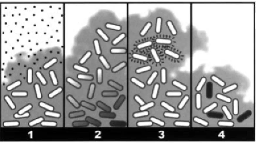
Cystic Fibrosis is correlated with increased proinfammatory signals; however this does not result in efficient clearance of bacteria. In fact, the failure to clear infection causes cyclic neutrophil influx which releases oxidants and proteases (15). This environment is characterized by low level of oxygen that triggers the change in the morphology of Pseudomonas aeruginosa from planktonic (mobile) to biofilm form (figure 2 – diagrams d-e). In addition, the bacteria which were strictly aerobic develop the ability to undergo anaerobic respiration (16). Mutations enable the bacteria to produce an extracellular polysaccharide (EPS) matrix composed mainly of alginate, the biofilm (figure 5).
9
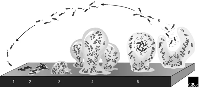
Figure 5: A model of the stages of bacterial biofilm development. At stage 1, the bacterial cells attach reversibly to the surface. Then, at stage 2, the cells attach irreversibly, a step mediated mainly by exopolymeric substances, and the cells lose their flagella-driven motility. At the next stage (3), the first maturation phase is reached, as indicated by early development of biofilm architecture. The second maturation phase is reached at stage 4 with fully mature biofilms, as indicated by the complex biofilm architecture. At the dispersion stage (5), single motile cells (dark cells on the figure) disperse from the microcolonies. Adapted from (17).Taken from reference (18).
In the biofilm form, the bacteria lose their flagellum which is recognized by the immunological system. In fact, P. aeruginosa isolated from chronically infected CF patients also have mutations enabling mucus production (mucoid strains) rendering them highly resistance to antibiotic and neutrophil-mediated killings. After maturation, these colonies of bacteria encased in the alginate matrix can return to the planktonic form (mobile) and disperse to colonize new niches (17). The genetic processes related to biofilm formation and dispersion has been extensively described in the literature (19).
1.2.4. Treatments
1.2.4.1. Treatment of the lung disease
The main goal of the treatment of the lung disease is to successfully eradicate microorganisms by antibiotic therapy. However, the establishment of chronic lung
10
inflammation is inevitable. Thus, other medications for chronic use are indicated to treat the symptoms due to pulmonary inflammation, as listed in table I (20).
Table I: Chronic medications for maintenance of lung health.
Class of drugs Examples Objective
Mucolytics Dornase alpha (inhalation)
Increase mucus fluidity, hence aiding mucus clearance. Recombinant human DNase degrades the residual DNA caused by neutrophils infiltration.
Osmotic agents
Hypertonic saline (inhalation)
Manitol (inhalation)
Reestablish the hydration condition of the ASL and improve the mucus transport rate. Osmotic agents draw water from the interstitium into the ASL which allows increased ciliary beat and cough clearance. β2-adrenergic receptor
agonists
Salbutamol, salmeterol
(inhalation) Treat reactive airway disease.
1.2.4.2. Treatment of lung infection caused by Pseudomonas aeruginosa Antibiotic therapy against P. aeruginosa aims at increasing the life expectancy of patients by preventing pulmonary exacerbations which can result in irreversible loss of pulmonary capacity. Thus, aggressive and early antibiotic treatment of P. aeruginosa is associated with increased life expectancy (21, 22), since in most cases resistant bacterial cells selected over time in chronic infection cannot be eradicated. Although the pulmonary route is always favored due to decreased level of side effects and increased concentrations, the concomitant administration of systemic drugs (orally or intravenously) may also be indicated, depending on the severity of the infection. Indeed clinical practice is the main resource to guide physicians to the appropriate choice of the antibiotic therapy. However, the antibiotic arsenal is finite and limited to combat P. aeruginosa which exhibits a strong capacity to mutate into resistant forms and is hyper adapted to the lungs of CF patients. In addition, the slow development of new classes of antimicrobials suggests that it is worthwhile to invest in the development of new formulation for current drugs. In fact, pharmacotechnical research allows changes in the drug’s pharmacokinetic properties which can be adapted to better target P. aeruginosas in the lungs. These innovative strategies
11
(presented in section 1.2.4.4), represent an important alternative to improve the bacterial eradication in cystic fibrosis, which is directly related to the longevity of CF patients.
1.2.4.3. Chronic Pseudomonas aeruginosa pulmonary infection
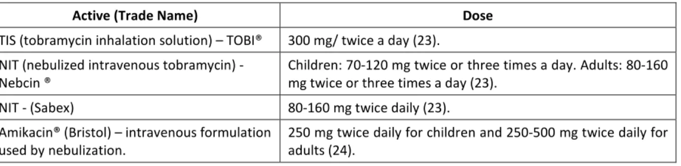
The use of aminoglycosides is recommended for the chronic suppressive therapy of P. aeruginosa in an alternating 28-day on/off regimen. Two different formulations are available including solution for inhalation and intravenous preparation also used for inhalation, as explained in table II.
Table II: Aerosolized aminoglycosides for the chronic suppressive therapy of P. aeruginosas.
Active (Trade Name) Dose
TIS (tobramycin inhalation solution) – TOBI® 300 mg/ twice a day (23).
NIT (nebulized intravenous tobramycin) - Nebcin ®
Children: 70-120 mg twice or three times a day. Adults: 80-160 mg twice or three times a day (23).
NIT - (Sabex) 80-160 mg twice daily (23).
Amikacin® (Bristol) – intravenous formulation used by nebulization.
250 mg twice daily for children and 250-500 mg twice daily for adults (24).
Although there are no thorough studies to demonstrate the clinical efficacy of colistimethate sodium, this drug can also be prescribed to treat multi-resistant strains (24, 25). Thus, colistin (Coly-Mycin® M Parenteral - ERFA Canada Inc.) is indicated as another option and is supplied as an aerosol preparation (powder for reconstitution to 150 mg/2 mL). Colistin is also available as an intravenous formulation for nebulization (Colobreathe ®). The aerosol dosage recommended for Colobreathe ® is 25-150 mg (intravenous dry powder) twice or three times daily (24).
Beta-lactams are also available to fight pseudomonal infections in CF patients. Aztreonam, carbapenems, cephalosporins (ceftazidime and cefapime) and penicilins are indicated for intravenous administration (26-28).
12
1.2.4.4. Recently approved therapies
Given the advantages of the pulmonary administration over systemic routes for the treatment of cystic fibrosis, recent research efforts have focused in the development of new antibiotic formulations such as powder for nebulization or powder for inhalation, as listed in table III.
Table III: Recently approved aerosolized antibiotics for the treatment of cystic fibrosis.
Drug Formulation Comments
CMSa (Colomycin®-Forest and Promixin®-Profile Pharma)
Intravenous formulation used for nebulization.
Composed by colistimethate sodium (CMS). Administered by Jet nebulizer. Administration time is 15 minutes. Formulation must be refrigerated (29)
CMSa (Colobreathe®) Dry powder for inhalation (DPI).
Composed by colistimethate sodium (CMS). Administrated with the Turbospin inhalation device. Administration time is approximately 1minute. Storage at room temperature (30).
Aztreonam lysine (AZLI, Cayston®, Gilead Sciences)
Powder for nebulization (aerosol).
Administrated with an electronic vibrating mesh nebulizer (Altera®). Administration time
is 3 minutes. Formulation must be
refrigerated. The CEDAC recommends the use of aztreonam inhalation solution for the treatment of chronic P. aeruginosa infection in patients with moderate to severe CF and
deteriorating clinical condition despite
treatment with inhaled tobramycin (31, 32). Tobramycin (TOB
Podhaler®- Novartis)c
DPI administrated with the Podhler (T-326) inhaler.
Tobramycin Inhalation Powder (TIP). Administration time is under 6 minutes. Storage at room temperature (33).
CEDAC: Canadian Expert Drug Advisory Committee: CF: Cystic Fibrosis.
a
Approved in the European Union (EU).
b
Approved for the treatment of CF patients infected with P aeruginosa in Australia and USA, and conditionally approved in Canada and EU. The Canadian Expert Drug Advisory Committee (CEDAC) recommends the use of aztreonam inhalation solution (28-day cycles) for the treatment of chronic P. aeruginosa infection in patients with moderate to severe cystic fibrosis and deteriorating clinical condition despite treatment with inhaled tobramycin.
c
Approved in European Union, Canada, Switzerland and other countries.
It has to be mentioned that the solution for inhalation requires the use of a nebulizer and compressor combination that can be noisy and difficult to transport. Additionally, nebulizers require long time for administration which may include the cleaning step of the equipments. Thus, the development of powder for inhalation must be encouraged since the advantage to
13
have rapid drug delivery with a portable inhaler includes potentially improving patient adherence.
All formulations discussed in table III are composed of free drugs. Indeed, they are aerosolized formulations which bring therapeutic improvements the treatment of CF since they enable the pulmonary administration of antibiotics. However, additional benefits would be achieved with the application of an aerosolized sustained delivery formulation (drug encapsulated into nanocarriers), as will be discussed afterwards.
1.2.5. Causes for treatment failure 1.2.5.1. Patient compliance
The new advances in the treatment of cystic fibrosis and aggressive management of lung disease have resulted in great improvements of the patient’s length and quality of life. However, as the number of treatments expands, the medical regimens become increasingly tiresome and time-consuming (34). Therefore, treatment burden for patients with cystic fibrosis is extremely high, and includes a range of inhaled and systemic medications, physiotherapy and exercises, often taking more than 2 hours a day (35).
Non adherence to treatment may cause accelerated disease and increased number of hospital admissions. The overall rate of treatment adherence in children with CF was found to be below 50% (36).
The most important reasons for patient non adherence to treatment include time management difficulties, oppositional behavior from children, poor taste when using breathing nebulizer for a long period, forgetting to administer the drug and embarrassment to take a lot of drugs or receive physiotherapy at school (37).
Therefore, decreasing the frequency of drug administration, providing safe drug association in the same formulation, offering easy and fast administration (important factors in the case
14
of drugs for lung administration) and developing formulations with tolerable taste may be promising goals to increase patient adherence and efficacy of antibiotic treatment. For instance, sustained drug release can be achieved by encapsulation of drug in a biodegradable carrier thus resulting in decreased frequency of drug administration. Such goal has been achieved by the liposomal formulation Amikacin® which is now in phase IV of clinical trials (38).
1.2.5.2. Pseudomonas aeruginosa resistance to antibiotics
In spite of aggressive antibiotic therapy, Pseudomonas aeruginosa has demonstrated the ability to evade the mechanism of action of antibiotics. Some adaptive mechanisms of defense against antibacterial drugs can be listed (table IV) such as changes in the bacterium expression of proteins which are target for drugs, lack of membrane porins which are important for antibiotic diffusion and expression of drug efflux mechanisms (39, 40). These mutations can partially explain their ability to survive and persist for years in the CF patient’s lungs.
Thus, as different classes of antibiotics show diverse modes of action, the rate and mechanisms of resistance vary according to the antimicrobial class as can be seen in the table IV.
15
Table IV: Main mechanisms of resistance of P. aeruginosa to certain classes of antibiotics.
Antibiotic Class Examples Mechanism of action Resistance mechanism
Fluoroquinolones Levofloxacin,
ciprofloxacin
Inhibition of DNA synthesis by inactivation of topoisomerases II and IV (enzymes essential for DNA replication).
Expression of drug efflux pumps. Mutations in the topoisomerases II and IV (39).
Aminoglycosides
Tobramycin, gentamicin, amikacin.
Inhibition of protein synthesis by antibiotic binding to bacterial
ribosomes and inhibition of
ribosomal enzymes.
Expression of drug efflux pumps. Reduction in the active transport through the
membrane. Modifying enzymes. Beta-lactams Imipenem, Meropenem, aztreonam, ceftazidime, cefapime.
Interference with cell wall
synthesis.
Production of β-lactamase enzymes that inactivate the drug. Multi-drug efflux (40) .
Polymyxins Colistin Disrupt the bacterial cell wall
through osmotic rupture.
Mutation in the lipid A
reducing binding of
polymyxins to
lipopolysaccharide (h) (41).
Polymyxins had already been considered an alternative for the treatment of multi-resistant strains of P. aeruginosa (42). Their bactericidal activity consists of cell membrane disruption leading to leakage of cell contents and bacterial death. This is achieved at least in part by binding to lipopolysaccharide (LPS), a major component of the Gram-negative cell surface, through interactions with phosphates and fatty acids of lipopolysaccharides core and lipid A moieties. However, spontaneous polymyxin-resistant mutants of Pseudomonas aeruginosa have already been isolated (41). Indeed, lipid A of these mutants contained aminoarabinose which reduces binding of polymyxins to LPS, whereby resistance arises (43).
The increase in bacterial resistance to antibiotics is also related to the route of administration. Thus, in the case of cystic fibrosis, the pulmonary route is preferred since it allows increased local concentration of antibiotics and longer contact time with the pathogen. Moreover, the local mechanism of action provided by pulmonary delivery decreases the occurrence of side-effects and the possible occurrence of sub-inhibitory drug levels, as can occur in other routes (oral, intravenous) which favors drug resistance (44). In
16
addition, the extended antibiotic release achieved with a nanocarrier would in theory also help to maintain the therapeutic concentration of drug in the lungs.
In addition, a previous study done by our group (45) showed an increased antifungal effect of itraconazole, voriconazole and amphotericin B loaded PEG-g-PLA nanoparticles compared with the free drug against resistant strains of Candida ssp and Aspergillus fumigates (these strains presented over expression of efflux pumps). These studies showed that PEG-g-PLA nanoparticles with small hydrodynamic diameter (˂200 nm) can be internalized in these yeast strains and might block the efflux pumps thereby overcoming fungal resistance to these drugs.
1.2.5.3. Antibiotic diffusion in the mucus
The mucus structure can interact with drugs hindering its diffusion through the mucus and the targeting of embedded bacteria. Various studies showed that positively charged, low-molecular weight drugs such as amikacin, tobramycin, gentamicin and some β-lactams antibiotics bind to negatively charged components in the mucus (46). Therefore, bacteria are exposed to sub-inhibitory level of antibiotics which may induce bacterial resistance and biofilm formation (44, 47).
1.2.5.4. Biofilm formation
The implications of biofilm formation in the resistance to cystic fibrosis treatments have been extensively reported. Firstly, the alginate envelope (biofilm matrix) serves as a direct barrier against phagocytic cells and effective opsonisation (22). Moreover, alginate has immunomodulatory properties stimulating the release of inflammatory cytokines which increases lung inflammation and subsequent destruction. Intensive inflammation favors the development of hypoxic areas in the lungs that can trigger biofilm formation. The polysaccharide matrix can also contribute to antibiotic resistance, since some antibiotics
17
cannot penetrate its structure and sub-inhibitory level of antibiotic can result and bacterial selection and resistance (48). Likewise, within the biofilm some cells are dormant escaping the mechanism of action of some antibiotics, e.g. penicillins (49). Moreover, the oxidative stress inside the biofilm structure results in numerous genetic mutations that are transferred horizontally between the cells and confers resistance to antibiotics (50) (51). The main mechanisms of antibiotic resistance in biofilms are described in figure 6.
Figure 6: Four hypothesized biofilm resistance mechanisms. 1) The antibiotic (squares) penetrates slowly or incompletely; 2) A concentration gradient of a metabolic substrate or product leads to zones of slow or non-growing bacteria (shaded cells); 3) An adaptive stress response is expressed by some of the cells (marked cells); 4) A small fraction of the cells differentiate into a highly protected persister state (dark cells).Taken from reference (52)
1.3. Nanotechnology for pulmonary drug delivery
1.3.1. Nanocarriers for drug delivery in the lungs
As discussed before, controlled-release formulations are important tools for drug delivery in the lungs since they may increase and sustain local drug concentrations which contribute to decrease dose frequency and systemic toxicity and result in better patient compliance and augmented treatment efficiency.
18
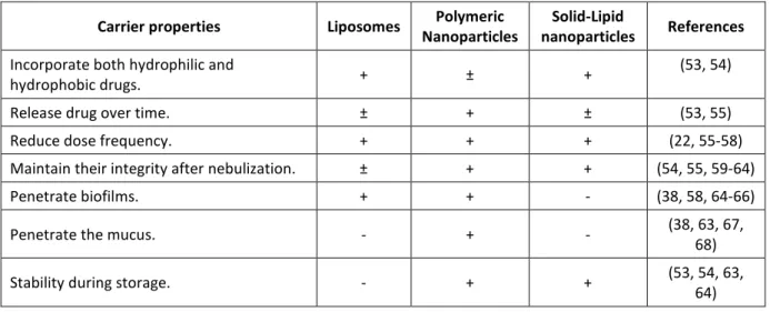
A number of nanocarriers have been developed for drug release in the lungs; therefore we have selected the most well-known classes and described their main characteristics in table V.
Table V: Properties of the main nanocarriers produced for drug delivery to the lungs
Carrier properties Liposomes Polymeric
Nanoparticles
Solid-Lipid
nanoparticles References
Incorporate both hydrophilic and
hydrophobic drugs. + ± +
(53, 54)
Release drug over time. ± + ± (53, 55)
Reduce dose frequency. + + + (22, 55-58)
Maintain their integrity after nebulization. ± + + (54, 55, 59-64)
Penetrate biofilms. + + - (38, 58, 64-66)
Penetrate the mucus. - + - (38, 63, 67,
68)
Stability during storage. - + + (53, 54, 63,
64)
The structural configuration of nanocarriers described in table V is displayed in figure 7.
Figure 7: Schematic illustration of four nanoparticle platforms for antimicrobial drug delivery: (a) liposome, (b) polymeric nanoparticle, (c) solid lipid nanoparticle, and (d) dendrimer. Black circles represent hydrophobic drugs; black squares represent hydrophilic drugs; and black triangles represent either hydrophobic or hydrophilic drugs. Taken from
19
Liposomes are formed by phospholipid dispersion in water solution that after saturation can form micelles able to entrap the drug added to this system. Liposomes are also the most investigated system for antibiotic controlled pulmonary delivery, since they may be prepared with phospholipids endogenous to the lungs. In addition, their lipid bilayer structure mimics the cell membrane and can fuse with infectious microbes to deliver high drug cargos into the cytoplasm, saturating their drug efflux pumps, therefore overcoming bacterial resistance (69, 70). Moreover, Meers et al have suggested that amikacin-loaded liposomes can also penetrate biofilms and infected mucus (38). Although studies have showed that liposomes positively charged can penetrate biofilms, positive charge appears to lead to more surface adsorption of liposomes at the expense of further penetration. In this respect, Meers et al hypothesized that the neutral or zwitterionic lipids used to prepare the amikacin-bearing liposomes in their study preclude strong ionic interactions and may help to enhance the penetration. Indeed, in this same study, inhaled liposomal amikacin formulation exhibited slow sustained released in normal rat lungs and was more efficacious than inhaled free amikacin in lungs infected by P. aeruginosas (38). However, liposomes are unstable during storage in the liquid and fragile carriers that can be physically destroyed in the nebulization process. Moreover, adjusting the drug release profile is a challenge if compared with other carriers such as nanoparticles. For instance, tobramycin liposome formulation showed increased both drug retention in the lung and antimicrobial activity compared with classical formulation. However, long-term efficacy could not be demonstrated by this formulation (22).
Dendrimers are defined as highly ordered and regularly branched globular macromolecules synthesized by stepwise iterative approaches. The structure of dendrimers consists of a core, layers of branched repeat units emerging from the core, and functional end groups on the outer layer of repeat units. Thus, hydrophobic drugs can be loaded inside the cavity core and the hydrophilic ones can be loaded in the outer layer through covalent conjugation or electrostatic interaction. Although some dendrimers exhibited sustained pulmonary drug release and antimicrobial activity itself (53, 56, 64), their activity was far from those of
20
commercialized drugs. In addition their synthesis and purification are time consuming, expensive and cumbersome, facts that justify the choice of other nanocarriers.
Solid lipid nanoparticles are carriers formed by solid lipids such as triglycerides, partial glycerides, fatty acids, steroids and waxes and they are able to encapsulate hydrophobic or hydrophilic drugs (66). However, they have not yet been fully exploited for pulmonary lung delivery. In addition, they can exhibit some drawbacks such as low drug loading and unpredictable drug release (58).
Conversely, polymeric nanoparticles (NP) are easier to formulate in order to reach pulmonary sustained release and its use for antimicrobial drug delivery has been extensively investigated, since this carriers offer several advantages. For instance, polymeric NP exhibit structural stability in biological fluids and under harsh and various conditions for formulation (such as spray drying, nebulization) and storage. In addition, by manipulating the formulation composition such as polymer chain lengths and concentration, surfactants and organic solvents, it is possible to tune polymeric NP properties such as size, charge, drug loading and drug release profiles. Furthermore, polymeric NP offer the possibility for insertion of chemical groups on its surface to improve mucus penetration and bacterial targeting, as will be discussed later.
1.3.2. Polymeric nanoparticles
A number of synthetic and natural polymers have been used for the synthesis of nanoparticles. However, synthetic polymers can be engineered to reach increased sustained release as compared with natural polymers such as albumin, gelatin, alginate, collagen, cyclodextrins and chitosan (71). Likewise, examples of synthetic polymers used for pulmonary applications include poly(lactic acid) (PLA), poly(glycolic acid)(PGA), poly(lactide-co-glycolide) (PLGA) and poly(ε-carprolactone) (PCL) (72). In addition, these polymers are considered non-toxic since they undergo hydrolysis upon implantation into the body, degrading into biologically compatible moieties (lactic acid and glycolic acid) which are
21
cleared from the body by the citric acid cycle. Therefore, degradation products are formed slowly assuring sustained release and do not affect normal cell function. Indeed, the polyesters PLA and PLGA are the most extensively investigated polymers for drug delivery (64). They have an hydrophobic core, therefore grafting of hydrophilic motifs such as poly(ethylene glycol) (PEG) on both polymer backbone have been studied (73, 74). The purposes for the PEG copolymerization are: 1) to facilitate the encapsulation of hydrophilic drugs, 2) to increase the resident time in the body, since hydrophobic particles are readily cleared by the mononuclear phagocytic system (MPS); 3) to modulate mucus adhesiveness in order to achieve mucus penetrating particles (64).
1.3.3. Engineered polymeric nanoparticles for drug delivery in the lungs 1.3.3.1. Mucus penetration particles
Although the proposal to develop an aerosolized sustained antibiotic release formulation for lung administration has the goal of achieving higher antibiotic levels in the lungs, the minimal inhibitory concentration (MIC) cannot be attained if the carrier does not diffuse in the mucus to target the bacteria. In fact, the build-up mucus acts as a physical, chemical and biological barrier to drug penetration, which also contributes to bacterial resistance to antibiotics. The mucus is composed mainly by mucins, but also contains DNA, lipids, ions, proteins, cells, cellular debris, and water (75). However, dysfunctions in the CFTR alter the mucus composition and generate increased viscosity which impairs the diffusion of drugs and carriers. Indeed, up to date no gene vector has been shown to penetrate the mucus in order to reach the epithelial cells, fact which explains the failure of clinical trials of gene therapy for cystic fibrosis (76). Therefore, our goal is to produce mucus inert polymeric nanoparticles as an antibiotic carrier able to penetrate the mucus.
Studies have elucidated the interaction between mucus structure and nanoparticle surface coating, in order to decrease NP retention in the mucus, therefore improving nanoparticle
22
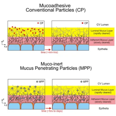
diffusion (77). In addition to hydrophilic sites with negative charges imparted by the presence of carboxyl or sulfate groups on the mucin proteoglycans, the mucus also exhibit hydrophobic regions along mucin strands, stabilized by multiple internal disulfide bonds. These findings explain why the diffusion of both hydrophobic and cationic drugs such as tobramycin can be hindered in the mucus (78). However, the mucus is not impenetrable, as some viruses such as the human papilloma virus (HPV) can traverse it. In fact, a closer study of the HPV structure revealed that they are densely coated with both positively and negatively charged groups, leading to a densely charged yet net neutral surface. Based on these findings, Hanes and co-workers (67) rationalized that mucus penetrating particles must possess a high density on the hydrophilic surface able to minimize hydrophobic entrapment of mucus and be small enough to preclude significant steric inhibition by the dense fiber mesh, since the mucus structure contains multiple pores. Finally, the PEG properties such as being strongly hydrophilic and having a neutral charge make it an appropriate nanoparticle coating. However, as studies have also evidenced mucoadhesiveness properties of PEG, the rationale was to use a PEG molecule with controlled molecular weight. Thus, Hanes et al (67) hypothesized that the PEG molecular weight must be low enough to prevent adhesion via polymer interpenetration in the mucus (hydroplilic interactions). In addition, PEG density must be sufficient to effectively shield the hydrophobic core common to many biodegradable polymers resulting in decreased hydrophobic interactions between polymeric nanocarriers and mucus (67) (figure 8).
23
Figure 8: Summary schematic illustrating the fate of mucus-penetrating particles (MPP) and conventional mucoadhesive particles (CP) administered to a mucosal surface. MPP readily penetrate the luminal mucus layer (LML) and enter the underlying adherent mucus layer (AML). In contrast, CP are largely immobilized in the LML. Because MPP can enter the AML and thus are in closer proximity to the cells, cells will be exposed to a greater dose of drug released from MPP compared to drug released from CP. As the LML layer is cleared, CP are removed along with the LML whereas MPP in the AML are retained, leading to prolonged residence time for MPP at the mucosal surface. In the respiratory airways, CP are mostly immobilized in the luminal stirred mucus gel layer, whereas MPP penetrate the mucus gel and enter the underlying periciliary layer. Upon mucociliary clearance, a significant fraction of MPP remains in the periciliary layer, resulting in prolonged retention. Taken from reference
(67).
1.3.3.2. Nanoparticles size and charge
Therefore, in order to prove the MPP theory, the nanoparticle diffusion in the mucus was evaluated by Hanes et al (77) as a function of PEG coating and nanoparticle size. Polystyrene particles as large as 200 nm in diameter that were densely coated with low molecular weight (2 KDa) polyethylene glycol (PEG) moved through undiluted CF sputum with average speeds up to 90-fold faster than similarly-sized uncoated polystyrene particles. Conversely, the transport of both coated and uncoated 500 nm particles was strongly hindered (77). These nanoparticles exhibited almost neutral surface (zeta potential higher than -10 mV) demonstrating that size (≤ 200 nm) and superficial charge (neutral) may also influence in the transport of nanoparticles in the mucus. Finally, this experiment proved that nanoparticles
24
with correct size, PEG coating and charge may overcome the mucus barrier for the transport of nanocarriers.
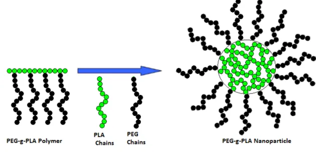
1.3.4. PEG-g-PLA nanoparticles as a model vector for pulmonary administration The synthesis and characterization of PEG-g-PLA polymer have been previously proposed by our group of research (74). Briefly, PEG is grafted in the PLA backbone to form an amphiphilic polymer. When the outer phase in the emulsification process for nanoparticle production is water, this polymer can form nanospheres which are comprised by a hydrophobic core mainly composed of PLA and a hydrophilic surface composed of PEG coating (figure 9). In polymeric nanocapsules, a polymeric membrane forms a shell with an inner space loaded with the drug which are solubilized in aqueous or oily solvents. In contrast, nanospheres are solid nanoparticles with the drug homogeneously distributed in the polymeric matrices of variable porosity, as expected with PEG-g-PLA nanoparticles (79).
Figure 9: Scheme to represent the structure of PEG-g-PLA nanoparticles. Taken from reference (79).
25
The copolymer of PEG and PLA is amphiphilic (have an hydrophobic and an hydrophilic portion in the same polymer unit), fact which favors the encapsulation of hydrophilic and hydrophobic drugs (54). Hammady and workers have already reached the co-encapsulation of paclitaxel (a hydrophobic drug with a Log P value of 3.96 and water solubility of less than 0.01 mg/mL) (80) and endotelin (a hydrophilic drug with solubility of 1 mg/mL) for the anti-angiogenic treatment of diseases related with an angiogenic component (e.g. solid tumors, arthritis, psoriasis, diabetic retinopathy and atherosclerosis) (81). The majority of the antibiotics available for the CF treatment are hydrophilic; therefore, we believe that PEG-g-PLA might be an appropriate polymer to produce a pulmonary carrier for the CF treatment.
The antimicrobial activity of PEG-g-PLA nanoparticles formulations have also been investigated, as compared with the free drug. Two different copolymers of PLA and PEG were studied by Essa et al; branched PEGylated polymer in which PEG was grafted on PLA back bone (PEG-g-PLA) and multiblock copolymer of PLA and PEG, (PLA–PEG–PLA) (57). In addition, PLA nanoparticles were also analyzed. Itraconazole (ITZ) loaded nanoparticles were produced with the cited polymers and their in vitro antifungal activity was evaluated against both Candida and Aspergillus species. All ITZ-NPs were nearly spherical with smooth surface with a size range of 185–285 nm and zeta potential measured values were close to neutrality. In addition, ITZ release showed an initial burst followed by a gradual release profile: over 5 days for PEG-g-PLA and over 2 days for PLA-PEG-PLA nanoparticles. Most importantly, ITZ-loaded PEG-g-PLA nanoparticles inhibited fungal growth more efficiently in specific fungus strains (Candida) than either free ITZ or ITZ-loaded PLA nanoparticles suggesting that PEG-g-PLA–ITZ could be used efficiently as a nanocarrier to enhance antifungal efficacy (82).
Likewise, studies carried out by our group revealed that PEG-g-PLA loaded voriconazole nanoparticle formulation significantly improved the in vitro antifungal activity against Candida ssp (MIC of free voriconazole = 2.917 ± 0.137 mg.L-1, n=3 and MIC of
voriconazole-26
loaded-PEG-g-PLA nanoparticles = 0.094 ± 0.01 mg.L-1, n=3). However, just a slightly improvement was found in the same test against Candida ssp biofilms (45).
1.3.5. Recent drug carriers and other formulations under development
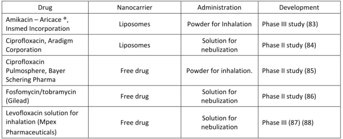
The new therapies under development for the treatment of cystic fibrosis are described in table VI.
Table VI: Recent formulations under development for CF treatment.
Drug Nanocarrier Administration Development
Amikacin – Aricace ®,
Insmed Incorporation Liposomes Powder for Inhalation Phase III study (83)
Ciprofloxacin, Aradigm
Corporation Liposomes
Solution for
nebulization Phase II study (84)
Ciprofloxacin Pulmosphere, Bayer Schering Pharma
Free drug Powder for inhalation. Phase II study (85)
Fosfomycin/tobramycin
(Gilead) Free drug
Solution for
nebulization Phase II study (86)
Levofloxacin solution for inhalation (Mpex Pharmaceuticals)
Free drug Solution for
nebulization Phase III (87) (88)
The most innovative formulas under development are the amikacin and ciprofloxacin liposomal formulations, since the use of a nanocarrier may enable sustained drug release (providing decreased side effects and reduced frequency of drug administration). In addition, nanocarries may enable mucus penetration thus preventing sub-inhibitory level of antibiotics and resulting in decreased bacterial resistance and biofilm formation. These benefits provided by nanocarriers may increase the efficacy of the antimicrobial formulation against the targeting bacteria; thereby improving the treatment of CF. However the other formulations improve the treatment by providing pulmonary administration as compared to oral or intravenous administration.
27
CHAPTER 2:
28
2.1. Hypothesis
Based on the given facts, our hypothesis is that antibiotic-loaded PEG-g-PLA nanoparticles may improve the efficiency of the treatment against Pseudomonas aeruginosa in the lungs of CF patients, as compared with the free drug. The drugs chosen as models are colistin sulfate, tobramycin and levofloxacin, as an attempt to test three different classes of antibiotics extensively used for the treatment of CF.
We predict that PEG-g-PLA engineered nanoparticles may improve the treatment against Pseudomonas aeruginosa in Cystic Fibrosis patients based on the following concepts:
1) Engineered PEG-g-PLA nanoparticles with suitable size and charge are potentially mucus penetrating and might increase the targeting of mucus embedded Pseudomonas aeruginosa;
2) PEG-g-PLA nanoparticles might provide extended release of the encapsulated drug therefore increasing the exposure of Pseudomonas aeruginosa to therapeutic doses of drug and reducing bacterial resistance
3) PEG-g-PLA nanoparticles have the potential to circumvent bacterial resistance if the main mechanism of resistance presented by the strains under analysis is drug efflux (as in the case of P. aeruginosa), as have been showed in a precious research carried in our laboratory in Candida ssp. and Aspergillus fumigatus strains (45).
2.2. Objectives
Our main goal is to develop poly(ethylene glycol)-g-poly(lactic acid) (PEG-g-PLA) antibiotic- loaded nanoparticles with suitable physicochemical properties for antibiotic delivery in the lungs in order to increase the antimicrobial efficiency against Pseudomonas aeruginosa. In addition, PEG-g-PLA nanoparticles will be engineered to be mucus inert hence favoring