To link to this article: DOI:10.2298/ABS1104047S http://dx.doi.org/10.2298/ABS1104047S
This is an author-deposited version published in: http://oatao.univ-toulouse.fr/
Eprints ID: 5835
To cite this version: Smaoui, Slim and Mathieu, Florence and Fourati Ben Fguira, Lilia and Merlina, Georges and Mellouli, Lofti Taxonomy and antimicrobial activities of a new Streptomyces sp. TN17 isolated in the soil from an oasis in Tunis. (2011) Archives of Biological Sciences, vol. 63 (n°4). pp. 1047-1056. ISSN 0354-4664
O
pen
A
rchive
T
oulouse
A
rchive
O
uverte (
OATAO
)
OATAO is an open access repository that collects the work of Toulouse researchers and makes it freely available over the web where possible.
Any correspondence concerning this service should be sent to the repository administrator: [email protected]
Arch. Biol. Sci., Belgrade, 63 (4), 1047-1056, 2011 DOI:10.2298/ABS1104047S
1047
TAXONOMY AND ANTIMICROBIAL ACTIVITIES OF A NEW STREPTOMYCES SP. TN17 ISOLATED IN THE SOIL FROM AN OASIS IN TUNIS
SLIM SMAOUI1, FLORENCE MATHIEU2, LILIA FOURATI BEN FGUIRA1,
GEORGES MERLINA3 and LOTFI MELLOULI1*
1 Laboratory of Microorganisms and Biomoleculesat the Centre of Biotechnology of Sfax, Road of Sidi Mansour Km 6,
P.O. Box 1177, 3018 Sfax, Tunisia
2 University of Toulouse, Laboratory of Chemical Engineering UMR 5503 (CNRS/INPT/UPS), Bioprocess and Microbial
Systems Department INP-ENSAT, 1 Av, de l’Agrobiopôle, BP 32607, 31326 Castanet-Tolosan France
3 CNRS, EcoLab, ENSAT, 31326 Castanet-Tolosan, Cedex France
Abstract - An actinomycete strain referred to as TN17 was screened for its antimicrobial activities. The taxonomic status
of this strain was established. The organism was found to have morphological and chemotaxonomic characteristics typical of Streptomycetes. Based on the 16S rRNA nucleotide sequences, Streptomyces sp. TN17 was found to have a relationship with Streptomyces lilaceus, Streptomyces gobitricini and Streptomyces lavendofoliae. Combined analysis of the 16 S rRNA gene sequence (FN687757), phylogenetic analysis, fatty acids profile and physiological tests indicated that there are geno-typic and phenogeno-typic differences between TN17 and neighboring Streptomyces species’ neighbors. Therefore, TN17 is a novel species: Streptomyces sp. TN17 (=DSM 42020T=CTM50229T). A cultured extract of this strain inhibits the growth of several Gram positive and Gram negative bacteria and fungi.
Key words: Actinomycete, polyphasic taxonomy, 16S rRNA gene, Streptomyces sp. TN17 (=CTM50229T=DSM 42020T),
antimicrobial activities
UDC 579.873.7(611-13):602.6
INTRODUCTION
Actinomycetes are gram-positive bacteria that are free-living, saprophytic, widely distributed in soil, water; they also colonize plants, exhibit, marked chemical and morphological diversity and form a distinct evolutionary line of organisms (Goodfellow and O’Donnell, 1989).
At least 90% of actinomycetes isolated from soil have been reported to be Streptomyces spp (Ander-son and Wellington, 2001). The genus Streptomyces was proposed by Waksman and Henrici (1943) and classified in the family Streptomycetaceae on the ba-sis of morphology and subsequent characterized cell
wall chemotype. Streptomycete systematics, notably the delineation of species, is becoming increasingly objective due to the application of the polyphasic tax-onomic approach. However, the classification of the genus Streptomyces in the current edition of Bergey’s Manual of Systematic Bacteriology (Willimas et al., 1989) is based not on a combination of genotypic and phenotypic properties, but on the extensive numeri-cal taxonomic survey of Williams et al. (1989). Given the presence of a phylogenetic branching pattern, a combination of properties such as wall chemotype, peptidoglycan type, whole-cell sugars, fatty acid and phospholipid profiles and menaquinones facilitates the delineation of actinomycete genera from neigh-boring taxa (Kroppenstedt et al., 1990). Streptomyces
species can be distinguished from other actinomyc-etes by their cell wall type which is characterized as Type I sensu (Lechevalier and Lechevalier, 1970). The presence of L-L diaminopimelic acid and glycine and the absence of characteristic sugars are typical of this cell wall type (Uchida and Seino, 1997). The analysis of 16S rRNA has proved to be a very important tool in Streptomyces systematics, as well as helpful in as-signing the newly isolated strain to the genus
Strepto-myces. There is increasing interest in the isolation of
a novel Streptomyces species as they are very potent producers of active secondary metabolites (Mellouli et al., 2003). Streptomyces have been the most fruitful source of microorganisms for all types of bioactive metabolites that have important applications in hu-man medicine and in agriculture fields (Watve et al., 2001).
During our routine screening program for the isolation of novel actinomycete bacteria producing bioactive compounds, an interesting bacterium, re-ferred to as TN17, was isolated from Tunisian oasis soil samples and selected for its capacity to produce antimicrobial molecules. In the present work, we describe the identification of a Streptomyces strain, designated as TN17, isolated from a Tunisian oasis soil sample by conventional and molecular methods, as well as the antimicrobial activities of the culture extract of this strain.
MATERIALS AND METHODS
Strain isolation and conservation
Strain TN17 was isolated by the dilution agar plait-ing method from oasis soil from southern Tunisia. The strain was maintained by cultivation on an ISP 2 agar medium that contained (per liter) 4 g glucose, 4 g yeast extract, 5 g malt extract and a vitamin/ amino acid mixture (1 mg vitamin B1; 1 mg vita-min B2; 1 mg vitavita-min B6; 1 mg biotin; 1 mg nico-tinic acid; 1 mg phenylalanine; 0.3 g alanine) at pH 7.2, incubated at 28°C for two weeks. The strain was maintained on a yeast extract-malt extract-dextrose (YMD) agar medium at 4°C (Williams and Cross, 1971).
Phenotypic characterization
The cultural characteristics and the colors of the ma-ture sporulating aerial mycelium and the substrate mycelium of the isolated TN17 were monitored in 7, 14 and 21 day-old cultures grown in different agar media as follows: the Four (ISP 2-5) International Streptomyces Project (ISP) media recommended by Shirling and Gottlieb (1966); the Bennett agar me-dium; the Nutrient agar medium and the Sabouraud agar medium. Melanin production was tested in the peptone-yeast extract-iron (ISP6 medium) agar and trypsin (ISP7 medium) agar.
Chemotaxonomic studies
Sufficient biomass for chemotaxonomic studies was obtained after incubation at 28°C for 3 days by growing in shake-flasks in ISP 2 broth. The isomer-ic form of diaminopimelisomer-ic acid (DAP), glycine and sugars in the whole cell hydrolyzates were analyzed by TLC (Staneck and Roberts, 1974). Phospholip-ids were examined by two-dimensional TLC and identified using several spray reagents and by co-migration with standards (Collins and Jones, 1980; Minnikin et al., 1979). Fatty acids were extracted, methylated and analyzed by gas chromatography (GC) using the standard Microbial Identification System (MIDI) (Sasser 1990).
Physiological tests
The ability of the strains to utilize 15 compounds as sole carbon sources and 16 compounds as sole ni-trogen sources for energy and growth was examined on specimens grown on ISP medium 9 for 3 days at 28°C. Each source was added at a final concentration of 1% (w/v) and 0.1% (w/v), respectively. The utiliza-tion of sole carbon and sole nitrogen sources was in-vestigated according to Shirling and Gottlieb (1966). Sodium salts (acetate, alginate, benzoate, butyrate, citrate, desoxycholate, hydrogen carbonate, nitrate, oxalate, perchlorate, propionate, pyruvate, succinate, sulfite, tartrate, tetraborate and thiosulfate) were added at a final concentration of 0.1% (w/v) (Gor-don et al., 1974).
TAXONOMY AND ANTIMICROBIAL ACTIVITIES OF A NEW STREPTOMYCES 1049
Esculin and arbutin (1.0%, w/v) degradation was determined by the methods of Williams et al. (1989) and examined after 3 days incubation of the TN17 strain in ISP2 solid medium. The degradation of ca-sein (1.0%, w/v) was detected in the ISP2 agar after either 3, 7 and 14 incubation days and clearing under and around the colonies’ growth areas was scored as positive. Gelatin (0.4%, w/v) and starch (1.0%, w/v) degradation was read after 3 days in the ISP2 agar by flooding the plates with trichloroacetic acid (3.0%, v/v) and iodine solutions respectively, and scoring the zones of clearing as positive (Willimas et al., 1989).
The degradation of tyrosine, hypoxanthine, xan-thine, adenine and guanine (1.0%, w/v) was investi-gated according to Gordon et al. (1974) and the hy-drolysis of Tween 80 was measured using the method of Sierra (1957).
The effects of salt on growth were determined in TSB media supplemented with graded doses of sodium chloride (1, 4, 5, 7 and 10% w/v). Maximum sodium chloride concentration in the medium al-lowing any growth was recorded (Williams et al., 1989).
The growth at various temperatures was tested using TSA plates incubated at 4, 10, 15, 25, 30, 37, 40 and 45°C. The effects of pH on growth were tested in pH-adjusted TSB media (pH 4.0~10.0 in 0.5 unit increments).
Tolerance to lysozyme (0.005%), phenol (0.05, 0.1%, 0.2%, 0.5% and 1.0%), and sodium azide (0.001 and 0.01%) was tested using GYEA media (Athalye et al., 1985).
Resistance to antibiotics was examined with erythromycin, streptomycin, penicillin at (10 mg/l), rifampicin, gentamicin, vancomycin at (5 mg/l) and chloramphenicol, oxytetracycline, kanamycin at (25 mg/l) incorporated into the glucose yeast extract agar (Lechevalier and Lechevalier a, b, 1970) as a basal medium. Readings were taken at 1, 3, 7 and 14 days of growth. Organisms were scored as
resist-ant (+) when growth on the test plates was greater or equal to that on positive control plates lacking inhibitors.
Genotypic characterization
The genomic DNA of strain TN17 was isolated as described by Hopwood et al. (1985). PCR ampli-fication of the 16S rRNA gene of strain TN17 was performed in an automated thermocycler (Perkin Elmer) using two primers 5’-AGAGTTTGATC-CTGGCTCAG-3’ and 5’-AAGGAGGTGATC-CAGCCGCA-3’ as described by Edwards et al. (1989) and according to the amplification profile described by Elleuch et al. (2010). The PCR reac-tion mix was analyzed by agarose gel electrophore-sis and DNA of the expected size was purified and then cloned into a pCR-Blunt vector. The nucleotide sequence of the 16S rRNA gene of strain TN17 was determined on both strands by an automated 3100 Genetic Analyzer (Applied Biosystems) using spe-cific primers. For phylogenetic analysis, reference strains were chosen from the BLAST (Altschul et al., 1997) results. The nucleotide sequence of the whole 16S rRNA gene (1521 pb) of TN17 strain has been assigned in GenBank (EMBL) under accession number FN687757. Multiple sequence alignment was carried out using CLUSTAL W (Thompson et al., 1997) at the European Bioinformatics Insti-tute website (http://www.ebi.ac.uk/clustalw/). Phy-logenetic analyses were performed using programs from the PHYLIP package (Felsentein, 1985) and a phylogenetic tree was constructed by the neighbor joining (NJ) algorithm (Saitou and Nei, 1987) us-ing Kimura 10-parameter distance. The robustness of the inferred tree was evaluated by bootstrap (100 replications).
Antimicrobial activities determination
Indicator microorganisms were grown overnight in LB medium at 30°C for Micrococcus luteus LB14110 and at 37°C for Staphylococcus aureus ATCC 6538 (Gram-positive bacteria), Pseudomonas aeruginosa ATCC 49189, Salmonella enterica ATCC43972 and Escherichia coli ATCC 8739 (Gram-negative
bacte-ria) and then diluted 1:100 in LB medium and incu-bated for 5 h under constant agitation of 200 rpm at the appropriate temperature. Fusarium sp. was grown in a potato dextrose agar (PDA) for 7 days at 30°C. Spores were collected in sterile distilled water and then adjusted to a spore density of approximately 104
spores/ml. Candida tropicalis R2 CIP203 was grown in YP10 medium (10 g/l yeast extract, 10 g/l peptone, 100 g/l glucose, 15 ml of 2 g/l adenine solution) at 30°C for 24 h in an orbital incubator with shaking at 200 rpm.
Spores of Strain TN17 at 107/ml were used to
inoculate a 500 ml Erlenmeyer flask with four in-dents containing 100 ml of TSB (Tryptic Soy Broth) medium at 30 g/l. After incubation at 28°C for 24 h, this pre-culture was used to inoculate at 1/10 (v/v) 1000 ml Erlenmeyer flask with four indents, con-taining 200 ml of TSB medium. After incubation at 28°C for 72 h in an orbital incubator with shaking at 200 rpm, the culture broth was centrifuged to remove the biomass. The cell-free supernatant was extracted with ethyl acetate (2×) and the obtained organic extract concentrated in vacuo to dryness. The resulting dry extract was recuperated in 2 ml of ethyl acetate and assayed against indicator mi-croorganisms. Antimicrobial activities were deter-mined by the agar diffusion test: a paper disk (8mm Ø) was impregnated with 50 µl of the correspond-ing sample and then laid on the surface of an agar plate containing 3ml of top agar seeded by 40 µl of a 5-h-old culture of the corresponding microorgan-ism. For antifungal activity against the Fusarium sp., 100 µl of spore suspension was added to 3 ml of the top agar. After 2 h at 4°C, plates were incubated overnight at the appropriate growth temperature of the corresponding indicator microorganism. Plates were examined for evidence of antimicrobial activi-ties represented by a zone of inhibition of growth of the studied indicator cell around the paper disk. The experiment was carried out simultaneously three times under the same conditions. In each case, all obtained diameters of inhibition zones were similar and the reported inhibition zones (mm) are the av-erage from three experiments.
RESULTS AND DISCUSSION
Morphological and physiological characteristics
Strain TN17 is a Gram-positive bacterium. Mor-phological observation of the 7–15 day-old cul-ture of this strain grown on yeast extract-malt extract agar (ISP2) (Shirling and Gottlieb, 1966) revealed that both aerial and vegetative hyphae were abundant. The isolate developed well on several media, including ISP2, ISP3, ISP4, ISP5, Bennett agar and nutrient agar media. The de-tailed cultural characteristics of strain TN17are given in Table 1. The aerial mycelium was abun-dant, well-developed and varied from white to gray on all tested media. The substrate hyphae varied from yellowish-white to yellowish-brown. Diffusible pigments were not produced on any test media and melanin was not produced. The physiological features are indicated in Table 2 and in the species description.
The strain Streptomyces sp. TN17 has been de-posited in the DSMZ and CTM under the numbers DSM 42020T and CTM50229T, respectively.
Chemotaxonomic analysis
The cell wall peptidoglycan of strain TN17 con-tained only LL-diaminopimelic acid and glycine, indicating that this strain has a chemotype cell-wall type I (Lechevalier and Lechevalier a, b, 1970). Whole-cell hydrolyzates contained mainly glucose and small quantities of xylose, galactose and arab-inose. The diagnostic phospholipid was phosphati-dylethanolamine (PE) (phospholipids type II sensu (Lechevalier and Lechevalier, 1970b). The fatty acid profile included mainly saturated iso- and anteiso-branched-chains and straight-chain fatty acids “fatty acid type 2c sensu” (Kroppenstedt, 1985). The ma-jor cellular fatty acids were isoC15:0 (23.58%), anteiso
C15:0 (29.88%) and isoC16:0 (28.52%), and smaller
amounts of C12:0 3-OH (1.61%), C15:0 (3.12%), C16:0
(4.49%), C16:1 w 9 (3.57%) and isoC17:0 (3.89%) were
TAXONOMY AND ANTIMICROBIAL ACTIVITIES OF A NEW STREPTOMYCES 1051
The chemical and morphological properties of strain TN17 are clearly consistent with its assignment to the genus Streptomyces (Williams et al., 1989).
Phylogenetic analysis
The nucleotide sequence of the 16S rRNA gene of strain TN17 was determined on both strands. The nucleotide sequence of the whole 16S rRNA gene (1521 pb) of TN17 strain has been assigned GenBank (EMBL) under accession number FN687757. This sequence was subjected to similarity searches against public databases to infer possible phylogenetic rela-tionships of strain TN17. The phylogenetic tree (Fig. 1) from representative strains of the related species indicated that strain TN17should be placed in the genus Streptomyces. In the comparison of 16S rRNA gene sequences, TN17 was mostly related with S.
li-laceus NBRC 13676T (99.79%), S. gobitricini NBRC
15419T (99.66%), and S. lavendofoliae NBRC 12882T
(99.52%).
Physiological characteristics
A comparative study between strain TN17and close-ly related species of the genus Streptomyces revealed that it differed from S. lilaceus NBRC 13676T, S.
gobitricini NBRC 15419T and S. lavendofoliae NBRC
12882T in morphological, cultural, and
physiologi-cal characteristics as summarized in Table 2. In ad-dition, the aerial mycelium of strain TN17varied from white to gray and soluble pigments were not produced. In contrast, the substrate mycelium of S.
lilaceus NBRC 13676T is grayish reddish brown to
strong brown, and soluble yellow or red pigments are produced. The aerial mycelium of S. lavendofoliae NBRC 12882T and S. gobitricini NBRC 15419T are
grayish yellow and soluble pigments were not pro-duced.
For comparative studies, the physiological characteristics of related type strains were tested together with that of strain TN17. On the basal Fig. 1. Phylogenetic trees of the Streptomyces sp. TN17 strain.
100 53 37 41 89 37 17 15 25 35 77 78 100 50 45 100 94 70 100 S. melanogenes NRRL B-2072T S. roseoverticillatus NBRC 3726T S. werraensis NRRL B-5317T S. netropsis NRRL B-1831T S. kitasatoensis NBRC S. eurocidicus NRRL-S. amakusaensis NRRL B-3351T S. lavendofoliae NBRC 12882T S. gobitricini NBRC 15419T S. lilaceus NBRC 13676T Streptomyces TN17T S. diastatochromogenes ATCC 12309T S. aurantiacogriseus NBRC 13668T S. mauvecolor NBRC 13854T S. chryseus NBRC 13377T S. laurentii NBRC 15422T S. bikiniensis NBRC 14598T S. castaneus NBRC 13670T S. badius NRRL-B 2567T S. ostreogriseus NBRC 13423T S. raminifaciens NBRC 13455T(AB 100 53 37 41 89 37 17 15 25 35 77 78 100 50 45 100 94 70 100 S. melanogenes NRRL B-2072T (DQ442527) S. roseoverticillatus NBRC 3726T (AB184794) S. werraensis NRRL B-5317T (DQ442558) S. netropsis NRRL B-1831T (DQ442512) S. kitasatoensis NBRC 13686T S. eurocidicus NRRL-B1676T S. amakusaensis NRRL B-3351T (AY999781) S. lavendofoliae NBRC 12882T (AB184217) S. gobitricini NBRC 15419T (AB184666) S. lilaceus NBRC 13676T (AB184457) Strain TN 17 S. diastatochromogenes ATCC 12309T(NR025867) S. aurantiacogriseus NBRC 13668T (AB184451) S. mauvecolor NBRC 13854T (AB184532) S. chryseus NBRC 13377T (AB184876) S. laurentii NBRC 15422T (AB184669) S. bikiniensis NBRC 14598T (AB184602) S. castaneus NBRC 13670T (AB184453) S. badius NRRL-B 2567T (AY999783) S. ostreogriseus NBRC 13423T (AB184392) S. graminifaciens NBRC 13455T (AB 184416)
Table1. Culture characteristics of strain TN17 in different media.
Medium Growth Sporulation Aerial mycelium Substrate mycelium
Yeast -malt extract agar
(ISPT medium 2) Good Good Gray Soft yellowish brown
Oatmeal agar
(ISPT medium 3) Good Good White Pale yellow
Inorganic salt-starch agar
(ISPT medium 4) Good Good White Soft yellowish white
Glycerol-asparagine agar
(ISPT medium 5) Good Good Gray Moderate brown
Bennett agar Good Good Gray Pale yellow
Nutrient agar Good Moderate Gray Soft yellow
Sabouraud agar Moderate Moderate Gray Pale yellow
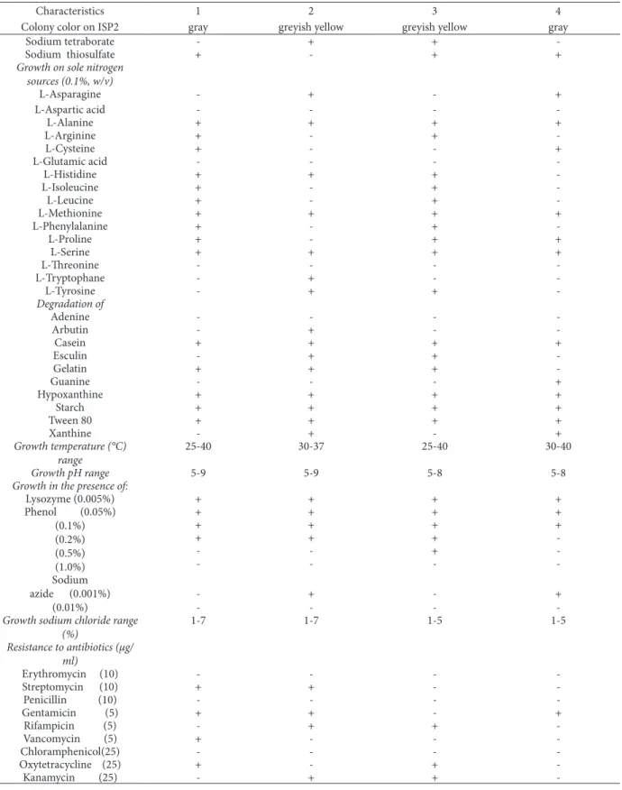
Table 2. Physiological properties separating strain TN17 from related Streptomyces species. Strains: 1, TN17; 2, Streptomyces
lav-endofoliae NBRC 12882T; 3, Streptomyces lilaceus NBRC 13676T and 4, Streptomyces gobitricini NBRC 15419T.
Characteristics 1 2 3 4
Colony color on ISP2 gray greyish yellow greyish yellow gray Production of diffusible
pigment - - -
-Melanin production on ISP6 - - +
-Melanin production on ISP7 - - +
-Melanoid pigment on
tryptone-yeast extract broth - - +
-Nitrate reduction + + + +
Growth on sole carbon sources (1%, w/v) L-Arabinose + + - + D-Fructose - + - -D-Galactose + - + + Glucose + + + + Glycerol + - + + Meso-Inositol - + + -D-Lactose - - + + Maltose + + + + D-Mannose - - - + D-Raffinose - - - -L-Rhamnose - - - -D-Ribose + - - + Sucrose + - - -D-Trehalose + - - + D-Xylose - + - +
Growth on sole energy sources (0.1%, w/v) Sodium acetate + + + -Sodium alginate - + - -Sodium benzoate - + - -Sodium butyrate - - + + Sodium citrate + + + + Sodium desoxycholate - + +
-Sodium hydrogen carbonate - + -
-Sodium nitrate - - + + Sodium oxalate - + - + Sodium perchlorate + - + + Sodium propionate - - + + Sodium pyruvate - + + -Sodium succinate + + - -Sodium sulfite - + -
-TAXONOMY AND ANTIMICROBIAL ACTIVITIES OF A NEW STREPTOMYCES 1053
Table 2. Continued
Characteristics 1 2 3 4
Colony color on ISP2 gray greyish yellow greyish yellow gray
Sodium tetraborate - + +
-Sodium thiosulfate + - + +
Growth on sole nitrogen sources (0.1%, w/v) L-Asparagine - + - + L-Aspartic acid - - - -L-Alanine + + + + L-Arginine + - + -L-Cysteine + - - + L-Glutamic acid - - - -L-Histidine + + + -L-Isoleucine + - + -L-Leucine + - + -L-Methionine + + + + L-Phenylalanine + - + -L-Proline + - + + L-Serine + + + + L-Threonine - - - -L-Tryptophane - + - -L-Tyrosine - + + -Degradation of Adenine - - - -Arbutin - + - -Casein + + + + Esculin - + + -Gelatin + + + -Guanine - - - + Hypoxanthine + + + + Starch + + + + Tween 80 + + + + Xanthine - + - + Growth temperature (°C) range 25-40 30-37 25-40 30-40 Growth pH range 5-9 5-9 5-8 5-8
Growth in the presence of:
Lysozyme (0.005%) + + + + Phenol (0.05%) (0.1%) (0.2%) (0.5%) (1.0%) + + + + + + + + + + + -- - + -- - - -Sodium azide (0.001%) (0.01%) -- +- -- +
-Growth sodium chloride range
(%) 1-7 1-7 1-5 1-5 Resistance to antibiotics (µg/ ml) Erythromycin (10) - - - -Streptomycin (10) + + - -Penicillin (10) - - - -Gentamicin (5) + + - + Rifampicin (5) - + + -Vancomycin (5) + - - -Chloramphenicol(25) - - - -Oxytetracycline (25) + - + -Kanamycin (25) - + +
-medium (Pridham and Gottlieb, 1984), TN17 uti-lizedL-arabinose, D-galactose, glucose, glycerol, maltose, D-ribose, sucrose and D-trehalose but not D-fructose, meso-inositol, D-lactose, D-man-nose, D-raffiD-man-nose, D-rhamnose and D-xylose as
sole carbon sources. This utilization was different from the patterns of all strains used for compari-son (Table 2). The four strains of Streptomyces have the sameas physiological characters such as the ability to utilize sole carbon and nitrogen sourc-Table 3. Cellular fatty acid contents (%). 1 - strain TN17; 2 - Streptomyces lavendofoliae NBRC 12882T ; 3 - Streptomyces lilaceus NBRC 13676T; 4 - Streptomyces gobitricini NBRC 15419T Fatty acids 1 2 3 4 C10:0 2-OH - - - -C11:0 - 0.753 - -C12:0 0.094 0.1244 0.358 -C12:0 2-OH 0.026 0.466 - -C12:0 3-OH 1.611 - 1.344 2.221 C13:0 - 1.447 0.145 0.285 C14:0 0.453 0.26 0,615 0.634 C14:0 2-OH 0.152 0235 - -C14:0 3-OH - - - -C15:0 3.120 0.126 0.9272 1.344 IsoC15:0 23.583 43.728 23.193 24.382 anteisoC15:0 29.889 44.306 29.859 31.467 C16:0 4.490 1.356 5.404 3.896 IsoC16:0 28.529 3.994 29.595 24.454 C16:0 2-OH - - - -C16:1 w 9 3.578 - 3.488 4.029 C17:0 0.146 - - 4.228 IsoC17:0 3.891 2.533 4.344 2.802 anteisoC17:0 0.3508 - 0.3715 0.2535 C18:0 - - 0.352 -C18:1 w 9 (cis) 0.0109 - - -C18:1 w 9 (trans) - - - -C18:2 w 9,12 0.0701 - - -C19:0 - - - -IsoC19:0 - - - -C20:0 - - -
-Table 4. Antimicrobial activities of the crude extract of the supernatant culture of Streptomyces sp. TN17
Test organism Diameter of inhibition zones (mm)
Micrococcus luteus LB14110 21
Staphylococcus aureus ATCC 6538 18
Salmonella enterica ATCC43972 14
Escherichia coli ATCC 8739
-Pseudomonas aeruginosa ATCC 49189
-Fusarium sp. 18
TAXONOMY AND ANTIMICROBIAL ACTIVITIES OF A NEW STREPTOMYCES 1055
es: glucose, maltose, D-mannose, D-raffinose and L-rhamnose (sole carbon sources) and L-aspartic acid, alanine, glutamic acid, methionine, L-serine and L-threonine (sole nitrogen sources).
Growth of strain TN17 was observed at a wide range of temperature (25–40°C), although the op-timal temperature range was between 25 and 30°C. The initial pH range for which growth of strain TN17 was observed was between pH 5-9; however, the op-timal pH value for growth was determined to be 7.5. Strain TN17 was also capable of growth in the pres-ence of 7% NaCl and 0.2% of phenol. In addition, the strain TN17 reduced nitrate to nitrite. Casein, gelatin, starch, hypoxanthine and Tween 80 were degraded by Streptomyces sp. TN17 but not adenine, arbutin, esculin, guanine and xanthine.
The detailed fatty acid profile of strain TN17 given in Table 3 was clearly different from that of
Streptomyces lavendofoliae NBRC 12882T,
Strep-tomyces lilaceus NBRC 13676T and Streptomyces
gobitricini NBRC 15419T. For Streptomyces lilaceus
NBRC 13676T, the major cellular fatty acids were
iso C15:0 (23.19%), anteiso C15:0 (29.85%) and iso C16:0
(29.59%) and smaller amounts of C16:0 (5.4%), C16 :1
w 9 (3.48%) and IsoC17:0 (4.34%). For Streptomyces
gobitricini NBRC 15419T, the major cellular fatty
acids were iso C15:0 (24.38%), anteiso C15:0 (31.46%)
and iso C16:0 (24.45%) and smaller amounts of C12:0
3-OH (2.22%), C16:0 (3.89%), C16:1 w 9 (4.02%), C17:0
(4.22%) and iso C17:0 (2.8%). For Streptomyces
laven-dofoliae NBRC 12882T the major fatty acids were iso
C15:0 (43.72%) and anteisoC15:0 (44.3%) and smaller
amounts of iso C16:0 (3.99%) and iso C17:0 (2.53%).
Based on the genotypic and phenotypic evidence, it is suggested that strain TN17 is a novel species of the genus Streptomyces, for which the name is
Strep-tomyces sp. TN17 (=DSM 42020T=CTM50229T).
Biological activities
As shown in Table 4, the ethyl acetate extract of the supernatant culture of the Streptomyces sp TN17 ex-hibited an inhibitory effect against M. luteus LB14110,
S. aureus ATCC 6538 (Gram positive bacteria), S. enterica ATCC43972 (Gram negative bacterium), Fusarium sp. (filamentous fungus) and C. tropicalis
R2 CIP203 (Yeast).
CONCLUSION
An aerobic bacterium TN17 was isolated from the oasis soil of Tunisia. The bacterium has morphological characteristics and chemotaxonomic properties consistent with its assignment to the genus
Streptomyces. This strain was compared
phenotypi-cally and phylogenetiphenotypi-cally with the nearest species in the genus Streptomyces: this comparison suggests that they are different from Streptomyces species.
Streptomyces sp. TN17 (=DSM 42020T=
CT-M50229T) showed antimicrobial activity against
Gram-positive and Gram-negative bacteria and fun-gi.
Acknowledgment - This work was supported by the CMCU project N°: 06/S 0901 2006-2009 “MELLOULI/AIGLE”.
REFERENCES
Altschul, S.F., Madden, T.L., Schäffer, A.A., Zhang, J., Zhang, Z., Miller, W., and D.J. Lipman (1997). Gapped BLAST
and PSI-BLAST: a new generation of protein database search programs. Nucl. Acids Res. 25, 3389–3402.
Anderson, A.S., and E.M. Wellington (2001). The taxonomy of Streptomyces and related genera. Int. J. Syst. Evol. Micr.
51, 797–814.
Athalye, M., Goodfellow, M., Lacey, J., and R.P. White (1985).
Numerical classification of Actinomadura and
Nocardi-opsis. Int. J. Syst. Evol. Micr. 35, 86–98.
Collins, M.D., and D. Jones (1980). Lipids in the classification
and identification of coryneform bacteria containing peptidoglycan based on 2,4-diaminobutyric acid. J. Appl
Bacteriol. 48, 459–470.
Edwards, U., Rogall, T., Blöcker, H., Emde, M., and E.C. Böt-tger (1989). Isolation and direct complete nucleotide
determination of entire genes. Characterization of a gene coding for 16S ribosomal RNA. Nucl. Acids Res. 17, 7843–7853.
Elleuch, L., Shaaban, M., Smaoui, S., Mellouli, L., Karray-Rebai, I., Fourati Ben Fguira, L., Shaaban, K.A., and H.
Laatsch (2010). Bioactive Secondary Metabolites from a
New Terrestrial Streptomyces sp. TN262. Appl Biochem
Biotechnol. 162, 579–593.
Felsentein, J. (1985). Confidence limits on phylogenies: an
ap-proach using the bootstrap. Evolution. 39, 783–789.
Goodfellow, M., and A.G. O’Donnell (1989), Search and
dis-covery of industrially-significant actinomycetes: Pro-ceeding of the 44th Symposium on Society for General Microbiology, (SCGM’89), Cambridge University Press, Cambridge, 343–383.
Gordon, R.E., Barnett, D.A., Handarhan, J.E., and C. Hor-Nay-Pang (1974). Nocardia coeliaca, Nocardia autotrophica
and the nocardin strains. Int. J. Syst. Evol. Micr. 24, 54–63.
Hopwood, D.A., Bibb, M.J., Chater, K.F., Kieser, T., Bruton, C.J., Kieser, H.M., Lydiate, D.J., Smith, C.P., Ward, J.M., and H. Schremph (1985). Genetic Manipulation of Strepto-myces: A Laboratory Manual. Norwich, UK: John Innes
Foundation.
Kroppenstedt, R.M. (1985). Fatty acid and menaquinone
analysis of actinomycetes and related organisms. In Chemical Methods in Bacterial Systematics, Edited by M. Goodfellow & D. E. Minnikin. London: Academic Press, 173–199.
Kroppenstedt, R., Stackebrandt, E., and M. Goodfellow (1990).
Taxonomic revision of the actinomycete genera
Actino-mudura and Microtetruspora. Syst. Appl. Microbiol. 13,
148–160.
Lechevalier, M.P., and H. Lechevalier (1970). Chemical
com-position as a criterion in the classification of aerobic ac-tinomycetes. Int. J. Syst. Evol. Micr. 20, 435–443.
Lechevalier, M.P., and H.A. Lechevalier (1970a). Composition
of whole-cell hydrolysates as a criterion in the classifica-tion of aerobic actinomycetes. In: The Actinomycetales. Prauser H. (Eds.) G. Fisher Verlag, Jena, 311–316.
Lechevalier, H.A., and M.P. Lechevalier (1970b). A critical
evaluation of genera of aerobic actinomycetes. In: The
Actinomycetales. Prauser H. (Eds.). G. Fisher Verlag,
Jena, 393–405.
Minnikin, D.E., Collins, M.D., and M. Goodfellow (1979). Fatty
acid and polar lipid composition in the classification of Cellulomonas, Oerskovia and related taxa. J. Appl
Bac-teriol. 47, 87–95.
Mellouli, L., Ameur-Mehdi, R.B., Sioud, S., Salem, M., and S. Bejar (2003). Isolation, purification and partial
charac-terization of antibacterial activities produced by a newly isolated Streptomyces sp. US24 strain. Res. Microbiol. 154, 345–352.
Pridham, T.G., and D. Gottlieb (1984). The utilization of
car-bon compounds by some Actinomycetales as an aid for species determination. J. Bacteriol. 56, 107–114.
Saitou, N., and M. Nei (1987). The neighbour-joining method:
a new method for reconstructing phylogenetic tree.
Mol. Biol. Evol. 4, 406–425.
Sasser, M. (1990). Identification of bacteria by Gas
Chroma-tography of cellular fatty acids. USFCC Newslett. 20, 16.
Shirling, E.B., and D. Gottlieb (1966). Methods for
character-ization of Streptomyces species. Int. J. Syst. Bacteriol. 16, 313–340.
Sierra, G. (1957). A Simple method for the detection of
lipoly-tic activity of microorganisms and some observations on the influence of the contact between cells and fatty substrates. Anton. Leeuw. Int. J. G. 23, 15-22.
Staneck, J.L, and G.D. Roberts (1974). Simplified approach to
the identification of aerobic actinomycetes by thin-layer chromatography. Appl. Microbiol. 28, 226–231.
Thompson, J.D., Gibson, T.J., Plewniak, F., Jeanmougin, F., and D.G. Higgins (1997). The Clustal X windows interface:
flexible strategies for multiple sequence alignment aid-ed by quality analysis tools. Nucl. Acids Res. 24, 4876– 4888.
Uchida, K., and A. Seino (1997). Intra- and intergeneric
rela-tionships of various actinomycete strains based on the acyl types of the muramyl residue in cell wall peptido-glycans examined in a glycolate test. Int. J. Syst.
Bacte-riol. 47, 182–190.
Waksman, S.A., and A.T. Henrici (1943). The nomenclature
and classification of the actinomycetes. J. Bacteriol. 46, 337–341.
Watve, M.G., Tickoo, R., Jog, M.M., and B.D. Bhole (2001). How
many antibiotics are produced by the genus
Streptomy-ces? Arch Microbiol. 176, 386–390.
Williams, S.T., and T. Cross (1971). Isolation, purification,
cul-tivation and preservation of actinomycetes. Methods
Microbiol. 4, 295–334.
Williams, S.T., Sharpe, M.E., and J.G. Holt (1989). Bergey’s
Manual of Systematic Bacteriology, Williams and Wilkins Company: Baltimore, 4.