POSTNATAL DEVELOPMENT 0F GLUTAMATERGIC RECEPTOR MEDIATED EXCITATORY POSTSYNAPTIC CURRENTS AND THEIR
MODULATIONS BY ACH AND DOPAMINE IN NUCLEUS ACCUMBENS
Par Liming Zhang
Département de Physiologie faculté de Médecine
Thèse présentée à la faculté des Études Supérieures en vue de l’obtention du grade de
Philosophiae Doctor (Ph.D.) en Sciences Physiologiques Septembre, 2002 ©Liming Zhang, 2002 / Gtade conféré (\ â compter du OO4 AVR,
o
1u
o
de Montréal
Direction des bibliothèques
AVIS
L’auteur a autorisé l’Université de Montréal à reproduite et diffuser, en totalité ou en partie, par quelque moyen que ce soit et sur quelque support que ce soit, et exclusivement à des fins non lucratives d’enseignement et de recherche, des copies de ce mémoire ou de cette thèse.
L’auteur et les coauteurs le cas échéant conservent la propriété du droit d’auteur et des droits moraux qui protègent ce document. Ni la thèse ou le mémoire, ni des extraits substantiels de ce document, ne doivent être imprimés ou autrement reproduits sans l’autorisation de l’auteur.
Afin de se conformer à la Loi canadienne sur la protection des renseignements personnels, quelques formulaires secondaires, coordonnées ou signatures intégrées au texte ont pu être enlevés de ce document. Bien que cela ait pu affecter la pagination, il n’y a aucun contenu manquant.
NOTICE
The author of this thesis or dissertation has granted a nonexclusive license allowing Université de Montréal to reproduce and publish the document, in part or in whole, and in any format, solely for noncommercial educational and research purposes.
The author and co-authors if applicable retain copyright ownership and moral rights in this document. Neither the whole thesis or dissertation, nor substantial extracts from it, may be printed or otherwise reproduced without the author’s permission.
In compliance with the Canadian Privacy Act some supporting forms, contact information or signatures may have been removed from the document. While this may affect the document page count, it does not represent any loss of content from the document.
Université de Montréal Faculté des Etudes Supérieures
Cette thèse intitulée
POSTNATAL DEVELOPMENT 0F GLUTAMATERGIC RECEPTOR MEDIATED EXCITATORY POSTSYNAPTIC CURRENTS AND THEIR MODULATIONS BY ACH AND DOPAMINE IN NUCLEUS ACCUMBENS
Présentée par
Liming Zhang
a été évaluée par un jury composé des personnes suivantes
Dr. Jacques de Champlain Dr. Jean-Claude Lacaille Dr. Ariel Agmon
Dr. Richard A Warren
représentant de la F.E.S.
Ce travail décrit le développement postnatal des courants postsynaptiques
excitateurs (CPSEs) dans les neurones épineux moyens (MS) du noyau accumbens
(nAcb) du rat in vitro ainsi que les effets neuromodulateurs de l’acétylcholine (ACh) et
de la dopamine (DA) en utilisant la technique whole-cell patch-cÏamp.
Les CPSEs, évoqués par une stimulation électrique, ont été enregistrés dans plus
de 500 neurones du nAcb pendant le développement postnatal à partir du jour de la
naissance (jour postnatal 0; P0) jusqu’à P71. Un CPSE a été identifié dans tous les neurones enregistrés et à tous les âges, démontrant que des synapses excitatrices fonctionnelles étaient déjà présentes dans le nAcb le jour de la naissance. Dans la majorité de neurones (80%), les CPSEs avaient deux composantes distinctes. La première atteignait un maximum entre 4 et 2lms après le début de stimulus, avait une relation ‘R
VM linéaire et était sensible au CNQX. La seconde composante pouvait être mesurée entre 20 et 138 ms après le début de stimulus, avait une relation IR-VM en y avec un
maximum autour de —40 mV et était sensible à l’APV. Ces caractéristiques montrent que
la composante précoce des CPSEs étaient médiée par des récepteurs de type AMPA/KA
tandis que la deuxième était médiée par des récepteurs de type NMDA.
Pendant les premiers jours suivant la naissance, l’amplitude des CP$Es était relativement petite. Par la suite, les CPSEs ont augmenté progressivement jusquTà la fin
de la deuxième semaine postnatale.
À
partir de ce moment, l’amplitude de la composante précoce s’est stabilisée jusqu’à l’âge adulte alors que celle de la composante tardive a commencé à diminuer pour devenir virtuellement nulle dans les préparations provenant d’animaux âgés de plus de 3 semaines. Le rapport entre l’amplitude de la composanteiv
tardive et celle de la composante précoce a augmenté graduellement durant les deux premières semaines et a par la suite diminué de façon marquée. Ces résultats suggèrent
que l’expression de CPSEs médiés par les récepteurs NMDA est prédominante durant la
deuxième semaine du développement postnatal dans le nAcb.
Nous avons trouvé que l’Adi produisait deux effets médiés par différents types
de récepteurs sur les CPSEs. L’ACh diminuait les CPSEs en agissant sur des récepteurs
muscariniques tandis qu’elle augmentait les CPSEs en agissant sur des récepteurs nicotiniques. Cependant, l’effet excitateur produit par l’activation des récepteurs nicotiniques était généralement masqué par les effets inhibiteurs muscarmniques en
absence d’un antagoniste de ces derniers. Donc, l’activation des interneurones
cholinergiques dans le nAcb pourrait produire une excitation nicotinique rapide et une inhibition muscarmnique plus lente.
La DA, par une action sur les récepteurs de la famille Dl, produisait une
diminution importante du rapport entre les CPSEs médiés par des récepteurs NMDA et ceux médiés par les récepteurs AMPAJKA en inhibant davantage les CPSEs médiés par
les récepteurs NMDA. En effet, l’inhibition produite par la DA sur les CPSEs NMDA
était comparable à celle produite par l’APV, un antagoniste spécifique des récepteurs NMDA. Les effets de la DA sur les CPSEs ne semble pas impliquer la protéine kinase A
ni la protéine kinase C, parce que cette action était résistante aux inhibiteurs de protéines
kinases H89 et Ro-32-0432.
Les agonistes cholinergiques et dopaminergiques ont changé le rapport des
réponses à des stimuli pairés (paired pulse ratio) sans toutefois modifié la conductance membranaire ni la réponse des neurones au glutamate en présence de TTX, suggérant que
leurs effets sur les CPSEs étaient principalement médiés par des mécanismes présynaptiques. Cependant, les effets postsynaptiques de l’ACh et et de la DA ont pu être masqués par la présence de QX-314 dans la pipette d’enregistrement car cette substance bloque certains canaux ioniques K et Na qui auraient pu être modulés par l’Adi et la DA.
En résumé, cette recherche démontre que les réponses synaptiques médiées par les récepteurs NMDA atteignent leur maximum pendant la seconde semaine du développement postnatal et pourraient jouer un rôle important dans les processus développementaux dépendant de l’activité. La modulation des CPSEs NMDA et AMPAJKA pendant cette période par la DA et l’ACh suggèrent que ces substances pourraient jouer un rôle déterminant aussi pendant cette période.
Mots-clés : CPSE, développement postnatal, Acétylcholine, dopamine, récepteur AMPA!KA, récepteur NMDA, noyau accumbens, courant postsynaptique excitateur
SUMMARY
This work describes the postnatal development of excïtatory postsynaptic currents (EPSCs) of medïum spiny neurons (MS) in nucleus accumbens (nAcb) suces of rat as
well as the modulations of two classic neurotransmitters dopamine (DA) and
acetylcholine (ACh) on EPSCs using whole-cell patch-clamp technique.
EPSCs were evoked by local electricai stimulation in 509 nAcb neurons during postnatal development from the day of birth (postnatal day 0; P0) to P71. An EPSC was found in ah recorded neurons of ail ages, showing that functional excitatory synapses
were aiready present in the nAcb on the day of birth. In majority of neurons (80%) the
EPSCs had two distinct components: an early component with a peak between 4 and 21
ms after stimulus onset, linear IR-Vm and sensitive to CNQX and a late one that was
found from 20 to 138 ms after stimulus onset, had a V-shape IR-Vm relationship with a peak around —40 mV and sensitive to APV. These characteristics demonstrate the eariy
and late components of the EPSC were mediated by AMPA and NMDA receptors
respectively. During the first few days after birth, the amplitudes of both early and late components of the EPSCs were relatively smail and then started to increase untii the end of the second postnatal week. Whereas the early component of the EPSC appeared to stabilize from that point on, the late component began to decrease in samples from animais aging more than 3-week-old. In addition, the ratio between the amplitudes of
late and early components followed a developmental pattem gradualiy increased during
the first two postnatal weeks followed by a decrease dramatically. Together, these results
show that there is a dominant expression of NMDA receptor-mediated EPSC during the
Two remarkable differential actions of ACh were found on the EPSCs by two types of ACh receptors. ACh depressed EPSCs through muscarinic Ml receptors, whereas it enhanced EP$Cs through nicotinic receptors, suggesting that nAcb cholinergic interneurons may produce a fast nicotinic excitation and slow muscarinic inhibition. However, nicotinic receptor-mediated effects were usually masked by muscarinic receptor-mediated actions in our experimental condition in vitro. Moreover, we also found that the inhibitory effects of ACh on NMDA receptor- but not on AMPA receptor mediated EPSCs significantly increased during the first two postnatal weeks.
DA, through an action on D1-like receptor, distinctly decreased the ratio of NMDA receptor- to AMPA receptor-mediated EP$Cs, rnimicked the effect of APV on EPSCs and abolished almost completely NMDA receptor-mediated EP$Cs with minimal effect on AMPA receptor-mediated EPSCs. The DA-induced depression of EPSCs did not involve either protein kinase A or protein kinase C, because this action was resistant to the protein kinase inhibitors H29 and Ro-32-0432.
Both cholinergic and dopaminergic agonists altered the ratio of paired-pulse stimulation-evoked EPSCs, but did not change input conductance of membrane of MS neurons. Also both of transmitters had no effect on glutamate injection-evoked EP$Cs in the presence of TTX and QX-3 14, suggesting their modulation on EPSCs is mainly presynaptic in the postnatal development of nAcb. However, in accordance with the effect of QX-3 14 on membrane potentials during the modulation of ACh, and distinct change in the ratio of NMDARIAMPAR-EPSCs during the modulation of DA, it is suggested that there are postsynaptic effects of ACh and DA either in the nAcb during the postnatal development. This work shows that NMDA receptor-mediated synaptic
vil’
responses are dominant during postnatal development and that DA and ACh effectively modulate NMDA receptor-mediated currents, indicating that NMDA receptor-mediated synaptic events may play important roles in postnatal development. The modulations of NMDA receptor- and AMPA receptor-mediated EPSCs by DA and ACh may be important for activity-dependent developmental processes or other plasticity in the nAcb. In addition, quite significant modulation of NMDA receptor-mediated EPSCs by ACh and DA during postnatal development could be important for the study in the etiology of schizophrenia.
Key words: Nucleus accumbens, Excitatory postsynaptic currents, Postnatal developrnent, Cholinergic and dopaminergic modulation, NMDA receptor, AMPA receptor, Pre and postsynaptic modulations.
SOMMAIRE.iii
SUMMARY.vi
LIST 0F FIGURES xiii
LIST 0F TABLES xviii
LIST 0F ABBREVIATIONS xix
DEDICATION xxi
ACKNOWLEDGEMENTS xxii
CHAPTER I: INTRODUCTION
1. Overview 2
2. Anatomical and functional characteristics ofthe nucleus accumbens 4
2.1. Composition ofthe nucleus accumbens 4
2.1.1. Innervations and projections 4
2.1.2. Neuron types $
2.1.2.1 Medium spiny neurons 2
2.1.2.2 Large aspiny neurons 9
2.1.3. Synaptic framework in the nucleus accumbens 10
2.2.Membrane properties of MS neurons during postnatal development 12
3. An overview ofionotropic glutamate receptors and their properties 16
3.1. Glutamatergic receptors-mediated EPSCs 17
3.2. AMPA receptors 17
3.2.1. AMPA subunits and flip or flop isoforms during development 19
3.3. KA receptors 20
3.4. NMDA receptors 21
3.4.1. Glycine, a co-agonist 22
3.4.2. NMDA receptor subunits and developmental regulation 22 3.4.3. Components of subunits determine properties ofNMDA receptors 24
3.4.4. Single channel properties during developrnent 26
4. Cholinergic receptors 27
4.1 Cellular localization of cholinergic receptors 22
4.1.1. Muscarinic ACh receptors 28
4.1.2. Nicotinic ACh receptors 30
4.1.2.1. Subunit composition of functional nicotinic ACh receptors 31 5. Muscarinic ACh receptors and synaptic transmission in the nucleus accumbens... 32
X
5.1. Activities ofmuscarinic receptors modulate the functions ofMS neurons 32 5.1.1. Modulation of ionic conductance by activating muscarinic receptor 32 5.2. Activation ofmuscarinic receptor modulates the release of glutamate 34 5.3. ACh release regulating by muscarinic receptors in LA neurons 35
6. Activation of nicotinic ACh receptors facilitates synaptic transmission in CNS 36 6.1. Facilitation of glutamate release by presynaptic nicotinic receptors 36 6.2. Facilitation ofdopamine release by presynaptic nicotinic receptors 38
6.3. Facilitation ofACh release 40
6.4. Pre- but flot post-synaptic nicotinic receptors mediates the facilitation 41
7. Dopaminergic modulation of neuronal excitability in the nucleus accumbens 41
7.1. Doparnine receptor subtypes 41
7.2. Dopamine receptor expression in the nucleus accumbens 42
7.3. Dopaminergic modulation of neuronal activities in the nucleus accumbens 42
7.3.1. Modulation of ionic conductance 43
7.3.2. Modulation of synaptic transmission 44
8. Aims ofthe present study 46
CHAPTER II (ARTICLE 1): POSTNATAL DEVELOPMENT 0F EXCITATORY POSTSYNAPTIC CURRENTS IN NUCLEUS ACCUMBENS
Acknowledgements 49 Abstract 50 Résumé 51 Introduction 53 Methods 55 Suce preparation 55
Recording and electrical stimulation 55
Analysis 56
Morphology 57
Results 58
Characteristics and nature ofthe EPSCs 58
Developmental characteristics of postsynaptic currents 65
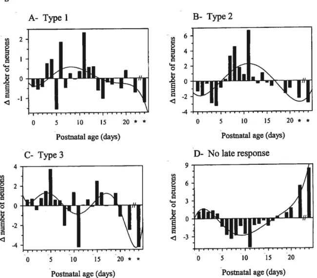
Type I response 65
Type II response 70
Type III response 70
No late response 71
Morphology 76
Discussion $1
Validity ofthe observations 82
Functional significance 86
CHAPTER III (ARTICLE 2): MUSCARINIC AND NICOTINIC PRESYNAPTIC
MODULATION 0F EPSCS IN THE NUCLEUS ACCUMBENS DURING
POSTNATAL DEVELOPMENT Résumé 94 Abstract 96 Introduction 96 Methods 97 S lice preparation 97 Recording 97 Synaptic stimulation 97 Pharmacological agents 9$ Statistics 98 Resuits 9$
Characteristics of glutamatergic EPSCs 98
Effects ofcholinergic receptor agonists 99
Effects ofmuscarinic receptor antagonists 100
Effects ofthe nicotinic receptor agonist 101
Effects of ACh as function of postnatal age 102
Locus ofthe cholinergic modulations ofevoked EPSCs 102
Discussion 103
Locus of cholinergic receptors 105
Muscarinic depression ofEPSCs 106
Nicotinic potentiation ofEPSCs 106
Functional considerations 107
Acknowledgements 107
References 107
CHAPTER IV (ARTICLE 3): PREFERENTIAL INHIBITION 0F NMDA
RECEPTOR-MEDIATED EPSCS BY DOPAMINE DURING DEVELOPMENT IN NUCLEUS ACCUMBENS
Abstract 114
Résumé 116
Introduction 118
Materials and methods 121
Slice preparation 121
Recording 121
Synaptic stimulation and drugs application 122
Currents measurement 124
Statistics 124
Results 125
Characterization of the glutamatergic EPSCs 125
xii
Dopamine reduced the ratio ofNMDAR-EPSCs
to AMPAIKAR-EPSCs 130
DA inhibited both isoÏated NMDAR-EP$Cs and
AMPAJKAR-EPSCs 133
Inhibition ofDA on EPSCs is mediated by a D1-like receptor 135 Pre and postsynaptic mechanism are involved
in the attenuation of EPSCs by DA 139
Discussion 143
DA inhibited both NMDAR-EP$Cs and AMPA/KAR-EPSCs
inthenAcb 143
Both pre- and postsynaptic rnechanism were involved
in inhibition of EPSCs by DA 145
The inhibitory effect of DA on EPSCs did flot
involve PKA and PKC pathways 148
Functional implications 151
Acknowledgments 153
References 154
CHAPTER V: GENERAL DISCUSSION
1. Experimental aim and resuits 164
2. Expression ofNMDA receptor subunits and the performances ofEPSCs 165
3. NMDA receptors and synapsogenesis 167
4. Paired-pulse ratio and presynaptic mechanisms 169 5. Direct interaction between NMDA receptor and Dl receptor 171 6. NMDA receptor trafficking and use-dependent expression 173 7. Interaction oftransmitters in the nucleus accumbens 174
2. NMDA receptors and shizophrenia 177
CHAPTER I (INTRODUCTION)
Figure Y. The anatomical location of the nAcb 5
Figure 2. Diagram with inputs and outputs ofthe nAcb 7
Figure 3. Diagram with synaptic organization 13
CHAPTER II (ARTICLE 1)
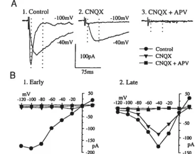
Figure 1. Characteristics and nature ofthe EPSCs 60
figure 2. Effects of ionotropic glutamatergic antagonists on the EP$Cs 64 Figure 3. Classification ofneurons according to the relative size of
the late response 67
f igure 4. Distribution of type I, II, III and no late response neurons
as a function of postnatal age 69
f igure 5. Ratio between the late and early components ofthe EPSCs
as a function of postnatal age 73
Figure 6. Characteristics ofthe early and late response
as a function of postnatal age 75
(A) Early response delay 75
(B) Early response decay 75
(C) Late response delay 75
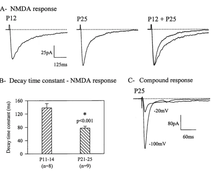
figure 7. NMDA responses 77
(A) NMDA receptor-mediated EPSCs 77
(B) NMDA EPSC decay constant 77
(C) Compound response 77
xiv
(A)P2 .79
(B)P7 .79
(C)P14 79
(D)P22 79
Figure 9. Graphs for the measurements of neuronal development 80
(A) Celi body perimeter $0
(B) Ceil body area 80
(C) Number ofprimary dendrites 80
CHAPTER III (ARTICLE 2)
Figure 1. Nature ofthe EP$C evoked by local electrical stimulus in the
presence ofBMI 98
(A) Isolaiton ofEPSCs 98
(B) I-V relationship in the presence or absence of CNQX
andAPV 98
Figure 2. Effect of cholinergic receptor agonists on the EPSCs 99
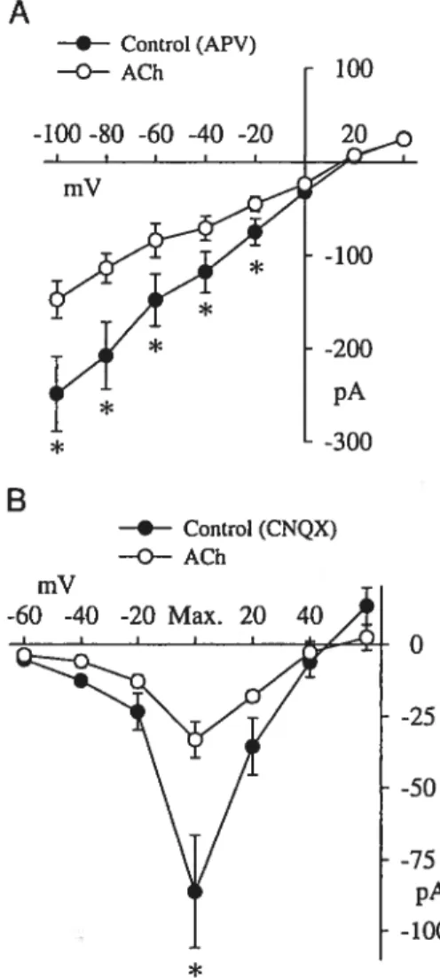
(A) Representative traces 99
(B) I-V relationship in the presence ofCCh 99
(C) I-V relationship in the presence of ACh 99
Figure 3. Effects of ACh on the EPSCs 100
(A) Effects ofACh on AMPA!KA receptor-mediated EPSCs 100
(B) Effects ofACh on NMDA receptor-mediated EPSCs 100
figure 4. Effects ofACh on the EPSCs in the presence ofatropine 101
(A) Effects of atropine itself on EPSCs 101
(C) Effect of ACh on I-V relationship in presence of atropine 101
figure 5. Effects ofnicotinic agonist and antagonist on the early and late
EPSCs in the presence ofatropine 102
(A) Effect ofDMPP on EPSCs 102
(B) Effect ofDMPP on the I-V relationship 102
(C) Mecamylamine blocked the effect of DMPP on EPSCs 102
figure 6. Effects ofACh as a function of postnatal age 103
(A) ACh on early response 103
(B) ACh on late response 103
figure 7. Effects of cholinergic agonists on holding membrane currents 103
(A) Effects ofACh 103
(B) Effect of nicotinic receptor activation 103
figure 8. Locus ofthe effects ofcholinergic agonist on EPSCs 104
(A) Effect of ACh on PPR 104
(B) Effect ofDMPP on PPR 104
(C) Effect ofACh on glutamate injection-evoked EP$Cs 104
(D) Effect of CCh on membrane potential and firing properties
ofMS neurons 104
CHAPTER IV (ARTICLE 3)
figure 1. Pharmacologically isolated EPSCs 127
(A) Two distinct components ofthe EPSCs 127
(B) I-V relationship ofthe evoked EPSCs 127
f igure 2. Effect ofdopaminergic agonist on the EPSCs 129
xvi
(B) Average I-V relationship ofthe early and late responses 129 Figure 3. DA decreased ratio of late to early component ofthe EPSC 131
(A) Representative traces 131
(B) Statistical comparison 131
(C) Late to early component ratio as a function of postnatal age 131 Figure 4. The inhibitory effects of DA and APV on late component of
compound EP$Cs 132
(A) Comparison for inhibitory efficacy ofDA and APV 132 (B) I-V relationship for the representative neuron 132 (C) ¾ inhibitions produced by DA and APV on late EPSCs 132
Figure 5. DA inhibited both NMDAR-EPSCs and AMPA/KAR EPSCs 134
(A) A representative NMDAR-EPSC 134
(B) A representative AMPA!KAR-EPSC 134
(C) I-V relationship for NMDAR-EPSC with or without dopamine 134 (D) Time course ofDA inhibition ofAMPA!KAR-EPSCs 134 (E) Sumrnary for the effect of dopamine on AMPA!KAR-EPSCs 134 Figure 6. The inhibitory effect of DA on the EPSCs was mediated
by D1-like receptor 137
(A) SKF 38393 mimicked the inhibitory effect ofDA 137 (B) SCH23390 blocked the inhibitory effect ofDA 137
(C) Summary for the blockade ofSCH2339O 137
Figure 7. Locus ofthe effects of dopaminergic agonist on EPSCs 142 (A) Representative traces from PPR at —lOOmV 142
(A’) Summary for 12 celis at —lOOmV 142
(B) Representative traces from PPR at —4OmV 142
(B’) Summary for 12 cells at —4OmV 142
(C) The effect ofDA on membrane current conductance 142
(D) The effect ofDA on glutamate injection-induced EPSCs 142
(D’) Summary of 5 ceils for glutamate-injection 142
CHAPTER V (GENERAL DISCUSSION)
f igure 1. A diagram for the proposed mechanisms of ACh and DA effects
LIST 0F TABLES
Table I (in article 1) Characteristics of different types ofresponse 68 Table I (in article 3) The effects of DA on EPSC amplitudes in the presence of
ACh Acetylcholine
AChR Acetylcholine receptor
ACSf Artificial cerebrospinal fluid
AMP Adenosine monophosphate
AMPA u-amino-3 -hydroxy-5 -methyl-4-isoxazolepropionate
AMPA/KA AMPA/kainate
AMPAR- AMPA receptor-mediated
APV : D(-)2-amino-5-phosphonopentanoic acid
ATP Adenosine triphosphate
BAPTA Bis(2-aminophenoxy)ethane-N,N,N’, N’ -tetraacetate
BMI Bicuculline methiodide
Ca2 : Calcium
cAMP Cyclic adenosine monophosphate
CCh Carbachol
ChAT Choline acetyl transferase
C1 Chloride
7C1 Kyn 7-chlorokynurenic
CNQX 6-cyano-7-nitroquinoxaline-2,3 -dione
CNS Central nervous system
CTZ Cyclothiazide DA Dopamine DAG Diacylglycerol Dl-113E Dihydro-13-erythoidine DMPP 1,1 -dimethyÏ-4-phenylpiperazinium DMSO Dimethlysulfoxide
DOPAC 3 ,4-dihydroxyphenylacetic acid
EPSCs Excitatory postsynaptic currents EPSP Excitatory postsynaptic potential
FS fast spiking
GABA y-aminobutyric acid
GluRs Glutamate receptors
G-protein Guanine nucleotide binding protein
GTP Guanine triphosphate
HVA homovanillic acid
1P3 Inositol 1,4,5 triphosphate
IP$C Inhibitory postsynaptic current
IPSP Inhibitory postsynaptic potential
K Potassium
KA Kainate
LA Large aspiny
LT$ Low threshold spike
xx
McN-A-3 43 : 4- [[[(3 -chlorophenyl)amino] carbonyl] oxy]
-N,N,N-trimethyl-2-butyn- 1 -aminium chloride
MLA Methyllycaconitine citrate
MS Medium spiny
Na Sodium
nAcb : Nucleus accumbens
nAChR Nicotinic ACh receptor
NMDA N-methyl-D-aspartate
NMDAR- NMDA receptor-mediated
NO Nitric oxide
PFC Prefrontal cortex
PKA Protein kinase A
PKC Protein kinase C
PLC Phospholipase C
PP 1 Phosphatase 1
RMP Resting membrane potential
TTX Tetrodotoxin
C:
I would like to thank Dr. Richard A. Warren for the confidence he put in me and
for bis generous support, encouragement and invaluable help during the time I worked
with him. His enthusiasm for research and innovative ideas has been a continuous
motivation for me. Perhaps, most importantly, his deference and his optimistic spirit have created an environment in the laboratory where it was aiways a pleasure to work
and I could flot have accompÏished the present work without that during ail the years of
my studies.
As wefl, I thank Dr. Vincent F. Castellucci who recommended me to Dr. Warren’s
laboratory for deepening my expertise in neuroscience research fields.
I would like to express my great gratitude to Dr. Robert W. Dykes for supporting
my studies in neuroscience.
I wish to express my sincere gratitude and appreciation to Dr. Guichun Yu, my
wife, for everything, particularly for the initiative and inspiration that allowed me to
complete my Ph.D at Université de Montréal, Canada.
I am also particularly gratefiul to Miss Xiaosi Zhang, my daughter and a medical
student at Université de Montréal, for her support and helping me to understand every letter in french as well as revising the final version ofthis thesis.
2 INTRODUCTION
1. Overview
The nucleus accumbens (nAcb) is an important point of convergence for different afferents originating in limbic structures (Lopes da Silva, 1984; Pennartz and Kitai, 1991; Pennartz et al., 1994; O’Donnell and Grace, 1995; Finch, 1996). Several of these pathways are thought to be glutamatergic and to provide an excitatory drive by activating NMDA andlor AMPA!KA receptors necessary to trigger firing activity in nAcb neurons (Kombian and Malenka, 1994). In addition, the nAcb receives a dense dopaminergic input from the ventral tegmental area (VTA), and this system lias been implicated in drug addiction and other neuropsychiatric disorders (Ungerstedt, 1971; Schiistrom et al., 1 998a,b). The nAcb also contains a small population of cholinergic interneurons, whicli play an important role in modulating glutarnatergic transmission (Sugita, et al., 1991; Hersch, et al., 1994; Zhang and Warren, 2002; de Rover et al., 2002).
The nAcb lias been proposed to serve as an interface between the limbic system and the extrapyramidal motor system (Mogenson et al., 1980; Mogenson and Yim, 1981; Poweli and Leman, 1976; Yang and Mogenson, 1984; Yim and Mogenson, 1982). Studies have provided evidence for the involvement of the nAcb in a number of functions including motivation (Mogenson et al., 1980; Robbins and Everitt, 1996; Swerdlow and Koob, 1987), attention (Solomon and Staton, 1982; van den Bos et al., 1991), and reward (Apicella et al., 1991; Colle and Wise, 1988; Robbins and Everitt, 1996; fantin and Bottecchia, 1984; OÏds, 1990). Recent studies have shown its involvement in learning and plasticity (Parkinson et al., 2000). Moreover, the nAcb may be involved in mediating some of the therapeutic actions of antipsychotic drugs that inactivate the mesencephalic
dopaminergic ceils that project to this region when administered chronically (Chiodo and Bunney, 1983; White and Wang, 1983). Chaotic neurotransmissions in the nAcb could
be a critical determinant in some neuropsychiatrie disorders, including schizophrenia,
Tourett’s syndrome and drug addiction (Koob and Nestier, 1997; Wise, 1998).
furthermore, selective loss of cholinergic interneurons in the nAcb has also been observed in schizophrenia (Hoit et al., 1999) and Alzheimer’s disease (Lehéricy et al.,
1989). Additionally, an alteration of dopaminergic transmission is thought to play a key
role in psychiatrie disorders (Sokoloff et al., 1992), such as, dopaminergic hyperfunction
has been implicated in schizopbrenia (Gray et al., 1995; Joyce, 1993; Joyce and Meador
Woodruff, 1997). Interest in understanding cholinergic and dopaminergic mechanisms in controlling or regulating motor and psychological function in mammals has been growing
since acetylcholine (ACh) and dopamine (DA) were postulated to play a role in the
pathophysiology described above.
Ionotropic glutamate receptors-mediated events including excitatory postsynaptic currents (EPSC) play a crucial role in synaptogenesis and formation of neuronal circuitry,
as well as in synaptic plasticity including long-term potentiation (LTP) and long-term
depression (LTD). However, excessive activation of glutamate receptors might induce excitotoxic neuronal celi death, which is thought to contribute to neurodegeneration
(Choi, 1992; Lipton and Rosenberg, 1994). Defects in glutamatergic transmission in the
nAcb are thought to be involved in the pathophysiology of schizophrenia (Carlsson and Clarlsson, 1990a, b; Olney and Farber, 1995; Meador-Woodruff and Healy, 2000). However, during postnatal development, the characteristics of EP$C and its modulation
4 by classic neurotransmitters ACh and DA in the nAcb remain largely enigmatic. The present thesis atternpts to provide some answers to these questions.
2. Anatomical and functional characteristics of the nucleus accumbens
Because of its similarities including cytoarchitecture, neurochernistry and afferent and efferent connections with the dorsal striatum, the nAcb is usually considered as a ventromedial extent of the striatum or the ‘ventral striatum” (Heimer and Wllson, 1975; Heimer et al., 1997; Swanson and Cowan, 1975). Based on studies of its connectivity and distribution of neurotransmitters and chemical markers, the nAcb can be divided into two teilitories (Groenewegen and Russchen, 1984; Heimer et al., 1991; Brog et al., 1993; Zahm and Heimer, 1993). The portion ofthe nAcb surrounding the anterior commissure is known as the core where enkephalin and opioid receptors are rich, and strong immunoreactivity for calcium-binding protein, calbindin D28k (CaB) is found. The rnedio-ventral region of the nAcb is called the sheil in which dense concentrations of substance P (SP), dynorphin, and tyrosine hydroxylase (TH) overlaping enkephalin- or CaB-poor zones are exhibited (Zahm and Brog, 1992; Jongen-Relo et al., 1994).
[Figure 1. The anatomical location ofthe nAcb]
2.1. Composition oftlie nucleus accumbens
2.1.1. Innervations and projections
The major afferents of the nAcb arise primarily from limbic structures including the prefrontal cortex (PfC), hippocampus, basal amygdaloid complex and midline thalamic nuclei (Groenewegen et al., 1982, 1987; Jayaraman, 1985; Kelley and
Figure 1. An anatomical indication ofthe nAcb and its subterritories
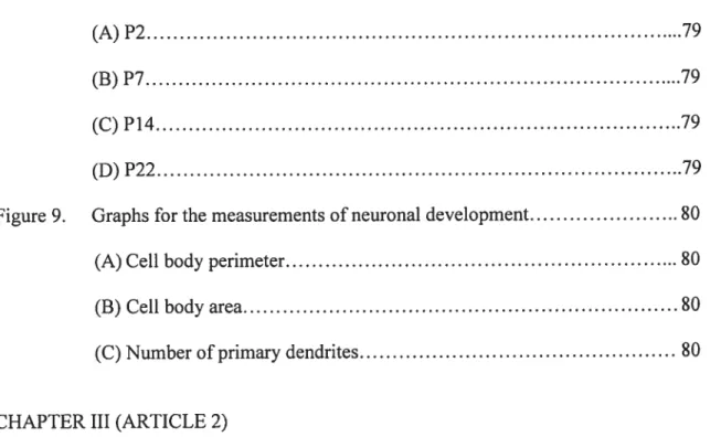
A. A parasagittal section of rat whote twain. B. A drawing shows mainly parts offorebrain to match the sagittal section. nAcb, nucleus accumbens (highlight areas); aca, anterior
commissure; VP, ventral pallidum; CPu, caudate-putamen. The circles flhled with white color indicate the sites where recordings were done in nAcb. The circles fllled with black color show the places in which ttie stimuli were given for evoked responses. C. Coronal section through the forebrain ofthe rat showing SP immunoreactivity. Note that a border between the core(Co) and the dorsally adjacent main part ofthe striatal complex, CPu can not be identifled, whereas the border between the Co and the shell(Sh) is distinct (arrowheads). (Paxions and Watson, 1986; De Olmos and Heimer,1999).
Figure 1.
A
‘4
/
B
acaJ
djC
t 2 aca_-Sh
Co
.1
CPuSh
6 Domesick, 1982; KeIIey and Stinus, 1984; Kelley et al., 1982; Krayniak et aI., 1981; Newman and Winans, 1980; Phillipson and Griffiths, 1985; Meredith et al., 1990). These inputs are ail thought to be glutamatergic. The nAcb also receives inputs from the ventral pallidum, dopaminergic VTA, serotonergic median raphe nucleus, and the noradrenergic celi group located in the nucleus ofthe solitary tract (Groenewegen et al., 1987; Brog et al., 1993; Berendse et al., 1992). The output of the nAcb is GABAergic and is primarily directed to the ventral pallidum (Hakan et al., 1992; Yang and Mogenson, 1985; Zahm and Heimer, 1990), which is involved in the activation of voluntary movements (Heirner et al., 1994; $werdlow and Koob, 1987). This input-output organization suggests that the nAcb somehow provides a centre for Ïimbic integration with motor systems driven by the ventral pallidum (Beninger et al., 1983; Lopes da Silva et al., 1984; Mogenson et al., 1980). In addition, the nAcb also projects to the VTA and the media! part of the substantia nigra pars compacta (Heimer et al., 1991; Mogenson et al., 1983; Nauta et al.,
1978; Swanson and Cowan, 1975).
The core of the nAcb has been reported to receive its main cortical input from the prelimbic PFC (Brog et al., 1993; Sesack et aÏ., 1989; Berendse et al., 1992; Montaron et al., 1996), and dorsal subiculum (Brog et al., 1993). It also appears to project to the dorsal portion of the ventral pallidum (Zahm and Heimer, 1990; Heimer et al., 1991). The sheli receives its major inputs from the infralimbic PFC and the ventral subiculum (Kelley and Domesick, 1982; Brog et al, 1993; Yang and Mogenson, 1984; Sesack and Picel, 1990; Aylward and Totterdel!, 1993) and sends projections to the ventro-medial part ofthe ventral pallidum.
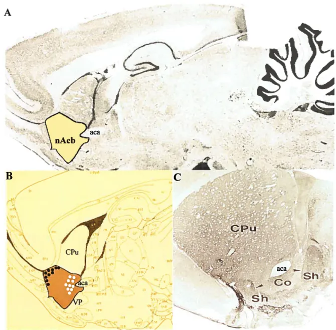
Figure 2.
A B hippocampus prefrontal cortex amygdala nigra amygdalaFigure 2. Diagram with inputs and outputs of the nAcb
Schematic representation of the input-output relationships of clusters of neurons in the nAcb.
A. Different clusters of neurons (ensembles?) receive different combinations of converging
inputs and project to distinct targets, including the ventral pallidum, the lateral hypothalamus,
and the ventral mesencephalon. B. The various (limbic) cortical and subcortical structures
that project to the nAcb are strongly and, in a number of cases, reciprocally interconnected. Note that the nAcb, via the ventral pallidum and the mediodorsal thalamic nucleus is involved
in a closed thalamocortical-basal ganglia loop. nAcb, nucleus accumbens; MD, mediodorsal
thalamic nucleus; P’vÇ paraventricular thalamic nucleus. (Groenewegen et al., 1999). ventral pallidum
prefrontal cortex
8 2.1.2. Neuron types
As described in series of remarkable studies of the neostriatum (Bolam et al., 1984; Kawaguchi, 1992, 1993; Kawaguchi et al., 1989, 1990), the nAcb also consists of five different types of neurons in accordance with the neurochemical, morphological and physiological characteristics (Chang and Kital, 1985; Hedreen, 1981). Two types ofthe celis have been described with some details (Sesack and Pickel, 1990; O’Donnell and Grace, 1993). They are the GABAergic medium spiny (MS) neurons, which are the only identified projection neurons in the nAcb, and the large aspiny (LA) neurons, which are cholinergic intemeurons. In addition to MS neurons, two other types of GABAergic interneurons have been identified: the fast spiking (FS) neurons and cairetinin containing fleurons. A third type, identified as the low threshold spike (LTS), which could also be GABAergic, contains somatostatin, neuropeptide Y and NO as co-transmitters.
2.1.2.1. MS neurons
The principal neurons in the nAcb are the projection MS neurons (8-15 tm), which make up approximately 95% of all neurons (Chang and Kital, 1985). MS neurons appear to use GABA as one oftheir primary neurotransmitters (Fisher et aI., 1986) along with several peptides as co-transmitters such as $P and neurotensin (Penny et al., 1986). MS neurons are by far the most frequently encountered celi type during physiological recordings and are recognizable by their strikingly large inward rectification during application of hyperpolarizing current pulses and their low RMP (around —80 mV). When recorded in vivo, MS neurons show a pattem of spontaneous activity consisting of long periods of silence separated by brief episodes of firing. Intracellular recordings in vivo in the nAcb (O’Donnell and Grace, 1995; Yim and Mogenson, 1988; Finch, 1996)
have shown that the suent and active episodes correspond to two different stable states of the membrane potential 10-20 mV apart: a hyperpolarized suent state around -$0 mV and a depolarized active state around -60 rnV. The shifis in membrane potential are relatively rapid (5 mV/s or more), of large amplitude, appear spontaneously in vivo and can last 100-500 ms. MS neurons fire only during the depolarized state with the spikes often occurring in bursts.
In nAcb suces maintained in vitro, the RMP of M$ neurons is around -$0 to -90 mV, corresponding to that of the hyperpolarized periods seen in vivo in normal animais and no depolarizing episodes are observed (Belleau and Warren, 1995, 2000). Because in the preparations in vitro neurons receive much iess synaptic input than they do in vivo, it is unlikely that the membrane potential on MS neurons is maintained by barrages of inhibitory postsynaptic potential (IPSP). This suggests that the hyperpolarized state observed in vivo is flot due to tonic inhibition, but rather because of a lack of excitation. furthermore, Belleau and Warren (1995) found that GABAA receptor mediated IPSPs are depolarizing at RMP in vitro, showing that the hyperpolarized membrane potential of MS neurons is below the chloride ion equilibrium and may not resuit from GABAA mediated inhibition. Following lesion or reversible inactivation of subicular inputs in vivo, MS neurons remains in the hyperpolarized state. This suggests that the depolarized state requires the integrity of hippocampal inputs in the nAcb (O’Donnell and Grace, 1995).
2.1.2.2. LA neurons
LA neurons are actually a group of giant cholinergic cells. This neuronal subtype has long been recognized as a separate cell type since it has a large somatic size
(20-10 60 cm) and an extensive aspiny dendritic tree that is much larger than those of MS neurons (Meredith et al., 1989; Kawaguchi, 1992; Zhou et aÏ., 2002). An important step for their identification as intemeurons was the discovery that they were the only source of ACh and choline acetyltransferase (ChAT) in the nAcb, since ventral and dorsal striatum inputs are devoid of any other cholinergic afferent (McGeer et al., 1971; Bolam et al., 1984). The RMP of LA neurons is more depolarized and doser to firing threshold than that of M$ neurons. Consequently, they will fire more readily when injected with depolarizing current. Their firing pattems show littie adaptation, but their firing ftequency is limited by a large and long afierhyperpolarization (Beleau and Warren, 2000). Presumably, when they fire action potentials, they release ACh, which in turn modulate the excitability of the other neuronal elements of the nAcb by acting on ACh receptors. LA neurons are recognizable by their time-dependent rectification causing a large depolarizing sag in response to hyperpolarizing current pulses and the presence of a large and long duration spike afterhyperpolarization. The axonal fields of LA neurons are also more extensive than that of other accumbal neuronal elements and make most of their synapses with MS neurons (Izzo and Bolam, 198$; Phelps et al., 1985). In the dorsal striatum, LA neurons also receive convergent excitatory postsynaptic potentials (EP$Ps) from cortical and thalamic stimulations (Wilson et al., 1990; Lapper and Bolam, 1992) that are probably mediated by both NMDA and AMPA types of glutamatergic receptors (Kawaguchi, 1992).
2.1.3. Synaptic framework in the nucleus accumbens
In the nAcb, DA and glutamate, which come from extrinsic sources, and ACh and GABA from local circuit fleurons, are all capable of in±luencing the activity of accumbal
MS neurons. In the core region, the inputs onto spines and distal dendrites generally arise from extrinsic sources, whereas the synapses situated more proximally on dendrites or perikarya corne frorn intrinsic sources (Meredith, 1999). By contrast, in the caudal medjal sheli M$ neurons receive a mixture of intrinsic and extrinsic contacts, both distally and proximally. An important part of the intrinsic innervation of MS neurons is from other MS ceils or from the local circuit neurons, such as LA neurons.
The primary asymmetrical input is from excitatory, presumably glutamatergic axons of cortical and thalamic origins. Inputs to both the core and sheli regions arise frorn the amygdala and prefrontal area (Sesack and Pickel, 1992; Jolmson et al., 1994). The lateral or medial entorhinal areas project prirnariÏy to the core, whereas the hippocampus innervates neurons primarily in the shell (Meredith, 1999; Meredith et al., 1993). Neurons in both midline and intralaminar thalarnic nuclei project topographically to the core and sheÏl, where they make asyrnmetrical contacts with dendrites and spines (Groenewegen et al., 1991; Meredith et al., 1993; Dube et al., 1988). Extrinsic inputs frorn dopaminergic centres, or intrinsic contacts such as those containing ACh provide additional but minor asymmetrical axospinous innervations to MS neurons. Glutarnatergic nerve terminals make asymmetrical synaptic contacts with MS neurons, and asymmetrical synaptic specializations occur most commonly on the heads of dendritic spines and symmetrical inputs, along dendritic shafis, at the necks of spines, and on perikaryal membranes (Bolam, 1984; Meredith et al., 1993).
MS neurons also represent the main synaptic target of LA neurons (Graybiel, 1990; Izzo and Bolam, 1988). Cholinergic nerve terminals frequently form symmetrical synapses on their perikarya, dendrites and spines of MS neurons (Meredith and Chang,
12 1994), whereas the DA terminais are aiways found in a position proximal to those with glutamate, and, as such, are effective in gating signais from widely separated cortical areas. However, ultrastructural studies of cholinergic neuron innervation in the nAcb also suggest that ACh might regulate the release of other transmitters via presynaptic rnechanisms, through a non-junctional or volumic mode of transmission (Contant et al.,
1996).
The converging projections from glutamatergic and dopaminergic sources have been shown to synapse concurrently on dendrites of the same MS output neurons and axo-axonal juxtaposition between converging terminals bas also been found (Bouyer et ai., 1984; Totterdel and Smith, 1989; Sesack and Pickel, 1990). Neuroanatomicai investigations demonstrate clearly that most dopaminergic afferents end directly on the MS neurons (freund et al., 1984), although cholinergic interneurones do also receive some dopaminergic inputs (Chang, 1988; Kubota et al., 1987).
[Figure3. A diagram with synaptic organization]
2.2. Membrane properties of MS neurons during postnatal development
The morphologicai and functional maturation of the nAcb probably depends on the interaction between the maturation of its neuronal elements and its innervation by extrinsic glutamatergic and other neuromodulatory inputs. It is iikely that the disturbance of one or the other of these elements during a critical developmentai period could lead to pathological states (Lipska et al., 1993, 1998; Weinberger and Lipska, 1995). Recent findings by Beiieau and Warren (2000) reveaied that around the time of birth and during the first postnatal weeks, the membrane and firing characteristics of MS neurons are quite
Figure 3.
A- Core
B- Sheil
local
collateral axons
29% ofTH EAA rrn.I 3111 npritMSNs 111111111SNs Up k,1D. I(N 2fl. t—
extrinsic target
C-extrinsic target
Figure
3. Adiagram
with synaptic organizationSchematic diagrams of the synaptic wiring of typical cote (A) and sheil (B) neurons in nAcb. Rectangular boxes surround extrinsic inputs, and dotted unes surround connections that originate tocaily, either from other medium spiny neurons (MSNs) or local circuit neurons (LCNs). Enkephalin (ENK)-positive terminais end signiflcantly more often on spine necks in the sheli than in the core. Excitatory amino acids (EAA) are used by cortical and thalamic (thai) inputs. Tyrosine hydroxylase (TH) represents the presumed dopaminergic input. Note that proximal synapses in the cote arise predorninately from local neutons (principal and intemeurons) and distai connections.
ChAT-immunoreactive neurons, which are ptesumabiy cholinergic, receive cortical inputs (EAA) onto distal small dendrites but thalamic aiso (EAA) terminais proximaiiy on the celI body or proximal dendrites. ChAT-positive endings contact other ChAT-positive dendrites. The cholinergic interneurons aiso contact the dendrites ofMSN (C). GAD, glutamate decarboxylase; hippo, hippocampus (Meredith, 1999).
14 different from those observed later. These characteristics changed rapidly during first 3 postnatal weeks, at which point they resembie those found in aduits. Both whole-cell membrane resistance and membrane time constant decreased more than four-foid during this period. During the first postnatal week, the current-voltage relationship of ail encountered MS neurons was linear over a wide range of membrane potentials above and below RMP. Through the second postnatal week, the proportion of neurons displaying inward rectification in the hyperpolarized range increased steadily. Afier P15, ail recorded MS neurons displayed significant inward rectification. At ail ages, inward
rectification was blocked by extracellular cesium and tetra-ethylammonium (TEA) but
flot by 4-aminopyridine (4-AP), suggesting that inward rectification was mediated by the
same currents in young and mature MS neurons. M$ neurons fired single and repetitive N&’/K action potentials as early as Pi. Spike threshold and amplitude remained constant tbroughout development in contrast to spike duration, which decreased significantly over the same period. Depolarizing current pulse from rest showed that immature MS neurons fired action potentials more easily than their older counterparts.
The resuits suggest that young and adult nAcb MS neurons integrate excitatory
synaptic inputs differentiy because of differences in their membrane and firing properties. These findings provide important insights into signal processing within nAcb during this critical period of development (Belleau and Warren, 2000).
Characteristics such as absence of inward rectification and consequent higher input resistance have aiso been demonstrated in cat M$ neurons in dorsal striatum during
the neonatal period (Cepeda et al., 1991). The maturation of these properties has a
postnatal week. Tepper and colleague (199$) found that the proportion of neurons that exhibited inward rectification increased steadily throughout postnatal development and reached a plateau by the end of the third postnatal week but stiil had not reached aduit levels by the fifih postnatal week in rat striatum. The investigations from various groups have suggested that K currents were responsible for inward and outward rectification in MS neurons (Nisenbaum et al., 1994; Nisenbaum and Wilson, 1995; Belleau and Warren, 2000). In addition, the mean RMP of MS neurons in the nAcb are —60 mV during the first postnatal week and below —$0 mV after P21 (Belleau and Warren, 2000).
Compared to adults, membrane input resistance of MS neurons in neonatal rats is higher in the nAcb. However, input resistance decreased with postnatal development in MS neurons in the nAcb is closely correlated with inward rectification. The concomitant decrease in input resistance with age implies an increase in ion channel density. Cells displaying inward rectification display significantly more negative RMP than that of neurons lacking inward rectification (Belleau and Warren, 2000).
Despite their more depolarized membrane potential, no spontaneous activity in MS neurons in neostriatum in younger animals has been observed in vitro (Tepper et al.,
1998; Napier et al., 1985; Tepper and Trent, 1993).
Discrete up and down states, as described in adult MS neurons in vivo (O’Donnell et al., 1999; Stem et aÏ., 1997; Wilson and Kawaguchi, 1996) are absent in young neonates. A nominal condition for the appearance of membrane potential bistabiÏity in MS neurons is the presence of a more negative RMP such as that found in vitro in mature MS neurons (Tepper et al., 199$). The functional differences between young and mature
16 MS fleurons could be important throughout a period during which activity-dependent development and stabilization of synaptic inputs is probably occurring in the nAcb. The nAcb receives putative excitatory glutamatergic input from various sources that are flot fully developed at birth, so the nAcb is likely to complete its development in parallel with those structures (Belleau and Warren, 2000).
3. An overview of ionotropic glutamate receptors and their properties
Glutamate receptors mediate most of the excitatory synaptic transmission and play a crucial role in synaptogenesis and formation of neuronal circuitry as well as in synaptic plasticity including LTP and LTD. However, excessive activation of glutamate receptors might induce excitotoxic neuronal celI death and is also thought to contribute to neurodegeneration following a wide range of neurological insuits including ischemia, trauma and epileptic seizures (Lipton and Rosenberg, 1994; Ozawa at al., 199$). The glutamate receptors are divided into two distinct groups, ionotropic and metabotropic receptors (Nakanishi, 1992; Seeburg, 1993; Hollmann and Heinemann, 1994). The ionotropic receptors are further subdivided into three groups: u-amino-3-hydroxy-5-methyl-4-isoxazolepropionate (AMPA), kainate (KA) and N-methyl-D-aspartate (NMDA) receptor channels on the basis of agonist specificities. However, since neither agonist nor antagonist clearly distinguished between AMPA and KA receptors in earlier time, they were ofien collectively referred to as non-NMDA receptors. AMPA receptors mediate the majority of fast excitatory synaptic transmissions (Hollmann and Heinemann, 1994; Borges and Dingledine, 199$; Dingledine et al., 1999). KA receptors contribute to postsynaptic responses at excitatory synapses and can also modulate presynaptic neurotransmitter release at some synapses (Frerking and Nicoil, 2000), whereas NMDA
receptors are crucial for the induction of specific forms of synaptic plasticity and play
important roles in modulating synaptic strength, ceil death and in several
neuropsychiatric disorders (Hollmann and Heinemaim, 1994; Dingledine et al., 1999; Malenka and NicolI, 1999). The metabotropic receptors are coupled to G-proteins, and regulate the production of intracellular messengers (Ozawa et al., 199$).
3.1. Glutamatergic receptors-mediated EPSCs
At most central synapses, both AMPA and NMDA receptors are activated during
synaptic transmission. Several lines of evidence suggest that AMPA and NMDA
receptors are co-localized and commonly activated by glutamate liberated into the
synaptic cleft (Jones and Baughman, 1991; Clements et al., 1992). EP$Cs commonly
have both AMPA and NMDA receptor-mediated components. The AMPA receptor
mediated EP$C (AMPAR-EPSC) bas rapid kinetics of channel gating, whereas NMDA receptor-mediated EPSC (NMDAR-EPSC) has much siower rise and decay times relative
to the AMPAR-EPSC (Hestrin et aI., 1990; Lester et al., 1990; Keller et al., 1991).
According to these different kinetics, it is believable that the AMPA receptor has a relativeÏy low affinity for glutamate and becomes unbound very quickly afier the clearance of the transmitter, whereas the NMDA receptor has much higher affinity, resulting in prolonged binding during which the channel can open repeatedly (Hestrin et
al., 1990; Lester et al., 1990; Lester and Jahr, 1992).
3.2. AMPA receptors
AMPA receptors are ligand-gated channels usually considered to be permeable to Na and K and mediate fast excitatory synaptic transmission in central neurons
18 (Hollmann and Heinemann, 1994). AMPA receptors are encoded by four genes designated G1uRÏ through GIuR4 and exist in Ca2timpermeable and Ca2-permeable forms. AMPA receptors assembled from G1uR1, G1uR3 and G1uR4 alone or in combination are permeable to Ca2 and have doubly rectifying current-voltage relationships. The presence of G1uR2 subunits render heteromeric AMPA receptor Ca2-impermeable (Hollmann et al., 1991, 1989; Verdoorn et al., 1991; Nakanishi, 1992; Seeburg, 1993; Hollmann and Heinemann, 1994). G1uR2 subunits forming channels with other GIuR subunits are Ca2timpermeable and electrically linear or outwardly rectifying (Pellegrini-Giampietro et al., 1997). The dominance of the G1uR2 subunit in determining permeability to Ca2 and other divalent ions is attributed to the presence of a positively charged arginine in place of a glutamine residue within the M2 domain (Hume et al., 1991; Bumashev et al., 1992). Thus, the GIuRY and GluR3 to GIuR2 ratio may be taken as a predictor of formation of Ca2-permeable AMPA receptors (Pellegrini-Giampietro et al., 1992). Different studies have demonstrated marked developmental changes in glutamate receptor subunit expression in rat brain (Pellegrini-Giampietro et al., 1991; Standley et al., 1995). In the neocortex, striatum and cerebellum, the G1uR1+GIuR3/GÏuR2 ratio is high at early postnatal stages and decreases monotonically
2+
with age, suggesting that a larger proportion of Ca -permeable channels is formed during early neonatal than during aduit life. In the hippocampus, the ratio increases from P7-P21, afier which time it declines. Thus, the synthesis of G1uR2 could provide a developmental mechanism regulating Ca2 permeable AMPA receptors at crucial times (Pellegrini-Giampietro et al., 1997).
2,3-benzodiazepines act as AMPA receptor-selective antagonists. 0f these
compounds, the drug GYK153655 stands out as the most selective (Paternain et al., 1995; Wilding and Huettner, 1995). Quinoxalinediones such as 6-cyano-7-nitroquinoxaïine-2,3-dione (CNQX) and 6,7-dinitroquinoxaline-6-cyano-7-nitroquinoxaïine-2,3-dione (DNQX) are potent competitive antagonists at non-NMDA receptors (Honoré et al., 1988). In addition, 2,3-Dioxo-6-nitro- 1,2,3 ,4-tetrahydrobenzo [f] quinoxaline-7-sulfonmide (NBQX) is a potent, selective and competitive AMPA receptor antagonist.
3.2.1. AMPA subunits and flip or flop isoforms during development
Each of the GIuRÏ-G1uR4 subunits exists in two different forms, “flip” and “flop”, created by alternative spiicing of a 115-base pair region immediately preceding the M4 segment (Sommer et al., 1990). DeveloprnentaÏ and regional differences in expression of the alternative spiice variants, flip and flop, have been demonstrated using in situ hybridization histochemistry (Sommer et al., 1990; Monyer et al., 1991).
AMPA receptor subunits are expressed predominantly in the flip form in embryonic brains. The flop form is expressed at low levels prior to P8, and gradually increases throughout the brain, reaching aduit levels by P14, and then co-expresses with the flip form in several structures. Thus, excitatory neurotransmission in the adult brain appears to be mediated mainly by AMPA receptors carrying the flop module. AMPA receptors of the flip form are more resistant to desensitization than those of the flop form (Partin et al., 1995; f leck et al., 1996), whereas receptors ofthe flop form show a faster desensitization rate than those with the flip form (Mosbacher et al., 1994). Recently, Seifert et al. (2000) have observed that the lowest sensitivity of AMPA receptors to KA
20 and NBQX began at P18, and suggested that these changes reflect a lower abundance of G1uR1 at that developmental stage. A decrease of potentiation of receptor currents by cyclothiazide (CTZ), a selective AMPA receptor modulator to distinguish functionally between flip/flop variants, an acceleration of the recovery from CTZ potentiation and a faster and more complete desensitization of glutamate-evoked currents suggest an up regulation of flop spiice variants with increasing age in P3-45 rats in hippocampal CAl neurons (Seifert et al., 2000). Several unes of evidence suggest that a reduction in flip form expression is likely to explain the developmental changes of AMPA receptor kinetics (Lawrence and Trusseil, 2000).
3.3. KA receptors
The KA receptors are encoded by two gene families, KAI/KA2 and GIuR5-7. Both of which have signfficant structural homology to AMPA receptors, U1uR1-4 (Contractor et al., 2000). KA receptors that contain edited G1uR5 subunits display a significantly reduced Ca2 permeability, a linear or slightly outwardly rectifying current voltage relationship and a single low conductance as well as a highly significant increase in the permeability to chloride ions (Chittajallu et al., 1999). KA receptors contribute to the EPSCs in response to glutamate and have been proposed to modulate synaptic transmission through an inhibitory presynaptic action (frerking and NicolI, 2000; Frerking et al., 2001). In the nAcb, functional KA receptors are abundantly expressed and can be activated by exogenous application of KA, but they do flot directÏy participate in glutarnatergic synaptic transmission evoked by electrical stimulation of cortical afferent fibers in the MS neurons. Activation of KA receptors in the nAcb inhibits
excitatory synaptic transmission including AMPAR- and NMDAR-EPSCs via a presynaptic mechanism (Casassus and Mulle, 2002; Crowder and Weiner, 2002).
Selective antagonists for KA receptors including NS-102, GYK152466, GYKI 53655, LY29355$ and LY294486 are providing novel pharmacological tools to allow the differentiation not only between AMPA and KA receptors but also between individual KA receptors comprising or containing G1uR5 subunits (Chittajallu et al., 1999).
3.4. NMDA receptors
The NMDA receptor is a heteromeric protein complex constituting a cationic channel and several modulatory sites. It is a ligand-gated and voltage-dependant channel, which is highly permeable to Ca2. NMDA receptors are characterized by voltage dependent block by Mg2, and at RMP it remains largely blocked by Mg2. In addition to the membrane depolarization required to remove the Mg2 block, NMDA receptors require the simultaneous binding of both glutamate and the co-agonist glycine for efficient gating (Ravenscrofi and Brotchie, 2000). Tonic currents through the receptor only occur when the neuronal membrane is depolarized (Mayer et al., 1984; Nowak et al., 1984) with a high permeability to Ca2 (MacDermott et al., 1986; Mayer and Westbrook, 1987) and slow gating kinetics, i.e., NMDAR-synaptic transmission occurs slowly and lasts for a prolonged period compared to AMPAR-currents (Lester et al., 1990). During synaptic transmission, EPSC generated by NMDA receptor activation occurs with slow rise and an exceptionally slow decay time, which exceeds that of AMPAR-EPSC by several orders of magnitude. NMDA channels first open about 1 Oms afier glutamate is
22
released into the synaptic clefi, and then last hundreds of milliseconds until glutamate unbinds from the receptor (Behe et al., 1999; Dzubay and Jahr, 1996).
In extemal medium containing physiological concentrations of Mg2 (1 mM),
the NMDAR-current is maximal between —20 and —30 mV, and is reduced at more
hyperpolarized potentials despite the increased electrical driving force. The inward
current is negligible at —$0 mV, and the I-V relationship of the NMDA response thus
exhibits a clear negative siope conductance between —$0 and —30 mV. The negative
slope conductance is eliminated by removing Mg2 from the extemal solution (Mayer et
al., 1984; Nowak et al., 1984).
3.4.1. Glycine, a co-agonist
Glycine is a co-agonist of the NMDA receptor (Johnson and Ascher, 1987). The NMDA response is markedly potentiated by glycine in cultured central neurons. NMDA responses were flot detected without glycine in the external solution. This implies that glycine is not simply a strong potentiator of the NMDA response, but is absolutely necessary to enable the NMDA receptor channel to enter the open state, it thus plays a
role as a co-agonist. However, NMDA responses are stili detectable because of the
presence of endogenous glycine. 7Cl Kyn could almost completely abolish NMDA responses by competitively displacing glycine from its binding site (Kemp et al., 1988; Vyklicky et al., 1990).
The NMDA receptor consists of a NRI, NR2 and two NR3 subunits. The NR2
includes NR2A, NR2B, NR2C and NR2D subunits (Hoiimann, 1999; Moriyoshi et ai.,
1991; Das et al., 1998; Ikeda et ai., 1992; Kutsuwada et al., 1992; Meguro et ai., 1992;
Monyer et ai., 1992; Ishui et al., 1993). Ail NMDA receptors appear to function as
heteromeric assembiies composed of multiple NR1 subunits in combination with at ieast
one type of NR2. The NR3 subunit does flot form ftmctionai receptors aione, but co
assembles with NR1/NR2 complexes (Das et ai, 1998; Perez-Otano et ai, 2001).
The time course of decay of NMDAR-EPSCs and the apparent affinity of the
receptors for glutamate are both strongiy influenced by the identity of the NR2 subunits invoived. The functionai properties of NMDA receptor channels such as the degree of voitage-dependent Mg2 biock and deactivation kinetics depend on which of the four NR2 is assembied. Diheteromeric NMDA receptors containing NR2A or NR2B subunits generate ‘high-conductance’ channel openings with a high sensitivity to biockade by Mg2, whereas NR2C- or NR2D-containing receptors give rise to ‘low-conductance’ openings with a iower sensitivity to extracelluiar Mg2. The deactivation times follow the sequence: NR2A<2C2B«2D. Thus, a brief application of glutamate onto NR1/NR2A assembiies generates a macroscopic current with a deactivation time constant of tens of
ms, compared with severai seconds for NR1/NR2D receptors (Monyer et al., 1994;
Wyllie et ai., 1998; Vicini et al., 199$).
The NR2 subunits are regulated developmentaiiy in rodent brains (Watanabe et
al., 1992; Monyer et ai., 1991). NR2B and NR2D subunits predominate in the neonatal
brain, but over the course of development these are suppiemented with, or replaced by
24 1994). At embryonic stages, the NR2B subunit is found in most brain regions, whereas the NR2D subunit is present in the diencephalon and brainstem. Soon afier birth, NR2A mRNA is found in most regions, whereas NR2C appears later and is predominant in the cerebellum (Monyer et al., 1994; Akazawa et al., 1994). A general trend shows that the contribution ofNR2B subunit is decreasing during development, which is associated with an increasing contribution of NR2A-containing NMDA receptors to synaptic current. Additionally, when NR2A is expressed, it almost aiways co-expresses with NR2B at P3-P9 ages in some brain regions. Neurons expressing NR2A subunit mRNA have faster NMDAR-EPSCs than celis not expressing this subunit, regardless of postnatal age. Expression of NR2A subunit mRNA in cortical neurons at even low levels seems sufficient to alter the NMDA receptor time course. Generally, the proportion of celis expressing NR2A and displaying fast NMDAR-EPSCs increase developmentally (Flint et al., 1997).
3.4.3. Components of subunits determine properties of NMDA receptors
Mg2 inhibition of NMDA currents (Nowak et al., 1984) is almost exclusively displayed by receptors containing NR1/NR2A or NR1/NR2B subunits (Kutsuwada et al., 1992; Monyer et aI., 1992). NMDA receptors containing NR2C exhibit a low sensitivity to Mg2, which would be expected to allow these NMDA receptors to operate at more negative membrane potentials than conventional NR2A/B-containing receptors. This difference may explain, in part, the ability of antagonists with moderate selectivity for NR2AJB- or NR2C!D-containing receptors to differentially block LTP and LTD in the hippocampus (Hrabetova et al., 2000). In external solution containing 1 mM Mg2, the current response was largest at —25 mV in the NR2A/B two-receptor channels, whereas it
was around —45 mV in the NR2C/D two receptor channels. Furthermore, the blockage by Mg2 on the inward current is much stronger in the range between —25 and —20 mV for
the former than the latter (Monyer et al., 1994). The main functional properties, such as
the sensitivity to Mg2 and ifenprodil, and the time course of synaptic current suggest that
most receptors are composed ofNRl and NR2B subunits (Plant et al., 1997).
Recent studies found that NR1 subunits also strongly influence NMDA receptor properties. For example, the pH sensitivity of NMDA receptors is determined by the presence of exon 5 of the NR1 subunits. At physiological pH, spiice variants that include exon 5 are fiully active, whereas those lacking exon 5 are partially blocked (Traynelis et
al., 1995). Most importantly, it lias been shown that spiicing of exon 5 can influence the
deactivation properties of NMDA receptors (Rumbaugh et al., 2000). Unlike
NR2A-containing receptors (Vicini et al., 199$), the deactivation time of recombinant NR2B-containing receptors is dependent on whether or not NR1 contains the exon 5 insert. The deactivation rate is roughiy four times faster for NRI-lb/NR2B (exon-5-containing) receptors than for the NR1-YaJNR2B (exon-5-iacking) receptors. This observation may
well be relevant to the change in time course of the NMDAR-EPSC decay that occurs at
many synapses during development (Laurie and Seeburg, 1994).
A graduai replacement or suppiementation of NR2B by NR2A during postnatal
development has been implicated in the speeding of NMDAR-EPSC decay—a phenomenon ofien linked with the ability of neuronal circuits to exhibit experience dependent synaptic plasticity (Constantine-Paton and Cline, 199$). for example, the NMDAR-EPSCs in the visual cortex are sensitive to NR2B-selective antagonists, ifenprodil, when the NMDAR-EPSC decay is slow at P3-P5. This sensitivity is lost by
26 P7 when the NMDAR-EPSC decays more rapidly. Therefore, to test EPSC sensitivity to
ifenprodil has been a reliable indicator for NR2B subunit-containing NMDA receptors. Reversibly, NR2A/NR2B subunits ratio appears to be an indicator of NMDAR-EPSCs decay (Quinlan et al., 1 999a).
Investigation suggests that native nAcb NMDA receptors are composed of NR1/NR2B and maybe to a lesser extent ofNRl/NR2A subunits, although a combination of these three subunits is also possible (Chazot and Stephenson, 1997; Le Greves et al.,
1997).
3.4.4. Single channel properties during development
The native NMDA receptor channel has 5O pS conductance levels when open
and 4OpS sublevels in various central neurons (Nowak et al., 1984; Jahr and Stevens,
1987; Ascher et al., 1988). farrant and colleagues (1994) have shown that the single
chaimel properties of the NMDA receptor in cerebellar granule celis markedly changes
during early developrnent. At an early stage (before P13), most openings were of the
5OI4OpS state. In contrast, the majority of channel openings (65%) were to the lower
conductance state (—33I2OpS) at P19-23. Expression studies have shown that both
NR1/NR2A and NR1/NR2B NMDA receptors have 5O/4O pS openings (Stem et al.,
1992; Tsuzuki et al., 1994), whereas NRÏ/ NR2C and NR1/NR2D receptor openings
have lower conductance (—35/2O pS) (Stem et al., 1992; Wyllie et al., 1996). These
resuits strongly suggest that changes in single channel properties of the NMDA receptor during early developrnent is due to developmental changes in expressions of the NR2 subunits. These findings are consistent with the resuits from cerebellar granule ceils in
which at P7 the single channel conductance of the NMDA receptor is predominantly -50/40 pS. At P30, the iow-conductance (—34/1 8pS) channels become dominant, and very few high-conductance chaimels are detected (Takahashi et ai., 1996).
It is most likeiy that the high-conductance channeis are produced predominantly by NR1/NR2B and NR1/NR2A combinations in immature and mature animais, respectively, since in situ hybridization studies have shown that NR2A is expressed relatively late postnataliy whereas NR2B is expressed transiently during the eariier stage in cerebeliar granule celis (Watanabie et ai., 1992; Monyer et ai., 1994).
D-2-amino-5-phosphonovalerate (D-APV), 7-chlorokynurenic acid (7C1 Kyn), Mg2, and MK-$01 are different NMDA receptor antagonists (Ozawa et ai., 199$). Additionally, Ifenprodii and a group of reiated compounds, such as, Haloperidol and CP 101, 606 are selective antagonists of NR2B-containing NMDA receptors, whereas PPDA is a competitive inhibitor ofNR2C and NR2D subunits (Culi-Candy et ai., 2001). Ifenprodii seiectively block NR2B-containing NMDA receptors in a non-competitive, voltage-independent, activity-dependent manner (Constantine-Paton and Cime, 199$; Moriyoshi et ai., 1991; Sugihara et al., 1992).
4. Cholinergic receptors
The neurotransmitter ACh is reieased from vesicies in presynaptic nerve terminais and influences functionai and behaviorai states through its actions at metabotropic muscarinic ACh receptors (mAChRs) andlor ionotropic nicotinic ACh receptors (nAChRs) (Guo and Chiappineiii, 2000). f ive mAChR subtypes (M1-M5) have been identified by molecular cioning (Cauifieid and Birdsall, 199$). Although a number of
2$ subunits of neuronal nAChRs (u2-Œ9, f32-f34) have been cloned, and several subtypes of nAChRs are known in brain, a complete nomenclature has flot yet been achieved due to the numerous combinations of these subunits that can form native nAChRs (Sargent, 1993). Responses mediated by nAChRs are aiways excitatory, occur rapidly, and are blocked by d-tubocurarine. In contrast, muscarinic responses can be either excitatory or inhibitory, depending on the mAChR subtype mediating the response and the type of G protein to which the mAChR is coupled (Brown et al., 1997; felder, 1995). These responses have longer latency of onset and can be blocked by atropine or scopolamine. In some brain regions, mAChRs are co-localized with nAChRs, suggesting that cholinergic modulation is complex in the CN$ (Quirion et al., 1994). One important function of ACh receptors (AChRs) localized on or near presynaptic terminals is its role in modulating neurotransmitter release (Caulfield, 1993; Wonnacott, 1997).
4.1. Cellular localization of cholinergic receptors
4.1.1. Muscarinic ACli receptors
Ml receptors are distinctly expressed in the nAcb (Mash and Potter, 1986; Biake et al., 1991; Kohler et al., 1995; Kushida et al., 1995; Adem et al., 1997). Immunocytochemistry and in situ hybridization studies of mAChRs in the neostriatum including nAcb suggest that probabiy all neurons express mAChRs but different types of neurons appear to express different types of mAChRs. Studies agree that Ml mAChR is the most abundant, being detected in the soma of 7$-85% neurons displaying the characteristics of MS neurons (Bemard et al., 1992; Hersch et al., 1994; Weiner et al., 1990). M4 receptors are found in a subset of putative MS neurons: ail those containing