HAL Id: dumas-01721804
https://dumas.ccsd.cnrs.fr/dumas-01721804
Submitted on 2 Mar 2018
HAL is a multi-disciplinary open access
archive for the deposit and dissemination of sci-entific research documents, whether they are pub-lished or not. The documents may come from teaching and research institutions in France or abroad, or from public or private research centers.
L’archive ouverte pluridisciplinaire HAL, est destinée au dépôt et à la diffusion de documents scientifiques de niveau recherche, publiés ou non, émanant des établissements d’enseignement et de recherche français ou étrangers, des laboratoires publics ou privés.
Drug-resistant tuberculosis in Île-de-France children
under eighteen years of age : descriptive observational
retrospective study in Île-de-France over a period of five
years
Annabelle Brunelin-Bourassi
To cite this version:
Annabelle Brunelin-Bourassi. Drug-resistant tuberculosis in Île-de-France children under eighteen years of age : descriptive observational retrospective study in Île-de-France over a period of five years. Pediatrics. 2017. �dumas-01721804�
ET DE LA GUYANE HYACINTHE BASTARAUD
2017 N° 2017ANTI0205
DRUG-RESISTANT TUBERCULOSIS
IN ILE-DE-FRANCE CHILDREN
UNDER EIGHTEEN YEARS OF AGE :
Descriptive observational retrospective study in Ile de France over a period
of five years
THESE
Présentée et soutenue publiquement à la Faculté de Médecine Hyacinthe BASTARAUD Des Antilles et de la Guyane
Et examinée par les Enseignants de la dite Faculté Le 23 OCTOBRE 2017
Pour obtenir le grade de
DOCTEUR EN MEDECINE
Par
BRUNELIN-BOURASSI Annabelle
Examinateurs de la thèse :
Pr DJOSSOU Felix, Président, PU-PH, CHAR, Cayenne
Pr FAYE Albert, Directeur de thèse, PU-PH, CHU Robert Debré, Paris
Pr HOEN Bruno, PU-PH, CHU de Pointe à Pitre
Dr ELENGA Narcisse, MCU-PH, CHAR, Cayenne
1
UNIVERSITE DES ANTILLES
FACULTE DE MEDECINE HYACINTHE BASTARAUD
ANNEE : 2017 N°2017ANTI0205/Thèse
DIPLOME
DU DIPLOME D’ETUDES SPECIALISEES (D.E.S.)
DE BIOLOGIE MEDICALE
Qui, conformément aux dispositions du
Décret n°90-810 du 10 septembre 1990 (Article 11, 5) Tient lieu de :
THESE
POUR LE DIPLOME D’ETAT DE DOCTEUR EN MEDECINE
PRESENTE ET SOUTENU
LE 23 OCTOBRE 2017
PARMme BRUNELIN-BOURASSI Annabelle
DRUG-RESISTANT TUBERCULOSIS
IN ILE-DE-FRANCE CHILDREN
UNDER EIGHTEEN YEARS OF AGE :
Descriptive observational retrospective study in Ile de France over a period
of five years
Article soumis à publication
JURY
Président : Professeur DJOSSOU Felix, PU-PH, CHAR, Cayenne Membres :
Professeur FAYE Albert, Directeur de thèse, PU-PH, CHU Robert Debré, Paris Professeur HOEN Bruno, PU-PH, CHU de Pointe à Pitre
Docteur ELENGA Narcisse, MCU-PH, CHAR, Cayenne
2
REMERCIEMENTS
Tout d’abord,
Merci à tous ces enfants rencontrés, petits et grands, qui font mon admiration et mon amour et sans qui je ne serais sûrement jamais arrivée au bout.
A ma directrice de thèse,
Madame le docteur Virginie POMMELET,
Je te remercie d’avoir accepté de m’encadrer pour mon premier mémoire et ma thèse alors que tu ne me connaissais que très peu. Merci de tes conseils si formateurs, de tes commentaires toujours encourageants, de ta patiente et ta disponibilité indéniable malgré la distance, de ta rigueur et de ton intelligence.
A mon co-directeur de thèse,
Monsieur le Professeur Albert FAYE,
Merci de m’avoir fait l’honneur de représenter cette thèse et de m’avoir toujours soutenue pour écrire mon article. Merci de votre aide, de votre écoute, de votre disponibilité depuis les débuts de ce sujet, ainsi que de votre accueil toujours bienveillant lors de mes passages furtifs parisiens.
A mon président de jury,
Monsieur le Professeur Felix DJOSSOU,
Je vous remercie d’avoir accepté si spontanément de présider ce jury et d’avoir mis du cœur pour être présent à ma soutenance.
A Monsieur le docteur Narcisse ELENGA,
Merci d’avoir accepté d’être membre de mon jury. Merci d’avoir toujours cru en moi et de m’avoir donné la passion de la pédiatrie en Guyane, jusqu’à m’accepter dans ce service de pédiatrie si cher à mon coeur.
A Monsieur le docteur Guillaume THOUVENIN,
Je te remercie d’avoir accepté d’être membre de mon jury. Merci de m’avoir laissée accéder aux dossiers des enfants du service de pneumologie à Trousseau, en occupant ton bureau de longues journées. Merci pour tes conseils avisés.
A monsieur le Professeur HOEN,
Je vous remercie d’avoir accepté d’être également un membre de mon jury malgré les difficultés pratiques de la réalisation de la soutenance.
3 Au Professeur Nicolas VEZIRIS du Centre National de Référence des Mycobactéries de l’Hôpital Pitié-Salpêtrière, au docteur Katarina CHADELAT du service de pneumologie pédiatrique de l’Hôpital Armand Trousseau, au Professeur Loïc DEPONTUAL du service de pédiatrie générale de l’Hôpital Jean Verdier,
Merci de votre collaboration dans la réalisation de cette thèse ainsi que pour vos conseils et vos directives respectives.
Au Professeur Christian DELACOURT du service de pneumologie pédiatrique de NEM, au docteur Cécile CHARLOIS et l’équipe du CLAT 75, au docteur Elisabeth MARC et l’équipe du CLAT 94, à l’équipe du CLAT 93, à l’équipe du Professeur d’Emmanuelle CAMBAU du service de batériologie de l’hôpital Lariboisière, au docteur CHAKVETADZE du service de médecine polyvalente de Melun, à madame Nancy BUHET CARTRO et au service des archives de Jean Verdier, à Madame Isabelle HASCOET et au service des archives de Necker, à Madame Isabelle LESTRADE de l’hopital Trousseau, à Madame Camille DIALLO du département de médecine du Sanatorium de Bligny, à Madame Jessica TORRI du service de pneumologie d’Avicenne,
Je vous remercie à tous pour votre aide dans l’identification des dossiers de patients à inclure et pour l’accès aux dossiers et compte-rendus des enfants ayant été hospitalisés dans les différents hôpitaux.
A Véronique, l’infirmière référente-tuberculose de l’Hôpital Robert Debré,
Je vous remercie de votre gentillesse et votre aide. Merci de m’avoir permise de rester à votre bureau durant mes longues heures de recueil de données.
A Madame Isabelle LEMOAL de l’Hôpital Robert Debré,
Je vous remercie de votre aide précieuse et surtout de votre ferveur et votre enthousiasme à toujours m’avoir aidée à positiver quand je baissais un peu les bras. Merci pour tous les dossiers mis si rigoureusement à ma disponibilité.
A tous mes maîtres,
A tous les assistants, chefs de clinique, praticiens hospitaliers, chefs de service des Antilles, de la Guyane et de Paris qui m’ont acceuillie dans leur service pendant mon cursurs. Merci de votre patience, de votre dévouement, de m’avoir fait gardé la passion de cette spécialité, au cours de ces 4 années qui n’ont pas toujours été faciles pour moi.
A toutes les personnes rencontrées pendant ces années d’internat, aux équipes soignantes et non soignantes, aux secrétaires, professeurs, animateurs,…qui m’ont appris, chacun à leur manière une partie de mon métier dans le respect et la bienveillance. Grâce à vous et malgré tout j’ai toujours envie de croire à l’hôpital publique.
A tous mes cointernes pour ces semestres endiablés passés ensemble et cette complicité rare. A madinina : Nayelie, Jean-Olivier, Anaïs, Maxime, Jean-Eude, Dominique.
A mes amoureux de la Guyane : Martin, Rémi, Pauline, Lenaïc, Clémence, Elise, Alessia. A mes petites perles de Paris : mon Basilou, ma Gégé, ma Nora, Eleonore, Naïm, Nicolas, Marie, Isabelle, Tifenn, et puis Marion, Lucie, Marie et Marie.
A mes basse-terriens : Eléa et Alexis.
Et pour finir, aux guyanais du renouveau : Greta, Florent, Alexandra, Benjamin et, surtout mon petit soleil lumineux, Charlène.
4 A ma mère,
Tu as toujours cru en moi et m’a soutenue à chacun de mes pas. Tu as toujours su être si présente, au-delà des kilomètres endurés, et je te dois la plus grande partie de ce premier aboutissement.
A mon Riad, ma moitié, l’amour de ma vie,
Je n’aurais jamais assez de mots pour te dire merci. Je n’aurais jamais réussi sans toi. Merci d’être un mari si extraordinaire, des plus dévoués et aimants qui soit. Merci d’avoir supporté tous mes doutes, mes humeurs, mes angoisses et surtout, de m’avoir aidée à les surmonter. Merci d’être mon admirateur secret préféré…
A mon père,
Merci de tes conseils et encouragements sincères. Merci d’avoir toujours été certain de ma réussite.
A Stéphanie,
Je te remercie de la sœur que tu es pour moi ; tu as su transformer tant de mes larmes en sourires. Merci de toute la force que tu m’as donnée tout au long de ces études, au travers de ta foi en moi, indéniable.
A ma petite sœur,
Qui est si grande et belle aujourd’hui…Merci d’avoir toujours eu confiance en moi. A mon parrain Jean-Louis et ma marraine Daisy,
Merci d’être des guides si aimants et si tolérants envers moi. Merci de m’avoir accompagnée avec des ondes bienveillantes et positives durant toutes mes études et mon apprentissage…qui n’est pas terminé !
A Elisabeth,
Merci pour ton soutien, ta douceur, ta sincérité qui m’ont aidée à garder la tête haute et à y croire toujours.
A Pépé et Françoise,
Merci d’être mes plus fidèles admirateurs et d’avoir toujours eu des mots réconfortants pour moi. Merci de prendre soin de moi à toute épreuve, et de m’avoir donné tant de courage. A ma Mamyvette,
Je te remercie de m’aimer et d’avoir foi en moi comme tu le fais, avec une fierté que tu sais bien me témoigner.
A Arthur et Maxime, mes cousins,
Vous m’en avez fait voir de belles quand j’étais petite, mais vous avez toujours eu un regard, un mot, un échange confiant dans ce que je faisais. Merci de m’avoir encouragée à votre manière.
5 A Virginia ma princesse et Joelle ma deuxième maman,
Merci d’être ma famille de cœur qui me soutient aussi généreusement à chaque pas. Vous avez su me montrer qu’on peut être capable de beaucoup, même dans la fragilité et la vulnérabilité. Merci ma princesse de tout l’amour que tu me donnes.
A ma cousine Stéphanie et ma tante Sylvie,
Vous m’avez appelé « docteur » avant l’heure et grâce à vous, j’ai toujours eu envie d’y croire. Merci de votre engagement envers moi.
A mon Julien, mon deuxième amour de vie,
Merci de m’avoir fait tant de fois relativiser et de m’avoir montré que j’avais le droit de croire un peu en moi, et d’y arriver.
A mes diamants, Hortense, Justine et Manon,
Merci d’être des amies si extraordinaires et aimantes, d’avoir cru toujours en votre petite Anna, et de m’avoir poussée encore et encore, quand même moi je ne voyais plus le bout. Merci d’avoir été là dans les bons et les mauvais moments.
A vous, Muriel, Thomas et « petit bout », Mach, Nico et Samuel, Caro et Pierre, Anne Lise, Romain et Marius, Vincent et Elod, Pox, Pauline et Kevin, Lucas, Nabilou, Lionel, Cyprine et Simon, Léna, Agathe, Tex, Arnaud et Camille, Nico, Laeti, Lolo et Paulo, Chlo, Elise et Louis, Benou, Raph, Marie et Charles, Alizée et Bertrand, Samy, Laura et Laina, Nabilah, Agnès, Flora, Flore, Marlène, Olivier et Laurence, la Gaufrette,….et tous ceux que j’aurais fait l’affront d’oublier !
Merci d’être mes amis, des vrais, des uniques qui n’ont cessent de croire en moi. Comme je suis chanceuse de vous avoir…!
Merci au reste de ma famille réunionnaise et métropolitaine,
Aussi recomposée soit-elle, vous êtes le métissage de mon cœur. Merci d’être toujours là. A toute ma belle famille chère à mon cœur,
Merci de votre compréhension pour toutes mes absences et de votre soutien généreux et aimant.
Enfin, je rends hommage à mon frère, là-haut, que je ne cesse d’avoir dans le cœur et à qui je dédie mon chemin parcouru et la foi de ma réussite future, je l’espère.
6
LISTE DES ENSEIGNANTS (2016-2017) :
Le Président de l'Université des Antilles : Jacky NARAYANINSAMY Doyen de la Faculté de Médecine : Raymond CESAIRE
Vice-Doyen de la Faculté de Médecine: Suzy DUFLO
Professeurs des Universités - Praticiens Hospitaliers
Bruno HOEN Maladies Infectieuses
CHU de POINTE- À -PITRE/ABYMES Tel : 05 90 89 15 45
Pascal BLANCHET Chirurgie Urologique
CHU de POINTE- À -PITRE/ABYMES Tel : 05 90 89 13 95 André-Pierre UZEL Chirurgie Orthopédique et Traumatologie CHU de POINTE-A-PITRE/ABYMES Tel : 05 90 89 14 66
Pierre COUPPIE CH de CAYENNE Dermatologie
Tel : 05 94 39 53 39 Thierry DAVID CHU de POINTE-A-PITRE/ABYMES Ophtalmologie Tel : 05 90 89 14 55 Suzy DUFLO CHU de POINTE-A-PITRE/ABYMES ORL – Chirurgie Cervico-Faciale
Tel : 05 90 93 46 16 Eustase JANKY CHU de POINTE-A-PITRE/ABYMES Gynécologie-Obstétrique Tel 05 90 89 13 89
François ROQUES Chirurgie Thoracique et Cardiovasculaire
CHU de FORT- DE - FRANCE Tel : 05 96 55 22 71 Jean ROUDIE
Chirurgie Digestive CHU de FORT- DE - FRANCE Tel : 05 96 55 21 01 - Tel : 05 96 55 22 71 Jean-Louis ROUVILLAIN CHU de FORT- DE - FRANCE Chirurgie Orthopédique Tel : 05 96 55 22 28 André CABIE
Maladies Infectieuses CHU de FORT- DE - FRANCE Tel : 05 96 55 23 01
Philippe CABRE CHU de FORT- DE - FRANCE Neurologie
Tel : 05 96 55 22 61 Vincent MOLINIE CHU de FORT- DE - FRANCE Anatomopathologie Tel : 05 96 55 23 50 Raymond CESAIRE
Bactériologie-Virologie-Hygiène option virologie CHU de FORT- DE - FRANCE Tel : 05 96 55 24 11
7
Professeurs des Universités - Praticiens Hospitaliers
(Suite)
Philippe DABADIE
Anesthésiologie/Réanimation CHU de POINTE- À -PITRE/ABYMES Tel : 05 96 89 11 82
Maryvonne DUEYMES-BODENES Immunologie
CHU de FORT- DE - FRANCE Tel : 05 96 55 24 24 Régis DUVAUFERRIER
Radiologie et imagerie Médicale CHU de FORT- DE - FRANCE Tel : 05 96 55 21 84 Annie LANNUZEL
Neurologie CHU de POINTE- À -PITRE/ABYMES Tel : 05 90 89 14 13 Louis JEHEL
Psychiatrie Adulte CHU de FORT- DE - FRANCE Tel : 05 96 55 20 44 Mathieu NACHER
Epidémiologie, Economie de la Santé et Prévention CH de CAYENNE Tel : 05 94 93 50 24 Guillaume THIERY Réanimation CHU de POINTE-A-PITRE/BYMES Tel : 05 90 89 17 74 Magalie DEMAR - PIERRE
Parasitologie et Infectiologue CH de CAYENNE Tel : 05 94 39 53 09 Vincent MOLINIE
Anatomie Cytologie Pathologique CHU de FORT DE FRANCE Tel : 05 96 55 20 85/55 23 50 Philippe KADHEL Gynécologie-Obstétrique CHU de POINTE-A-PITRE/ABYMES Tel : 05 90 89 13 20 Michel DEBANDT Rhumatologie CHU de FORT- DE - FRANCE Tel : 05 96 55 23 52 Jeannie HELENE-PELAGE
Médecine Générale CHU de Pointe-à-Pitre / Cabinet libéral
Tel : 05 90 84 44 40 Karim FARID
Médecine Nucléaire CHU de FORT- DE - FRANCE Tel : 05 96 55 21 67 Mehdi MEJDOUBI
Radiodiagnostic et imagerie Médicale CHU de FORT- DE - FRANCE Tel : 05 96 55 21 84 Rémi NEVIERE
Physiologie CHU de FORT- DE - FRANCE Tel : 05 96 55 .. .. . Christian SAINTE-ROSE
Radiodiagnostic et imagerie Médicale CHU de FORT- DE - FRANCE Tel : 05 96 55 .. .. .
8
Professeurs Associés de Médecine Générale
Franciane GANE-TROPLENT
Médecine générale Cabinet libéral les Abymes Tel : 05 90 20 39 37
Maître de Conférences des Universités - Praticiens Hospitaliers
Christophe DELIGNY Médecine Interne
CHU de FORT- DE - FRANCE Tel : 05 96 55 22 55
Jocelyn INAMO
Cardiologie
CHU de FORT- DE - FRANCE
Tel : 05 96 55 23 72 - Fax : 05 96 75 84 38 Fritz-Line VELAYOUDOM épse CEPHISE Endocrinologie CHU de POINTE- À -PITRE/ABYMES
Tel : 05 90 89 13 03
Marie-Laure LALANNE-MISTRIH Nutrition CHU de POINTE- À -PITRE/ABYMES Tel : 05 90 89 13 00
Sébastien BREUREC Bactériologie &Vénérologie CHU de POINTE- À -PITRE/ABYMES Tel : 05 90 89 12 80
Narcisse ELENGA Pédiatrie
CH de CAYENNE Tel : 05 94 39 77 37 Moana GELU-SIMEON Gastroentérologie hépatologie CHU de POINTE-A-PITRE/ABYMES Tel : 05 90
Chefs de Clinique des Universités - Assistants des Hôpitaux
BANCEL Paul ORL/Chirurgie maxillo faciale CHU de Pointe-à-Pitre Tél. : Tél. : 0590 89 14 60 BORJA DE MOZOTA Daphné
Gynécologie-Obstétrique CHU de POINTE- À -PITRE/ABYMES
Tél. : 0590 89 19 89 DARCHE Louis
Chirurgie Digestive et Viscérale CHU de Martinique Tél. : 0596 55 DE RIVOYRE Benoit Ophtalmologie CHU de Pointe-à-Pitre Tél. : 0590 89 14 50 DEBBAGH Hassan Chirurgie thoracique CHU de Martinique Tél. : 0596 55 22 71 DOURNON Nathalie Maladies infectieuses CHU de Pointe-à-Pitre Tel : 05 90 89 10 10
GALLI-DARCHE Paola Neurologie
9
Chefs de Clinique des Universités - Assistants des Hôpitaux
(Suite) GHASSANI Ali Gynécologie-Obstétrique CHU de Pointe-à-Pitre Tél. : 0590 89 19 89 JACQUES-ROUSSEAU Natacha Anesthésie-Réanimation CHU de Pointe-à-Pitre Tél. : 0590 89 11 82 MARY Julia Rhumatologie CHU de Martinique Tél. : 0596 55 23 52 MOINET Florence Rhumatologie-médecine interne CHU de Martinique Tél. : 0596 55 22 55 MONFORT Astrid Cardiologie CHU de Martinique Tél. : 0596 55 23 72 MOUREAUX Clément Urologie CHU de Pointe-à-Pitre Tél. : 0590 89 13 95 NABET Cécile Parasitologie et Mycologie
CH “Andrée ROSEMON” de Cayenne Tél. : 0594 39 53 59 PARIS Eric Réanimation CHU de Pointe-à-Pitre Tél. : 0590 89 10 10 PIERRE-JUSTIN Aurélie Neurologie CHU de Pointe-à-Pitre Tél. : 0590 89 13 40
SAJIN Ana Maria
Psychiatrie CHU de Martinique Tél. : 0596 55 20 44 SEVERYNS Mathieu Chirurgie orthopédique CHU de Martinique Tél. : 0596 55 22 28
Chefs de Clinique des Universités – Médecine Générale
CARRERE Philippe
Médecine Générale
CHU de Pointe-à-Pitre /Cabinet Tél. : 0690 99 99 11
PLACIDE Axiane
Médecine Générale
CHU de Martinique / Cabinet
NIEMETZKI Florence
Médecine Générale
CH « Andrée Rosemon » de Cayenne/Cabinet
MOUNSAMY Josué
Médecine Générale
10 Professeurs EMERITES CHARLES-NICOLAS Aimé Psychiatrie Adulte Georges JEAN-BAPTISTE Rhumatologie CHU de FORT- DE - FRANCE Tel : 05 96 55 23 52 - Fax : 05 96 75 84 44 Serge ARFI
Médecine interne CHU de FORT- DE – France Tel : 05 96 55 22 55 - Fax : 05 96 75 84 45
11
RÉSUMÉ
Introduction
La tuberculose résistante (TBR) chez l’enfant est une préoccupation négligée et les recommandations, fondées sur des preuves scientifiques, concernant la conduite à tenir sont rares. Le diagnostic microbiologique de confirmation est difficile et une grande proportion des cas pédiatriques est ainsi diagnostiquée sur des critères cliniques et anamnestiques. De plus, la littérature n’offre qu’une faible évaluation de la tolérance des traitements anti-tuberculeux de deuxième-ligne chez l’enfant.
Matériel et méthodes
Dans une étude de cohorte descriptive rétrospective menée dans des hôpitaux de Paris et d'île de France, nous avons analysé les aspects démographiques, cliniques, microbiologiques et radiologiques, la prise en charge et l’évolution d’enfants traités pour une TBR. Tous les patients âgés de moins de 18 ans ayant un diagnostic confirmé ou probable ont été inclus du 1er janvier 2011 au 31 décembre 2015 avec un suivi documenté jusqu'au 31 juin 2017. Les données ont été recueillies à partir des dossiers médicaux et de la base de données microbiologiques du CNR-MyrMA. Pour chaque patient, le cas index associé, s'il existait, était décrit. Des définitions standardisées ont été utilisées pour décrire diagnostic de la maladie, effets indésirables et résultats de traitement.
Résultats
Les 24 enfants atteints de TBR inclus avaient un âge médian de 10,7 ans (IQ, 3,4-14,2 ans) avec un sexe ratio 1/1; 3 (12,5%) avaient des antécédents de tuberculose, 17 (70,9%) déclaraient être vaccinés contre le BCG, aucun n'avait le VIH. Douze patients (50,0%) étaient symptomatiques mais la plupart des cas (62,5%) ont été diagnostiqués au cours d’un dépistage systématique. Sur les 16 souches résistantes, 8 (50,0% des souches ; 10 enfants sur 24 soit 41,6%) ont été importées de pays à incidence croissante de TBR.
Treize enfants (54,2%) ont eu un diagnostic confirmé et 11 (45,8%) un diagnostic probable; 15 enfants (62,5%) étaient porteurs de souche MDR, 8 (33,3%) de souches monorésistantes (mDR) à l’isoniazide et 1 (4,2%) d’une souche pré-XDR. Le délai médian entre le diagnostic de confirmation microbiologique et le début de traitement adapté était de 78,0 jours (IQ, 40,5-119,5) pour tous les cas ; onze cas index ont été identifiés et décrits.
Dix enfants (41,7%) ont reçu un traitement injectable et la durée médiane de traitement total était de 12,0 mois (IQR 9,3-16,0) avec 18,6 mois pour le cas de souche pré-XDR, 13,3 mois (IQ, 12,0-18,0) pour les cas porteurs de souches MDR et 8,1 mois (IQ, 6.0-9.3) pour les cas porteurs de souche mDR.
Douze enfants (50,0%) ont été guéris, 11 (45,8%) ont eu une guérison probable ; 5 (20,8%) ont été perdus de vue. Aucun décès n'a été rapporté.
Conclusion
Une TBR ne peut être confirmée dans tous les cas, mais cela ne doit pas retarder le traitement chez l’enfant atteint. Le dépistage systématique autour du cas index et la surveillance des cas-contacts sont nécessaires pour la prise en charge de la TBR chez l’enfant. Plus de 90% des enfants atteints de TBR peuvent être traités avec succès ; cependant des études de pharmococinétique manquent pour la compréhension du métabolisme de l'enfant et l'adaptation de la posologie des traitements.
12
ABSTRACT
Background
Paediatric drug-resistant tuberculosis (DR-TB) is a neglected concern and evidence-based guidance regarding management and outcome is lacking. Microbiologically-confirmed diagnosis is hard and a large proportion of pediatric cases is thus diagnosed on anamnestic and clinical criteria. Furthermore, litterature offers limited assessment of the second-line anti-tuberculosis drugs safety in children.
Methods
In a retrospective descriptive cohort study conducted in Paris and île de France hospitals, we analysed demographic, clinical, microbiological and radiological features, management and outcome in children treated for drug-resistant tuberculosis (DR-TB). All patients under 18 years of age with a confirmed or probable diagnosis were included from 1 january 2011 to 31 december 2015, with a follow-up documented until 31 June 2017. Data were collected through folder review and CNR-MyrMA microbiological database. For each patient, the related DR-TB index case, if existed, was reported. Standardised definitions were used for diagnosis, adverse reactions and outcome.
Results
The 24 children with DR-TB included had a median age of 10,7 years old (interquartile range IQR, 3.4-14.2 years) with a sex ratio 1/1; 3 (12.5%) had tuberculosis history, 17 (70.9%) declared BCG vaccinated, none had HIV infection. Twelve patients (50.0%) were symptomatic but most of cases (62.5%) were diagnosed by systematic contact tracing. Of 16 drug-resistant strains, 8 (50.0% of strains – 10 children of 24, 41.6%) were imported from countries with an increasing incidence of DR-TB.
A confirmed diagnosis was made in 13 children (54.2%) and 11 (45.8%) had probable diagnosis; 15 children (62.5%) had multidrug-resistant (MDR) strain, 8 (33.3%) monoresistant(mDR)-to-isoniazid strains and 1 a pre-extensively drug-resistant (pre-XDR) strain. The median delay from microbiological diagnosis to onset of appropriate DR-TB treatment was 78.0 days (IQR 40.5-119.5) for all cases ; Eleven source cases were found and reported.
Ten children (41.7%) were treated with an injectable drug and the total treatment duration was a median of 12.0 months (IQR 9.3-16.0) with 18.6 months for the pre-XDR case, 13.3 months (IQR 12.0-18.0) for MDR cases and 8.1 months (IQR 6.0-9.3) for mDR cases.
Twelve children (50.0%) were cured, 11 (45.8%) probably cured. Five (20.8%) were lost to follow-up. No death was reported.
Conclusion
A confirmed diagnosis of DR-TB is not possible in all cases but should not impede the treatment of DR-TB in children. Systematic screening around source case and contact-cases follow-up is crucial for DR-TB management in paediatric cases. More than 90% of children with DR-TB can be successfully treated. However, pharmacokinetic studies are missing for the understanding of child's metabolism and the adaptation of treatment’s doses.
13
Table des Matières
INTRODUCTION ...14
PATIENTS AND METHODS ...18
Study Design and Definitions ...18
Mycobacterial culture and drug susceptibility testing ...18
Data collection ...19
Outcome ...20
Statistical analysis ...20
RESULTS ...21
Demographic characteristics of study population...21
Final diagnosis of DR-TB...22
Clinical and radiological findings...22
Three cases with extrapulmonary involvement…...23
Susceptibility patterns for isolates of children and source cases...24
Twenty fours cases and sixteen strains...25
Management and monitoring of children with DR-TB...25
Outcome of children with MDR-TB...27
DISCUSSION ...29
REFERENCES ...36
TABLES AND FIGURES ...41
14
INTRODUCTION
The emergence and spread of bacilli resistant to first-line antituberculosis drugs - rifampicin and isoniazid, defined as multidrug-resistant (MDR) strains - and second-line - at least one fluoroquinolone and one injectable drug, defined as extensively drugresistant (XDR) strains -bring significant challenges in disease control and management.
The World Health Organization (WHO) estimates that these strains account for 3.5% of new cases and 20.5% of previously treated cases. Access to diagnosis and treatment of these new drug-resistant tuberculosis (DR-TB) cases is still very limited and an estimated 480,000 new cases of multidrug-resistant tuberculosis (MDR-TB) occurred worldwide by 2014 (approximately 5 % of all cases), only one quarter - or 123,000 - was detected and reported and only 50% cured (3,6). More than half of these reported cases occur in India, in the People's Republic of China and in Russian Federation. However, the most affected regions today are South-East Asia, the Western Pacific and Eastern Europe (3,7,9) where access to screening and treatment remains limited (7,10).
In France, reported tuberculosis cases’ number decreased steadily for several decades. With an incidence rate of 7.5 cases per 100 000 inhabitants in 2013 (11), France is considered as a low-incidence country. But strong regional and population disparities remain. Thus, incidence rates in three departments -île-de-France, Mayotte and French Guyana- are 1.5 to 2 fold those of other french regions (1). And, it’s higher among people from tuberculosis high incidence regions (or with increased incidence) and homeless people (1). About one hundred cases of drug-resistant tuberculosis occurred in 2012 were managed in various french hospitals.
In France, increase in primary resistance to isoniazid is confirmed. The number of MDR strains, stable from 2006 to 2010 around 35 to 49 per year, has increased since 2011 in France, even doubled between 2006 and 2013. The number of XDR strains, which was 1 to 2 per year from 2006 to 2008, has increased by 10 to 20 in 2013.
The importation’s increase of strains isolated from patients born in higher MDR tuberculosis incidence countries, such as the ex-USSR (1), could explain mainly these.
15 In children, fewer than 500 MDR-TB cases have been described in the literature (12), in small series done in the United Kingdom, the United States, Canada, Asia and South Africa (13,14,15,16). This is due, mostly, to the difficulty in obtaining drug-resistance diagnosis, wich requires mycobacterial culture and drug susceptibility testing (DST), in children.
A large proportion of paediatric cases is thus diagnosed on anamnestic and clinical criteria. Indeed, obtaining respiratoy secretions, such as sputum or gastric aspirates, or specimen of extrapulmonary tuberculosis, more common in young children, makes microbiological confirmation challenging, not to mention the fact that half of children with a clinical diagnosis of tuberculosis, are smear-negative or culture-negative (paucibacillary form). Also, when information is available, child is treated from index case or source case’s microbiological and mycobacterial profiles (17,18).
Drug-resistant tuberculosis therapeutic management is long and complex. A confirmed microbiological diagnosis, where possible, is essential to best adapt the treatment.
Paediatric drug-resistant tuberculosis is a neglected concern, and the guidelines and recommendations about treatment are rare.
A microbiologically confirmed diagnosis is only available in 20-40% of cases. Clinicians are often reluctant to start treatment for DR-TB in children because of the lengthy use of more toxic second-line drugs, significant adverse events, and prolonged hospital admission involved.
According to the last WHO treatment guidelines for DR-tuberculosis in 2016, update of 2011, in patients with rifampicin-resistant tuberculosis (RR-TB) or MDR-TB, a regimen with at least 5 effective TB medicines during the intensive phase is recommended, including pyrazinamide (PZA) (first line oral anti-TB drug) and 4 second-line anti-TB medicines : a fluoroquinolone (later generation are preferred), an injectable agent (aminoglycoside including kanamycin, amikacin or capreomycin), and at least two drugs, among ethionamide (or prothionamide), cycloserine (or terizidone), linezolid or clofazimine ; and no more ethionamide and either cycloserine or para-aminosalicylic acid (PAS), as specified in 2011. Since 2016, if the minimum number of effective TB drugs cannot be composed as given above, an agent as bedaquiline, delamanid, or even PAS or others from group D3 may be added to bring the total to five (5,19,20) (appendix).
16 According to 2011 guidelines report, The best chance of drug regimens’ success is observed with an intensive phase (initial part of treatment using a parenteral agent) of 8 months duration and a total treatment duration of more than 20 months (using a minimum of 3 drugs, likely to be effective, of each different class), in patients without any previous MDR-TB treatment (6,19).
With 2016 update, based on a set of criteria, for those same patients, whom resistance to fluoroquinolones and second-line injectable agents was excluded or is considered highly unlikely, a shorter regimen of 12 months may be used, with an intensive phase of 4 months (extended up to a maximum of 6 months in case of lack of sputum smear conversion), followed by a continuation phase of 5 months (using at least 4 drugs, likely to be effective). It is newly recommend that children with confirmed RR-TB or MDR-TB be given the same consideration for treatment with a shorter MDR-TB treatment regimen as adults (20).
As for extensively drug-resistant tuberculosis (XDR-TB), an addition of a minimum of six anti-TB drugs, known to be effective, during the intensive phase and four anti-TB drugs during the remaining treatment time are recommended, following similar treatment durations to MDR-TB 2011 recommendations (meaning up to 9 months for the intensive phase, using parenteral agent, and over a total duration between 21 to 25 months) (6,19).
A recent meta-analysis about MDR tuberculosis treatment in children, made on eight different studies including 315 children, highlighted, despite the wide variation in treatment regimens approaches and choices, the possibility to treat children successfully in similar strategies to those carried out in adults. However, no better demonstration is available to date and recommendations concerning the optimization of a child-specific treatment regimens remain largely missing. All the more so because children have a broader and more diverse spectrum of disease. Some experts suggest treating limited disease less aggressively in order to limit toxicity and long hospitalisation, but once again, the evidence supporting this theory is still too much lacking.
In France, today’s litterature can not offer studies about DR-TB management and monitoring in children. Furthermore, available data regarding second-line anti-TB drugs’ tolerance in this vulnerable population are scarce to date, and yet crucial to find the most appropriate treatment for these DR-TB children.
17 In view of uncertainties in pediatrics, a better knowledge of these drug-resistant tuberculosis remains a major challenge and, above all, a necessity for ensuring adequate management and effective treatment of children resistant tuberculosis. In order to improve our knowledge on this subject, and to learn more about the epidemiological, clinical and radiological presentation, mycobacterial profiles of drug-resistant tuberculosis in France, we conducted an analysis to describe clinical presentation, microbiological and radiological features, management and outcome in children treated for drug-resistant tuberculosis, in hospitals in Paris and Ile-de-France.
18
PATIENTS AND METHODS
Study Design and Definitions.
We reviewed data from all patients under 18 years of age, diagnosed with an active (confirmed or probable) Drug-Resistant (DR)-TB, admitted in 7 Parisian and Ile de France Hospital Centers (Robert Debre Hospital, Armand Trousseau Hospital, Jean Verdier Hospital, Necker Hospital, Avicenne Hospital, Saint Camille Hospital, Delafontaine Hospital), from January 2011 to December 2015.
According to the French Agence Régionale de la Santé’s (ARS) mandatory reporting criteria, microbiologically-confirmed case was defined as the presence of at least 1 clinical specimen positive for Mycobacterium tuberculosis (Mtb) on culture, or positive Acid-Fast Bacilli (AFB) smear microscopy, or 1 histology sample positive for caseating granulomas, or nucleic acid amplification test positive for Mtb. Also, we defined a probable case as a child with clinical symptoms, signs and/or radiology of TB, with documented close exposure to DR-TB and a decision to treat patient with anti-TB treatment.
For each patient, the reported DR-TB source case was associated, if existed. The source case was defined as a confirmed case of DR-TB with close contact to the child within the prior 24 months. Timing and duration of contact exposure should be recorded when available. As documentation of proof of exposure is often unrealistic in the field, a verbal report of close contact to a source case was acceptable.
Mycobacterial culture and drug susceptibility testing.
Mycobacterial culture (samples of affected and source cases) was completed at the Centre National de Référence des Mycobactéries et de la Résistance des Mycobactéries aux Antituberculeux (CNR-MyRMA) and through the « sentinel network » (« Azay Mycobactérie ») of Microbiology Department of each hospital Center.
Microscopic examination was screening by auramine staining and confirmed by Ziehl-Neelsen staining. Primary biological samples were first decontaminated and then cultured using solid media (Löwenstein-Jensen and Coletsos solid phase medium or solid agar medium 7H10)- or liquid medium through Mycobacterial Growth Indicator Tube (MGIT) 960 system. The presence of Mtb complex was confirmed by PCR amplification.
Drug susceptibility was determined, through antibiogram, with use of the absolute concentration method with Löwenstein-Jensen medium. Drugs tested were isoniazid (low and high level resistance), rifampin, ethambutol, amikacin, kanamycin, moxifloxacin, ofloxacin, cycloserine, para-aminosalicylic acid.
19 Genotypic drug susceptibility testing (DST) to isoniazid and rifampicin was initially carried out using line probe assay (GenoType MTBDRplus, Hain Lifescience, Nehren, Germany), according to the manufacturer’s instructions. All the isolates found to be MDR or Rifampicin-resistant were tested for resistance to second-line drugs using the GenoType MTBDRsl (Hain Lifesciences, Nehren, Germany) based on mutations or non-binding wild-type probes for the gyrA and gyrB gene (fluoroquinolones), the rrs gene (aminoglycosides/cyclic peptides), and embB gene (ethambutol). Further DST against other second line drugs (streptomycin, ethionamide…) were performed and eighteen antibiotics were tested to the maximum.
Mtb resistance was categorized as resistance based on isoniazid or/and rifampin resistance. Mono-resistance (mDR) was resistance to one first line anti-TB drug; multi-drug resistance (MDR) was resistance to both isoniazid and rifampin; extensively drug-resistant (XDR) was resistance to isoniazid, rifampin, and to at least one fluoroquinolone and at least one of the three main injectable agents (amikacin, kanamycin, capreomycin); the term pre-XDR resistance was used to describe resistance to isoniazid, rifampin, and to one fluoroquinolone or one of the three main injectable agents.
Data Collection.
Detailed data regarding demographic and epidemiological characteristics, medical history, BCG immunization status, mode of discovery and illness course, source of microbiological diagnosis, clinical and radiological features, microbiological findings, medical management and outcome were collected through the medical and laboratory records of the Hospital Centers.
Research was supplemented with data from the Centres de lutte antituberculeux (CLAT) of Paris and the Paris crown (Bobigny 93, Créteil 94). Finally, most biological data were transmitted from the CNR-MyRMA database.
Data, concerning patients and their source cases when available, were collected retrospectively from 1 January 2011 to 31 December 2015. This is why we based on the 2011 WHO guidelines with five groups of treatment, since the 2016 classification update was available after the start of treatment of our cases. Follow-up was documented until 31 June 2017.
Disease was classified as pulmonary if there were any chest radiographic changes attributable to tuberculosis or if any thoracic samples were positive for Mtb. Extrapulmonary disease was classified if any imaging demonstrated extrathoracic tuberculosis or if a microbiological sample confirmed tuberculosis at a site other than the lungs.
20 Outcome.
Clinical response was observed with disappearance or improvement of initial symptomatology. Delay between drug-resistance diagnosis (patient’ or source case’) and the beginning of the effective treatment was reported.
Adverse reactions were recorded during each child course of treatment. Following the Common Terminology Criteria for Adverse Events (CTCAE) and WHO guidelines for DR-TB, a serious adverse event (SAE) was defined, in our study, as an “adverse reaction which is fatal, life-threatening, disabling, incapacitating, or which results in or prolongs hospitalization”, requiring additional care in addition to stopping drug.
Treatment outcomes were further classified into 5 items : treatment completed ; cure, defined in this study as clinical and radiological improvement with negative results of 3 consecutive respiratory cultures, with no positive culture result after the first negative result, in presence of treatment completion ; favorable course defined as cure without microbiological results’ negativation ; transferred out and/or lost to follow-up and death.
Statistical analysis.
Data set was compiled anonymously into Microsoft Excel® software as a relational database. Statistics were made from Microsoft Excel® and BioStat TGV® software. Continuous variables were summarized using means, medians and interquartile ranges (IQRs, 25th-75th percentile), and categorical variables were summarized using frequencies and percentages with their 95% confidence intervals (CIs).
This study was declared to the Commission Nationale et Informatique des libertés (CNIL) and obtained a favorable opinion from the Robert Debré Hospital’s Ethics Committee.
21
RESULTS
Demographic characteristics of study population.
Twenty-four children with drug-resistant (DR) tuberculosis (TB) were included in the analysis (Figure 1), with a median age of 10.7 years old (interquartile range [IQR], 3.4-14.2 years old; min-max 1.3-17.8). Ten (41.7%) were under five years of age and 9 (37.5%) were between 10 and 15.
Three patients (12.5%) had history of TB. Two of them had completed their treatment abroad and were considered as cured. In the third case, the patient was still on treatment when he arrived in France.
Among the 19 children whose vaccination status was known, 17 (89.5%) were declared BCG vaccinated. None had HIV infection. Demographic and clinical characteristics at baseline are described on Table 1.
Seventeen children (70.8%) had been in contact with a source case known to be infected with a drug-resistant strain. Eleven source cases were identified in the study, of which 2 were under 18 and also included as cases in the study. The median age of the source cases was 23.7 years old (IQR 21.1-37.2 ; min-max 17.7-67.3).
The relationship of source case to contact ranged between the first and the fourth degree of affinity. Eight lived in same house than the child. Duration of contact was more than 40 hours for 10 source cases. Three of them had significant comorbidities : two had HIV infection ; the last, with XDR strain, had multiples cardiovascular risk factors (with diabetes, high blood pressure and coronary artery disease).
A previous TB treatment was reported for 2 source cases, each in a country with increased incidence of DR-TB : one, 54 years before in Algeria, and one, who is also included as a case, 3 years before in India.
Seven children (29.2%) were born in countries where DR-TB’s incidence is increasing (4 in Tibet, South East Asia (SEA), 3 in Romania, Eastern Europe (EE)). Of the thirteen children (54.2%) born in France and Western Europe, 2 of them (8.3% of study’s cases) had source cases also born in SEA and EE. As shown on Table 2, 8 DR-TB strains (50.0% of study’s strains), including 3 MDR-TB strains and 5 mDR-TB strains, were from countries with an increasing incidence of DR-TB.
22 Finally, twelve children (50.0%), representing 6 study’s strains of DR-TB, were born in Maghreb or sub-Saharan African countries, or had african origin through their source case (among those born in France).
Final diagnosis of DR-TB
We identified 15 children (62.5%) with multidrug-resistant (MDR) tuberculosis, including 10 microbiologically confirmed cases (3 without source cases, 5 others related to 5 different source cases and 2 who are index cases themselves, under 18 years old) and 5 probable cases (related to 5 different source cases).
Eight (33.3%) children had monoresistant(mDR)-to isoniazid tuberculosis, including 6 probable cases related to 6 different source cases and 2 confirmed cases without a source case identified.
One patient (4.2%) was identified as a pre-XDR case, related to an XDR source case. This child had a history of tuberculous pleurisy treated 6 years before in Algeria.
Clinical and radiological findings.
Fifteen cases (62.5%), were diagnosed through systematic contact tracing. Twelve children (50.0%) were symptomatic.
Median time between onset of symptoms and onset of management was 14.5 days (IQR 11.3-21.3). Median delay from onset of symptoms to onset of appropriate treatment (adapted to the resistance pattern) was 120.0 days (IQR 81.8-159.0).
All children had chest X-ray and chest CT at diagnosis. Their main characteristics at initial presentation are summarized on Table 1. Five children (20.8%) with normal chest X-ray had CT performed because of the existence of a close-contact case with microbiologically proved DR-TB and a positive TST. Bronchial fibroscopy was considered necessary for 16 children (66.7%). It was described as normal in 9 cases (56.3%) and 3 children (18.8%) had partial bronchial compression with lobar orifice reduction between 10 to 50%.
Among the 21 intra-thoracic tuberculosis (87.5%), 4 (19.0%) were exclusively pulmonary, 4 (19.0%) had pulmonary localization with mediastinal and hilar lymph node involvement, 6 (28.6%) had pulmonary localization with only peri-hilar lymph node involvement, 3 (14.3%) pulmonary localization with only mediastinal lymph node involvement ; the remaining 4 children (19.0%) had exclusive lymph node involvement.
23 Three cases with extrapulmonary involvement
Three patients had an extra-pulmonary localization (12.5%). Two of them (8.3% - including the pre-XDR strain) had both intrathoracic and cerebral localization.
The first child, infected with a pre-XDR strain, had mediastinal and latero-tracheal lymph nodes and extensive bilateral nodules, including calcifications. Two days after onset of adapted treatment, he presented isolated paraesthesias of both hands without motor deficit with concomitant thighs myalgia. Lumbar puncture (LP) showed high WBC count (including 96% neutrophils, 2% lymphocytes, 2% other cells), low glucose level, elevated protein level ; spinal MRI showed hyperintensities of thoracic nerve roots and cerebral MRI small increase of brain volume of peri-cerebellar, peri-cerebral spaces and of the lateral and third ventricles, revealing strong suspicion of tuberculous meningitis associated.
The second child presented at initial care in Romania generalized tonic-clonic seizure and peripheral facial palsy. Meningitis was confirmed on LP and MRI. PCR done on the cerebral spinal fluid (CSF) was positive for Mtb with rpoB mutation. At arrival in France, MRI, done after two months of second-line treatment initiated in Romania, found tuberculous meningoencephalitis with basilar meningitis and several small hemispherical strokes. Then, PCR on the CSF in France was negative at that time.
The third patient presented a 5 centimeters-cervical lymphadenopathy with a phlyctenular reaction to TST. The cervical and pelvic-thoraco-abdomino CT showed under mandibular, jugular, supraclavicular (with mass effect on adjacent arteriovenous vessels), mediastinal and mesenteric multiple adenopathies. Cerebral CT and MRI performed secondarily for paralysis of the left abducens, revealed tuberculous osteomyelitis of the sella turcica. Moreover, sellar mass’s biopsy completed showed typical granulomas with epithelioid and giganto-cellular cells, one containing caseous necrosis next to bone lamella, but no AFB. The PCR made on this was positive to Mtb. In addition, microbiological diagnosis with genotypic and
24 Susceptibility patterns for isolates of children and source cases.
The diagnosis of active TB was microbiologically confirmed (by Ziehl–Neelsen staining, culture or PCR) in 13 cases (54.2%), with a median age of 14.1 years (IQR 13.9-15.5; min-max 3.3-17.8). In particular, culture was positive in 12 (50.0%) cases, Ziehl–Neelsen staining in 5 (20.8%) and PCR, from positive sputum smears, in 3 (12.5%) cases.
The eleven children (45.8%) with probable TB had a median age of 3.4 years old (IQR 2.1-3.4 min-max 1.3-12.3).
Among the 13 confirmed cases, 5 children (38.5%), related to 3 MDR strains, one pre-XDR strain and one mDR strain, had positive acid-fast bacilli (AFB) smears and cultures for Mtb ; 7 children (53.8%), related to 6 MDR and 1 mDR strains, had negative AFB smears but positive cultures for Mtb. The last one (7.7%) was diagnosed MDR case only with a positive PCR from CSF done in Romania.
Then, among the 12 children (50.0% of cases) with positive Mtb-culture, 6 were related to 5 different source cases and 2 were both source and study’s cases. Seven (58.3%) had positive culture obtained on respiratory sputum before or without fibroscopy. Four (33.3%) had strains isolated only on post-fibroscopy respiratory sputum, and one (8.3%) on bronchoalveolar lavage (BAL) sample.
The remaining confirmed case had only positive PCR on CSF carried out in Romania (see description above) with a positive rpoB mutation. Data about complete antibiogram and DST made in Romania were missing.
As shown on Table 1, PCR performed in 13 samples, whose 12 from confirmed cases, was positive for 10 cases (76.9% of confirmed cases) (Table1) and made the diagnosis of drug-resistance in 9 cases (69.2%). Three were performed on positive sputum smear. Three PCR from positive sputum culture, one from positive BAL culture and one from bone biopsy sample were obtained before culture’s results, allowing the DR’s diagnosis. The last one from CSF was the only diagnosis test.
Of the 11 source cases, 2 were included cases of study. All were sputum smear positive, with cavity on thoracic CT for 8 of them (missing data for the remaining 3). Nine source cases had a culture-confirmed diagnosis (missing data for the last two).
According to DST reported, 5 sources cases (45.5%) had MDR strain, 5 (45.5%) mDR strain and the last one (9.1%) had XDR strain (Table1).
25 Twenty-four cases and sixteen strains
Features of the 16 resistant tuberculosis strains related to the 24 cases analyzed are presented on Table 2.
Four of these (25.0 % of strains) were regarded as imported from Eastern Europe (in this case, Romania) and 4 others (25.0%) from South East Asia (including India), consistent with cases or source cases’ place of birth. In the same way, 5 (31.3%) strains were related to sub Saharan Africa, 1 (6.3%) to North Africa, 1 (6.3%) to France, with a notion of recent travelling in Algeria for source case, and 1 (6.3%) to Caribbean (in Haïti).
Among the 5 cases related to the third strain, 3 confirmed cases had phenotypic profile showing rifampin susceptibility but genotypic DST with rpoB mutation involved, supporting rifampin resistance, then attesting to multidrug resistance as shown for the first and second strain.
Only one mDR case, related to strain 11, had an ethambutol resistant strain, modifying his appropriate DR-TB treatment.
Management and monitoring of children with DR-TB.
As shown on Table 3, all except two children were hospitalized to start treatment and during the intensive phase if an injectable agent was given. The first was a probable case with mDR strain, diagnosed through systematic contact tracing. As she was asymptomatic and her younger brother followed at hospital, she initiated her treatment at day-hospital, achieved follow-up with favorable course. Another mDR case had also his treatment initiated during day-hospital, with same favorable course.
Then, children were treated at home with regular hospital outpatient follow-up and consultation.
One MDR case was treated with aminoglycoside injections twice over two different period of three months, for a total duration of six months, decided mostly because of poor treatment compliance associated to unfavorable clinical and radiological course.
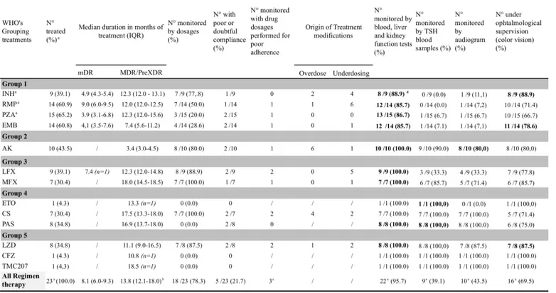
The TB drugs used and their characteristics are documented on Table 4.
Median delay from onset of management to onset of appropriate treatment (adapted to the resistance pattern), calculated for 22 children (because of missing data and 1 no-treated child) was 75.5 days (IQR 37.5-100.5).
26 Among the 15 children with MDR strains, 7 (46.7%) were treated with a combined regimen using 4 drugs from 5 different groups (2011 WHO classification) including a fluoroquinolone, an injectable drug, one or two drugs from group 4 and one from group 5 (linezolid (LZD)); 1
(6.7 %) child received the same regimen without LZD; 5 (33.3%) received a combined therapy including 4 first-line drugs and a fluoroquinolone; the last one (6.7%) received a therapy including 3 first-line drugs, one fluoroquinolone and one injectable drug (switched to rifampicin due to bad adherence).
One child was not treated but only followed. Indeed, during a first family screening, he had been treated with a first line three months tritherapy (without ethambutol) decided on a phlyctenular TST and isolated nodules on thoracic CT scan, while he was asymptomatic with normal chest X-ray.
During a second family screening revealing his sister’s confirmed MDR strain, he had normal clinical examination, negative microbiological results and the repeated CT scan was normal. He was only followed clinically and radiologically for 12.1 months and considered as cured at the end of follow-up.
The case with a pre-XDR strain was treated with a 5 drugs regimen including a fluoroquinolone, an injectable agent, one drug from group 4 and 3 drugs from group 5 (including one new anti-TB drug, bedaquiline TMC207).
Of the 8 mono-resistant to isoniazid strains, two children (25.0%) were treated with first-line quadritherapy, all two considered at risk of MDR strain because of their origin from countries with increasing incidence of DR-TB ; one case (12.5%) was treated with 2 first-line drugs in addition to a fluoroquinolone because of his ethambutol resistant strain. The remaining 5 children (62.5%) were treated with a first-line tritherapy without isoniazid. None received injectable drug.
Children monitoring is described on Table 4. Indeed, clinical examination; Blood test following hematologic, liver, kidney and thyroid functions; hearing monitoring -performed mostly for patients treated with amikacin- ; ophtalmological supervision -done mostly for patients treated with izoniazid (INH), ethambutol (EMB) and/or linezolid (LZD)-, were carried out regularly for each patient, with weekly monitoring to monthly, then quarterly or, more rarely, semi-annual. All patients treated with ethionamid or p-aminosalicylic acid had TSH blood tests. Eighty percent of children receiving amikacin were monitored with audiogram. Then, 88.9%, 78.6% and 87.5% of patients treated with, respectively, INH, EMB, or LZD were under ophtalmological supervision.
27 Outcome of children with MDR-TB.
Follow-up was documented until 31 June 2017. As notified on Table 3, 23 patients (95.8%) have completed their treatment. The last one was lost to follow-up before completing treatment. Among them, 12 (52.2% of completed treatment cases and 50.0% of all cases) were still followed-up and 7 (30.4% of completed treatment cases and 29.2% of all cases) have completed their follow-up, at the end of the study period.
Finally, 12 children (50.0%) were cured and 11 (45.8%) had favorable course; 5 (20.8%) were lost to follow-up before completing treatment for one of them (missing data) and at the end for 4 of them. No death was reported (Table 3).
The median duration of respiratory isolation, estimated on 8 microbiologically-confirmed cases (66.7%) over 12 with positive culture, was 5.2 months (IQR 4.0-8.0). Two cases, whose one with positive sputum smear, were not isolated because of poor compliance ; isolation duration’s data missed for the last two.
The median delay to culture conversion, estimated on 9 of the 13 confirmed cases, was 1.8 months (IQR 1.4-2.3).
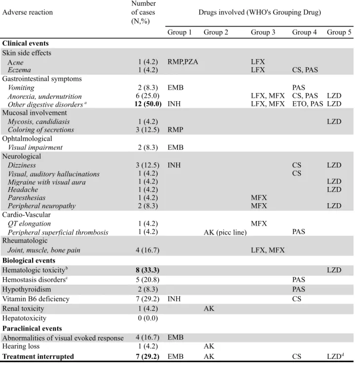
Monitoring of adverse reactions is documented on Table 4 and main adverse reactions reflected on Table 5.
Serious adverse events (SAE) were observed in 12 cases (50.0%), as followed. Four cases were affected by undernutrition despite adequate nutritional management.
Over fourteen children (58.3%) treated with ethambutol (EMB), drug was interrupted for 4 and the dosage was decreased for 1 of them because of visual disturbances or visual evoked response (VER) impairment and / or pathological electroretinogram. In four out of five cases, situation normalized after the treatment was interrupted or decreased. The last one had mild impairement on VER prior to the treatment initiation, which worsened during the treatment and despite stopping it. No visual pathway impairement was individualized on MRI; however, the adverse reaction’s cause was probably not related to the TB treatment itself, but worsened by the treatment. This child was still being monitored for her probably constitutional lesions that have stabilized at the end of documented follow-up.
Two other children had more or less profound hearing loss. Ototoxicity was strongly imputable to aminoglycoside use for one and to linezolide (LZD) for the second, revealed and monitored in both cases by audiogram. Hearing loss at the end of treatment is not reported for one case since he was lost to follow-up.
28 Among the seven children (29.2%) receiving cycloserine (CS), one child presented SEA as disabling visual and auditory hallucinations which leaded to treatment interruption, allowing discontinuation of adverse event.
Of 10 children (41.7%) with combined regimen including injectable drug, amikacin was stopped for only one child because of parent’s patient and patient’s refusal to prosecute. Finally, one patient who received LZD had to interrupt his treatment for hematologic toxicity, solved after one month.
The most commonly described clinical AR is reported as "digestive disorders", imputable to fluoroquinolones, ethionamide and PAS (Group 4), and LZD (Group 5). Half of children (50.0%) said they were prone to these as abdominal pain, diarrhea, nausea or heatburn.
During follow-up, sequelae have been reported in 11 cases (45.8%), including the lost to follow-up patient with ototoxicity described above. Among the 7 cases who have completed their follow-up, 3 (12.5% of all cases) presented sequelae as cutaneous due to nodal puncture’s scar, peripheral neuropathy linked to LZD or arthralgia and tendinopathies due to fluoroquinolones (FQ).
Among the 12 still followed-up at the end of study, 7 (29.2% of all cases) presented sequelae : undernutrition for 3 of them; hematologic toxicity due to LZD and QT elongation linked to moxifloxacin (MFX) for 1; thrombosis scar of picc line and temporary tendinopathies related to MFX for another 1 ; hematologic toxicity and transitional tendinitis due to LZD for 1 ; mild impairement on VER for the child described above.
Finally, the last case with pre-XDR strain presented three severe sequelae related to treatment: migraine with phosphenes, peripheral neuropathy with dysesthesia in all limbs and ototoxicity with deafness in the treble, all two related to LZD ; and two others related to TB disease : atelectasis of both upper lobe and a stable restrictive lung disease.
Nevertheless, we noted that after discontinuation of treatment, clinical adverse reactions decreased and supplements introduced, as thyroid hormones and / or B6 vitamin and / or K vitamin would no longer be necessary.
29
DISCUSSION
Data on the management of drug-resistant tuberculosis (DR-TB) in children in Europe are scarce (21). To our knowledge, this study is one of the only paediatric studies about this subject, particularly emphasizing on management and drug toxicity, carried out in France. The analysis of 24 cases and 11 related source cases, brings to describe 16 DR Mycobacterium Tuberculosis strain with different resistance profiles (Table 2), in a region with one of the highest incidence rate of DR-TB in France.
As we reported, 8 DR-TB strains (50.0%) related to 10 cases (41.7% of study’s cases) were described as imported from countries with an increasing incidence of DR-TB (3,7,9) : 4 in South East Asia (SEA), including India, and 4 in Eastern Europe (EE) ; linked to the case’s or source’s case place of birth.
In the same way, 5 strains (31.3%) related to 11 children (45.8 % of cases) were imported from sub Saharan Africa, 1 (6.3%), the pre-XDR strain, from North Africa and 1 (6.3%) from Caribbean. The last one (6.3%) was a “french” strain related to an index case who had only travelled in North Africa recently. In Africa (sub-Saharan and Maghreb), where the tuberculosis burden is high, data about estimated incidence of DR-TB are still disparate or missing (1,4,9).
This finding is consistent with literature showing a link between increase of DR-TB strains in France and importation’s increase of strains isolated from patients born in countries with a higher incidence of MDR-TB (5,9). Therefore, it is essential to ask a TB-case about his origin and trips but moreover, about origins and recent trips of child’s relatives.
Since microbiological confirmation is not always possible in children (2,6,13,21), the current cohort would be more representative of all children with DR-TB and has greater relevance to clinicians. Our work described both 13 confirmed cases (54.2%), whose 8 had an index case (including 2 children both cases and index cases) and 11 probable cases (45.8%) each related to a source case. In addition, more children had been diagnosed through systematic contact tracing than on clinical call point (62.5% vs 33.3%).
As suggested in several reports, a significant proportion, often exceeding 50%, received a presumptive treatment, based on clinical and radiological arguments with microbiological diagnosis of a DR-TB index case or poor clinical response after first-line therapy onset (6,10,18).
30 The broad clinical and radiological spectrum of TB presentation in children combined with the poor frequency of positive sputum smear form in young children (2,10,13, 18) or with the rarer extra-thoracic forms, whose investigations are hard to carry out (21,23), make biological confirmation diagnosis difficult.
Of the twelve symptomatic children (50.0%), 10 were confirmed cases with a median age of 14.4 years old (IQR 11.9-16.5 ; min-max 3.3-17.8) and two presumed cases, with a younger median age of 6.8 years (IQR 5.1-8.4 ; min-max 3.4-10.1).
Furthermore, a culture confirmed diagnosis was made for three asymptomatic children, with a median age of 14.0 years old (IQR 13.9-14.4). It can be seen that older child has an easier microbiological diagnosis (13).
Otherwise, research is currently supporting development of new biomarkers and biological molecular tests to improve microbiological diagnosis of children TB. Thus, we would avoid, on one hand, “over-diagnosis” with significant presumptive therapy use and, on the other hand, “under-diagnosis”, which is still the cause of TB morbidity and mortality (23).
The median delay from microbiological diagnosis to onset of appropriate DR-TB treatment was 78.0 days (IQR 40.5-119.5) for all cases, and 71.0 days (IQR 53.0-97.8) for MDR-TB cases.
According to a recent meta-analysis, published in 2012 by D.Ettehad et al, including eight different studies carried out in developed and developing countries, describing the treatment outcome of 315 children, this delay varied from 2 days to 46 months for the treatment of MDR tuberculosis (21). Then, it seems short in our territory of study, compared to other reports conducted abroad (14,15,16,21).
This is probably related to french systematic contact tracing around a TB case (regardless DST), allowing earlier diagnosis of children then earlier treatement with presomptive therapy, even if they are asymptomatic or probable cases. Also, a second-line treatment can be started quicker, adapted on index case’s strain resistance profile, even obtaining child’s own microbiological diagnosis (21,34).
The absence of an identified adult MDR-tuberculosis index case was strongly associated with delay in initiation of appropriate therapy in children (13,15). Among 8 of the 13 confirmed cases reported, with known index cases, second-line tuberculosis treatment was introduced earlier in 7 cases from the source case’s resistance profile obtained before their (including two children both cases and index cases).
31 It shows the importance of systematic contact tracing around adult index case often smear-positive, whose microbiological confirmation diagnosis is more frequent, and also the need for a regular follow-up of contact cases, avoiding more severe diseases in children.
Moreover, for principles of good clinical practice and need for improved paediatric surveillance data, developing effective tools to enable a microbiological diagnosis in children remains crucial (12,21). These missions aim to improve DR-TB treatment’s adaptation and to reduce drugs’ tocixity in children.
DR-TB therapeutic management in children varies widely according to literature (6,15,21,40) although codified according to WHO adult recommendations (1,4,5,19,20). The appropriate treatment duration of DR-TB in children remains unclear given the broad spectrum of disease and the limited reports about second-line drugs toxicity in children (21).
In our work, children received individualized therapy based on case’s or source case’s DST. Median duration of injectable drug use was 3.4 months (IQR 3.0 – 5,5), well below that recommended by the 2011 WHO adult guidelines followed during the study period (5,19) - except for the pre-XDR case whose median duration was 9,0 months - but close to results in previously literature reviews in children (10,13). Scientific journals or expert opinion (18) endorse their use thanks to convincing results of MDR TB treatment in children.
All MDR children of our study were treated with an addition of four second-line anti-TB drugs including at least one later generation fluoroquinolone with injectable drug for 60.0% of them.
Regimen for pre-XDR case included a new therapeutic agent, as Bedaquilin TMC207, usually less available in children and being studied in recent clinical trials (16).
Before last 2016 uptdate, the advice from the WHO and most TB programmes is that all patients newly diagnosed with DR-TB (i.e. not previously treated dor MDR-TB) are treated for a standardised (long) duration over 20 months for MDR and XDR cases (5,19).
According to the meta-analysis by D.Ettehad et al, treatment duration ranged from 6 to 34 months, but for most studies included, treatment was given for a minimum of 18 months (21). Regardless of the strain’s resistance, the median duration of treatment reported in our work was shorter than recommended in adult, before 2016 (1,5,6,19).
The median duration result of 13.3 months (IQR 12.0-18.0) for MDR cases is similar in comparison to the 2013 descriptive cohort study of 149 children started on MDR-TB treatment from Cape Town (13). A majority of 95.8% cases were regarded as treated successfully, including the 12 children cured and the 11 with favorable course.