The DmpA aminopeptidase from Ochrobactrum anthropi LMG7991 is the
prototype of a new terminal nucleophile hydrolase family
Laurence FANUEL*, Colette GOFFIN*, Abdellatif CHEGGOUR*, Bart DEVREESE†, Gonzales V
ANDRIESSCHE†, Bernard JORIS*,
Jozef V
ANBEEUMEN† and Jean-Marie FRE!RE *
1*Laboratoire d’Enzymologie et Centre d’Inge! nierie des Prote!ines, Universite! de Lie"ge, Institut de Chimie, B6, B-4000 Sart Tilman, Belgium, and †Vakgroep Biochemie, Fysiologie en Microbiologie, Laboratorium voor Eiwitbiochemie en Eiwitengineering, Rijksuniversiteit-Gent, K. L. Ledeganckstraat, 35, B-9000 Gent, Belgium
The DmpA (-aminopeptidase A) protein produced by
Ochro-bactrum anthropi hydrolyses p-nitroanilide derivatives of glycine
and -alanine more efficiently than that of -alanine. When regular peptides are utilized as substrates, the enzyme behaves as an aminopeptidase with a preference for N-terminal residues in an configuration, thus exemplifying an interesting case of stereospecificity reversal. The best-hydrolysed substrate is -Ala-Gly-Gly, but tetra- and penta-peptides are also efficiently hydro-lysed. The gene encodes a 375-residue precursor, but the active enzyme contains two polypeptides corresponding to residues 2–249 (α-subunit) and 250–375 (β-subunit) of the precursor. Residues 249 and 250 are a Gly and a Ser respectively, and various substitutions performed by site-directed mutagenesis
INTRODUCTION
Ochrobactrum anthropi LMG7991 contains two distinct
intra-cellular enzymes that hydrolyse -alanyl-p-nitroanilide (-Ala-p-Na) [1]. One of them, DmpB (-aminopeptidase B), is homo-logous to the -alanyl aminopeptidase described by Asano et al. [2,3], and has very similar catalytic properties. The second enzyme, DmpA, is produced in much smaller quantities by the original strain. A DNA fragment was isolated that encoded an original 375-residue open reading frame (ORF) and whose integration in the pUC18 plasmid downstream of the lacZ promoter resulted in the production of large amounts of
-Ala-p-Na-hydrolysing activity. These experiments suggested strongly
that this ORF represented the DmpA structural gene [1]. In this study, we describe the purification of the cloned DmpA protein and demonstrate that the 375-residue precursor is activated by a probably autocatalytic cleavage between residues 249 and 250. A detailed analysis of this cleavage site indicates that its properties are strikingly similar to those of N-terminal nucleophile (N-tn) amidohydrolases. Exploration of the databases (available at the website of the National Center of Biotechnology Information, http :!!www.ncbi.nlm.nih.gov) shows that DmpA is the pro-totype of a new family of N-tn hydrolases. The catalytic properties of the enzyme are also analysed.
MATERIALS AND METHODS
Molecular biology, oligonucleotides, kits, enzymes, chemicals and
other materials
Components of culture media were from Difco (Detroit, MI, U.S.A.) and Biome! rieux (Marcy-l’Etoile, France). Sequencing
Abbreviations used : Dmp,D-aminopeptidase ; ORF, open reading frame ;D-Ala-p-Na,D-alanyl-p-nitroanilide ; N-tn ; N-terminal nucleophile ; LB,
Luria–Bertani ; QSFF, Q–Sepharose Fast-Flow.
1 To whom correspondence should be addressed (e-mail jmfrere!ulg.ac.be).
result in the production of an uncleaved and inactive protein. The N-terminal Ser residue of the β-subunit is followed by a hydrophobic peptide, which is predicted to form a β-strand structure. All these properties strongly suggest that DmpA is an N-terminal amidohydrolase. An exploration of the databases highlights the presence of a number of open reading frames encoding related proteins in various bacterial genomes. Thus DmpA is very probably the prototype of an original family of N-terminal hydrolases.
Key words : N-terminal nucleophile amidohydrolase, peptidase, protease precursor, stereospecificity.
kits, oligonucleotides, plasmids, purification supports and columns were purchased from Pharmacia Biotech (Uppsala, Sweden). The QuickChange Site-Directed Mutagenesis kit was a Stratagene product (La Jolla, CA, U.S.A.). The p-nitroanilide and peptide substrates were from Bachem (Bubendorf, Switzer-land) and Sigma (Bornem, Belgium), and protease inhibitors were from Boehringer Mannheim (Mannheim, Germany) or Worthington (Stoke-on-Trent, Staffs, U.K.). The DNA laser sequenators were from Pharmacia Biotech and EMBL (Heidel-berg, Germany). The Cybertech CS-1 system (Cybertech, Berlin, Germany) was used to quantify proteins on gels after SDS! PAGE. The Constant Basic System Disintegrator was from Inceltech (Toulouse, France). Protein purification was performed with the help of an A"kta Explorer apparatus (Pharmacia Biotech). The VG Bio-Q Electrospray Triple Quadrupole Mass analyser, upgraded with a platform source, was from Micromass (Altrincham, Cheshire, U.K.), and was connected to a Harvard 11 syringe pump (Harvard Instruments, South Natick, MA, U.S.A.). The TLC plates (Silica gel 60F250) were from Merck (Darmstadt, Germany).
Purification of the DmpA protein produced by Escherichia coli
DH5α
After transformation with pDML1102 [1], 2–3 colonies pro-ducing -Ala-p-Na-hydrolysing activity were inoculated in 250 ml of Luria–Bertani (LB) medium containing ampicillin (50 µg!ml) and cultured during 16 h at 37!C. Aliquots of this preculture were diluted 500 times in the same medium and the culture incubated for 16 h at 37!C. The presence of inclusion
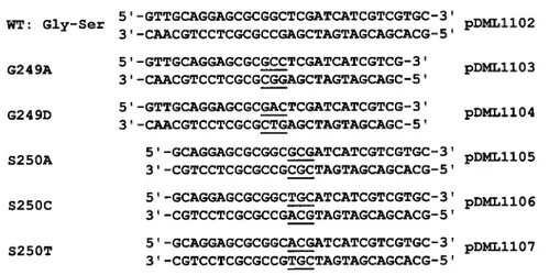
Figure 1 Oligonucleotides designed for mutagenesis of the DmpA cleavage site and plasmids utilized for production of the mutants
WT, wild type.
bodies in the E. coli cells was detected by examination of the cells by phase-contrast microscopy.
Extraction of inclusion bodies
Culture (50 ml) was centrifuged and cells were resuspended in 10 ml of 10 mM Tris!HCl buffer, pH 8.0, containing 1 mM EDTA (buffer A). Inclusion bodies were then extracted as described by Goraj et al. [4].
Purification of the soluble active protein
The soluble fraction of DmpA was purified to more than 95 % as follows : 1 litre of culture was centrifuged and cells were resus-pended in 50 ml of buffer A. Cells were disrupted with the Constant Basic System Disintegrator and the suspension centri-fuged. Activity towards -Ala-p-Na was detected in the super-natant. After dialysis against 50 mM potassium phosphate, pH 6.0, the protein solution was loaded on to a 200-ml Q– Sepharose Fast-Flow (QSFF) column (40 cm"2.5 cm) equili-brated in the same buffer, and eluted with a 0–0.3 M NaCl linear gradient over a volume of 2 litres. Active fractions were pooled and dialysed against buffer A or diluted twice with 20 mM Tris!HCl, pH 8.0. The sample was then loaded on to the QSFF column equilibrated with buffer A and eluted with a 0.2–0.5 M NaCl linear gradient under the same conditions as above. Active fractions were pooled and the pure protein (60 mg) dialysed against 50 mM potassium phosphate, pH 7.0, concen-trated and stored at#20 !C.
Immunodetection of the DmpA
Rabbit antibodies against the purified DmpA enzyme produced in E. coli were prepared by Gamma (Lie# ge, Belgium). After SDS!PAGE, electroblotting on to a nitrocellulose membrane and treatment with antibodies, positive bands were revealed with the help of the Bio-Rad ImmunoBlot Alkaline Phosphatase Assay System (Bio-Rad, Nazareth, Belgium). After blotting, standard proteins were stained with 0.1 % (w!v) Ponceau S Red
in 5 % (v!v) acetic acid and the membrane was rinsed with water. Prestained standard proteins (low-range, Bio-Rad) were also used.
Site-directed mutagenesis of the DmpA cleavage site
To assess the importance of the two residues constituting the cleavage site, the Gly-249 residue was replaced successively by Ala (the least-disturbing possible modification) and Asp. Con-versely, the Ser-250 residue was replaced by residues possessing potential nucleophilic groups in their side chains and sometimes encountered in N-tn hydrolases (Thr or Cys) and by a residue devoid of such properties (Ala). The experiments were performed with the help of the QuickChange Site-Directed Mutagenesis kit (Stratagene), according to the supplied protocol. Oligo-nucleotides used for the mutagenesis reaction are described in Figure 1. The PCR reactions introducing the mutations were performed directly with the pDML1102 plasmid as a template. The reaction mixture contained 10 ng of DNA matrix (40 ng for the S250T mutation), 125 ng of each oligonucleotide (the S250T oligonucleotides were incubated previously for 10 min at 95!C to denature the secondary structures), all dNTPs at 0.05 mM and 2.5 Pfu polymerase units, in a total volume of 50 µl. After heating for 30 s at 95!C, PCR reactions were performed as follows : 1 min at 55!C, 8 min at 68 !C, 12 cycles to replace one base, 16 cycles to replace two bases and for the S250T mutation. After completion of the PCR reactions and digestion of the non-mutated DNA matrix with the DpnI enzyme, 1 µl of each PCR mixture was used to directly transform XL1Blue E. coli super-competent cells purchased with the mutagenesis kit. Trans-formed cells were plated on LB$ampicillin and grown for 16 h at 37!C. Plasmids extracted from the various clones were analysed by restriction (the G249A mutation created a second
BssHII GCGCGC site in the dmpA gene) and sequenced on both
strands for verification of the presence of the mutation. Plasmids containing the mutated dmpA gene were named pDML1103–1107 according to the mutation (see Figure 1) and were then used to transform E. coli DH5α cells to produce the corresponding proteins.
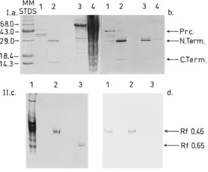
Figure 2 Electrophoretic analysis of DmpA samples
Panel I, Coomassie Brilliant Blue staining of the gel (a) and immunoblot revealed with rabbit anti-DmpA antibodies (b) after SDS/PAGE (18% polyacrylamide) of the following samples: MM STDS, Gibco-BRL protein molecular-mass standards (kDa) ; lanes 1, inclusion bodies fromE. coli containing the uncleaved putative DmpA precursor (Prc.). Note that the intensity of the bands appearing at lowerMrvalues was found to increase with time upon strorage of the inclusion bodies, a result which suggested that these bands might represent degradation products of the 45000-Mr antibody-recognized protein. Lanes 2, active soluble DmpA purified fromE. coli (N. Term. and C. Term., N- and C-terminal fragments) ; lanes 3, active DmpA partially purified from O. anthropi LMG7991 (fraction containing the highest activity after chromatography on the Superdex 75 molecular-sieve column [1]) ; lanes 4, soluble fraction of the total cell extract ofO. anthropi LMG7991. Panel II, Coomassie Brilliant Blue staining of the gel (c) and immunoblot revealed with rabbit anti-DmpA antibodies (d) after electrophoresis on non-denaturing 8% polyacrylamide gel of the following samples: lanes 1, partially purified DmpA fromO. anthropi LMG7991 (fraction containing the highest activity after chromatography on the Superdex 75 molecular-sieve column) ; lanes 2, active soluble DmpA purified fromE. coli ; lanes 3, active soluble DmpB purified from O. anthropi LMG7991. Identical RFvalues were obtained with the zymogram method usingD-Ala-p-Na as a substrate.
Production of the wild-type and modified enzymes
The wild-type protein was purified as described above. For each mutant, 10 ml of LB$ampicillin medium were inoculated with a few colonies and the culture grown for 8 h at 28!C. These precultures were diluted in 250 ml of the same medium and grown for 16 h at 28!C. Cells were collected by centrifugation, resuspended in 15 ml of buffer A and disrupted. The DNA was digested with benzonase (16 h at 4!C) in the presence of 2 mM MgCl
!. Cell extracts were then centrifuged, and the supernatants analysed by dot-blot with rabbit anti-DmpA antibodies and by SDS!PAGE (15% polyacrylamide) followed by electroblotting and immunodetection as described above.
Chemical procedures
N-terminal sequences were determined as described previously [5] on a 477A pulsed-liquid sequenator. Approximate Mrvalues were obtained by SDS!PAGE (15% polyacrylamide), and ac-curate values by electrospray MS. Isoelectric points were meas-ured by isoelectric focusing on Ampholine PAGplates, pH 3.5–9.5, detection of the active bands with the -Ala-p-Na substrate [1] and measurement of the pH at the position of the active protein. The activity could also be detected directly after electrophoresis on non-denaturing 8 % polyacrylamide gels by the same zymogram technique. Protein concentrations were estimated on the basis of the absorbance at 280 nm or with the help of the BCA protein assay kit (Pierce, Rockford, IL, U.S.A.). After SDS!PAGE, the intensities of Coomassie Brilliant Blue-stained bands were compared with those of protein standards.
Gel scanning was done using the Cybertech CS-1 System. Substrate acetylation was performed as described previously [1].
Kinetic measurements
The enzyme activity was measured in 50 mM potassium phos-phate, pH 8.0, or 100 mM Tris, pH 8.0, at 30!C. For substrates containing a p-nitroaniline leaving group, variations of ab-sorbance were monitored with the help of HP8452A or Uvikon spectrophotometers, at 405 nm for #
" measurements (∆ε% 11 500 M−#"cm−#) and at 440 nm (∆ε% 2250 M−#"cm−#) for com-plete time-course analysis. With substrates containing an N-terminal -alanine, the released -alanine was quantified by the -amino-acid oxidase method [6]. Liberation of -alanine from the peptide N-terminus was measured by oxidation with -alanine dehydrogenase [7] or by quantification on TLC (see below). The k
catand Kmparameters were obtained by non-linear
regressions of the #
" values using the ENZFITTER software package (Elsevier Biosoft, Cambridge, U.K.) or by analysis of the complete time-courses with the help of the integrated Henri–Michaelis equation [8]. Estimated errors on #
"values were ! 10%. The degradation of non-chromogenic substrates was monitored by withdrawing samples at various times, separating the substrates and products by TLC at 20!C and detecting their amino groups using ninhydrin [9]. Degradation of unstable substrates in the absence of enzyme was also monitored to account for their spontaneous degradation. The TLC solvent was n-butanol!acetic acid!5% NH$OH in water (5.5 : 3 : 1.5, v!v!v). After ninhydrin revelation, the resulting picture was digitalized and analysed by densitometry with the Cybertech CS-1 system. Quantification was done by comparison with known
quantities of standard peptides and amino acids corresponding to the products expected in the reaction mixture. The estimated errors on product quantification were&20%.
RESULTS
Production and purification of DmpA
E. coli DH5α cells harbouring the pDML1102 plasmid contained
inclusion bodies but also a high amount of soluble -Ala-p-Na-hydrolysing activity. After purification to more than 95 % homogeneity, only one protein band was detected upon non-denaturing PAGE (Figure 2, panel II) and electrofocusing (results not shown, pI% 5.0), but gel electrophoresis in the presence of SDS revealed two polypeptides with respective M
r values of
about 30 000 and 15 000 (Figure 2, panel I). These were electro-transferred on to a PVDF membrane and submitted to N-terminal amino acid sequencing, which yielded the TSQTPTR-KPR and SIIVVLATDL sequences for the large and small peptides, respectively. These corresponded to residues 2–11 and 250–259 of the protein deduced from the sequence of the cloned gene. Antibodies were raised against the purified protein.
Presence of a potential precursor in the inclusion bodies
Inclusion bodies were solubilized and submitted to SDS!PAGE. The highest-Mr protein revealed by Coomassie Brilliant Blue
Figure 3 Sequence of the DmpA amidohydrolase
Hydrolysis of the Gly-249–Ser-250 peptide bond (boxed) cleaves the polypeptide precursor into two subunits : an α N-terminal and a β C-terminal peptides (by analogy with penicillin acylase). Note that the N-terminal M residue is also absent in the mature active protein (arrow).Mrvalues were determined by electrospray MS.
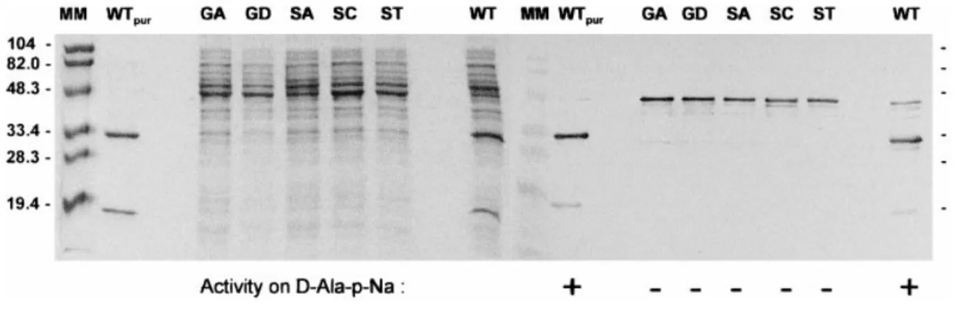
Figure 4 Electrophoretic analysis of DmpA mutants and correlation with activity usingD-Ala-p-Na as a substrate
Coomassie Brilliant Blue-stained gel (left) and immunoblots revealed with anti-DmpA antibodies (right) after SDS/PAGE (15 % polyacrylamide) of the following samples : MM, pre-stained molecular-mass standards (Bio-Rad) ; WTpur, wild-type purified DmpA protein (1.5 µg) produced by E. coli DH5α ; GA, GD, SA, SC, ST and WT, 15 µl of 10-fold diluted cell supernatants (equivalent to 25 µl
of culture) containing the various mutants (G249A, G249D, S250A, S250C and S250T) and the non-mutated (WT) DmpA enzyme.$, activity found; #, no activity found.
staining reacted with the antibodies raised against the soluble and active DmpA (Figure 2, panel I). Its migration rate corres-ponded to an M
rvalue of about 45 000, indicating that it might
represent a precursor of the active enzyme. This hypothesis was corroborated by the identification of the first 10 N-terminal residues of the solubilized protein. These corresponded exactly with the N-terminal sequence of the large polypeptide, which was preceded by the Met residue corresponding to the first ATG codon of the gene. Attempts to obtain active enzyme by successive denaturation of the inclusion bodies and renaturation remained unsuccessful. The active protein was submitted to electrospray MS and two peptides exhibiting Mrvalues of 26 564.3&2.6 and 13 736.8&0.6 were found. These values corresponded well with the Mr values calculated on the basis of the sequences for residues 2–249 (M
r 26 565) and 250–375 (Mr 13 737). These
results suggested that the active protein was derived from the precursor by elimination of the N-terminal Met residue and cleavage of the Gly-249–Ser-250 peptide bond without additional loss of residues (Figure 3).
Absence of detectable precursor in O. anthropi
A sample of partially purified enzyme from an O. anthropi culture [1] was submitted to non-denaturing PAGE and to polyacrylamide-gel isoelectric focusing. In both cases, a zymo-gram revealed the presence of -Ala-p-Na-hydrolysing activities
Table 1 Activity of DmpA on Xaa-p-Na substrates
Values have S.D.s of&10%. The following substrates were also hydrolysed by DmpA:L -Leu-p-Na (0.5 % of Gly--Leu-p-Na), L-Met-p-Na (0.2 %) and L-Val-p-Na (0.02 %). Acetyl-D-Ala-p-Na, D-Leu-p-Na andD-Phe-p-Na were not hydrolysed significantly (! 0.005%). ND, not determined.
DmpA Substrate kcat(s−1) K m(mM ) kcat/Km(M−1"s−1) Gly-p-Na* 70 3 23 000 Gly-p-Na† 70 3 23 000 L-Ala-p-Na* 0.5 0.6 800 L-Ala-p-Na† 0.56 0.36 1550 D-Ala-p-Na* 4.0 0.54 7500 D-Ala-p-Na† 3.3 0.52 6300 L-Lys-p-Na† 0.11 0.4 275 L-Arg-p-Na† 0.14 0.4 350 L-Phe-p-Na† ND ND 17 * In 100 mM Tris/HCl, pH 8.0, at 30!C. † In 50 mM potassium phosphate, pH 8.0, at 30!C.
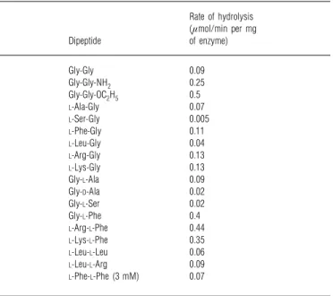
Table 2 Hydrolysis of dipeptides by DmpA
The substrate concentrations were 10 mM unless otherwise stated. The rate values have S.D.s of&20%. The following compounds were not hydrolysed significantly (! 0.005 µmol/min per mg of enzyme) : acetyl-Gly-Gly,D-Ala-Gly,L-His-Gly,L-Asp-Gly, benzoyl-Gly-L-Ala, phenyl-acetyl-Gly-L-Ala, Gly-D-Phe, Gly-L-Asp, Gly-L-Gln andL-Trp-L-Phe.
Rate of hydrolysis (µmol/min per mg Dipeptide of enzyme) Gly-Gly 0.09 Gly-Gly-NH2 0.25 Gly-Gly-OC2H5 0.5 L-Ala-Gly 0.07 L-Ser-Gly 0.005 L-Phe-Gly 0.11 L-Leu-Gly 0.04 L-Arg-Gly 0.13 L-Lys-Gly 0.13 Gly-L-Ala 0.09 Gly-D-Ala 0.02 Gly-L-Ser 0.02 Gly-L-Phe 0.4 L-Arg-L-Phe 0.44 L-Lys-L-Phe 0.35 L-Leu-L-Leu 0.06 L-Leu-L-Arg 0.09 L-Phe-L-Phe (3 mM) 0.07
in positions identical to those observed with the fully purified DmpA produced by E. coli. In the first case, a positive response to the antibodies after electroblotting was also observed (Figure 2, panel IId). After SDS!PAGE, a similar analysis of the same partially purified fractions highlighted two polypeptides in the same positions as those observed with the purified E. coli protein (Figure 2, panel Ib). The active enzymes produced by E. coli and
O. anthropi also behaved similarly upon molecular-sieve filtration
on Superdex 75 and chromatography on QSFF. By contrast, the 45 000-Mrinactive precursor was never observed in O. anthropi cell extracts.
Table 3 Hydrolysis of tripeptides by DmpA
The substrate concentrations were 10 mM unless otherwise stated. The rate values have S.D.s of&20%. The following compounds were not hydrolysed significantly: acetyl-Gly-Gly-Gly, acetyl-L-Ala-Gly-Gly andD-Leu-Gly-Gly.
Initial rate of hydrolysis (µmol/min per mg Tripeptide of enzyme) L-Ala-Gly-Gly 1.25 D-Ala-Gly-Gly 0.04 Gly-Gly-L-Ala 0.70 L-Phe-Gly-Gly 0.50 L-Leu-Gly-Gly 0.60
L-Ser-L-Ser-L-Ser 0.65
L-Leu-L-Leu-L-Leu 0.13
L-Phe-L-Phe-L-Phe (1 mM) 0.18
Table 4 Influence of the length of the peptide chain on the activity of DmpA
The substrate concentration was 2 mM in all cases. Values have S.D.s of&20%. Initial rate of hydrolysis
(µmol/min per mg Substrate of enzyme) Gly2 0.02 Gly3 0.11 Gly4 0.22 Gly5 0.14
DmpA mutant analysis
At the stationary phase of growth, all E. coli cells producing the wild-type enzyme and the five mutants contained inclusion bodies, which were visualized by phase-contrast microscopy. Moreover, SDS!PAGE analysis of cell supernatants followed by Western-blot detection with anti-DmpA antibodies showed that all the mutant-producing cells contained a soluble protein whose electrophoretic mobility corresponded to that of the wild-type DmpA precursor, a 45 kDa protein, and which was recognized by the antibodies (Figure 4). By contrast, under the same conditions, the supernatant from cells producing the wild-type DmpA contained both uncleaved soluble precursor and cleaved protein. Incubating all cell extracts for several hours at 30!C alone or in the presence of purified active wild-type DmpA failed to further cleave the soluble precursors. Of all samples, only the cell extract containing the wild-type enzyme was active on the -Ala-p-Na substrate. This showed that cleavage and activity were related phenomena and that the intact Gly-249–Ser-250 site was essential for processing of the precursor into an active enzyme. Note that host cells that did not contain the plasmid or which harboured a plasmid devoid of the dmpA insert did not produce inclusion bodies and that the corresponding supernatants were devoid of proteins yielding positive Western-blot responses.
Kinetic characterization of DmpA
Activity was first measured towards Xaa-p-Na chromogenic substrates (Table 1). For these artificial peptide analogues, the highest activity was recorded with Gly and -Ala derivatives. An activity profile was also established with a set of peptide substrates (Tables 2–4). Hydrolysis of tripeptides exhibiting different resi-dues at their N- and C-termini was monitored to determine the
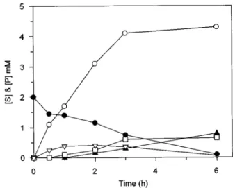
Figure 5 Hydrolysis of 10 mM tripeptides: Gly-Gly-L-Ala (a),L-Ala-Gly-Gly
(b) andD-Ala-Gly-Gly (c) by 9.2 µM DmpA at 30 !C in 50 mM potassium
phosphate, pH 8.0
Products were quantified after TLC and treatment of the plates with ninhydrin, as explained in the text. Gly and Gly-L-Ala (a) could not be well separated, and were quantified by comparison
with the Gly standard. (a) !, Gly$Gly-L-Ala ;", Gly-Gly-L-Ala ;#,L-Ala. (b) !, Gly; $,
Gly-Gly ;",L-Ala-Gly-Gly ;#,L-Ala. (c) !, Gly; ",D-Ala-Gly-Gly ;#,D-Ala.
preferential site of DmpA action. Figure 5 shows that the first products of tripeptide hydrolysis were always the N-terminal residues, and that the release of C-terminal residues was signifi-cantly delayed when compared with the release of N-terminal residues. These results suggest that C-terminal residues were only liberated when the dipeptide formed during the first reaction step was hydrolysed by DmpA. Note that tripeptides were better substrates than dipeptides (Tables 2 and 3), with the exception of -Ala-Gly-Gly, which was a very poor substrate : in this case, the Gly-Gly dipeptide was not an observable product and was thus
Figure 6 Hydrolysis of 2 mM Gly5by 2.1 µM DmpA in 50 mM potassium
phosphate, pH 8.0, at 30 !C
The accumulation of the various intermediates is also shown :!, Gly; %, Gly2;$, Gly3;&,
Gly4;", Gly5.
Table 5 Activity of DmpA on simple ester and amide substrates
The substrate concentration was 10 mM in all cases. The values have S.D.s of&20%. Initial rate of hydrolysis
(µmol/min per mg Substrate of enzyme) Gly-NH2 1.4 L-Ala-NH2 0.09 D-Ala-NH2 0.23 Gly-OCH3 1.2 L-Ala-OCH3 0.09 D-Ala-OCH3 1.2
hydrolysed as soon as it was formed. Peptide degradation studies in the Glynseries showed that the products appeared as the result of the release of one Gly from one extremity (exoprotease) and that the better substrates were tetra-, penta- and tripeptides (in that order, see Table 4 and Figure 6), whereas the dipeptide accumulated. An activity profile was also determined for residues in the N1 (the first N-terminal residue) and N2 (the following residue) positions, their configuration and their number (Tables 2–4). Acetylation of the substrate N-terminus suppresses the activity, a result showing that DmpA only recognizes substrates with a free N-terminal amino group. DmpA thus belongs to the aminopeptidase group of enzymes (EC 3.4.11). However, DmpA was also active towards -Ala-NH
!, -Ala-OCH%(Table 5) and -Ala-p-Na (Table 1). Hence, DmpA is also a amidase-esterase. Note that the specific activity of DmpA towards
Ala-p-Na was much weaker than that of the Dap enzyme, a strict
-aminopeptidase [2].
DmpA inhibition
The following protease inhibitors were tested unsuccessfully (30 min pre-incubation at 30!C, at the concentration advised by
the supplier but also with 10-fold higher concentrations) : anti-pain, aprotinin, bestatin, chymostatin, trans-epoxysuccinyl--leucylamido-(4-guanidino)butane (E-64), EDTA, leupeptin, pefabloc SC and 1,10-phenanthroline.
DISCUSSION
DmpA as a new N-tn amidohydrolase
The sequence of the purified DmpA protein confirmed that its structural gene was the ORF identified downstream of the lacZ promoter on the pDML1102 plasmid that encoded a 375-residue polypeptide. However, the soluble active protein produced in E.
coli consisted of two distinct polypeptides corresponding to
residues 2–249 (α-subunit) and 250–375 (β-subunit), as shown by N-terminal sequencing and electrospray MS (Figure 3). The absence of signal peptide was in agreement with the intracellular location of the protein. The enzyme was not purified from the original O. anthropi strain, where the quantities produced were too small, but the analysis of a crude preparation by immuno-logical methods indicated the presence of two polypeptides of sizes similar to those of the α- and β-subunits. Upon over-production in E. coli, large quantities of inclusion bodies that contained the complete and uncleaved polypeptide accumulated, but failed to yield active enzyme after solubilization. In some cases, small quantities of the large polypeptide were obtained in a soluble form (Figure 4), but it was not further processed upon incubation of the crude extract. In the mature protein, the two subunits remain associated by non-covalent bonds, since the sequence highlights only one Cys residue at position 180 of the precursor.
The cleavage of the 249–250 peptide bond could result from either the action of a cytoplasmic protease or self-processing. The facts that this cleavage, although very specific (see below), occurred in both E. coli and O. anthropi, and that the unprocessed protein was not observed in the latter strain, argued in favour of the second hypothesis. The cleaved Gly–Ser peptide bond is identical to the autocatalytic processing sites of the AcyII cephalosporin acylases produced by various Pseudomonas strains [10,11], and is generally similar to that found in N-tn amido-hydrolases (Table 6).
Numerous site-directed mutagenesis results have been obtained with various N-tn hydrolases : 20S proteasomes [12,13], penicillin acylases [14,15], Bacillus subtilis PRPP amidotransferase [16,17],
Fla#obacterium meningosepticum aspartyl glucosaminidase [18]
and the MIP (intein-extein) in #itro splicing system [19,20]. By analogy, five mutants were produced at the DmpA cleavage site. All the modified proteins were produced with similar good yields but remained both uncleaved and inactive, even when Ser-250 (position n) was replaced by other potential nucleophilic residues such as Thr or Cys. With the other N-tn hydrolases, mutations performed either at the n#1 or n positions, and which prevent cleavage, always result in a loss of activity [12,14,15,17,18,30]. Conversely, in position n, substitution by a residue with a nucleophilic side-chain sometimes allows processing but only yields an active enzyme in the case of the 20 S proteasome Thr# Ser mutant [12]. This suggests that the geometries necessary for cleavage and activity are different, and this is not surprising, since the free α-amino group of residue n that acts as a general base in the enzymic activity cannot play the same role in the cleavage reaction.
An analysis of the DmpA sequence with the help of the Chou and Fasman algorithm [31] shows that the N-terminal Ser of the β-subunit is located at the N-terminus of a β-sheet structure, in exactly the same way as the active nucleophile residue in N-tn hydrolases. The N-terminal Ser-1–Lys-18 peptide of the small
β-subunit is highly hydrophobic (results not shown) and the fact that this subunit is not well recognized by antibodies raised against the native protein (Figure 2) suggests that most of it is buried inside the protein core. All these results indicate that DmpA might be the first representative of a new subfamily of N-tn hydrolases. The crystal structure of DmpA, which has now been solved at a resolution of 1.82 A" [C. Bompard-Gilles, V. Villeret, L. Fanuel, J.-M. Fre# re and J. Van Beeumen, unpub-lished work], confirms this assumption and shows a clear similarity between the fold of DmpA and that of the N-tn amidohydrolases of known structure [32].
A search of the databases (see the Introduction) revealed that the genomes of Pyrococcus horikoshii, Pseudomonas aeruginosa,
Ps. fluorescens, Bordetella pertussis and Neisseria meningitidis
contained ORFs encoding putative proteins very similar to DmpA (Table 6). When these sequences are compared with that of DmpA, Gly and Ser residues align with the position-249–250 Gly!Ser dyad of DmpA. Moreover, the sequences downstream of this dyad are nearly identical (Table 6) but, as in other N-tn hydrolases, the sequences upstream of the cleavage site are much less conserved. Lower degrees of similarity were found with putative proteins encoded by ORFs in the genomes of
Myco-bacterium leprae and M. tuberculosis. In these cases, alignments
highlight striking similarities downstream of a potential cleavage site formed by an Asn!Thr dyad, which is reminiscent of the Gln!Thr or Asp!Thr cleavage sites in other N-tn hydrolases (Table 6). Surprisingly, these two putative mycobacterial proteins exhibit a significant similarity with the well-characterized amidase NylC, a nylon hydrolase produced by Fla#obacterium sp. K172 [29]. It is thus very tempting to assume that all these proteins are cleavage-activated N-tn hydrolases and that the DmpA family can be divided into two subfamilies, containing either Gly!Ser-or Asn!Thr-cleavage sites. It can also be expected that all these proteins, when isolated, will exhibit amidohydrolase activities, as already demonstrated for the NylC enzyme, and that the members of the ‘ Gly!Ser’ subfamily will behave as aminopeptidases.
It should be noted finally that the consequences of the elimination of the N-terminal Met of DmpA on the enzyme’s activity remain undetermined.
Enzymic characterization of DmpA
DmpA liberates the N-terminal residues from peptide substrates with an efficiency depending on the peptide length : Xaa
&" Xaa$ " Xaa
%# Xaa!. Tripeptides of the form Xaa-Gly-Gly are hydro-lysed with a consistently higher efficiency than the corresponding Xaa-Gly dipeptides ; in the Gly
nseries, the tetrapeptide is the
best substrate. To allow recognition, the substrate α-amino group must be free (peptides whose N-terminus has been modified are not degraded) and optimal activity requires an N-terminal residue in an -configuration. Thus DmpA may possess a negative charge in the substrate-binding pocket that could stabilize the N-terminal positive charge of the peptide substrate and position it in a catalytically productive geometry, as observed in the PepC aminopeptidase [33]. Its catalytic profile for residues at the first N-terminal position in dipeptides is : basic amino acid (Arg, Lys) $ Phe $ aliphatic amino acid (Leu, Gly, Ala) $ hydroxylated amino acid (Ser). An acidic residue in the first or second N-terminal position precludes hydrolysis. This underlines the impor-tance of the N-terminal charge of the substrate on its hydrolysis : a double positive charge (basic amino acid) favours substrate binding and hydrolysis, whereas an ionized acidic group is detrimental to the activity. Dipeptides exhibiting a His or a Trp residue at the N-terminus are not hydrolysed. A Phe at the second position from the N-terminus increases the DmpA activity
Table 6 Alignment of the cleavage sites of N-tn and potential N-tn amidohydrolases
The N- and C-terminal residues of the cleaved peptide bond are shown in bold. The cleavage either eliminates an N-terminal propeptide of variable length or yields two subunits with or without the concomitant elimination of a propeptide or an internal peptide (both processes occur in penicillin acylase [25]). Some other enzymes might also be related to N-tn hydrolases [26] :Pseudomonas 7A glutaminase-asparaginase [27] andLactobacillus 30a histidine decarboxylase [28]. aa, amino acids. Addresses in data banks : a, DDBJ$AB009466; b, gnl$PAGP$P. aeruginosa%contig 163; c, gb$AF004848; d, gnl$Sanger $B. pertussis%contig 578; e, gnl$TIGR $GNMCQ43R; f , gb$U00014$M. leprae cosmid B1549%C2%208; g, EMBL$Z73902$M. tuberculosis H37Rv.
Enzyme
Cleavage site and position in the precursor Precursor length Result of cleavage Reference Proteasome 20 S (β-subunit)
Thermoplasma acidophilum * 1 MNQTLETGTTTVGITLKDAV 20 211 aa Propeptide [12]
Human (Z)† 36 LPKVRKTGTTIAG 48 Propeptide [12,13]
Glutamine PRPP amidotransferase
Bacillus subtilis* 4 EIKGLNEECGVFG 16 465 aa Propeptide [16]
Saccharomyces cerevisiae † 1 MCGILG 6 Propeptide [16]
Chicken† 4 EELGIREECGVFG 16 Propeptide [16]
Aspartylglucosaminidase or glycosylasparaginase
Human* 198 TEDDRGHDTIGMVVIHKTGH 217 346 aa 2 Subunits [21]
Flavobacterium meningosepticum † 189 IVNIENHDTIGMIALDAQGN 208 340 aa 2 Subunits [18] γ-Glutamyltranspeptidase‡
E. coli 383 LAPYESNQTTHYSVVDKDGN 402 580 aa 2 Subunits [22]
Human 374 YTP-DDGGTAHLSVVAEDGS 392 580 aa 2 Subunits [22]
Penicillin acylase
E. coli * 282 GLAGYPTTSNMWVIGKSKAQ 301 846 aa 2 Subunits [14,15,23]
Kluyvera citrophila† 282 GLAGYPTTSNMWVIGKNKAQ 301 844 aa 2 Subunits [24]
Cephalosporine acylase‡
AcyIPseudomonas sp. SE83 360 LSGGESADTTHVTVADAMGN 379 558 aa 2 Subunits [10] AcyIIPseudomonas sp. SE83 232 ASDAAGGGSNNWAVAPGRTA 251 774 aa 2 Subunits [10] Pseudomonas sp. SY-77-1 191 PPDLADQGSNSWAVAPGKTA 210 311 aa 2 Subunits [11] DmpA or putative proteins
potentially related to DmpA
O. anthropi 242 QSQLQERGSIIVVLATDLPL 261 375 aa 2 Subunits This study
Pyococcus horikoshii 231 GRGGEGKGSIIMIIATDAPL 250 361 aa ? (a)
Ps. aeruginosa 233 EEGTPGMGSIVVILATDAPL 252 370 aa ? (b)
Ps. fluorescens§ ERGTPGMGSIVVIIATDAPL ? (c)
Bordetella pertussis¶ PAGQPEKGSIILLLATDAPL ? (d)
Neisseria meningitidis ¶ TMMQENGSLIVWHGQDKPL ? (e)
Mycobacterium leprae 236 KSPLSALNTTIGVVATDATL 255 362 aa ? (f)
Mycobact. tuberculosis H37rv 225 GAFNTPFNTTIGVIACDAAL 244 344 aa ? (g)
6-Aminohexanoate oligomer hydrolase (nylon hydrolase)
Flavobacterium 259 PPVTEAGNTTISAIVTNVRM 278 355 aa ? [29]
* Enzymes whose three-dimensional structure have been determined and for which the involvement of the β-subunit N-terminal residue in the catalytic process has been demonstrated. † Enzymes exhibiting catalytic properties and sequence analogies with the first group.
‡ Potential N-tn hydrolases [32]. § Incomplete ORF.
¶ Complete ORF not available.
towards dipeptide substrates and the best-hydrolysed dipeptide substrate was -Arg--Phe. It is also interesting to note that the stereospecificity requirements do not appear to be absolute, since the Gly--Ala dipeptide and the -Ala-Gly-Gly tripeptide were hydrolysed significantly, although 4- and 30-fold less efficiently than the -isomers, respectively. Among all the peptides tested, -Ala-Gly-Gly was the best substrate.
Simple amino acid amides and ester derivatives were also hydrolysed by DmpA, with Gly derivatives as the best substrates. However, in this case, marked differences were observed when compared with the ‘ normal ’ peptides. First, the p-nitroanilide derivatives of -Arg, -Lys, -Leu and -Phe were much poorer substrates than Gly-p-Na, in sharp contrast to the results obtained with the dipeptides. Secondly, and more strikingly, the -Ala derivatives were hydrolysed 3–10-fold faster than their -Ala counterparts. However, this does not hold for larger amino acids, since -Leu-p-Na and -Phe-p-Na were not hydrolysed at
all. The recognition of -Xaa derivatives thus seems to be limited to -Ala : DmpA is thus also a -Ala-amidase-esterase. We verified that two well-characterized -aminopeptidases, micro-somal leucine aminopeptidase from pig kidney and amino-peptidase I from Streptomyces griseus, were nearly inactive on the -Ala-p-Na stereoisomer (-Ala-p-Na!-Ala-p-Na % 0.005 and 0.01, respectively). Moreover, no mention of this type of -stereospecific activity for -aminopeptidases is found in the literature [33]. This double -aminopeptidase!-amidase-esterase activity seems to be an original property of the DmpA enzyme, although it has been shown that the exact structure of the substrate might influence the stereospecificity of lipases [34,35] and -peptidases [36], a phenomenon that is generally attributed to steric hindrance or to the quality of the leaving group.
We demonstrate here that the specificity profile for chromo-genic p-nitroanilide substrates reflects the activity of the enzyme on simple amide and ester derivatives rather than on ‘ normal ’
peptides. The former compounds probably bind into the enzyme active site in a somewhat different way, resulting in a modified position of their scissile bonds relative to the enzyme catalytic groups. More detailed hypotheses attempting to explain such a ‘ changing ’ stereospecificity will have to rely on the detailed analysis of the DmpA protein’s three-dimensional structure. Nevertheless, the kinetic results show that attempts to charac-terize a new peptidase with chromogenic substrates can only be hazardous, as also noted by Niven [37,38].
Possible physiological roles of DmpA and DmpB
Thus O. anthropi produces both DmpA, an -aminopeptidase, and DmpB [1] or Dap [2], two very similar -aminopeptidases. To date, this latter activity has only been found in this species, which has been recently separated from the Achromobacter genus [39] and is still poorly characterized. These - and -amino-peptidases are probably parts of the general peptidase pool of O.
anthropi, enzymes used by these highly proteolytic bacteria to
extract amino acids from peptides present in the medium. In this respect, it is interesting to note that DmpA appears to exhibit a rather wide specificity profile and might thus contribute to the hydrolysis of a vast number of small peptides transported into the cell. Similarly, Lactococcus lactis possesses a set of proteases, among which are 8–10 intracellular or membrane-bound amino-peptidases, that produce the essential amino acids from milk proteins (for instance, see [40]). The physiological role of the DmpA-like proteins whose encoding gene was found in other species remains mysterious.
Conversely, bacteria producing enzymes that act on molecules containing amino acids in the -configuration, such as DmpB, could hydrolyse bacterial cell-wall-degradation products present in the medium. Some strains of Bacillus, a highly proteolytic bacterial genus, also produce enzymes acting on -amino acid-containing peptides [41,42]. However, it seems that these enzymes do not take part in intracellular peptidoglycan recycling. So far, no enzymic activity has been discovered that liberates a peptido-glycan-degradation product exhibiting an N-terminal -amino acid residue in the cell cytoplasm. A -Ala--Ala dipeptidase (VanX) involved in vancomycin resistance has been described and well studied [43], but its sequence does not exhibit any similarity with those of Dap and DmpB.
This work was supported, in part, by the Belgian Program of Interuniversity Poles of Attraction (PAI no. P4/03), by a grant of the Fonds National de la Recherche Scientifique (FNRS, Brussels, Belgium), which allowed the purchase of the A#kta Explorer apparatus, an Action Concerte! e with the Ministe"re de l’Education, de la Recherche et de la Formation (ARC 93/98-170) and a Geconcerteerde Onderzoeksactie of the Flemish Government (120.522.93). L. F. is Aspirant and B. J. and C. G. are Chercheurs qualifie! s of the FNRS. We thank C. Duez for her help and suggestions in molecular-genetic methods. B. D. is a Post-Doctoral Fellow of the FWO–Vlaanderen.
REFERENCES
1 Fanuel, L., Thamm, I., Kostanjevecki, V., Samyn, B., Joris, B., Goffin, C., Brannigan, J., Van Beeumen, J. and Fre" re, J.-M. (1999) Cell. Mol. Life Sci. 55, 812–818 2 Asano, Y., Nakazawa, A., Kato, Y. and Kondo, K. (1989) J. Biol. Chem.264,
14233–14239
Received 4 January 1999/6 April 1999 ; accepted 27 April 1999
3 Asano, Y., Kato, Y., Yamada, A. and Kondo, K. (1992) Biochemistry31, 2316–2328 4 Goraj, K., Renard, A. and Martial, J. A. (1990) Protein Eng.3, 259–266
5 Ledent, P., Duez, C., Vanhove, M., Lejeune, A., Fonze! , E., Charlier, P., Rhazi-Filali, F., Thamm, I., Guillaume, G., Samyn, B. et al. (1997) FEBS Lett.413, 194–196 6 Fre" re, J.-M., Leyh-Bouille, M., Ghuysen, J. M., Nieto, M. and Perkins, H. R. (1976)
Methods Enzymol.45, 610–636
7 Williamson, D. H. (1996) in Methods of Enzymatic Analysis (Bergmeyer, H. U., ed.), Verlag Chemie, Weinheim
8 De Meester, F., Joris, B., Reckinger, G., Bellefroid-Bourguignon, C., Fre" re, J.-M. and Waley, S. G. (1987) Biochem. Pharmacol.36, 2393–2403
9 Randerath, K. (1966) Thin Layer Chromatography, 2nd edn., Verlag Chemie and Academic Press, New York and London
10 Matsuda, A., Toma, K. and Komatsu, K.-I. (1987) J. Bacteriol.169, 5821–5826 11 Matsuda, A. and Komatsu, K.-I. (1985) J. Bacteriol.163, 1222–1228
12 Seemu$ ller, E., Lupas, A. and Baumeister, W. (1996) Nature (London) 382, 468–470 13 Zwickl, P., Grziwa, A., Pu$ hler, G., Dahlmann, B., Lottspeich, F. and Baumeister, W.
(1992) Biochemistry31, 964–972
14 Slade, A., Horrocks, A. J., Lindsay, C. D., Dunbar, B. and Virden, R. (1991) Eur. J. Biochem.197, 75–80
15 Choi, K. S., Kim, J. A. and Kang, H. S. (1992) J. Bacteriol.174, 6270–6276 16 Zhou, G., Broyles, S. S., Dixon, J. E. and Zalkin, H. (1992) J. Biol. Chem267,
7936–7942
17 Ma$ ntsa$la$, P. and Zalkin, H. (1984) J. Biol. Chem. 259, 14230–14236 18 Guan, C., Cui, T., Rao, V., Liao, W., Benner, J., Lin, C.-L. and Comb, D. (1996)
J. Biol. Chem.271, 1732–1737
19 Shao, Y., Xu, M.-Q. and Paulus, H. (1996) Biochemistry35, 3810–3815 20 Xu, M.-Q. and Perler, F. B. (1996) EMBO J.15, 5146–5153
21 Oinonen, C., Tikkanen, R., Rouvinen, J. and Peltonen, L. (1995) Nat. Struct. Biol.2, 1102–1108
22 Suzuki, H., Kumagai, H., Echigo, T. and Tochikura, T. (1989) J. Bacteriol.171, 5169–5172
23 Schumacher, G., Sizmann, D., Haug, H., Buckel, P. and Bo$ ck, A. (1986) Nucleic Acids Res.14, 5713–5727
24 Barbero, J. L., Buesa, J. M., de Buitrago, G. G., Mendez, E., Perez-Aranda, A. and Garcia, J. L. (1986) Gene49, 69–80
25 Sizmann, D., Keilmann, C. and Bock, A. (1990) Eur. J. Biochem.192, 143–151 26 Pietrokovski, S. (1994) Protein Sci.3, 2340–2350
27 Lubkowski, J., Wlodawer, A., Ammon, H. L., Copeland, T. D. and Swain, A. L. (1994) Biochemistry33, 10257–10265
28 Gallagher, T., Rozwarski, D. A., Ernst, S. R. and Hackert, M. L. (1993) J. Mol. Biol. 230, 516–528
29 Negoro, S., Kakudo, S., Urabe, I. and Okada, H. (1992) J. Bacteriol.174, 7948–7953
30 Souciet, J.-L., Hermodson, M. A. and Zalkin, H. (1988) J. Biol. Chem.263, 3323–3327
31 Chou, P. Y. and Fasman, G. D. (1978) Adv. Enzymol. Relat. Areas Mol. Biol.47, 45–148
32 Brannigan, J. A., Dodson, G., Duggleby, H. J., Moody, P. C. E., Smith, J. L., Tomchick, D. R. and Murzin, A. G. (1995) Nature (London)378, 416–419 33 Barrett, A. J. and Rawlings, N. D. (1998) Handbook of Proteolytic Enzymes, Academic
Press, London
34 Holmquist, M., Martinelle, M., Berglund, P., Groth Clausen, I., Patkar, S., Svendsen, A. and Hult, K. (1993) J. Prot. Chem.12, 749–757
35 Kazlauskas, R. J. (1994) Trends Biotechnol.12, 464–472
36 Damblon, C., Ledent, P., Zhao, G.-H., Jamin, M., Dubus, A., Vanhove, M., Raquet, X., Christiaens, L. and Fre" re, J.-M. (1995) Lett. Pep. Sci. 2, 212–216
37 Niven, G. W. (1995) Biochem. Biophys. Acta1253, 193–198 38 Niven, G. W. (1991) J. Gen. Microbiol.137, 1207–1212
39 Holmes, B., Popoff, M., Kiredjian, M. and Kersters, K. (1988) Int. J. Syst. Bacteriol. 38, 406–416
40 Niven, G. W., Holder, S. A. and Stroman, P. (1995) Appl. Microbiol. Biotechnol.44, 100–105
41 Tanizawa, K., Asano, S., Masu, Y., Kuramitsu, S., Kagamiyama, H., Tanaka, H. and Soda, K. (1989) J. Biol. Chem.264, 2450–2454
42 Asano, Y., Ito, H., Dairi, T. and Kato, Y. (1996) J. Biol. Chem.271, 30256–30262 43 Reynolds, P. E., Depardieu, F., Dutka-Malen, S., Arthur, M. and Courvalin, P. (1994)