Full Terms & Conditions of access and use can be found at
https://www.tandfonline.com/action/journalInformation?journalCode=tjpi20
Journal of Plant Interactions
ISSN: 1742-9145 (Print) 1742-9153 (Online) Journal homepage: https://www.tandfonline.com/loi/tjpi20
Plant defense in response to chewing insects:
proteome analysis of Arabidopsis thaliana
damaged by Plutella xylostella
Dieu-Hien Truong, Hoang Chinh Nguyen, Julien Bauwens, Gabriel
Mazzucchelli, Georges Lognay & Frédéric Francis
To cite this article: Dieu-Hien Truong, Hoang Chinh Nguyen, Julien Bauwens, Gabriel
Mazzucchelli, Georges Lognay & Frédéric Francis (2018) Plant defense in response to chewing insects: proteome analysis of Arabidopsis�thaliana damaged by Plutella�xylostella, Journal of Plant Interactions, 13:1, 30-36, DOI: 10.1080/17429145.2017.1414320
To link to this article: https://doi.org/10.1080/17429145.2017.1414320
© 2017 The Author(s). Published by Informa UK Limited, trading as Taylor & Francis Group
Published online: 11 Dec 2017.
Submit your article to this journal
Article views: 1101
View related articles
RESEARCH ARTICLE
Plant defense in response to chewing insects: proteome analysis of Arabidopsis
thaliana damaged by Plutella xylostella
Dieu-Hien Truonga, Hoang Chinh Nguyena, Julien Bauwensb, Gabriel Mazzucchellic, Georges Lognaydand Frédéric Francisb
a
Faculty of Applied Sciences, Ton Duc Thang University, Ho Chi Minh city, Vietnam;bFunctional & Evolutionary Entomology, University of Liège, Gembloux Agro-Bio Tech, Wallonia, Belgium;cMass Spectrometry Laboratory, University of Liège, Liège, Belgium;dAnalytical Chemistry Laboratory, University of Liège, Gembloux Agro-Bio Tech, Wallonia, Belgium
ABSTRACT
The interactions between Arabidopsis thaliana and Plutella xylostella have been considered as a model system to unravel the responses of plants to herbivorous insects. Here, we use a 2-DE proteome approach to detect protein expression changes in the leaves of Arabidopsis plants exposed to P. xylostella larval infestation at 27°C within 8 h. Approximately 450 protein spots were reproducibly detected on gels. Of these, comparing healthy and infested leaves, we identified 18 differentially expressed protein spots. Thirteen proteins were successfully identified by MALDI-TOF/MS and LC-ESI-MS/MS. Functional classification analysis indicated that the differentially identified proteins were associated with amino acid, carbohydrate, energy, lipid metabolism, and photosynthesis. In addition, their relative abundances were assessed according to larval pest feeding on Arabidopsis leaves. These data provide valuable new insights for further works in plant-biotic and environmental stress interaction.
ARTICLE HISTORY
Received 31 August 2017 Accepted 4 December 2017
KEYWORDS
Arabidopsis thaliana; Plutella xylostella; proteomic expression; 2-DE; MALDI-TOF MS; LC-ESI-MS/MS
Introduction
Throughout the growing season, plants are often challenged by voracious insects, which triggers a broad range of molecu-lar defense mechanisms, including profound alterations in gene/protein expression (Lippert et al. 2007; Atkinson and Urwin 2012; Duceppe et al. 2012; Kilian et al. 2012; Louis and Shah 2013; Chuang et al. 2014). It is well established that the infestation of chewing insects leads to extensive damage to plant cells, resulting in the upregulation or down-regulation of gene/protein expression (Collins et al.2010; Liu et al. 2010; Ali and Agrawal 2012; Silva and Furlong 2012; Pineda et al. 2017). Among the chewing insects, Plutella xylostella (L.) (diamondback moth) specializes in the Brassi-caceae family, including Arabidopsis thaliana, and induces differential expression of host genes/proteins (Sarfraz et al.
2006; Barker et al. 2007; Ehlting et al. 2008; Collins et al.
2010; Liu et al.2010; Silva and Furlong2012).
The A. thaliana–insect interaction is a model system used to demonstrate the defense resistance of plants to leaf chewers, particularly the analysis of cellular changes at gene/proteomic levels (Kliebenstein et al. 2002; Collins et al.2010; Truong et al.2015; Kroes et al.2017). Recently, Kroes et al. (2017) observed the upregulation of various JA-responsive genes in response to feeding by P. xylostella caterpillar, by a microarray analysis. The protein expression changes in A. thaliana leaves due to P. xylostella infestation were identified by two-dimensional gel electrophoresis (2-DE) coupled with mass spectrometry in several studies (Collins et al.2010; Liu et al. 2010). However, such studies were only conducted at the optimum temperature for Ara-bidopsis growth (22°C) (Herde et al. 2008; Antoun and Ouellet 2013), even if changes in temperature in the
environment can lead to altered protein profiles in plants (Amme et al. 2006; Loreto and Schnitzler 2010; Rocco et al. 2013; ČErnÝ et al.2014). Therefore, here we aimed to detect changes in the proteins expressed in Arabidopsis leaves infested by larvae at a temperature higher than 22° C. To do this, we used a 2-DE approach coupled with mass spectrometry (MALDI-TOF MS or LC-ESI-MS/MS). P. xylostella larvae were placed on A. thaliana leaves at 27°C during 8 h. The gathered data provide valuable new insights into the complex response of plants to chewing insects at different temperatures.
Materials and methods
Plant material and growth condition
All of the experimental procedures were performed with 5-week-old A. thaliana (L.) Heynh (Col-0). Seeds (Lehle Com-pany, TX, USA) were individually sown in plastic pots (0.2 l) with potting soil and were stored for three days at 4°C in the dark before being transferred to a growth chamber (21.8 ± 0.5°C). After germination, the seedlings were grown at 21.8 ± 0.5°C, 16L:8D (LED lighting: 43 µmol m−2s−1 photo-synthetically active radiation during the light period) and 66.2 ± 1.7% relative humidity (RH). The plants were watered twice a week (tap water, 10–20 ml/pot) for 5 weeks.
Insect rearing
Diamondback moth, P. xylostella (L.) larvae (2nd–3rd instar) were used in this study. Insects were kept in net cages in a controlled temperature room at 22 ± 2°C under a 16 h light regime and 50–70% RH. To maintain P. xylostella population,
© 2017 The Author(s). Published by Informa UK Limited, trading as Taylor & Francis Group
This is an Open Access article distributed under the terms of the Creative Commons Attribution License (http://creativecommons.org/licenses/by/4.0/), which permits unrestricted use, distribution, and reproduction in any medium, provided the original work is properly cited.
CONTACT Dieu-Hien Truong truongthidieuhien@tdt.edu.vn 2018, VOL. 13, NO. 1, 30–36
pupae were collected, transferred to a gauze cage, and pro-vided with 15% sugar solution as a food source in a climate room. Eggs laid by the adults obtained from the field were grown on cabbage plants (Brassica oleracea L.) until hatching into larvae.
Plant treatment
For feeding experiments, nine larvae (2nd–3rd instar) were applied per Arabidopsis plant at 27°C within 8 h according to Truong et al. (2014). Non-infested plants at 27°C were used as controls. Infestation by 9 larvae on Arabidopsis plant at 22°C over 8 h was also considered as controls. Larvae were removed, and all plant leaves were carefully frozen in liquid nitrogen and stored at−80°C until used. Three biologi-cal replicates were used for the control and larval pest-infested plants.
Protein extraction
Proteins were extracted according to the phenol-based extrac-tion method for Arabidopsis plants of Huang et al. (2011), with some modifications. Independent, frozen Arabidopsis leaves (0.3–0.4 g fresh mass) were finely powdered in liquid nitrogen using a pestle and mortar, and suspended in 2.5-volumes of extraction buffer (0.9 M sucrose, 0.5 M Tris, 0.005 M EDTA, 0.1 M KCl, and 1% W/V DTT) by vortexing until obtaining a thick paste. Prior to the addition of an equal volume of Tris-saturated phenol, pH 8.0, the suspension was sonicated in the ice-cold sonication bath (4°C) for 5 min two times. The mixture was vortexed, agitated for 10 min at room temperature in a Thermomixer (1000 RPM), and then centri-fuged at 5000g for 10 min at 4°C. After removing the upper phenol phase (dark-green phase), the mixture was extracted again with the extraction buffer, vortexed, and centrifuged in the same conditions. The proteins contained in the phenol phase were precipitated by the addition of five volumes of saturated ammonium acetate in methanol (0.1 M; precipi-tation solution), and kept overnight at −80°C. Precipitated proteins were centrifuged again at 5000g for 10 min at 4°C and supernatants were discarded. The pellet was washed with five volumes of precipitation solution, and then by five volumes of ice-cold (4°C) 80% acetone, centrifuged at 5000g for 10 min at 4°C. Two replicates were conducted for each step. The protein suspensions were stored at −80°C until a 2-DE analysis.
Protein-cyanine dye labeling and 2-D gel electrophoresis
The protein extracts were labeled with one of three Cydyes (GE Healthcare) according to the standard DIGE protocol (50 µg protein for each Cydye). Two samples corresponding to two different groups (Arabidopsis plants were subjected or not to larval pest feeding at 27°C within 8 h) labeled either with Cy3 or Cy5 and were mixed with an internal reference protein standard (pooled from equal aliquots from all of the experimental samples) labeled with Cy2. A conventional dye swap for DIGE was performed by labeling two replicates from each treatment group with one dye (Cy3 or Cy5) and the third replicate with the other of the two Cydyes. This mix of labeled proteins was adjusted to a final volume of 250 µl and loaded onto a 24 cm, pH 3–10, IPG strips
(pH3–10NL, GE Healthcare, Little Chalfont, UK) for 12 h at 20°C and a constant voltage of 50 V. Isoelectric focusing (IEF) was carried out at 200 V for 200 Vh, 500 V for 5 00 Vh, 1000 V for 1000 Vh, and 8000 V for 60.000 Vh at 20°C and a maximum current setting of 50 mA/strip in an IEF unit from GE Healthcare. Following IEF, the IPG strips were equilibrated for 15 min in 375 mM Tris (pH 8.8) con-taining 6 M urea, 20% (v/v) glycerol, 2% (w/v) SDS, and 130 mM DTT and then for a further 15 min in the same buf-fer except that DTT was replaced with 135 mM iodoaceta-mide. Active rehydration was carried out on the IPGphor (GE Healthcare) under the following conditions: 30 V during 1 h, 300 V during 3 h, gradient to 1000 V in 6 h, gradient to 8000 V in 3 h, 8000 V until 100,000 Vh. The IEF was con-ducted at 15°C. The 2-DE was performed at 20°C in Ettan Dalt-six electrophoresis unit (GE Healthcare) at 25 W/gel for 5 h.
Gel scanning, image analyses, and protein digestion The 2-DE gel images were scanned with an Ettan DALT-six System (GE Healthcare) at wavelengths corresponding to each Cydye. Images were then analyzed with SameSpots soft-ware version 3.2 (Nonlinear Ltd, Newcastle) according to the manufacturer’s instructions. Protein spots were excised (based on their significant expression changes among the treatments) from the gel using an Ettan spot picker robot (GE Healthcare).
Selected gel pieces were collected in 96-well plates designed for the Proteineer dp automated Digester (Bruker, Bremen, Germany). Briefly, gel pieces were washed with three incubations in 100% of 50 mM ammonium bicarbon-ate, and a mix of 50% acetonitrile plus 50% of 50 mM ammonium bicarbonate. Two additional washes were per-formed with 100% acetonitrile to dehydrate the gel. Freshly activated trypsin (Roche, porcine, proteomics grade) was used to rehydrate the gel pieces at 8 C for 30 min. Trypsin digestions were performed for 3 h at 30°C. Peptide extrac-tions were performed with 10μl of 1% formic acid for 30 min at 20°C.
Protein identification by mass spectrometry MALDI-TOF/MS
Protein digests (3μl) were adsorbed for 3 min on pre-spotted Anchorchips (R) using the Proteineer dp automaton. Spots were washed on-target using 10 mM ammonium dihydrogen phosphate in 0.1% TFA and MilliQ water (Millipore) to remove salts. High throughput spectra were acquired using an Ultraflex II MALDI mass spectrometer (Bruker) in posi-tive reflectron mode with close calibration enabled. The Smartbeam laser focus was set to medium, and the laser flu-ency setting was 65–72% of the maximum. Delayed extrac-tions were set to 30 ns. Spectra in the range of 860–3800 Da were acquired at a 200 Hz laser shot frequency with auto-mated evaluation of intensity, resolution, and mass range. Six hundred successful spectra per sample were summed, treated, and de-isotoped in line with an automated SNAP algorithm using Flex Analysis 2.4 software (Bruker), and sub-sequently submitted in batch mode to the Biotools 3.0 soft-ware suite (Bruker) with an in-house hosted MASCOT search engine (www.MatrixScience.com).
Liquid chromatography-electrospray ionization-ion-trap tandem mass spectrometry (LC-ESI-MS/MS)
Peptide separation by reversed-phase liquid chromatography was performed on an Ultimate LC system (LC Packings) complete with Famous autosampler and Switchos II micro-column switching device for sample clean-up and pre-con-centration. The sample (30 ml) was loaded at aflow rate of 200 nl/min on a micro-pre-column cartridge (300 mm i.d. × 5 mm, packed with 5 mm C18 100A PepMap). After 5 min, the pre-column was connected with the separating nano-column (75 mm i.d × 150 mm, packed with C18 Pep-Map100, 3 mm, 100 Å) and the gradient started. Elution gra-dient varied from 0% to 30% buffer B over 30 min, buffer A is 0.1% formic acid in acetonitrile/water 2:98 (vol/vol) and buf-fer B is 0.1% formic acid in acetonitrile/water 20:80 (vol/vol). The outlet of the LC system was directly connected to the nanoelectrospray source of an Esquire HCT ion-trap mass spectrometer (Bruker Daltonics, Germany). Mass data acqui-sition was performed in the mass range of 50–1700 m/z using the Standard-Enhanced mode (8100 m/z/s). For each mass scan, a data-dependent scheme picked the four most intense doubly or triply charged ions to be selectively isolated and fragmented in the trap and the resulting fragments were mass analyzed using the Ultra-Scan mode (50–3000 m/z at 26,000 m/z/s).
Identification
For identification, we used the public National Center for Biotechnology Information (NCBI) non-redundant database with parameters set for Viridiplants. A mass tolerance of 80 ppm with close calibration and one missing cleavage site were allowed. Partial oxidation of methionine residues and complete carbamoylation of cysteine residues were con-sidered. The probability score calculated by the software was used as one criterion for correct identification. In order
to confirm the identifications, experimental molecular weights (MW) and pI were compared to the predicted values resulting from the MASCOT analysis.
The significant interpretation was also correlated with the identified organism (mainly A. thaliana) and protein nature and function in the studied biological matrix. Proteins were classified based on the literature and information available in the Swiss-Prot/TrEMBL, Kegg pathways and Gene Ontol-ogy databases.
Results
Diverse 2-D DIGE protein patterns of P. xylostella-infested A. thaliana
A proteomic work was conducted by 2-DE to monitor the global alterations in protein expression from two samples types: healthy Arabidopsis leaves and Arabidopsis leaves infected by pest larvae at 27°C within 8 h. Approximately 450 protein spots were reproducibly detected (Figure 1). Comparing healthy and infested leaves, our statistical evalu-ation of relative spot volumes identified 18 proteins differen-tially expressed. Of these, only six proteins were upregulated by insect attack (12 were downregulated). Thirteen of 18 picked spots were successfully identified by MALDI-TOF/ MS and LC-ESI-MS/MS. Nine positive identification results were derived from MALDI-TOF/MS data, whereas six were by LC-ESI-MS/MS. The list of identified protein species, together with their quantitative alterations as a result of insect infestation at high temperature, is presented inTable 1. Most of the differentially regulated proteins appeared to be of A. thaliana, and several other proteins appeared to be derived from other plant species (i.e. Citrus sinensis [spot 392]; Vitis vinifera [spot 655]; Cymbidium goeringii [spot 295]; Glycine max [spot 495]; and Aegilops tauschii [spot 709]) (Table 1).
Figure 1.A 2D-PAGE gel separation from A. thaliana uninfested and infested by P. xylostella larvae at 27°C within 8 h. Numbered spots correspond to significantly expressed proteins between uninfested and infested Arabidopsis leaves by larvae that picked to analyzed by MS. Data of protein identification for each particular spot number are given inTable 1when they are available.
Five differentially regulated proteins were associated with amino acid metabolism and transport (spots 244, 392, 522, 665, and 774). Among these, four proteins (hydroxymethyl-transferase, alpha/beta hydrolase, glutathione S-transferase L3, and metal-nicotianamine transporter YSL7) were down-regulated in larvae-infested leaves, whereas glutathione S-Transferase was upregulated. In contrast, all of the identified proteins related to carbohydrate (i.e. sedoheptulose-1,7-bisphosphatase [spot 243] and carbonic anhydrase, chloro-plastic [spot 522]) and energy (i.e. ATP synthase subunit beta, chloroplastic [spot 241] and ATP synthase beta subunit, partial [chloroplast] [spot 295]) metabolism were downregu-lated. Three proteins involved in photosynthesis (i.e. Ribulose bisphosphate [RuBis] carboxylase large chain [spot 241], oxy-gen-evolving enhancer protein 1–1, chloroplastic [spot 448], and Rubis carboxylase [spot 646]) and another protein related to lipid metabolism (Sn1-specific diacylglycerol lipase alpha [spot 709]) was downregulated due to insect feeding. How-ever, in these classes, oxygen-evolving enhancer protein 2– 1, chloroplastic (spot 548) and GDSL esterase/lipase (spot 495) expression were significantly increased in pest infested leaves (Table 1;Figure 2).
Discussion
Here, by 2-DE coupled with MALDI-TOF/MS and LC-ESI-MS/MS, we identified differentially expressed proteins of A. thaliana leaves comparing non-infested leaves with leaves
infested with P. xylostella larval. Proteomic results suggested that larval feeding on Arabidopsis leaves at 27°C within 8 h had a significant effect on proteins related to different meta-bolic pathways. In fact, 18 proteins were differentially regu-lated by insect attacks at 27°C. This result was different in comparison with Arabidopsis-infested larvae at 22°C over 8 h. In agreement with Liu et al. (2010), we observed that 38 protein spots show a significant different expression (p < .05), among which 27 proteins were upregulated after the infestation of larvae on Arabidopsis in comparison with unin-fested plants (data not shown).
More than half of the differentially expressed proteins obtained in the present study were those associated with photosynthesis (four proteins) and amino acid metabolism and transport (five proteins). The infestation of larval pests on Arabidopsis leaves led to the downregulation of such pro-teins, except for oxygen-evolving enhancer protein 2–1 (chloroplastic) and glutathione S-Transferase. Some of these proteins are known as either insect or abiotic responsive pro-teins (e.g. Rubisco, alpha/beta hydrolase, GST, and GST L3) (Fakae et al. 2000; Giri et al. 2006; Liu et al. 2010; Chen et al.2011; Dubey et al.2013).
Similar observations have been reported with herbivorous insects or temperature stress in Arabidopsis plants (Collins et al.2010; Rocco et al.2013). Photosynthesis-related proteins (RuBis carboxylase large chain, oxygen-evolving enhancer protein 1–1, chloroplastic, oxygen-evolving enhancer protein 2–1, chloroplastic, and RuBis carboxylase) were observed in
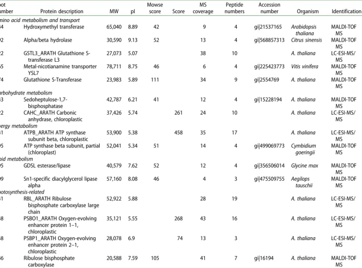
Table 1.List of identified proteins and related metabolic pathways in Arabidopsis leaves uninfested and infested by P. xylostella larvae at 27°C within 8 h. Spot
number Protein description MW pI
Mowse score Score MS coverage Peptide numbers Accession
number Organism Identification Amino acid metabolism and transport
244 Hydroxymethyl transferase 65,040 8.89 42 9 4 gi|21537165 Arabidopsis thaliana
MALDI-TOF MS 392 Alpha/beta hydrolase 30,590 9.13 52 13 4 gi|568857313 Citrus sinensis MALDI-TOF
MS 522 GSTL3_ARATH Glutathione S-transferase L3 27,073 5.07 38 10 A. thaliana LC-ESI-MS/ MS 665 Metal-nicotianamine transporter YSL7
78,711 8.75 46 6 4 gi|225423773 Vitis vinifera MALDI-TOF MS 774 Glutathione S-Transferase 23,983 5.89 111 34 9 gi|2554769 A. thaliana MALDI-TOF
MS Carbohydrate metabolism
243 Sedoheptulose-1,7-bisphosphatase
42,787 6.21 41 12 4 gi|15228194 A. thaliana MALDI-TOF
MS 522 CAHC_ARATH Carbonic anhydrase, chloroplastic 37,426 5.74 261 24 10 A. thaliana LC-ESI-MS/ MS Energy metabolism
241 ATPB_ARATH ATP synthase subunit beta, chloroplastic
53,900 5.38 458 35 17 A. thaliana LC-ESI-MS/
MS 295 ATP synthase beta subunit, partial
(chloroplast) 52,041 5.34 51 14 4 gi|499069773 Cymbidium goeringii MALDI-TOF MS Lipid metabolism
495 GDSL esterase/lipase 40,579 7.62 52 12 4 gi|356506014 Glycine max MALDI-TOF
MS 709 Sn1-specific diacylglycerol lipase
alpha 57,160 8.08 46 4 3 gi|475509755 Aegilops tauschii MALDI-TOF MS Photosynthesis-related 241 RBL_ARATH Ribulose
bisphosphate carboxylase large chain 52,922 5.88 28 19 A. thaliana LC-ESI-MS/ MS 448 PSBO1_ARATH Oxygen-evolving enhancer protein 1–1, chloroplastic 35,121 5.55 268 43 16 A. thaliana LC-ESI-MS/ MS 548 PSBP1_ARATH Oxygen-evolving enhancer protein 2–1, chloroplastic 28,078 6.9 74 13 3 A. thaliana LC-ESI-MS/ MS 646 Ribulose bisphosphate carboxylase
20,588 7.59 105 41 7 gi|16194 A. thaliana MALDI-TOF
MS Note: MW, molecular weight; pI, isoelectric point; Score, Mowse score according to Mascot search; Coverage, percentage of the protein sequence identified; Peptide
number, number of peptide hits for each protein.
this study. Existing data demonstrated that chewing insects caused the reduction of photosynthesis in plants following their infestation (Zangerl et al. 1997; Tang et al. 2009; Liu et al. 2010; Halitschke et al. 2011; Nabity et al. 2013). The downregulation of RuBis expression in insect-challenged plant leaves was found in some previous studies (Hermsmeier et al.2001; Giri et al.2006; Wei et al.2009). Moreover, it is indicated that heat stress causes downregulation of different key enzymes of the Calvin cycle in rice, including RuBis car-boxylase (Han et al.2009). The expression of RuBis carboxy-lase large chain was significantly downregulated in Arabidopsis leaves due to heat treatment (Rocco et al.
2013). Zou et al. (2011) indicated that RuBis carboxylase could be considered as a part of the plant adaptive response, to maintain CO2 fixation under stress factors. Based on the existing data, the downregulation of RuBis proteins observed in the present study may be a result of larval feeding on Ara-bidopsis leaves at high temperature (27°C compared to 22°C of Arabidopsis growth temperature).
With respect to amino acid metabolism and transport, five proteins from this class were identified in the present study (hydroxymethyltransferase, alpha/beta hydrolase, glutathione S-transferase, metal-nicotinamide transporter YSL7 and Glu-tathione S-Transferase). The amino acid is well-addressed as indicators of plants response to herbivorous insects’ attack on the plant (Schmelz et al. 2012; Sempruch et al. 2012). It is noted that glutathione plays a key role in detoxification of activated oxygen and can be up-regulated by jasmonates (JAs) (Sanchez-Sampedro et al.2007; Chen, et al. 2011). In addition, P. xylostella caterpillars induce JA-signaling in the
defense response of A. thaliana (Bidart-Bouzat and Klieben-stein2011; Savchenko et al.2013; Zhang et al.2013). There-fore, a glutathione S-transferase was here observed to be more abundant in the larvae-infested Arabidopsis leaves compared to uninfested plant samples. That may be related to insect behavior. This is in agreement with Collins et al. (2010), who found the upregulation of this protein in Arabidopsis leaves attacked by P. xylostella.
For carbohydrate and energy metabolism, herbivorous insect and heat stress negatively influence the glycolytic path-way and decrease energy production in Arabidopsis plants (Liu et al. 2010; Rocco et al. 2013). In agreement with this scenario, we obtained a down-representation of sedoheptu-lose-1,7-bisphosphatase, carbonic anhydrase, chloroplastic, ATP synthase subunit beta, chloroplastic, and ATP synthase beta subunit, partial (chloroplast) in Arabidopsis leaves attacked by larvae. Nabity et al. (2013) suggested that the alterations of such proteins consequently led to a reduction of photorespiration rate of plant leaves under invasive insects. Hence, it was noted that plants must use their energy econ-omically when challenging herbivorous insect infestation (Liu et al.2010).
In contrast to proteins related to carbohydrate and energy metabolism, a strong increase in GDSL esterase/lipase expression appeared in the proteome profile of larvae-infested Arabidopsis leaves in comparison to healthy leaves (14-fold change in its abundance). However, the expression of another protein, Sn1-specific diacylglycerol lipase alpha, was significantly downregulated due to larval infestation on leaves of Arabidopsis leaves. It has been suggested that
Figure 2.Comparison of protein expression between A. thaliana leaves infested and uninfested by P. xylostella larvae at 27°C within 8 h. Data of protein identification for each particular spot number are given inTable 1when they are available. Black and gray bars represented proteins up-regulated and downregulated by the larval attack, respectively. Labels on the right show the functional categories to which the proteins are assigned. *identified by MALDI-TOF/MS; **identified by LC-ESI-MS/ MS.
GSDL esterase/lipase plays an important role in rice (Oryza sativa L.) response to various environmental stress factors (Jiang et al. 2012). GSDL esterase/lipase often appears in plant response to environmental factors like cold, insect, or bacteria stresses, and its expression can be induced through JA-signaling (Chepyshko et al.2012).
Conclusions
This paper reports the characterization of the global pro-teome of A. thaliana leaves under larval pest infestation at high temperature for 8 h (27°C compared to 22°C of Arabi-dopsis growth temperature). By using 2-DE coupled with MALDI-TOF MS or LC-ESI-TRAP MS/MS, 13 of 18 differ-entially expressed protein spots were successfully identified. These proteins participate in multiple physiological pro-cesses. Functional classification analysis indicated that such proteins were associated with amino acid (5), carbohydrate (2), energy (2), and lipid (2) metabolism and photosynthesis (4). In addition, their relative abundance was up-regulated or downregulated according to larval pest feeding on Arabidop-sis leaves. Our data demonstrate that combined temperature and larvae stresses can alter the proteome in plant leaves. Acknowledgement
We thank the laboratory of Mass Spectrometry, University of Liège, for their excellent technical supports in protein identification.
Disclosure statement
No potential conflict of interest was reported by the authors.
References
Ali JG, Agrawal AA.2012. Specialist versus generalist insect herbivores and plant defense. Trends Plant Sci. 17:293–302.
Amme S, Matros A, Schlesier B, Mock HP.2006. Proteome analysis of cold stress response in Arabidopsis thaliana using DIGE-technology. J Exp Bot. 57:1537–1546. Epub 2006/04/01.
Antoun M, Ouellet F.2013. Growth temperature affects inflorescence architecture in Arabidopsis thaliana. Botany. 2013/09/01;91:642–651. Atkinson NJ, Urwin PE.2012. The interaction of plant biotic and abiotic stresses: from genes to the field. J Exp Bot. Jun;63:3523–3543. Epub 2012/04/03.
Barker JE, Poppy GM, Payne CC.2007. Suitability of Arabidopsis thali-ana as a model for host plant–Plutella xylostella–Cotesia plutellae interactions. Entomol Exp Appl. 122:17–26.
Bidart-Bouzat MG, Kliebenstein D. 2011. An ecological genomic approach challenging the paradigm of differential plant responses to specialist versus generalist insect herbivores. Oecologia. 167:677– 689. Epub 2011/06/01.
Chen Y, Pang Q, Dai S, Wang Y, Chen S, Yan X.2011. Proteomic identi-fication of differentially expressed proteins in Arabidopsis in response to methyl jasmonate. J Plant Physiol. Jul 01;168:995–1008. Epub 2011/03/08.
Chepyshko H, Lai C-P, Huang L-M, Liu J-H, Shaw J-F. 2012. Multifunctionality and diversity of GDSL esterase/lipase gene family in rice (Oryza sativa L. japonica) genome: new insights from bioinfor-matics analysis. BMC Genomics. 2012/07/15;13:1–19.
Chuang WP, Herde M, Ray S, Castano-Duque L, Howe GA, Luthe DS.
2014. Caterpillar attack triggers accumulation of the toxic maize protein RIP2. New Phytol. 201:928–939. Epub 2013/12/07. Collins RM, Afzal M, Ward DA, Prescott MC, Sait SM, Rees HH,
Tomsett AB. 2010. Differential proteomic analysis of Arabidopsis thaliana genotypes exhibiting resistance or susceptibility to the insect herbivore, Plutella xylostella. PLoS ONE. 5:e10103.
Dubey NK, Goel R, Ranjan A, Idris A, Singh SK, Bag SK, Chandrashekar K, Pandey KD, Singh PK, Sawant SV. 2013. Comparative
transcriptome analysis of Gossypium hirsutum L. in response to sap sucking insects: aphid and whitefly. BMC Genomics. 14:1471–2164. Duceppe MO, Cloutier C, Michaud D.2012. Wounding, insect chewing and phloem sap feeding differentially alter the leaf proteome of potato, Solanum tuberosum L. Proteome Sci. 73:10.
Ehlting J, Chowrira SG, Mattheus N, Aeschliman DS, Arimura G, Bohlmann J. 2008. Comparative transcriptome analysis of Arabidopsis thaliana infested by diamond back moth (Plutella xylos-tella) larvae reveals signatures of stress response, secondary metab-olism, and signalling. BMC Genomics. 9:1471–2164.
ČErnÝ M, JedelskÝ PL, NovÁK JAN, Schlosser A, BrzobohatÝ B.2014. Cytokinin modulates proteomic, transcriptomic and growth responses to temperature shocks in Arabidopsis. Plant Cell Environ. 37:1641–1655.
Fakae BB, Campbell AM, Barrett J, Scott IM, Teesdale-Spittle PH, Liebau E, Brophy PM.2000. Inhibition of glutathione S-transferases (GSTs) from parasitic nematodes by extracts from traditional Nigerian med-icinal plants. Phytotherapy Res PTR. 14:630–634. Epub 2000/12/13. Giri AP, Wunsche H, Mitra S, Zavala JA, Muck A, Svatos A, Baldwin IT.
2006. Molecular interactions between the specialist herbivore Manduca sexta (Lepidoptera, Sphingidae) and its natural host Nicotiana attenuata. VII. Changes in the plant’s proteome. Plant Physiol. 142:1621–1641.
Halitschke R, Hamilton JG, Kessler A.2011. Herbivore-specific elicita-tion of photosynthesis by mirid bug salivary secreelicita-tions in the wild tobacco Nicotiana attenuata. New Phytol. 191:528–535. Epub 2011/ 03/30.
Han F, Chen H, Li XJ, Yang MF, Liu GS, Shen SH.2009. A comparative proteomic analysis of rice seedlings under various high-temperature stresses. Biochim Biophys Acta. 11:25.
Herde M, Gartner K, Kollner TG, Fode B, Boland W, Gershenzon J, Gatz C, Tholl D. 2008. Identification and regulation of TPS04/GES, an Arabidopsis geranyllinalool synthase catalyzing the first step in the formation of the insect-induced volatile C16-homoterpene TMTT. Plant Cell. 20:1152–1168. Epub 2008/04/10.
Hermsmeier D, Schittko U, Baldwin IT.2001. Molecular interactions between the specialist herbivore Manduca sexta (Lepidoptera, Sphingidae) and its natural host Nicotiana attenuata. I. Large-scale changes in the accumulation of growth- and defense-related plant mRNAs. Plant Physiol. 125:683–700. Epub 2001/02/13.
Huang C, Verrillo F, Renzone G, Arena S, Rocco M, Scaloni A, Marra M.
2011. Response to biotic and oxidative stress in Arabidopsis thaliana: analysis of variably phosphorylated proteins. J Proteomics. 74:1934– 1949. Epub 2011/05/31.
Jiang Y, Chen R, Dong J, Xu Z, Gao X.2012. Analysis of GDSL lipase (GLIP) family genes in rice (Oryza sativa). POJ. 5:351–358. Kilian J, Peschke F, Berendzen KW, Harter K, Wanke D. 2012.
Prerequisites, performance and profits of transcriptional profiling the abiotic stress response. Biochim Biophys Acta. 1819:166–175. Kliebenstein D, Pedersen D, Barker B, Mitchell-Olds T. 2002.
Comparative analysis of quantitative trait loci controlling glucosino-lates, myrosinase and insect resistance in Arabidopsis thaliana. Genetics. 161:325–332. Epub 2002/05/23.
Kroes A, Broekgaarden C, Castellanos Uribe M, May S, van Loon JJA, Dicke M.2017. Brevicoryne brassicae aphids interfere with transcrip-tome responses of Arabidopsis thaliana to feeding by Plutella xylos-tella caterpillars in a density-dependent manner. Oecologia. 183:107–120. 10/2207/10/received10/16/accepted.
Lippert D, Chowrira S, Ralph SG, Zhuang J, Aeschliman D, Ritland C, Ritland K, Bohlmann J. 2007. Conifer defense against insects: pro-teome analysis of Sitka spruce (Picea sitchensis) bark induced by mechanical wounding or feeding by white pine weevils (Pissodes strobi). Proteomics. 7:248–270.
Liu LL, Zhang J, Zhang YF, Li YC, Xi JH, Li SY.2010. Proteomic analysis of differentially expressed proteins of Arabidopsis thaliana response to specialist herbivore Plutella xylostella. Chem Res Chin Univ. 26:958–963.
Loreto F, Schnitzler JP. 2010. Abiotic stresses and induced BVOCs. Trends Plant Sci. 15:154–166.
Louis J, Shah J.2013. Arabidopsis thaliana-Myzus persicae interaction: shaping the understanding of plant defense against phloem-feeding aphids. Front Plant Sci. 213:4.
Nabity PD, Zavala JA, DeLucia EH.2013. Herbivore induction of jasmo-nic acid and chemical defences reduce photosynthesis in Nicotiana attenuata. J Exp Bot. 64:685–694. Epub 2012/12/25.
Pineda ANA, Soler R, Pastor V, Li Y, Dicke M.2017. Plant-mediated species networks: the modulating role of herbivore density. Ecol Entomol. 42:449–457.
Rocco M, Arena S, Renzone G, Scippa GS, Lomaglio T, Verrillo F, Scaloni A, Marra M. 2013. Proteomic analysis of temperature stress-responsive proteins in Arabidopsis thaliana rosette leaves. Mol BioSyst. 9:1257–1267.
Sanchez-Sampedro A, Kim HK, Choi YH, Verpoorte R, Corchete P.
2007. Metabolomic alterations in elicitor treated Silybum marianum suspension cultures monitored by nuclear magnetic resonance spec-troscopy. J Biotechnol. 130:133–142. Epub 2007/05/04.
Sarfraz M, Dosdall LM, Keddie BA.2006. Diamondback moth–host plant interactions: implications for pest management. Crop Prot. 25:625–639. Savchenko T, Pearse IS, Ignatia L, Karban R, Dehesh K.2013. Insect her-bivores selectively suppress the HPL branch of the oxylipin pathway in host plants. Plant J. 73:653–662.
Schmelz EA, Huffaker A, Carroll MJ, Alborn HT, Ali JG, Teal PE.2012. An amino acid substitution inhibits specialist herbivore production of an antagonist effector and recovers insect-induced plant defenses. Plant Physiol. 160:1468–1478. Epub 2012/09/26.
Sempruch C, Leszczyński B, Chrzanowski G, Filipczuk A, Czerniewicz P, Wolska K.2012. Activity of aspartate aminotransferase and Alanine aminotransferase within winter triticale seedlings infested by grain aphid (Sitobion Avenae F.). J Plant Prot Res. 52:364–367.
Silva R, Furlong MJ.2012. Diamondback moth oviposition: effects of host plant and herbivory. Entomol Exp Appl. 143:218–230.
Tang J, Zielinski R, Aldea M, DeLucia E. 2009. Spatial association of photosynthesis and chemical defense in Arabidopsis thaliana follow-ing herbivory by Trichoplusia ni. Physiol Plant. 137:115–124. Truong DH, Bauwens J, Delaplace P, Mazzucchelli G, Lognay G, Francis
F., Leiss K. 2015. Proteomic analysis of Arabidopsis thaliana (L.) Heynh responses to a generalist sucking pest (Myzus persicae Sulzer). Plant Biol (Stuttg). 17:1210–1217. Epub 2015/07/15. Truong D-H, Delory BM, Brostaux Y, Heuskin S, Delaplace P, Francis F,
Lognay G.2014. Plutella xylostella (L.) infestations at varying temp-eratures induce the emission of specific volatile blends by Arabidopsis thaliana (L.) Heynh. Plant Signal Behav. 9:e973816.
Wei Z, Hu W, Lin Q, Cheng X, Tong M, Zhu L, Chen R, He G.2009. Understanding rice plant resistance to the Brown Planthopper (Nilaparvata lugens): a proteomic approach. Proteomics. 9:2798– 2808. Epub 2009/05/01.
Zangerl AR, Arntz AM, Berenbaum MR.1997. Physiological price of an induced chemical defense: photosynthesis, respiration, biosynthesis, and growth. Oecologia. 109:433–441.
Zhang PJ, Broekgaarden C, Zheng SJ, Snoeren TA, van Loon JJ, Gols R, Dicke M. 2013. Jasmonate and ethylene signaling mediate whitefly-induced interference with indirect plant defense in Arabidopsis thaliana. New Phytol. 197:1291–1299. Epub 2013/ 01/15.
Zou J, Liu C, Chen X.2011. Proteomics of rice in response to heat stress and advances in genetic engineering for heat tolerance in rice. Plant Cell Rep. 30:2155–2165. 2011/12/01.