HAL Id: hal-01620294
https://hal.archives-ouvertes.fr/hal-01620294
Submitted on 20 Dec 2017HAL is a multi-disciplinary open access
archive for the deposit and dissemination of sci-entific research documents, whether they are pub-lished or not. The documents may come from teaching and research institutions in France or abroad, or from public or private research centers.
L’archive ouverte pluridisciplinaire HAL, est destinée au dépôt et à la diffusion de documents scientifiques de niveau recherche, publiés ou non, émanant des établissements d’enseignement et de recherche français ou étrangers, des laboratoires publics ou privés.
Structural and magnetic properties of nano-sized
NiCuZn ferrites synthesized by co-precipitation method
with ultrasound irradiation
Hassen Harzali, Fairouz Saida, Arij Marzouki, Adel Megriche, Fabien Baillon,
Fabienne Espitalier, Arbi Mgaidi
To cite this version:
Hassen Harzali, Fairouz Saida, Arij Marzouki, Adel Megriche, Fabien Baillon, et al.. Structural and magnetic properties of nano-sized NiCuZn ferrites synthesized by co-precipitation method with ultrasound irradiation: Review. Journal of Magnetism and Magnetic Materials, Elsevier, 2016, 419, p. 50-56. �10.1016/j.jmmm.2016.05.084�. �hal-01620294�
Author’s Accepted Manuscript
Structural and magnetic properties of nano-sized
NiCuZn ferrites synthesized by co-precipitation
method with ultrasound irradiation
Hassen Harzali, Fairouz Saida, Arij Marzouki,
Adel
Megriche,
Fabien
Baillon,
Fabienne
Espitalier, Arbi Mgaidi
PII:
S0304-8853(16)30812-5
DOI:
http://dx.doi.org/10.1016/j.jmmm.2016.05.084
Reference:
MAGMA61498
To appear in:
Journal of Magnetism and Magnetic Materials
Received date: 27 February 2016
Revised date:
11 May 2016
Accepted date: 24 May 2016
Cite this article as: Hassen Harzali, Fairouz Saida, Arij Marzouki, Adel
Megriche, Fabien Baillon, Fabienne Espitalier and Arbi Mgaidi, Structural and
magnetic properties of nano-sized NiCuZn ferrites synthesized by
co-precipitation method with ultrasound irradiation,
Journal of Magnetism and
Magnetic Materials,
http://dx.doi.org/10.1016/j.jmmm.2016.05.084
This is a PDF file of an unedited manuscript that has been accepted for
publication. As a service to our customers we are providing this early version of
the manuscript. The manuscript will undergo copyediting, typesetting, and
review of the resulting galley proof before it is published in its final citable form.
Please note that during the production process errors may be discovered which
could affect the content, and all legal disclaimers that apply to the journal pertain.
1
Structural and magnetic properties of nano-sized NiCuZn ferrites
synthesized by co-precipitation method with ultrasound irradiation
Hassen Harzalia*, Fairouz Saidaa, Arij Marzoukia, Adel Megrichea, Fabien Baillonb, Fabienne Espitalierb, Arbi Mgaidia,c
a
Laboratory of Applied Mineral Chemistry, Faculty of Sciences, University Tunis ElManar, Campus university Farhat Hached El-Manar 2092 Tunis, Tunisia
bEcole des Mines d’Albi, Centre RAPSODEE, UMR CNRS 2392, 81013 ALBI Cedex 09, France
c
Taibah University, Faculty of Sciences & art Al Ula Saudi Arabia SA
E-mail address: harzali@mines-albi.fr
ABSTRACT
Sonochemically assisted co-precipitation has been used to prepare nano-sized Ni-Cu-Zn-ferrite powders. A suspension of constituent hydroxides was ultrasonically irradiated for various times at different temperatures with high intensity ultrasound radiation using a direct immersion titanium horn. Structural and magnetic properties were investigated using X-diffraction (XRD), FT-IR spectroscopy, transmission electron microscopy (TEM), Nitrogen adsorption at 77K (BET) and Vibrating sample magnetometer (VSM). Preliminary experimental results relative to optimal parameters showed that reaction time t=2h, temperature =90C and dissipated Power Pdiss= 46.27 watt. At these conditions, this work shows the formation of nanocrystalline single-phase structure with particle size 10-25 nm. Also, ours magnetic measurements proved that the sonochemistry method has a great influence on enhancing the magnetic properties of the ferrite.
Keywords: Ultrasound method; Ni0.4Cu0.2Zn0.4 ferrite nanoparticles ; Spinel structure; crystallite size, Magnetic properties.
.
1. Introduction
Commercially speaking, spinel ferrites are important materials because of their good magnetic-electrical properties [1,2]. Nanocrystalline spinel ferrites have been
2
investigated extensively due to their various applications in information storage systems, magnetic core, ferrofluids, microwave absorbers and medical diagnostics [3].
In recent years, extensive studies have been performed on the synthesis and magnetic characteristics of Ni-Cu-Zn ferrites because these materials present high electrical resistivity hard mechanical properties and also high Curie temperature and can be used for Multilayer Chip Inductor (MLCI) [4] and as transformer cores, inductors, recording heads, deflection yokes, etc [5]. According to Liu et al. [6], thin films of Ni0.4Cu0.2Zn0.4Fe2O4 can be integrated into radio frequency integrated inductors. In the present work we are interested in spinel ferrite nanoparticles with the following formula Ni0.4Cu0.2Zn0.4Fe2O4. We fixed Cu = 0.2, this value corresponds to good results of the saturation magnetization and electrical resistivity [7]. Moreover, we chose the molar ratios Ni/Zn=1 because the maximum of the saturation magnetization is obtained to this value [7].
Ni0.4Cu0.2Zn0.4 ferrites can be prepared through various wet chemical methods.
Sol-gel method and rapid thermal annealing (RTA) was used to synthesize thin films of Ni0.4Cu0.2Zn0.4 Fe2O4 [6].
Ni0.4Cu0.2Zn0.4Fe2O4 spinel nanoparticles were obtained using reverse micelle process [8].
Nanopowders of Ni0.4Cu0.2Zn0.4Fe2O4 were prepared by citrate precursor method followed by sintering at lower temperature [9].
Conventional solid state reaction oxide method and calcination at higher temperature was also used to prepare nanopowders of this ferrite [10].
Single-phase nano-crystalline Ni0.4Cu0.2Zn0.4Fe2O4 ferrites were successfully prepared through cheap and environmentally friend gelatin precursor method [11].
Nanocrystalline spinel ferrite MFe2O4 (M=Ni, Zn) powders were obtained via novel hydrothermal method using aloe vera plant extracted solution [12]. Authors conclude that hydrothermal route is very versatile for the synthesis of nanophase materials because reactions and phase formation occurred in closed systems at low temperatures (less than 200°C). Microwave assisted-hydrothermal was also tested as novel processing to synthesize ferromagnetic Ni0.9Fe2.1O4 and paramagnetic Zn1.1Fe1.09O4 ferrites [13].
3
In all methods used, the shape, the size distribution and the crystalinity of the final product can be precisely controlled through adjusting the reaction parameters such as temperature, time, solvent, and precursor type [14-17]. The physical properties of the spinel type nanoparticles such as magnetic, microwave and doping effects are determined by the particle size and shape as well as the type and the degree of defectiveness of the crystal lattice in which a total of 24 metal cations are partitioned between eight tetrahedral (8 Fe2+) and 16 octahedral (16 Fe3+ ) interstices.
To our knowledge no literature was found on sonochemistry synthesized NiCuZn ferrites. To avoid agglomeration of nanoparticles, sonochemical route was adopted to investigate the mechanism of Zn ferrite particles synthesized under ultrasound conditions [18].
Ni-Cu ferrite nanopowders were obtained by coprecipitation method with ultrasound irradiation [19]. This work concludes that only single phase of Cu1-xNixFe2O4 was obtained under these conditions otherwise the Cu2+ complexation will occurs.
In our previous work [20], we have elucidate the positive influence of ultrasound on crystallization processes through the reduction of the induction time, metastable zone width, the modification of the crystals size distribution and the increase in the number of crystals at equivalent supersaturation. So we concluded that the sonochemical process appears to be an attractive method for preparing several fine substances such as ferrites. Therefore, in the present work, it is aimed at carrying out a systematic study of nano-sized Ni0.4Cu0.2Zn0.4Fe2O4 ferrites synthesized by co-precipitation method with ultrasound irradiation. One of the motivations of this study is to show if a minimum energy and minimum synthesis duration exists with method assisted by ultrasound. For these reasons, the temperature effect and the time effect have been studied in this work.
2. Experimental
2.1. Preparation of powders
The initial chemicals were analytical grade nickel sulfate [NiSO4.6H2O], copper sulfate [CuSO4.5H2O], iron nitrate [Fe(NO3)3.9H2O] and zinc sulfate [ZnSO4.7H2O]. Stoichiometric amounts of each chemical necessary for the preparation of Ni0.4Cu0.2Zn0.4Fe2O4 were carefully dissolved in 10 g deionized water. After dissolution (the conductivity of solution was monitored), the obtained solutions were mixed. The pH of the final solution was adjusted to 12 using NaOH solution.
4
The experimental apparatus used for the synthesis is represented first by a thermostated vessel (100 mL) which was stirred with a magnetic stir bar at a constant rotation speed of about 200 rpm. Second, by ultrasound that was applied at the top of the liquid by a stainless tip transducer with a diameter of 1.1 cm, immersed at a 1 cm depth in the vessel. Three ultrasonic power levels, measured in water by the calorimetric method at 25 °C, were used corresponding to dissipated powers of 28.03 (Amplitude 30%), 38.23 (Amplitude 40%) and 46.27 Watt (Amplitude 50%) [21]. The solution temperature is measured with an instrumental resolution of 0.01 °C. Three temperature values were used in this work (70, 90 and 100 °C).
The resulting brown powders were collected by filtration and washed (hydrochloric acid and distilled water were used) to remove any possible impurities. The product was finally dried at 100 °C in an oven during 24 h.
2.2. Techniques
X-ray powder diffraction patterns are recorded using a panalyticalX’pert-pro X-ray diffractometer operating with Co Kα radiation.
Fourier transform infrared spectra (FT-IR) for the powders are recorded on a Perkin-Elmer 883 spectrophotometer from 4000 to 200 cm-1 by the KBr pellet method.
Morphological observation was performed using a transmission electron microscopy (TEM) system.Nanoparticles size and morphology were studied with a JEOL Model JEM 100 CX electron microscope having an acceleration voltage of 100 kV. High-resolution transmission electron microscopy observations were performed using a JEOL2010 microscope of 200 kV. Samples for TEM were prepared by placing a drop of NP solution on a carbon coated copper grid. The size distributions are determined with Image J software (1.37v) by measuring the diameter of around 100 nano-particules.
N2 adsorption isotherms of the dried products were measured at 77 K on a Micromeritics ASAP 2020 instrument. The samples were kept in vacuum at 323 K for 2 h to remove adsorbed gases before the measurement. The specific surface areas and the pore-size were determined using the Brunauer–Emmett–Teller (BET) and the Barrett–Joyner–Halenda (BJH) methods, respectively.
5
Magnetic properties such as saturation magnetization are investigated using vibrating sample magnetometer (VSM, LakeShore 7400) at room temperature.
3. Results and discussion 3.1. Dissipated power
The Cavitation intensity, can be thought of as the product of bubble collapse (or implosion) pressure times the number of bubbles collapsing, is linked to the local ultrasonic power which affects the cavitation regimes (dynamics of oscillating bubbles). The calorimetric method is used because all the energy entering into the calorimeter is expected to be transformed into heat.
In the present work, a temperature probe, with resolution of 0.01 °C, has been used in order to measure the dissipated power in our reactor.
The dissipated power, Pdiss (ultrasound power), was estimated by calorimetry and calculated using the following equation:
Pdiss = mCp (DT/Dt)t=0 (1)
Where m, CP and DT denote the solution mass, the specific heat of water and the initial slope of the curve of temperature versus time respectively. A known mass of water was added in the sono-reactor. Measurements of dissipated acoustic power in the sono-reactor were recorded for different amplitude (30, 40 and 50%). The calorimetric measurements have been done with water in the cooling jacket and with stirring during the measurements [21]. The obtained dissipted power Pdiss are summarized in table 1:
Table 1
3.2. X-ray diffraction
The time effect
The X-ray diffraction patterns recorded for the synthesized ferrite nanocrystals with variation of time at the same temperature and dissipated power (90 °C, 46.27 Watt) are shown in Fig. 1. The existence of the (220), (311), (400), (422), (511) and (440) major crystal plane in the XRD patterns confirms the formation of spinel cubic structure with the Fd3m space group. Weak diffraction peaks at 38 and 46 ° were observed for samples synthesized at 0.75 and 1 hour (Fig. 1). These peaks are attributed to hematite (α-Fe2O3) and to CuO [22, 23]. These peaks completely disappear when the synthesis duration begins high to 1 hour. From 1.5 hours of synthesis duration, the samples were considered to be single-phase spinel structure as
6
no extra peaks and no unreacted constituents were observed. In Table 2, we reported the optimal reaction time and temperature observed in the other chemical routes for NiCuZn ferrites synthesis.
Fig. 1 Table 2
The crystallite size for all samples estimated from XRD peak broadening using Scherrer’s formula:
d= 0.9λ/βcosθ (2)
Where d is the crystallite size, λ is the wavelength of X-ray radiation (Co Kα), θ is the Bragg angle and β is the full width at half maximum (FWHM) of all diffraction peaks.
The obtained lattice parameters, crystallite size and cell volume for the nanocrystals are listed in Table 3 [24]. The average crystallite size is increasing with a synthesis time duration.
Table 3
The temperature effect
Fig. 2 shows the XRD patterns collected on particles synthesized at 70, 90 and 100 °C (t=2 hours and dissipated power = 46.27 Watt). For all samples, the existence of the major crystal plane in the XRD patterns confirms the formation of spinel cubic structure with the Fd3m space group. The samples were considered to be single-phase spinel structure. We can notice that ultrasound has probably the major effect in the precipitation process compared to the temperature effect.
Fig. 2
Also, samples could be considered as a single-phase spinel structure. However as it can be seen there is a right shift diffractograms with increasing temperature. Several studies confirmed the increasing of crystallite size with temperature. Table 4 summarized the values of crystalliste size, the lattice parameters and the corresponding cell volume at different temperatures.
7
As it can be seen from this Table, the crystallite size increases with increasing temperature and the lattice parameters decrease slightly with temperature increasing. This result confirms the right shift of diffractograms XRD reported in Fig.2. In this work, we have chosen the optimal temperature =90C.
The dissipated power effect
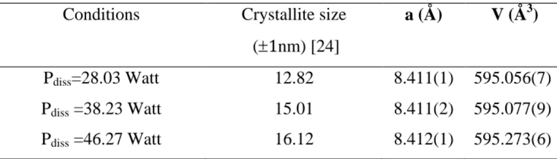
The X-ray diffraction patterns recorded for the synthesized ferrite nanocrystals with variation of dissipated power at the same temperature and duration time (90 °C, 2 hours) are shown in Fig. 3. The XRD results of figure 3 suggest that Ni0.4Cu0.2Zn0.4Fe2O4 samples crystallized in single-phase spinel structure with the group space (Fd3m).
Fig. 3
From this figure, we note that the peak (220) appears correctly from Pdiss = 38.23 and we have observed also that crystallite size increases with increasing dissipated power (see Table 5).
Table 5
3.3. FT-IR measurements
The obtained powders Ni0.4Cu0.2Zn0.4Fe2O4 were also investigated by Fourier transform infrared spectroscopy. The FTIR spectra in the frequency range 800-400 cm-1 are shown in Fig. 4.
Fig. 4
As it can be seen from this figure that FTIR spectra of the investigated samples exhibit two absorption bands around 400 and 600 cm-1. These bands are characteristic of spinel ferrites and attributed to the vibrations of the metal ion-oxygen complexes in the tetrahedral and octahedral sites [10]. The high frequency band appearing near 600 cm-1 was attributed to stretching mode of tetrahedral group (noted 1) and that lower band near the 420 cm-1 was assigned to octahedral cluster. We observed also change in the position band 1 which is related toincreasing of particle size [7].
8
3.4. Surface area and porosity measurements
The adsorption–desorption isotherm of nitrogen at 77K for Ni0.4Cu0.2Zn0.4Fe2O4 ferrites (Figure 5 and 6) are of Type IV isotherms with Type H2 hysteresis loops, according to the classification of the IUPAC [25], the initial part of the Type IV isotherm is attributed to monolayer–multilayer adsorption. Hysteresis appearing in the multilayer range of physisorption isotherms is usually associated with capillary condensation in mesopore structures. The H2 hysterisis loop is the characteristic of mesopore structures.
Fig. 5 Fig. 6
Textural parameters derived from nitrogen adsorption–desorption isotherm data are summarized in Table 6. As can be noted, the effect of temperature and dissipated power on Ni0.4Cu0.2Zn0.4Fe2O4 ferrites was accompanied by a marked increase in surface area and pore volume.
Table 6
3.5 Morphological study
Fig.7 shows the TEM image of Ni0.4Cu0.2Zn0.4Fe2O4 synthesized at T=90°C, t=2 hours and for Pdiss= 46.26 Watt deposited on carbon coated copper grid at 50 nm resolution. It can be seen, that powders have uniform shapes and well dispersed. The average particle sizes were measured in TEM micrographs and were found to be in the range of 10-25 nm. These values were, similar to the ones, calculated using the XRD measurements.
Figure 7
3.6 Magnetic properties
The magnetic properties are the most important properties of ferrites depending on the synthesis method, processing conditions, microstructure and chemical composition. The change in the magnetic properties can be attributed to the influence of the cationic stochiometry and their occupancy in the specific sites. In the spinel ferrites, it is well known that Zn2+ ions prefer the occupation of tetrahedral (A) sites [26,27], Ni2+ and Cu2+ ions prefer the occupation of octahedral (B) sites while Fe3+ ions partially occupy the A and B sites. Fe3+ (d5), Ni2+ (d8), Cu2+ (d9) and Zn2+ (d10) cations of the ferrite have 5, 2, 1, and 0 unpaired
9
electrons respectively and some magnetic moment in µB considering complete unquenched orbital moment.
For Ni0.4Cu0.2Zn0.4Fe2O4 nanopowders synthesized at different conditions, we have investigated their magnetic properties using a Magnetometer (VSM) at room temperature. Fig. 8 (a,b,c) shows the M-H curves of the ferrites at different conditions.
Fig. 8
From this figure, we can conclude that the saturation magnetization (Ms) values increases with the duration time (Fig. 8 a) and with dissipated power (fig. 8 c). The higher value of Ms (55.72 Am2.kg-1) is obtained for Ni0.4Cu0.2Zn0.4Fe2O4 synthesized at T=90°C, t=2 hours and for Pdiss= 46.27 Watt. The coercive field (Hc) is of the order of 3.9 10-3 T for all the ferrites which is indicating that the Ni0.4Cu0.2Zn0.4 ferrite could be used in high-density data storage devices [28].
Hysteresis loops in Fig. 8 are typical for soft magnetic materials and the “S” shape of the curves together with the negligible coercivity (Hc = 39 Oe) indicate the presence of small magnetic particles exhibiting superparamagnetic behaviors [29]. Literature data imply that when the diameter of particles is less than 30 nm, the particles show the character of superparamagnetism [29].
The annealing temperature dependence of saturation magnetization (Ms) and the coercivity Hc for the Ni0.4Cu0.2Zn0.4Fe2O4 was observed in earlier work [28]. The authors concluded changes in the magnetic properties of the samples can be attributed to the modification of the crystallite sizes. The increase in saturation magnetization was most likely attributed to the increasing of particle size and can be explained on the basis of changes in exchange interactions between tetrahedral and octahedral sub-lattices [29].
As can be noted, with the use of ultrasound in the synthesis (without calcination), we can obtain a good Ms compared to other methods (table 7).
Table 7
4. Conclusions
Sonochemically assisted co-precipitation method was used to prepare the NiCuZn ferrites. The structure and magnetic properties have been investigated and the following results have been obtained:
10
- A single-phase Ni-Cu-Zn ferrite with cubic spinel structure without any secondary phase was formed and no extra lines corresponding to any other phases could be detected.
- The crystallite size of synthesized nanocrystals was calculated using Scherrer’s formula and TEM images. The range of crystallite size lied between 10 and 25nm.
- The saturation of magnetization (Ms) increases with increasing the crystallite size.
-The optimal value of Ms was obtained for the sample synthesized at these initial conditions (reaction time =2 hours, temperature = 90°C and dissipated power Pdiss= 46 Watt).
References
[1] T. Abraham, J. Am. Ceramic. Soc. Bull. 73 (1994) 62.
[2] S.W. Lee, S.H. Yonn, S.Y. An, W.C. Kim, C.S. Kim, J. Magn. 4 (1999) 115.
[3] A. Baykal, N. Kasapoğlu, Y. Köseoğlu, M.S. Toprak, H. Bayrakdar, J. Alloys Compd. 464 (2008) 514.
[4] Z. Yue, L. Li, J. Zhou, H. Zhang, Z. Ma, Z. Gui, J. Mater. Lett. 44 (2000) 279.
[5] S. Ramesh, B. Chandra Sekhar, P.S.V. Subba Rao, B. Parvatheeswara Rao, Ceram Int. 40
(2014) 8729.
[6] F. Liu, T.Ren, C.Yang, L.Liu, A.Z.Wang, J.Yu, Materials letters 60 (2006) 1403. [7] J. H. Nam, S. J. Park, W. K. Kim, IEEE Trans. Magn. 39 (2003) 3139.
[8] A. Ghasmi, J. Magn. Magn.Mater. 360 (2014) 41.
[9] M. C. Dimri, A. Verma, S. C. Kashyap, D.C. Dube, O. P. Thakur, C.Prakash, Mater. Sci. Eng. B 133 (2006) 42.
[10] M.M. Eltabey∗, K.M. El-Shokrofy, S.A. Gharbia, J. Alloys Compd. 509 (2011) 2473. [11] M.A. Gabal, Y.M. Al Angari, A.Y. Obaid, A. Qusti, Advanced Powder Technology, 25
(2014) 457.
[12] S. Phumying, S. Labuayai, E. Swatsitang, V. Amornkitbamrung, S. Maensiri, Mat. Res. Bull. 48 (2013), 2060.
[13] R.S. Melo, F.C.Silva, K.R.M. Moura, A.S. de Menezes, F.S.M. Sinfrônio, J. Magn. Magn.Mater. 381(2015)109.
11
[15] N.Y. Mostafa, A.A. Shaltout, H. Omar, S.A. Abo-El-Enein, J. Alloys Compd. 467
(2009) 332.
[16] W.L. Suchanek, R.E. Riman, Adv. Sci. Tech. 45 (2006) 184.
[17] S. Kumar, R. Kumar, S.K. Sharma, V.R. Reddy, A. Banerjee, Alimuddin, J. Solid St. Comm. 142 (2007) 706.
[18] HA. Choudhury, A. Choudhary, M. Sivakumar, VS.Moholkar, Ultrason Sonochem. 1
(2013) 294.
[19] SG.Doh, EB. Kim, BH. Lee, JH. Oh, J. Magn. Magn. Mater. 3 (2003) 2238.
[20] H. Harzali, F. Baillon, O. louisnard, F. Espitalier, A. Mgaidi, Ultrason Sonochem. 18 (2011) 1097.
[21] H. Harzali, F. Baillon, O. louisnard, F. Espitalier, A. Mgaidi, Chem. Eng. J. 195 (2012) 332.
[22] D.P. Volanti, D. Keyson, L.S. Cavalcante, A.Z. Simoes, M.R. Joya, E. Longo, J.A. Varela, P.S. Pizani, A.G. Souza, J. Alloys Compd.459 (2008) 537.
[23] F. Liu, C.Yang, T. Ren, A.Z. Wang, J. Yu, L.Liu, J. Magn. Magn. Mater. 309 (2007) 75. [24] B.D. Cullity, 2nd ed., Addison Wesley, Reading, MA, (1978) 99.
[25] B. Abida , L. Chirchi, S. Baranton, T.W. Napporn, H. Kochkar, J.M. Léger, A.Ghorbel, Appl. Catal., B. 106 (2011) 609.
[26] M.A. Ahmed, E. Ateia, L.M. Salah, A.A. El-Gamal, J. Mater. Chem. Phys. 92 (2005) 310.
[27] A.M. El-Sayed, J. Mater. Chem. Phys. 82 (2003) 583.
[28] M. C. Dimri, A. Verma, S. C. Kashyap, D.C. Dube, O. P. Thakur, C.Prakash, Materials Science and Engineering. 133 (2006) 42.
[29] Manova E, Tsoncheva T, Estournes C, Paneva D, Tenchev K, Mitov I, Petrov L, Appl. Catal., A. 300 (2006) 170.
[29] Nawale AB, Kanhe NS, Patil KR, Bhoraskar SV, Mathe VL, Das AK, J. Alloys Compd., 509
12
13
Figure 2: X-ray diffraction patterns for Ni0.4Cu0.2Zn0.4Fe2O4 samples synthesized at 70, 90 and 100 °C for 2 hours (Pdiss = 46.27 Watt).
14
Figure 3: X-ray diffraction patterns for the samples Ni0.4Cu0.2Zn0.4Fe2O4 at different delivered power (table 1).
15
Figure 4: FT-IR spectra of Ni0.4Cu0.2Zn0.4Fe2O4 ferrites.
(a) time=1.5h; =90C; Pdiss=46.27Watt (b) time=2.0 h; =90C; Pdiss =46.27Watt (c) time=2.0h; =70C; Pdiss=46.27Watt (d) time=2.0h; =100C; Pdiss =46.27Watt (e) time=2.0h; =90C; Pdiss =38.23Watt (f) time=2.0 h; =90C; Pdiss =28.03Watt
16
17
18
Figure 7: TEM images and size histogram of Ni0.4Cu0.2Zn0.4Fe2O4 synthesized at T=90°C, t=2 hours and for Pdiss= 46.27 Watt (1, 2, and 3 represent different areas of the
sample; 4 the size histogram).
1
2
3
19
Figure 8: Hysteresis loop for the Ni0.4Cu0.2Zn0.4Fe2O4 (a)
(A) Room temperature hysteresis loop for the ferrites at different synthesis duration at 90°C (Dissipated power = 46.27 Watt). (B): Aggrandizement
(b)
Room temperature hysteresis loop for the ferrites synthesized at 70, 90 and 100°C for 2 hours (Dissipated power = 46.27 Watt).
(c)
Room temperature hysteresis loop for the ferrites at different dissipated power at 90°C for 2 hours.
Table 1: Dissipated power results for different amplitudes.
Apmlitude (%) Dissipated power (3 x 10‐4Watt) [21]
30 28.03
40 38.23
50 46.27
Table 2: Data obtained for Ni0.4Cu0.2Zn0.4 Fe2O4 ferrites nanostructures synthesized through different methods.
Ferrite Nature Synthesis Method Duration (hours) Temperature (°C) Reference Ni0.4Cu0.2Zn0.4Fe2O4 Sol-gel + RTA [6] Ni0.4Cu0.2Zn0.4Fe2O4 reverse micelle process >38 Calcination (600) [8]
Ni0.4Cu0.2Zn0.4Fe2O4 Citrate precursor 4 Calcination (600, 1h) + Sintering
(900,3h)
20
Ni0.4Cu0.2Zn0.4Fe2O4 Ceramic method 12 Calcination (900) [10]
Ni0.4Cu0.2Zn0.4Fe2O4 Gelatin precursor method 2 Ignition of precursor at 550 [11] Ni0.4Cu0.2Zn0.4Fe2O4 Ultrasound 2 Co precipitation at 90 This work
Table 3: Crystallite size, lattice parameters (a) and cell volume (V) of Ni0.4Cu0.2Zn0.4Fe2O4 nano-crystals for two synthesis duration.
Conditions Crystallite size (1nm) [24] a (Å) V (Å3) t=1.5 hours; T=90°C 12.10 8.411(1) 595.027(3) t=2 hours; T=90°C 16.12 (16-38) [11] 8.412(1) 8.37 [8] 595.273(6)
Table 4: Crystallite size, lattice parameters and cell volume (V) of Ni0.4Cu0.2Zn0.4Fe2O4 nano-crystals for three synthesis temperatures for 2 hours (Pdiss = 46.27 Watt).
Conditions Crystallite size (1nm) [24] a (Å) V (Å3) T=70°C 15.90 8.414(4) 595.757(8) T=90°C 16.12 8.412(1) 595.273(6) T=100°C 17.87 18.7 [23] 8.411(4) 595.111(7)
21
Table 5: Crystallite size, lattice parameters and cell volume of Ni0.4Cu0.2Zn0.4Fe2O4 nano-crystals at different dissipated power.
Conditions Crystallite size (1nm) [24]
a (Å) V (Å3)
Pdiss=28.03 Watt 12.82 8.411(1) 595.056(7) Pdiss =38.23 Watt 15.01 8.411(2) 595.077(9) Pdiss =46.27 Watt 16.12 8.412(1) 595.273(6)
Table 6: Specific surface area and average pore size of samples obtained at different initial
conditions
Initial conditions SBET (m2.g-1) Average pore size (nm) time=2.0h; =90C; Pdiss =46.27Watt 127 6.3
time=2.0h; =70C; Pdiss =46.27Watt 112 8.4 time=2.0h; =100C; Pdiss=46.27Watt 138 5.8 time=2.0h; =90C; Pdiss =38.23Watt 120 7.5 time=2.0h; =90C; Pdiss =28.03Watt 113 6.1
Table 7: Magnetic parameters of synthesized NiCuZn ferrites.
Ferrite Nature Synthesis Method Saturation magnetization Ms
Reference Ni0.4Cu0.2Zn0.4Fe2O4 Sol-gel + RTA * [6]
Ni0.4Cu0.2Zn0.4Fe2O4 reverse micelle process * [8] with Calcination Ni0.4Cu0.2Zn0.4Fe2O4 Citrate precursor 92 Am2.kg-1 [9] with Calcination Ni0.4Cu0.2Zn0.4Fe2O4 Ceramic method with Calcination 72 Am2.kg-1 [10]
22
Ni0.4Cu0.2Zn0.4Fe2O4 Gelatin precursor method * [11]
Ni0.4Cu0.2Zn0.4Fe2O4 Ultrasound without calcination
55.72 Am2.kg-1 This work
Coprecipitation experiments were carried out with
ultrasound.
The spinel ferrite NiCuZn was perfectly synthesized by ultrasound.