Presented to obtain the master’s diploma in applied physics
Texte intégral
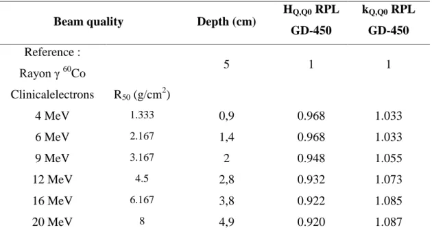
Figure
Documents relatifs
In order to study the nature of the reentrant superconducti- vity in ErRh4B4, we recently measured the variations of the superconducting critical temperature Tc, the
2014 I present a series of statistical results on the distribution of the size (i.e. the number of atoms) of clusters formed by atoms scattered randomly in a
Abstract : The x-rays imagining chains components from the source to the detector, rest on the first part of simulation to the energy production of x-rays emission (source),
In this work, we adopted the differential cross section of Mott and the total cross section of inner-shell ionization according to the formulation of Gryzinski, to simulate the
L’archive ouverte pluridisciplinaire HAL, est destinée au dépôt et à la diffusion de documents scientifiques de niveau recherche, publiés ou non, émanant des
Les alliages Sc-Gd respectent approximativement les lois d'kchelle, avec des &carts analogues aux verres de spins plus classiques : si l'on augmente la concentration, la
The main idea presented here is that it is possible to decompose a complex decision making problem (such as an optimization problem in a large search space) into a sequence
We can explain this by the following argument: below the transition temperature, there exists a single large cluster of lattice spins with some isolated ”defects” (i. clusters
