HAL Id: tel-02333812
https://pastel.archives-ouvertes.fr/tel-02333812
Submitted on 25 Oct 2019HAL is a multi-disciplinary open access archive for the deposit and dissemination of sci-entific research documents, whether they are pub-lished or not. The documents may come from teaching and research institutions in France or abroad, or from public or private research centers.
L’archive ouverte pluridisciplinaire HAL, est destinée au dépôt et à la diffusion de documents scientifiques de niveau recherche, publiés ou non, émanant des établissements d’enseignement et de recherche français ou étrangers, des laboratoires publics ou privés.
Early Steps of Ribosome Assembly Studied by Single
Molecule Force Measurements
Lena Melkonyan
To cite this version:
Lena Melkonyan. Early Steps of Ribosome Assembly Studied by Single Molecule Force Measurements. Physics [physics]. Université Paris sciences et lettres, 2018. English. �NNT : 2018PSLET026�. �tel-02333812�
!
THÈSE DE DOCTORAT
de l’Université de recherche Paris Sciences et Lettres
PSL Research University
Préparée à l’école supérieure de physique et chimie
industrielle
Early stages of ribosome assembly, studied by single-molecule force
measurements
Les premières étapes de l’assemblage du ribosome, étudiées par mesure de
force sur molécule unique
COMPOSITION DU JURY :
M. Berry Richard
Université d’Oxford, Rapporteur
M. Wuite Gijs
VU Amsterdam, Rapporteur
M. Allemand Jean-Francois
ENS Paris, Membre du jury
Mme. Bassereau Patricia
Institut Curie, Membre du jury
M. Bockelmann Ulrich
ESPCI, Membre du jury
M. Ueda Takuja
Université de Tokyo, Membre du jury
Soutenue par Lena
Melkonyan
le 13 décembre 2018
hEcole doctorale
n°
564
Physique en Ile-de-France
Spécialité
P
hysique
Dirigée par Ulrich Bockelmann
Acknowledgements
For the success of this work, I want to thank my PhD adviser Ulrich Bockelmann, for his support, patience, understanding and many fruitful discussions we had during these three years.
I would like to thank the members of the jury, Patricia Bassereau, Takuya Ueda, Jean François Allemand, Ulrich Bockelmann and especially the two referees, Gijs Wuite and Richard Berry for interesting discussions during the defense.
I would like to thank prof. Robert Menard and Maryse Brandt for their kindness and efficient support in finding a room for my PhD defense. Especially Maryse Brandt, for her efficiency, kindness and very nice human qualities.
I would like to thank my colleagues and friends: Thierry Bizebard for sharing his vast knowledge in molecular biology, for the preparation of DNA and RNA duplexes, for giving me insight into ribosomal proteins and their manipulation, and for being always available for a discussion. Andreas Biebricher for checking some of my results in the Peterman lab in the University of Amsterdam, for many useful discussions and experimental insights into optical tweezers. Mathilde Bercy for forming me in optical tweezers, preparation of the molecular construct, for her friendship and her support in difficult moments. Ismaïl Cissé for sharing with me his knowledge in molecular biology technics, for many interesting, entertaining discussions, for his friendship and his precious human qualities. Laurent Geffroy for his patience and help in finding good experimental configurations and alining the optical tweezers, for interesting and friendly discussions. Kokoura Mensah for taking care of me as his younger sister, for his sensitivity, attention, support and his successes in cheering me up. Jyoti Prakash for his very entertaining character, for numerous interesting stories about culture, habits, family values and nature of Nepal and India. Stefan Rouach for being friendly and supportive. Mehdi Vahdati for his friendship, support and comprehension. Artem Kovalenko for his friendship, support and sharing our cultural experiences. Armand Hakopian for his friendship, support, attention and warm heartedness.
I would also like to thank my family: Kay Wiese, my husband, for his love, patience, support, understanding and for always believing in me and giving me motivation to go forward in life. Lili Melkonyan, my three yers old niece, for her love, cheerfulness, tenderness and kindness, for being able to make my whole day with her smile. Vahan Melkonyan and Mariam Grigoryan, my brother and my sister in law, for their love, cheerfulness, kindness, support and help in difficult moments. Margarit and Varuzhan Melkonyan, my mother and my father, for their love, support and comprehension, for all
iv
the efforts they have taken to make me the person I am with my current achievements, for their eternal patience and for their faith in me.
Contents
Remerciements iii
Introduction 1
I Overstretching molecular duplexes made of DNA and RNA 3
I.1 DNA, RNA, DNA-RNA hybrid . . . 3
I.1.1 Chemical structure . . . 3
I.1.2 Physical structure . . . 6
I.2 Optical tweezers . . . 7
I.2.1 The principle of optical trapping . . . 7
I.2.2 Dual beam optical tweezers . . . 8
I.3 Four molecular constructs . . . 9
I.3.1 Preparation of dsDNA . . . 12
I.3.2 Preparation of the RNA-DNA hybrid . . . 13
I.3.3 Preparation of dsRNA . . . 13
I.3.4 Preparation of DNA-RNA hybrid . . . 14
I.3.5 Further treatment of the constructs . . . 16
Paper 1 18 Supplementary Information for Paper 1 35 II Early steps of ribosome assembly 43 III Résumé en Français 47 III.1 Introduction . . . 47
III.2 Comparaison entre quatre constructions moléculaires . . . 48
III.3 Influence des protines au repliement de l’ARN formant le ribosome . . 51
Paper 2 46 A Appendix 53 A.1 Preparation of the beads . . . 53
A.2 Preparation of dsDNA . . . 54
A.2.1 Polymerase chain reaction (PCR) . . . 54
vi CONTENTS
A.2.2 FseI restriction . . . 55
A.2.3 Ligation of sur_DNAbiot_MB and sur_compDNA2biot_MB oligonu-cleotides . . . 55
A.2.4 AflII restriction . . . 56
A.2.5 Klenow Fragment (exo-) treatment . . . 57
A.3 Preparation of RNA-DNA hybrid . . . 57
A.3.1 In vitro transcription of RNA leading strand . . . 57
A.3.2 Hybridization . . . 58
A.3.3 Ligation of sur_RNAbiot_MB Ph . . . 59
A.4 Preparation of dsRNA . . . 60
A.4.1 PCR . . . 60
A.4.2 In vitro transcription of RNA lagging strand . . . 61
A.4.3 Hybridization of DNA and ligation of RNA oligonucleotides . . 61
A.4.4 Hybridization of two RNA strands . . . 63
A.4.5 Ligation of sur_RNAbiot_MB Ph . . . 63
A.5 Preparation of DNA-RNA hybrid . . . 64
A.6 Preparation of 23S rRNA . . . 64
A.6.1 Polymerase chain reaction (PCR) . . . 64
A.6.2 In vitro transcription of 23S rRNA . . . 65
Introduction
Dear reader, this work aims at a better understanding of two problems that are im-portant, both from the point of view of fundamental physics, as of biology: First, the mechanical and structural behavior of biomolecules under an applied external force, and second, the early steps of ribosome assembly in Escherichia coli (E. coli).
Let us remind that forces play important roles in biology. They are exerted on DNA, RNA and hybrid molecules by molecular motors and enzymes [1, 2, 3, 4, 5] during biological processes, as DNA replication, homologous recombination, strand-repair, packaging, transcription, ribosome assembly, translation, cell division, and many more. These forces can result in topological, conformational or structural changes inducing extension, torsion or generation of a single strand. The latter is particularly important for biological processes, as it can lead to parasitic interactions and formation of non-native structures, which may affect biological processes involving duplexes composed of DNA or RNA. Therefore, the understanding of the mechanical response of DNA and RNA duplexes, as well as of DNA-RNA heteroduplexes (DNA-RNA hybrids) to an applied force is not only a fundamental problem in physics, but also of vital interest for biology. Beyond providing insight into molecular and cellular regulation, it may be useful for the development of applications in biotechnology and medecine.
Our second motivation is to better understand ribosome assembly. The ribosome is responsible for protein synthesis in all living organisms. The ribosome consists out of two subunits, a large and a small one. Here we study the large one. It is formed out of two RNA strands called 23S with 2904 nucleotides and 5S with 120 nucleotides, and proteins. The standard view is that 23S folds immediately after it is synthesised, that proteins are incorporated right away, and that the absence of proteins prevents it to fold properly. A key indication for this view is the large difference in efficiency of ribosome assembly in vivo as compared to that in vitro. While ribosome biogenesis in
vivo can occur efficiently at 37¶C in a couple of minutes, reconstruction of ribosomes
in vitro takes much longer: As an example, the large subunit folds in 90 minutes at
50¶C. Two major reasons have been put forward to explain this difference. One is that non-ribosomal factors are present in vivo that transiently interact with the nascent ribosome and assist its assembly. The second one is that in vivo ribosomal proteins and assembly factors bind to the RNA during its synthesis, facilitating its folding.
The idea of our experiment is the following: Using an RNA-DNA hybrid, we liberate the RNA with the sequence of the ribosomal RNA (i.e. 23S and 5S) at the rate at which it is synthesized in the cell, namely 40 base pairs per second. In this way we mimick as
2 CONTENTS
closely as possible RNA synthesis in living organisms, while at the same time keeping full control over the environment. More specifically, we compare folding of the rRNA in our standard buffer alone with the buffer augmented by the five proteins which are believed to bind first (early binders) [6]. This comparison is achieved by studying the force-extension curves in our optical tweezer experiments. Incorporation of the proteins is visible as a change in the hysteresis of the measured force-extension curves.
More specifically we ask ourselves the following questions: Problem 1.
What are the mechanical responses of deoxyribonucleic acid (DNA), ribonucleic acid (RNA) duplexes and DNA-RNA heteroduplexes to applied external forces? What is the difference between the response of different molecular duplexes?
Problem 2.
What is the effect of early binding ribosomal proteins uL4, uL24, uL22, uL13, bL20 on early steps of ribosome assembly?
These two questions will be treated in chapters I and II of the manuscript, re-spectively. In chapter I we also present the molecular objects of the study, the ex-perimental setup and afterwards the way to prepare the molecular constructs for the single-molecule force measurements. A summary in French is given in chapter III. Detailed protocols are provided in the annexA.
Chapter I
Overstretching molecular duplexes made
of DNA and RNA
In this work, we apply force on DNA and RNA duplexes, as well as DNA-RNA hybrids. In order to better understand the mechanical properties and force-induced structural modifications of these biopolymers, let us first refresh our knowledge on their chemical and physical structures.
I.1
DNA, RNA, DNA-RNA hybrid
I.1.1
Chemical structure
DNA
DNA is one of the main fundamental functional biopolymers and the only permanent carrier of genetic information in the cell. In native structure it is a double stranded helical molecule, each strand of which represents a polypeptide chain with a sequence of four different nucleotides called Adenine (A), Guanine (G), Cytosine (C), and Thymine (T). Each nucleotide consists of three different chemical groups: A phosphate group, 5 carbon sugars (deoxyribose for DNA) and a nitrogenous base, the latter being the only group by which the four nucleotides differ from each other. By establishing covalent bonds between the phosphate group of one and the sugar group of the other, these nucleotides form a single polypeptide chain of deoxyribonucleic acid (DNA) with a sugar-phosphate backbone and bases attached to it. Two DNA strands are joined together via hydrogen bonds between the bases of one strand and their complementary bases on the other strand forming base pairs. Adenine is always paired with Thymine (AT base pair) and Guanine with Cytosine (GC base pair). Two consecutive base pairs are held together by stacking interactions, see figure I.1.
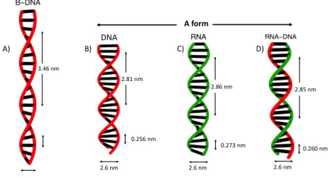
6Chapter I. Overstretching molecular duplexes made of DNA and RNA A form 3.46 nm A) 2.81 nm 2.6 nm 0.256 nm DNA B) 0.273 nm 2.6 nm 2.86 nm C) 2.85 nm 0.260 nm 2.6 nm D)
Figure I.4: Physical structures of dsDNA, dsRNA, RNA-DNA hybrid and differences of A and B form helices. In double stranded configuration all three molecules ex-hibit helical structure. Depending on external conditions dsDNA adopts A or B form, whereas dsRNA and RNA-DNA adopt only A form. Both A and B form helixes are right handed. Compared to B form, helixes in A form are shorter and wider. A) B-DNA has 2nm diameter and a distance of 0.346nm between two consecutive base pairs. One helical turn is 3.46nm corresponding thus to 10 base pairs. B) A-DNA has a diameter of 2.6nm and a distance between two consecutive base pairs is 0.256nm. One helical turn is 2.81nm corresponding thus to 11bp. C) A-RNA double helix has 2.6nm diameter and 0.273nm distance between two consecutive base pairs. One helical turn is 2.86nm corresponding thus to 10.5bp. D) RNA-DNA hybrid has 2.6nm diameter and 0.26nm distance between two consecutive base pairs. One helical turn is 2.85nm corresponding thus to 11bp.
between complementary bases, forming thus three different base pairs along the hybrid sequence: AT, GC, and AU, see figureI.4. As we saw above, all three molecules form, either completely or partially, a double helical structure in their native state. Let us see how similar and how different they are.
I.1.2
Physical structure
At room temperature, neutral pH and above 75% humidity, dsDNA is a double helix of B form (Fig.I.4A). It is right-handed with 2nm diameter and a helical turn of 3.46nm which corresponds to 10 base pairs. The distance between two adjacent base pairs is 0.346nm.
Compared to dsDNA, dsRNA and RNA-DNA hybrids are in A form. As one can see from Fig. I.4, unlike the B form which is thin and long, double helixes in A form are short and wide. A helix formed by dsRNA has a diameter of 2.6nm. The distance between two adjacent base pairs is 0.273nm. One complete helical turn is 2.86nm
I.2. Optical tweezers 7
corresponding to 11 base pairs per turn. The double helical structure of RNA-DNA hybrid is very close to that of dsRNA. It has a diameter of 2.6nm. The distance between adjacent base pairs is 0.26nm. One complete helical turn is 2.85nm corresponding to 10.5 base pairs per turn.
I.2
Optical tweezers
I.2.1
The principle of optical trapping
Over the last decade many different tools and techniques were developed to study the biological role and the mechanical properties of single molecules. Among them, dual-beam optical tweezers are of particular interest. Their principal is simple, and based on light-matter interaction. In our case, there is interaction between a focused laser beam and a polystyrene bead.
Comparing the wavelength of the laser, and the size of the object to be trapped, there are three regimes:
1. ⁄ ∫ d, the wavelength of light ⁄ is large as compared to the size d of the object (dipole-approximation regime).
2. ⁄ π d, the wavelength of light ⁄ is small compared to the size d of the object (ray-optics regime).
3. ⁄ ¥ d, the wavelength of the light ⁄ is comparable to the size d of the object.
In our experiments we deal with case 3, ⁄ ¥ d, which is well described by the generalized Lorentz-Mie theory [9]. In order to understand the basic principle of optical trapping and avoid complicated mathematics, we restrict our considerations to case 2, i.e. ⁄ π d. Light carries momentum, and as we know in scattering experiments the total mo-mentum is conserved. When interacting with matter, the photons of the laser beam are partially reflected and refracted by the matter thus loosing momentum. Let us de-note by ˛p the momentum of the photons before their interaction with the bead, by ˛p1 the momentum of those which have been refracted, and by p2 the momentum of those reflected by the bead. Then, the change in photon momentum, i.e., the momentum that the photons transfer to the bead due to their interaction, is
d˛p
dt = ˛p1+ ˛p2≠ ˛p . (I.1)
As stated, this difference is partially due to refraction and partially due to reflection of the photons. Thus, it can be represented as a sum of the momentum changes of the photons, due to refraction dprefr/dtand reflection dprefl/dt.
d˛p dt = d˛prefr dt + d˛prefl dt (I.2)
8Chapter I. Overstretching molecular duplexes made of DNA and RNA
According to Newtons second law, the force transferred to the bead is
d˛p
dt = ˛F . (I.3)
Therefore equation (I.2) can be written as
˛
F = ˛Fgrad+ ˛Fscat . (I.4) In equation (I.4) force ˛Fgrad corresponds to the force induced by the refracted lite and the force ˛Fscat is the force induced by the reflected lite. and For a Gaussian laser beam, the beam intensity is well approximated by a Gauss-function, and thus is highest in the center. If the bead is in the periphery of an unfocused laser (Fig.I.5a), the force
˛
Fb induced on the bead by beam b coming from the center dominates over the force
˛
Fa induced by the beam coming from the periphery. As beam b is refracted to the top, the resultant force ˛Fgrad will be directed down towards the center of the laser beam. It will thus move the bead to the center, while at the same time moving it in the direction of the incoming laser beam.
There is another force component acting on the bead. It is the scattering force
˛
Fscat, induced by the reflection of photons. Being directed along the propagation of the laser beam, ˛Fscat moves the bead along the laser beam. According to equation (I.4), the resultant force ˛F, which is the sum of ˛Fgrad and ˛Fscat, will attract the bead to the center of the beam and move it along its propagation axis.
Let us now consider a focused laser, see Fig. I.5b. If the bead is in the center of the beam, the forces ˛Fa and ˛Fb are equal in magnitude. They result in a force ˛Fgrad antiparallel to the direction of propagation of the beam, becoming therefore a force counteracting ˛Fscat. While ˛Fscat moves the bead along the laser beam, ˛Fgrad moves the beads towards the focal point, i.e., the point with the highest light intensity. This way the bead is stably trapped slightly behind the focal point [10, 11]. Another way to understand this is to realise that due to focussing, the incoming beam comes “from the side”, thus rotating the axes of the refracted beam s.t. it obtains a component moving in the opposite direction of the incoming beam.
I.2.2
Dual beam optical tweezers
To do our experiments we used dual beam optical tweezers (Fig.I.6). The laser beam emitted by a Nd:YVO4laser (1064nm, 10W) is first enlarged in diameter by a telescope composed of lenses L1 and L2. This allows us to reduce heating of the following optical components. The laser beam is then split into two independent beams of perpendicular polarization by a polarizing cube C1. A half wave plate ⁄/2 allows us to adjust the repartition of the power among the splited beams. One of the resulting beams is reflected by a mirror attached to a piezoelectric tilting stage creating a mobile beam. The other beam is reflected by a stable mirror, creating a fixed beam. After being recombined by a second polarizing cube C2, the two beams pass through a high-numerical aperture microscope objective (Nikon, 100x oil immersion, NA = 1.4)
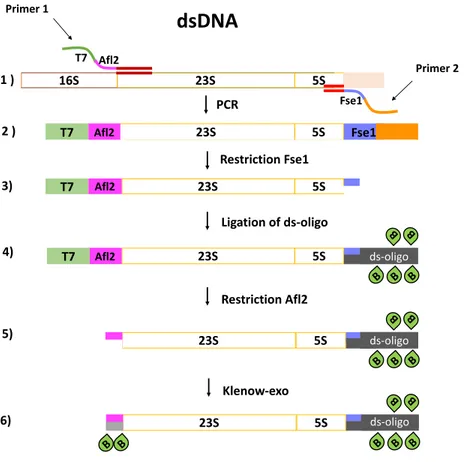
12Chapter I. Overstretching molecular duplexes made of DNA and RNA T7 Afl2 Fse1 23S Afl2 T7 5S Fse1 16S 23S 5S PCR
dsDNA
Primer 1 Primer 2 Restriction Fse1 23S Afl2 5S Ligation of ds-oligo Restriction Afl2 23S Afl2 5S B B B B B ds-oligo 23S 5S B B B B B ds-oligo 23S 5S B B B B B B B ds-oligo Klenow-exo 1 ) 5) 6) T7 T7 2 ) 3) 4)Figure I.9: Preparation of dsDNA
of murB, where murB plays the role of a spacer. The total length of our constructs is well-adapted for single-molecule manipulation, in particular it is convenient to capture the two beads linked by a molecule of this length (4050bp) in separate traps. We now describe the detailed steps needed to prepare the constructs, first for dsDNA, then for the RNA-DNA hybrid, dsRNA and for the DNA-RNA hybrid at the end.
I.3.1
Preparation of dsDNA
The sequence of interest is amplified by a polymerase chain reaction (PCR) from the plasmid pT7rrnB (Fig.I.9). The PCR primers (primer 1 and primer 2) were designed to introduce the sequence of a T7 polymerase promotor and an Afl2 restriction site (primer 1) at one extremity of the double-stranded PCR product, and the sequence of an Fse1 restriction site (primer 2) at the other extremity. The PCR product was then digested by the Fse1 restriction enzyme. The restricted piece was purified and replaced by a similar sequence carrying biotin via ligation. The resultant product was then digested by an Afl2 restriction enzyme generating a 4-nucleotide overhang at the 5’ end of the leading strand. The complementary nucleotides were filled in by Klenow DNA polymerase (Klenow exo-fragment). Some of them carry biotin groups
I.3. Four molecular constructs 13
for attachment.
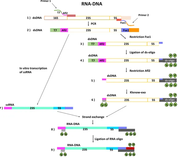
I.3.2
Preparation of the RNA-DNA hybrid
As for dsDNA, the sequence of interest was first amplified by a polymerase chain reaction (PCR, step1 Fig. I.10). The PCR primers (primer 1 and primer 2) were designed to introduce the sequence of a T7 polymerase promotor and an Afl2 restriction site (primer 1) at one extremity of the double-stranded PCR product, and the sequence of an Fse1 restriction site (primer 2) at the other extremity (Fig.I.10, step 2). A part of the PCR product (2µg) was conserved at ≠20¶C for in vitro RNA transcription and the rest was used to prepare dsDNA with biotin modifications at three of its extremities following the same steps (Figures N5, N6, steps 3-6) as described in sectionI.3.1. Once the dsDNA with biotin modifications was ready (Fig. I.10, step 6), the conserved PCR product was used to obtain in vitro transcribed RNA containing the sequences of 23S and 5S rRNAs (Fig. I.10, step 7). It is important to prepare fresh RNA for this step, since its stability is low and it degrades fast as compared to DNA. After preparation of ssRNA (Fig. I.10, step 7), the RNA and the dsDNA (Fig.I.10, step 6) were combined to do a strand exchange. This way, many copies of the RNA-DNA hybrid with biotin modifications at the two extremities of the DNA strand were obtained (Fig. I.10, step 8). The last biotin modification at the 3rd extremity of the RNA-DNA construct (3’ end of RNA strand) was introduced by a small RNA oligo of 20bp carrying two biotin-dT at its extremity. The remaining dsDNAs were degraded by an appropriate restriction enzyme to make sure that all the measurements were done on RNA-DNA hybrids only.
I.3.3
Preparation of dsRNA
Each strand of dsRNA was prepared separately. The leading strand was prepared as described in the previous section I.3.2. (Fig.I.10, steps 1, 2, 7, Fig.I.11, steps 1, 2, 3). The preparation of the lagging strand was done as follows. The sequence of interest (23S and 5S rRNA) was amplified by a polymerase chain reaction (PCR). The PCR primers 3 and 4 (Fig.I.11, step 1) were designed to introduce a random sequence at one extremity of the PCR product and a sequence of T7 polymerase promoter and a restriction site for FseI restriction enzyme at the other extremity of the PCR product (Fig. I.11, step 5). As one can see from Fig.I.11, to prepare the lagging strand the T7 polymerase promotor sequence is introduced at the opposite extremity of the PCR product (Fig. I.11, step 5) as compared to the preparation of the leading strand (Fig. I.11, step 2). After the PCR reaction, part of it was used to perform in vitro transcription of the lagging single stranded RNA (Fig.I.11, step 8). The latter was biotin-modified at both extremities via splint ligation. The idea of our particular splint ligation is the following. T4 RNA ligase 2 can ligate pieces of ssRNA (biotin-modified in our case) to DNA-RNA hybrids. This means that in order to ligate biotin-modified RNA oligonucleotides at two extremities of the ssRNA_lagg, it is necessary to create small regions of a DNA-RNA hybrid at both extremities. To do this, two DNA oligonucleotides (DNA1 and DNA2, Fig.I.11, step 7)
14Chapter I. Overstretching molecular duplexes made of DNA and RNA T7 Afl2 Fse1 23S Afl2 T7 5S Fse1 16S 23S 5S PCR RNA-DNA Primer 1 Primer 2 1 ) 2 ) 23S Afl2 5S Ligation of ds-oligo Restriction Afl2 23S Afl2 5S B B B B B ds-oligo 23S 5S B B B B B ds-oligo 23S 5S B B B B B B B ds-oligo Klenow-exo T7 T7 23S 5S B B B B B 23S 5S B B B B B 23S 5S B B 3 ) 4 ) 5 ) 6 ) 7 ) 8 ) 9 ) In vitro transcription of ssRNA Strand exchange ssRNA RNA-DNA RNA-DNA dsDNA dsDNA dsDNA dsDNA dsDNA dsDNA
Ligation of RNA oligo
Restriction Fse1
Figure I.10: Preparation of the RNA-DNA hybrid with biotin modifications.
having half of their sequence complementary to the ssRNA_lagg extremities and half to RNA oligonucleotides (biotin-modified) were hybridized to ssRNA_lagg, creating bridges for ligation of two RNA oligonucleotides (RNA1, RNA2, in red, Fig.I.11, step 9). After successful ligation, two RNA single strands (ssRNA_lead and ssRNA_lagg) were hybridized creating dsRNA with two biotin-modified edges (Fig. I.11, step 8). The final dsRNA construct with 3 biotin-modified extremities was obtained by ligation of the last RNA3 oligonucleotide (Fig. I.11, step 9). The latter is the same as for RNA-DNA hybrid (Fig.I.10, step 9).
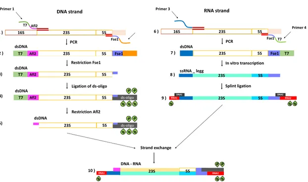
I.3.4
Preparation of DNA-RNA hybrid
In this case, the leading strand is DNA and the lagging strand is RNA. The prepa-ration of this construct is done in three steps: a prepaprepa-ration of dsDNA with three biotin-modified extremities (Fig.I.12, steps 1-5), preparation of the lagging RNA sin-gle strand with biotin modifications at its 5’ and 3’ ends (Fig. I.12, steps 6-9) and
16Chapter I. Overstretching molecular duplexes made of DNA and RNA Fse1 16S 23S 5S PCR RNA strand Primer 4 6 ) T7 23S 5S dsDNA 23S 5S Fse1 T7 23S 5S RNA2 RNA1 DNA1 DNA2 B B B Primer 3 In vitro transcription Splint ligation 7 ) 8 ) 9 ) ssRNA _ legg T7 Afl2 Fse1 23S Afl2 T7 5S Fse1 16S 23S 5S PCR DNA strand Primer 1 Restriction Fse1 23S Afl2 5S Ligation of ds-oligo Restriction Afl2 23S Afl2 5S B B B B B ds-oligo 23S 5S B B B B B ds-oligo 1 ) 5) T7 T7 2 ) 3) 4) RNA2 RNA1 B B B B B 23S 5S Strand exchange 10 ) dsDNA dsDNA dsDNA dsDNA DNA - RNA DNA - RNA
Figure I.12: Preparation of DNA-RNA hybrid with biotin modifications.
an exchange of the DNA-lagging strand by a RNA-lagging strand via a hybridization reaction (Fig.I.12, step 10). The preparation of dsDNA is the same as in sectionI.3.1
(Fig.I.12, steps 1-6) and the preparation of the lagging ssRNA is the same as described in sectionI.3.3 (Fig. I.11, steps 4-7).
I.3.5
Further treatment of the constructs
When the molecular constructs are prepared, part of them are conserved at ≠20¶C for later use, while part of them are mixed with streptavidin-coated polystyrene beads. The mixture is first centrifuged 6 min at 30G and then incubated at room temperature (25¶C) for one hour. Centrifugation allows the beads and the molecules to come close together, thus increasing their binding efficiency and reducing the incubation time from 3 hrs to 1 h. After incubation, the sample is loaded into a fluidics chamber composed of two glass coverslips sealed together by 2 parallel parafilm layers. After loading with the molecular construct the two open edges of the chamber are sealed with wax (Fig.I.13). Finally, the sample is installed on the microscope stage between two objectives (100x oil and 60x water, described above), a bead couple with a molecule attached in between is searched for, found, trapped and a force measurement is performed (Fig.I.14).
Overstretching double-stranded RNA, double-stranded DNA and
RNA-DNA duplexes
Submitted
L. Melkonyan1 , M. Bercy1 , T. Bizebard2 , and U. Bockelmann11 Nanobiophysique, ESPCI Paris 10 rue Vauquelin, 75005 Paris, France
2 Expression G´en´etique Microbienne, IBPC, CNRS UMR 8261 13 rue Pierre et Marie Curie,
75005 Paris, France
Using single-molecule force measurements, we compare the overstretching transition of the four types of duplexes composed of DNA or RNA strands. Three of the four extremities of each double helix are attached to two microscopic beads and a stretching force is applied with a dual-beam optical trapping interferometer. We find that overstretching occurs for all four duplexes with small differences between the plateau forces. Double-stranded RNA (dsRNA) exhibits a smooth transition, in contrast to the other three duplexes that show sawtooth patterns, the latter being a characteristic signature of peeling. This difference is observed for a wide range of experimental conditions. We present a theoretical description, which explains the difference and predicts that peeling and bubble formation do not occur in overstretching dsRNA. Formation of S-RNA is proposed, an overstretching mechanism that contrary to the other two does not generate single strands. We suggest that this singular RNA property helps RNA structures to assemble and play their essential roles in the biological cell.
I. INTRODUCTION
Forces act on DNA and RNA in the biological cell. They induce elastic deformation and torsion, can give rise to conformational and structural transitions and sometimes lead to base pair opening as well as profound modifications in base stacking and tertiary interactions. Generation of single-strands from duplexes containing DNA or RNA single-strands is particularly important, since it can lead to parasitic interactions and non-native structures. These duplexes are ubiquitous in the cell. Besides the DNA double helix being composed of two complementary single strands, most RNA molecules contain numerous helical parts and many of these local duplexes are essential elements of native RNA structures. Moreover, hetero-duplexes of DNA and RNA occur in DNA replication, DNA transcription, gene regulation and gene editing systems.
It has been shown by single-molecule measurements that mechanical force can generate single strands in different ways. In the unzipping configuration, forces pull the two strands of one duplex extremity in opposite directions and mechanically separate them [1–3]. In the peeling configura-tion, which occurs around 60 pN in overstretching of topological open nucleic acid (NA) duplexes, forces act along the helical axis from opposite duplex extremities and one strand peels off in a shear mode [4]. DNA overstretching was discovered about two decades ago by single-molecule force measurements [5, 6]. The experimental observations triggered many studies and
controver-19
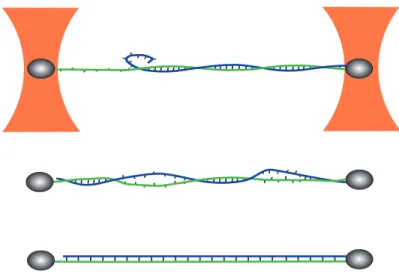
FIG. 1: Schematic representation of the measurement configuration and of the three overstretching mecha-nisms. The nucleic acid duplex is attached by three of its four single-strand extremities to two microscopic beads (beads and molecule are not at the same scale). The two beads of each dumbell are captured in separate optical traps (orange). Force versus displacement curves are obtained by measuring the position of one bead within the trap to nanometer precision, while the other trap is displaced. Peeling of the free strand, bubble formation and S-structure formation are presented from top to bottom. Single-strands under force and the S-structure exhibit a longer separation between adjacent nucleotides than the regular double helix. Base pairing is maintained in the S-structure, but base pair stacking and the number of helical turns are strongly reduced. Our molecular constructs are free to rotate around the axis of applied force, as on one side the bead is attached only to a single strand (green strand on the left-hand side of the figure).
sial discussions about the molecular mechanism underlying DNA overstretching, see [7, 8] and references therein. Recently, it has been shown experimentally that DNA overstretching can be caused by several mechanisms, including peeling, bubble formation as well as a structural transi-tion from the B-form helix to an S-DNA structure [4, 8–10]. These mechanisms are schematically represented in Fig.1.
While overstretching by bubble formation and by transition to S-DNA both show smooth plateaus in the force versus extension curve, peeling induces a characteristic sawtooth-shaped pattern. A sawtooth-shaped pattern can be observed only in the presence of a peeling front in dsNA. This peeling front can be either a free extremity of one of two strands under tension or a nick along one of the strands [8]. The absence of any of the latter always leads to smooth over-stretching [8]. The latter, can, however, be observed even when a peeling front is present, though special experimental conditions are required.
Here we investigate the overstretching transition of four different nucleic acid duplexes by single-molecule force measurements. These duplexes are stranded DNA (dsDNA), double-stranded RNA (dsRNA), a hetero-duplex with the DNA strand under tension (RNA-DNA) and a hetero-duplex with the RNA strand under tension (DNA-RNA), respectively. Strikingly, we find that dsRNA always exhibits a smooth overstretching signal, an observation that holds for a wide investigated range of salt conditions and pulling speeds. In contrast, the other three duplexes exhibit pronounced sawtooth-shaped signals during overstretching. Comparison between the ex-perimental data and a theoretical description based on the assumption of local thermal
equilib-20
rium indicates that peeling and bubble formation do not occur for dsRNA. Towards the end of the manuscript, we briefly discuss under which circumstances the absence of these single-strand generating mechanisms could be important in the biological cell.
II. MATERIALS AND METHODS A. Force measurement setup
Detailed descriptions of the dual-beam optical trapping interferometer and the sample prepa-ration steps immediately preceding the force measurement are published elsewhere [11]. The linearly polarized beam of a CW Nd:YVO4laser (Millenia IR, Spectra-Physics, 1064 nm, 10W)
is split with a polarizing cube beam splitter. One of the resulting beams is shifted in frequency by an acousto-optic frequency shifter. Then it is deflected by a piezoelectric mirror mount with an integrated position sensor operating in feedback loop and represents the mobile beam. The other beam remains fix. The two beams are combined with a second polarizing cube beam splitter before entering a microscope objective (Nikon, 100x, N.A. 1.4, oil immersion). This way, two optical traps of perpendicular linear polarization arise in the sample plane and the mobile trap can be laterally separated from the fixed trap with nanometer precision. The laser light passes through the sample and is collimated by a second objective (Olympus, 63x, N.A. 1.2, water immersion). A glan polarizer cube rejects the large majority of the light arising from the mobile beam. Force is deduced from the position of the bead in the fixed trap using back-focal plane interferometry [12, 13]. When a measurement cycle is completed, the molecular linkage between the two beads is broken and force is calibrated by recording the power spectral density of the bead in the fixed trap [13]. Unavoidable depolarization in the microscope objectives leads to some interference be-tween the fixed and mobile beams, which generates parasitic force signal at small distance bebe-tween the traps. The imposed frequency shift between the two laser beams avoids this parasitic signal [14]. We performed the experiments in a room of controlled temperature of 26 C. In the sample, the temperature is raised to about 33 C due to local heating by the trapping laser by a measured amount of∆T =7 C [11].
B. Preparation of the molecular constructs
All four duplexes contain 4050 base pairs and exhibit the same nucleotide sequence, corre-sponding to the sequence of a portion of the E. coli chromosome (strain K-12, substr. MG1655), starting at the first nucleotide of the rrlB gene (coding for 23S rRNA), encompassing the full gene sequences of rrlB and rrfB (coding for 5S rRNA) and ending in the middle of the murB gene. Preparation of the four different duplexes being related, we first describe the RNA-DNA case and then consider the other three duplexes.
The DNA sequence of interest is amplified by PCR from a plasmid (gift of K. Nierhaus) con-taining the full E. coli rrnB operon sequence. PCR primers were designed in order to introduce a T7 RNA polymerase promoter sequence followed by an AflII restriction site at one extremity of the PCR product, and a FseI restriction site at the other extremity. Part of the PCR product is in vitro transcribed using T7 RNAP, and the RNA product is conserved. AflII digestion of the rest of the PCR product followed by Klenow treatment in the presence of biotin-dATP allows to incorporate two biotin moieties close to the 3’ end of one strand. FseI digestion and ligation of a biotin-modified DNA oligonucleotide adds three biotins close to the 5’ end of the same strand.
21
The goal of the next step (strand-exchange step) is to replace the unmodified DNA strand by the in vitrotranscribed RNA. For this purpose, DNA and RNA are first denatured at high temperature and the resulting ss-strands are incubated together in temperature and solvent conditions that strongly favor RNA/DNA heteroduplex over dsDNA duplex formation [11, 15]. Subsequently, residual dsDNA duplexes are digested with EcoRI (to avoid any interference in the force experiments). Finally an RNA oligonucleotide, with two biotin modifications is ligated to the RNA 3’ end of the heteroduplex.
The DNA-RNA duplex is prepared similarly. PCR primers are designed to act on opposite plasmid strands compared to the RNA-DNA case. Oligonucleotides and ligations are adapted such that the RNA strand carries biotins close to both ends, while the DNA strand exhibits biotins close to its 3’ end only. Preparation of the dsDNA construct follows the protocol used for the RNA-DNA construct until the strand-exchange step. The latter is not required for dsDNA (and of course the EcoRI restriction step is omitted); the final dsDNA construct is obtained by ligating a DNA oligonucleotide carrying two biotin modifications. For the dsRNA construct, two PCRs and two in vitro transcriptions are performed to prepare two complementary RNA strands. The RNA strand intended to be biotinylated at each extremity (5’ and 3’) is in vitro transcribed in presence of GMP in large excess over GTP, i.e. to obtain a majority of RNA molecules with a single phosphate group at their 5’ extremity, thus ready to be ligated to the adequate oligonucleotide. Biotinylation of this RNA strand is performed using a DNA-splint ligation procedure and ligating biotinylated RNA oligonucleotides with T4 RNA ligase 2 [16]. The two RNA strands are then hybridized and the biotin groups at the 3’ extremity of the so far non-modified RNA strand are introduced using the same procedure than for the RNA-DNA duplex.
III. RESULTS
Four different double-stranded nucleic acid constructs have been prepared as described in the Materials and Methods section. They all contain exactly the same nucleotide sequence, except for the obvious T to U replacement when going from DNA to RNA (the sequence is described in the Materials and Methods section). Multiple biotin modifications were introduced at three of the four extremities of these duplexes and used for specific attachment to two streptavidin-coated beads. The beads are captured with two optical traps: the position of one optical trap is kept fixed to measure force by back focal plane interferometry, while the other trap is displaced with constant velocity to repeatedly strain and relax the investigated construct. This experimental configuration is schematically represented in Fig.1
A. The four duplexes at a common condition of salt and velocity
Below about 25 pN, the force-displacement curves of the four constructs exhibit rises of in-creasing slope, which remain identical upon relaxation (Fig.2). This part of each curve corre-sponds to a regime of entropic polymer elasticity at low forces, followed by a regime of enthalpic elasticity at intermediate forces. It is well described by the extensible worm-like chain model [19]. The curvature of the force-displacement curves changes sign around 25 pN. This softening has been observed for dsDNA before and has been attributed to twist-stretch coupling [4]. For all four duplexes the force-displacement relations measured below the overstretching plateau are well described by the twistable worm-like chain model, a theoretical description that takes twist-stretch coupling into account in terms of two phenomenological parameters (see SI). The force levelsFp
23 2.05 2.1 2.15 2.2 2.25 2.3 2.35 2.4 Displacement (μm) F o rce 5 pN
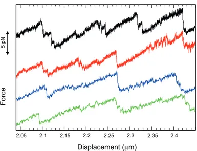
FIG. 3: Detailed view of four force-displacement curves measured on the overstretching plateau of the RNA-DNA hybrid. The lowest two curves correspond to two consecutive measurements of the same molecule and the two upper curves to two other molecules. The curves are shifted vertically for better visibility of the details. For the two lower curves, we used the buffer conditions and displacement veloc-ity of Fig.2, while the two upper curves were measured with smaller displacement velocveloc-ity (10 nm/s) and higher monovalent salt (400 mM KCl, 5 mM MgCl2, 20 mM Hepes, pH 7.6).
(about 1.8 pN standard deviation of the measured values) of the overstretching plateaus are close, but for the present case of equivalent base-sequence, same buffer and same displacement velocity we nevertheless can resolve a distinct order,
FpDN A/RN A'64.8 pN > FpRN A/DN A'60.0 pN > FpdsDN A'57.6 pN > FpdsRN A'55.2 pN. Surprisingly strong qualitative differences are observed between the overstretching curves of the dsRNA duplex on the one hand and the overstretching curves of the other three duplexes on the other hand. For dsRNA, the plateau is smooth and exhibits rather small hysteresis, whereas for the other three duplexes, the overstretching signal reveals a succession of sawtooth-shaped peaks and a strong hysteresis with deep decreases in force followed by sudden returns. In Fig.3 we present a zoom into the overstretching plateau measured upon stretching the RNA-DNA hybrid. The curves display successions of sawtooth-shaped peaks. Typically, a phase of slow increase in force is followed by a sudden force reduction. The same characteristic features are observed on the overstretching plateaus of the dsDNA and DNA-RNA constructs. The figure also shows that details of the sawtooth-shaped force signals can be similar from one pulling cycle to another and from one molecule to another.
B. Effects of salt concentrations and displacement velocity
It was shown that overstretching of dsDNA can involve different mechanisms and that the prevalence of one or another of these mechanisms depends on salt conditions and displacement velocity [8, 9]. Under the experimental conditions of Fig.2, we observe both sawtooth-like and smooth overstretching for dsDNA. A smooth region appears for instance at a displacement of about
26
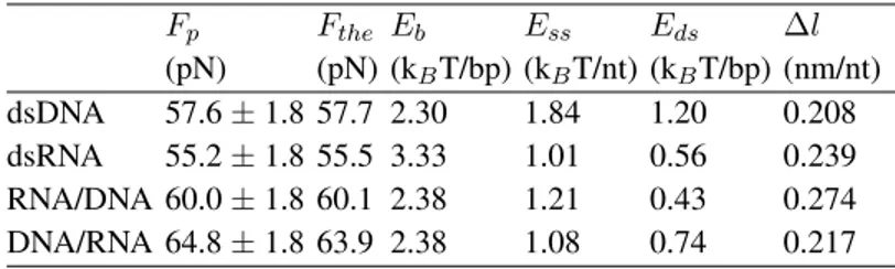
TABLE 1: Experimental and theoretical overstretching forces, together with energy and length values of the theoretical description.
Fp Fthe Eb Ess Eds ∆l (pN) (pN) (kBT/bp) (kBT/nt) (kBT/bp) (nm/nt) dsDNA 57.6 ± 1.8 57.7 2.30 1.84 1.20 0.208 dsRNA 55.2 ± 1.8 55.5 3.33 1.01 0.56 0.239 RNA/DNA 60.0 ± 1.8 60.1 2.38 1.21 0.43 0.274 DNA/RNA 64.8 ± 1.8 63.9 2.38 1.08 0.74 0.217
Experimental plateau values Fp are obtained by averaging 20-50 measured overstretching
plateaus for each duplex type. All these measurements were performed under the experimental conditions of Fig.2. The calculated forces Ftheverify E(Fthe) = 0, where E(F ) is defined
by Eq.1. The binding energies Ebare taken from the literature [20–22]. The elastic energies
Essand Edsand the length difference∆l = lss ldsare evaluated at force Fthe.
1. Overstretching by peeling
The force-induced peeling phenomenon can be described by a conversion of a double stranded nucleic acid into two single strands, only one of which stays under tension. This transition implies rupture of hydrogen bonds, modified stacking interactions as well as changes in elastic energy. We consider the free-energy difference E(F ) between a state (n + 1) exhibiting n + 1 peeled base pairs and a state (n) with n peeled base pairs at constant force F . The construct would peel progressively forE(F ) < 0, reanneal progressively for E(F ) > 0, and the states (n) and (n + 1) would have equal probability forE(F ) = 0.
E(F ) = Eb+ Ess Eds F (lss lds) (1)
Ebis an average energy required to open one base pair, which we obtained from published unified
nearest-neighbour ∆G0
37 parameters (see Table 1 and SI). For simplicity, we call this parameter
”base pair binding energy” in this paper. Essdenotes the energy per nucleotide required to stretch
a single-stranded nucleic acid (ssNA) from zero-force to a force F and Eds is the energy per
base pair required to stretch a double-stranded nucleic acid (dsNA) from zero to F . The term F (lss lds) describes the mechanical work (length change times force) , where lss and lds are
the length per nucleotide of a ssNA stretched to forceF and the length per base pair of a dsNA stretched to force F , respectively. We derived Ess,Eds,lssandlds from force measurements on
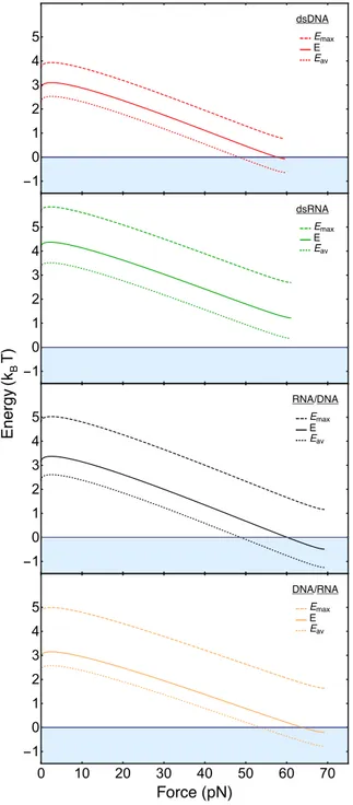
ssDNA and dsDNA. A detailed description of the model and its parameters is presented in SI. Equation (1) allows us to obtain a phase diagram, predicting the dsNA ! ssNA transition (Fig.7). It consists of two different regions: a region where the considered molecule has preference for a double stranded conformation (E > 0) and a region where it has preference for a single-stranded conformation (E < 0). Zero-energy defines the predicted force level of the overstretching plateau. AtF = 0, the energy E is simply given by Eb. Below about 2 pN, the energy versus force
curves E(F ) exhibit an initial increase with force, which is explained by a negative (lss lds).
At higher force,(lss lds) becomes positive, leading to a monotonic decrease of E(F ) . Largely
depending on its starting level Eb, the curve either penetrates (dsDNA, RNA-DNA, DNA-RNA)
27 ������ (�� � ) -1 0 1 2 3 4 5 ����� ���� � ��� -1 0 1 2 3 4 5 ����� ���� � ��� -1 0 1 2 3 4 5 ���/��� ���� � ��� 0 10 20 30 40 50 60 70 -1 0 1 2 3 4 5 ����� (��) ���/��� ���� � ���
FIG. 7: Energy difference E(F ) for overstretching by peeling, calculated using Eq.1. Energy versus force curves are presented for dsDNA (red), dsRNA (green), RNA-DNA (black) and DNA-RNA (orange). The energy diagram is divided into two regions: a region where the molecules are double-stranded (white) and a region where the peeled state is energetically favorable (light blue). For calculating the dotted lines, we used the literature binding energies Eb=Eav(F = 0) (Table 1). Emaxis the free energy of the strongest
base pair, Eavis the average free energy of all base pairs. For calculating the solid lines, we enhanced Eb
by small amounts that are 41 %, 37 %, 32 % and 24 % of the difference between Emaxand Eb(from top
to bottom). E = Eav+ Eadd. These enhancements are introduced in order to phenomenologically account
for sequence heterogeneity and out-of-equilibrium effects (see SI, section II. 2. Eadd term). They lead to
agreement between measured and theoretically predicted plateau forces for the three cases where peeling is observed. When the applied force reaches the threshold value for which the energy E is zero, dsDNA, RNA-DNA and DNA-RNA hybrids start to peel, whereas dsRNA always remains far away from the peeling region.
28
curves show slight positive curvature before stopping. The positive curvature is caused by the twist-stretch coupling term. At this point the molecule under tension completely unwinds (no twist is left) and the theoretical description of the twist-stretch coupling loses its physical meaning at forces aboveFmax(see SI). Application of the described model to our experimental data gives
two major results, (i) peeling is predicted only for three of the four molecular constructs and the significantly higher value ofEbis the main reason why peeling is not predicted for dsRNA.
2. Overstretching by melting bubble formation
Overstretching by melting bubble formation involves rupture of dsNA base pairs; the mech-anism is similar to peeling in this respect. As a difference, however, melting bubble formation results in single strands that both remain under tension, while one strand relaxes in the peeling case. The applied forceF is distributed among the two strands, either equally if the two strands are of the same nature (F1 = F2 = F/2; for dsDNA and dsRNA), or unequally if the two strands
are of different nature (FDN A 6= FRN A;FDN A+ FRN A = F ; for RNA-DNA and DNA-RNA).
The process can be described by equation 1 as for peeling, albeit the following modifications in the elastic energies of the single strands and the mechanical work.
E(F ) = Eb+ Ess⇤ Eds F (l⇤ss lds) (2) where E⇤ ss= 2EDN A ss (F/2) for dsDNA 2ERN A ss (F/2) for dsRNA EDN A
ss (FDN A) + EssRN A(FRN A) for DNA-RNA and RNA-DNA
l⇤ ss= lDN A ss (F/2) for dsDNA lRN A ss (F/2) for dsRNA lDN A
ss (FDN A) = lssRN A(FRN A) for DNA-RNA and RNA-DNA
Using equation 2 we constructed an equivalent to the peeling case phase diagram (Fig.8), again with two regions. In the lower region (E < 0) the molecule has a preference to form melting bub-bles along its NA chain. Zero energy indicates equilibrium between double stranded and melting bubble conformations. The energy versus force curves look alike to the peeling case, with a start value ofEband a round maximum around 5 pN that is followed by a continuous decrease. The free
energy versus force curves reach their end before theE = 0 phase boundary, which indicates that overstretching via melting bubble formation is energetically non-favorable for all four duplexes. For more details see SI.
IV. DISCUSSION
A. Force levels of overstretching by peeling
Under the conditions of section II.A, the dsDNA, DNA-RNA and RNA-DNA constructs show sawtooth-like force signals and pronounced hysteresis. These observations are clear signatures of peeling. We present in Table 1 the average values of the force plateaus Fp measured with
increasing displacement, and the calculated forcesFthefor overstretching by peeling. We observe
30 . ..
.
.
.
. ...
.
.
.
.
.
.
.
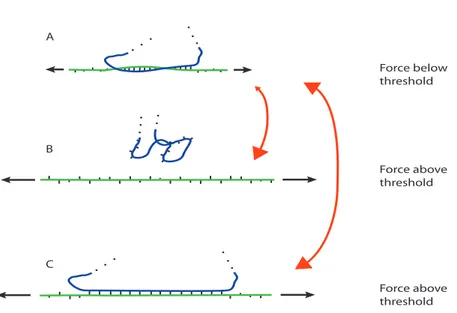
A B C Force below threshold Force above threshold Force above thresholdFIG. 9: Schematic representation of the implications of different force-induced overstretching modes. In panel A a single-strand (green) is submitted to a pulling force below the overstretching threshold (short black arrows). This strand is hybridized with a complementary base sequence (blue) forming a local duplex structure. When force increases above the overstretching threshold (long black arrows) there are two pos-sibilites. Overstretching might occur by peeling and/or bubble-formation and the hybridized motif unbinds (panel B). Alternatively, S-structure formation might occur and the hybridized structure remains bound (panel C). As explained in the text, the transition from A to B is widely irreversible when the force rede-creases below threshold, while the transition from A to C is reversible. This is illustrated by asymmetric and symmetric red double-arrows, respectively.
sequence with increasing GC content. During this ’stick’ phase force rises slowly. Subsequently a ’slip’ event occurs once the local energy barrier is overcome. Then the peeling front advances rapidly and the force drops. The energy landscape is determined by the base sequence, but the exact positions where the transitions occur exhibit stochastic variation. Flipping between discrete states is sometimes observed (for instance on the right part of the red curve), which is a signature of close-to-equilibrium dynamics. Sawtooth-shaped peaks and force flips are observed for the RNA-DNA, the dsDNA and the DNA-RNA constructs and these qualitative features agree with the observations of Gross et al [4], who studied a three-point-attachment dsDNA construct containing a pKYB1 sequence of 8393 base pairs.
C. RNA-DNA overstretching depends on displacement velocity and salt
Earlier studies showed that overstretching of dsDNA can be due to different mechanisms, lead-ing to a complex phase diagram that depends on salt conditions, displacement velocity, sequence and topology [4, 8–10]. In particular, it was shown that at high ionic strength S-DNA formation is favored over peeling in topologically open DNA [8]. In this case, Zhang et al observed peeling and S-DNA formation, but not bubble formation [9]. The present work indicates that the over-stretching phase diagram of an RNA-DNA heteroduplex is of similar complexity and qualitatively ressembles the one of dsDNA. In both cases, peeling dominates at low salt and velocity, while
31
overstretching with a smooth force signal occurs more frequently at high salt concentrations and high displacement velocities. However, we observe quantitative differences that are illustrated in Figs.4-6. Peeling remains the dominant overstretching mechanism for a much wider range of salt concentrations and displacement velocity in the RNA-DNA hybrid than in the dsDNA construct. We attribute the observed smooth overstretching to S-NA formation, because bubble formation is predicted to be energetically non-favorable (Fig.8). For the peeling mechanism, the calculated differences between the energies of RNA-DNA and dsDNA are small and the dsDNA energy lies slightly below the one of the RNA/DNA hybrid (Fig.7). The observation that RNA-DNA peeling dominates over a wider parameter space therefore suggests that the energy of the heteroduplex S-phase is higher than the one of the dsDNA S-S-phase. We do not know the reason for this difference and whether it is of structural or dynamical origin. Regarding structural difference, the RNA-DNA heteroduplex forms an A-type double-helix, which is more compact than the B-type DNA double helix [24]. As an example for different dynamical properties, some of us have shown previously that unfolding and refolding of hairpin structures under force occur faster and significantly closer to equilibrium in DNA than in RNA [25].
D. Peeling and bubble-formation do not occur in dsRNA
We observe remarkable qualitative differences between the force curves for overstretching dsRNA as compared to the curves for overstretching the other duplexes. They are illustrated in Fig.2: smooth plateau and weak hysteresis occur for dsRNA, while dsDNA, RNA-DNA and DNA-RNA show rapidly varying force-signals and pronounced hysteresis. We investigated a wide range of conditions, monovalent salt from 10 to 100 mM, divalent salt from 0 to 5 mM and displacement velocities from 10 to 100 nm/s, but for dsRNA we did not observe the characteristic signatures of peeling. Smooth plateaus were also observed in an earlier study, where dsRNA molecules were overstretched with a velocity of 500 nm/s in 150 mM, 300 mM and 500 mM NaCl [26]. Our theo-retical description predicts absence of peeling for dsRNA and indicates that this absence is caused by the higher base pair binding energy (Eb= 3.33 kBT) of dsRNA as compared to dsDNA and the
heteroduplexes (2.30 and 2.38 kBT, respectively). As described in SI section II.A.1, this
interpreta-tion holds for a wide range of salt concentrainterpreta-tions, including close-to-physiological salt condiinterpreta-tion. Bubble formation could explain smooth overstretching, but the results presented in Fig.8 suggest that it is not energetically favorable. We note that the remaining mechanism, the transition from an A-type helix to S-conformation, does not expose local single-stranded sequences of the RNA molecule. Biological implications of this result are discussed in the following subsection.
E. RNA overstretches without generating single strands: biological relevance
To what extent does the absence of single-strand generation attributed to our data on dsRNA help an RNA molecule achieve its biological role? Does this property match the functions of RNA, which differ from the ones of DNA?
Forces above 50 pN occur in the biological cell [27]. For instance, the maximum force the mitotic spindle exerts on a single moving chromosome in anaphase amounts to 700 pN. This force was measured in vivo [28]. During assembly of many viruses, a powerful molecular motor compacts the genome into a preassembled capsid. Forces of up to 100 pN were measured for bacteriophage φ29 DNA packaging in vitro [29, 30]. To the best of our knowledge, to date there exists neither a published report about an in vivo measurement of forces acting on RNA structures
32
nor one about a force measurement on viral RNA packaging. Unzipping of RNA double helices requires forces of 10-20 pN, depending on their base sequence [2, 25, 31, 32]. This mechanical opening and closing of RNA duplexes frequently occur close to thermal equilibrium; the opening fork breathes thermally, manifesting itself by flips in the observed signal. This equilibrium implies that formation of an RNA duplex from complementary strands generates forces of 10-20 pN. In view of the complex structures adopted by RNA, including numerous helices, tertiary structure interactions and sometimes also interactions with RNA-binding proteins, we think that major con-formational transitions are susceptible to generate transient forces that exceed the overstretching threshold at critical positions within an RNA structure.
To make overstretching relevant in vivo, it is also necessary that the force acts on a duplex in shear mode. When a single strand is hybridized with a short complementary NA sequence, peeling can occur and/or bubbles can form, as illustrated in Fig.1. When the force acting on the former strand exceeds the threshold for overstretching, the complementary strand is susceptible to dissociate and diffuse away. This is illustrated schematically in Fig.9, where it corresponds to the transition from panel A to panel B.
From the preceding two paragraphs, we are thus left with the idea that force-induced gener-ation of single-strands could occur from both the force magnitude and topology points of view. What would this possibility imply for RNA? The implications are schematically represented in Fig.9. If RNA duplexes were prone to peeling or bubble formation, whenF increases above the overstretching threshold, dissociation of the complementary RNA strand would occur. This would happen even if high force is reached only for a short moment. The two single-strands would sep-arate, move and engage binding with other RNA residues (or proteins), thus leading to either an irreversible or a long-lasting change of RNA structure, detrimental to its normal cellular activity. An asymmetric double arrow between panels A and B represents the irreversible behaviour. In contrast, if overstretching occurs via a mechanism that preserves base pairing, like the S-RNA formation that we attribute to our experimental observations, the initial RNA structure resumes readily when force falls below the overstretching threshold (symmetric double arrow between panels A and C). This general idea is further illustrated in SI section III, where we consider RNA action within the protein-synthesis machinery.
ACKNOWLEDGEMENTS
We are grateful to Marc Dreyfus for critical reading of the manuscript. This work has been supported by the Human Frontier Science Program [RGP008/2014].
[1] Essevaz-Roulet B, Bockelmann U, Heslot F (1997) Mechanical separation of the complementary strands of DNA. Proc. Natl. Acad. Sci. USA 94: 11935-11940.
[2] Liphardt J, Onoa B, Smith SB, Tinocco I, Bustamante C (2001) Reversible unfolding of single RNA molecules by mechanical force. Science 292: 733-737.
[3] Bockelmann U, Thomen P. Essevaz-Roulet B, Viasnoff V, Helot F (2002) Unzipping DNA with optical tweezers: high sequence sensitivity and force flips. Biophys J 82: 1537-1553.
[4] Gross P, Laurens N, Oddershede LB, Bockelmann U, Peterman EJG, Wuite GJL (2011) Quantifying how DNA stretches, melts and changes twist under tension. Nature Physics 7: 731-736.
33
[5] Cluzel P, Lebrun A, Heller C, Lavery R, Viovy JL, Chatenay D, Caron F (1996) DNA: an extensible molecule. Science 271: 792-794.
[6] Smith S B, Cui Y, Bustamante C (1996) Overstretching B-DNA: the elastic response of individual double-stranded and single-stranded DNA molecules. Science 271: 795-799.
[7] Bustamante C, Smith S B, Liphardt J, Smith D (2000) Single-molecule studies of DNA mechanics. Curr. Opin. Struct. Biol. 10: 279-285.
[8] King GA, Gross P, Bockelmann U, Modesti M, Wuite GJL, Peterman EJG (2013) Revealing the competition between peeled ssDNA, melting bubbles and S-DNA during DNA overstretching using fluorescence microscopy. Proc. Natl. Acad. Sci. USA 110: 3859-3864.
[9] Zhang X, Chen H, Le S, Rouzina I, Doyle P S, Yan J (2013) Revealing the competition between peeled ssDNA, melting bubbles, and S-DNA during DNA overstretching by single-molecule calorimetry. Proc. Natl. Acad. Sci. USA 110:3865-3870.
[10] Paik DH, Perkins TT (2011) Overstreching DNA at 65 pN does not require peeling from free ends or nicks. J. Am. Chem. Soc. 133: 3219-3221.
[11] Geffroy L, Mangeol P, Bizebard T, Bockelmann U (2017) RNA unzipping and force measurements with a dual optical trap. Methods Mol. Biol. 1665: 25-41.
[12] Gittes F, Schmidt C F (1998) Interference model for back-focal-plane displacement detection in optical tweezers. Opt. Lett. 23: 7-9.
[13] Neuman K, Block S M (2004) Optical trapping. Rev. Sci. Instrum. 75: 2787-2809.
[14] Mangeol P, Bockelmann U (2008) Interference and crosstalk in double optical tweezers using a single laser source. Rev. Sci. Instrum 79: 083103.
[15] Dean M (1987) Determining the hybridization temperature for Si nuclease mapping. Nucleic Acids Res. 15: 6754.
[16] Vilfan I D, Kamping W, van den Hout M, Candelli A, Hage S, Dekker N H (2007) An RNA toolbox for single-molecule force spectroscopy studies. Nucleic Acids Res. 35: 6625-6639.
[17] Fu H, Chen H, Zhang X, Qu Y, Marko J F, Yan J (2010) Transition dynamics and selection of the distinct S-DNA and strand unpeeling modes of double helix overstretching. Nucleic Acids Res. 39: 3473-3481.
[18] Arias-Gonzalez J R (2014) Single-molecule portrait of DNA and RNA double helices. Integr. Biol. 6: 904-925.
[19] Odijk T (1995) Stiff chains and filaments under tension. Macromolecules 28: 7016-7018.
[20] Xia T, SantaLucia Jr J, Burkard M E, Kierzek R, Schroeder S J, Jiao X, Cox C, Turner D H (1998) Thermodynamic parameters for an expanded nearest-neighbor model for formation of RNA duplexes with Watson-Crick base pairs. Biochemistry 37: 14719-735.
[21] SantaLucia Jr J (1998) A unified view of polymer, dumbbell, and oligonucleotide DNA nearest-neighbor thermodynamics. Proc. Natl. Acad. Sci. USA 95: 1460-65.
[22] Sugimoto N, Nakano S, Katoh M, Matsumura A, Nakamuta H, Ohmichi T, Yoneyama M, Sasaki M (1995) Thermodynamic parameters to predict stability of RNA/DNA hybrid duplexes. Biochemistry 34: 11211-216.
[23] Bockelmann U, Essevaz-Roulet B, Heslot F (1998) DNA strand separation studied by single molecule force measurements. Phys. Rev. E 58: 2386-2394.
[24] Marin-Gonzalez A, Vilhena J G, Perez R, Moreno-Herrero F (2017) Understanding the mechanical response of double-stranded DNA and RNA under constant stretching forces using all-atom molecular dynamics. Proc. Natl. Acad. Sci. USA 114: 7049-7054.
[25] Bercy M, Bockelmann U (2015) Hairpins under tension: RNA versus DNA. Nucleic Acids Res. 43: 9928-9936.
34
[26] Herrero-Gal´an E H, Fuentes-Perez M E, Carrasco C, Valpuesta J M, Carrascosa J L, Moreno-Herrero F, Arias-Gonzalez J R (2012) Mechanical identities of RNA and DNA double helices unveiled at the single-molecule level. J. Am. Chem. Soc. 135: 122-131.
[27] Roca-Cusachs P, Conte V, Trepat X (2017) Quantifying forces in cell biology. Nat. Cell Biol. 19: 742-751.
[28] Nicklas R B (1983) Measurements of the force produced by the mitotic spindle in anaphase. J. Cell Biol. 97: 542-548.
[29] Smith D E, Tans S J, Smith S B, Grimes S, Anderson D L, Bustamante C (2001) The bacteriophage φ29 portal motor can package DNA against a large internal force. Nature 413: 748-752.
[30] Rickgauer J P, Fuller D N, Grimes S., Jardine P J, Anderson D L, Smith D E (2008) Portal motor velocity and internal force resisting viral DNA packaging in bacteriophage φ29. Biophys. J. 94: 159-167.
[31] Harlepp S, Marchal T, Robert J, L´eger J-F, Xayaphoummine A, Isambert H, Chatenay D (2003) Prob-ing complex RNA structures by mechanical force. Eur. Phys. J. E 12: 605-615.
[32] Mangeol P, Bizebard T, Chiaruttini C, Dreyfus M, Springer M, Bockelmann U (2011) Probing riboso-mal protein-RNA interactions with an external force. Proc. Natl. Acad. Sci. 108: 18272-18276. [33] Kaczanowska M, Ryd´en-Aulin M (2007) Ribosome biogenesis and the translation process in
Es-cherichia coli. Microbiol. Mol. Biol. Rev. 71: 477-494.
[34] Lorsch J R (2002) RNA chaperones exist and DEAD box proteins get a life. Cell 109: 797-800. [35] Wen J-D, Lancaster L, Hodges C, Zeri A-C, Yoshimura S H, Noller H F, Bustamante C, Tinocco Jr I
(2008) Following translation by single ribosomes one codon at a time. Nature 452: 598-603.
[36] Liu T, Kaplan A, Alexander L, Yan S, Wen J-D, Lancaster L, Wickersham C E, Fredrick K, Noller H, Tinoco Jr I, Bustamante C J (2014) Direct measurement of the mechanical work during translocation by the ribosome. eLife 3: e03406.
[37] Schuwirth B S, Borovinskaya M A, Hau C W, Zhang W, Vila-Sanjurjo A, Holton J M, Cate J H D (2005) Structures of the bacterial ribosome at 3.5 ˚A resolution. Science 310: 827834.
Overstretching double-stranded RNA, double-stranded DNA and
RNA-DNA duplexes
– Supplementary Information –
L. Melkonyan1 , M. Bercy1 , T. Bizebard2 , and U. Bockelmann11 Nanobiophysique, ESPCI Paris 10 rue Vauquelin, 75005 Paris, France
2 Expression G´en´etique Microbienne, IBPC, CNRS UMR 8261 13 rue Pierre et Marie Curie,
75005 Paris, France
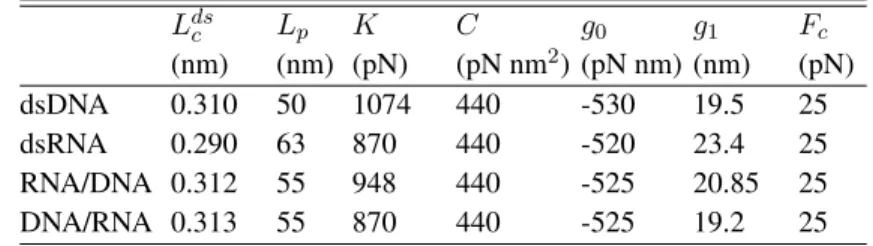
I. FITTING THE MEASURED FORCE-EXTENSION RELATIONS
In figure S1, we present average values and mean-square deviations of measured force-displacement curves for the four different constructs. The part below the overstretching plateau is fitted to the twistable worm-like chain model. The fit function is shown as a blue solid line. It relates the imposed displacement to the measured forceF . The displacement equals the sum of the lengthx of the molecule and the shifts F/ktrapof the beads compared to their equilibrium
positions in the optical traps. The length of the moleculex is the product of the number of base pairsNb(Nb = 4050 for our constructs) and the length per base pair lds. The latter is given by the
analytical expression [1, 2] lds(F ) = Ldsc 1 1/2 s kBT F Lp + F K g(F )2/C ! , (1) where Lds
c , Lp and K are respectively the crystallographic length per base pair, the persistence
length and the stretch modulus per base pair of the nucleic acid (NA) duplex. The twist-stretch coupling is parametrized by the twist rigidityC and the function g(F ). The latter is described by
g(F ) = ⇢
g0+ g1Fc for F Fc
g0+ g1F for F > Fc ,
(2)
with three parameters,g0,g1andFc. The parameters used to describe the average force-extension
relations are presented in Table S1.
Although we measured a significant number of force-displacement relations for each of the four NA duplexes, the experimental data do not allow determining the seven parameters in a unique way. The parameter set shown in Table S1 is consistent with the information available from the literature. Our lengthsLds
c andLpagree with published values for dsDNA and dsRNA [3–7]. The
parameters K, C, g0,g1 andFc affect the shape of the force-extension curve at high force. For
the twist rigidityC of all duplexes, we take a value reported in the literature for dsDNA [1, 8]. Moreover we assume a common critical forceFcthat is close to the value published for dsDNA [1].