Development and Assessment of Strategies for
Non-Invasive Prenatal Diagnosis Using Fetal Cells in
Maternal Blood
ParDr Ahmed Emad Service de génétique, Département de Pédiatrie
Thèse présentée à la Faculté de Médecine et des Sciences de la Santé en vue de l’obtention du grade de Philosophiae Doctor (Ph.D) en Biochimie
Sherbrooke, Québec, Canada Décembre, 2013
Membres du jury d’évaluation
Dr Jean Gekas, MD, PhD: Département de pédiatrie, Centre Hospitalier de l'Universitaire de Québec (CHUQ), Faculté de médecine, Université Laval, Québec, Québec, Canada, examinateur externe à l’Université de Sherbrooke
Dr Jean-Charles Pasquier, MD, PhD: professeur agrégé, Département d'obstétrique et gynécologie Université de Sherbrooke, Faculté de médecine et des sciences de la santé, Université de Sherbrooke, examinateur externe au programme
Dr Guylain Boissonneault, PhD : Professeur titulaire, Département de Biochimie, Faculté de médecine et des sciences de la santé, Université de Sherbrooke, examinateur interne au programme
Dre Chantal Bouffard, PhD : Professeure-chercheure Adjointe, Faculté de médecine et des sciences de la santé, Université de Sherbrooke, directrice de recherche
Dr François Corbin, MD, PhD : Professeur agrégé, FMSS Département de biochimie, Faculté de médecine et des sciences de la santé, Université de Sherbrooke, codirecteur de recherche
Par Ahmed Emad
Service de génétique, Département de Pédiatrie
Thèse présentée à la Faculté de Médecine et des Sciences de la Santé en vue de l’obtention du diplôme de Philosophiae Doctorat (Ph.D) en Biochimie, Faculté de Médecine et des Sciences
de la Santé, Université de Sherbrooke, Sherbrooke, Québec, Canada, J1H 5N4
Le diagnostic prénatal résulte encore aujourd’hui de procédures invasives, qui présentent des risques pour la grossesse. Le développement du diagnostic prénatal non-invasif (DPNI) changerait le rapport risque : bénéfice, rendant le diagnostic prénatal plus intéressant pour les femmes enceintes. Plusieurs chercheurs ont montré la présence de cellules fœtales dans le sang maternel et des travaux ont été entrepris afin de les cibler et de les utiliser éventuellement en DPNI. Toutefois, la faible concentration des cellules fœtales dans le sang maternel réduit les possibilités d’isolement ainsi que celles de leur utilisation en clinique. Un autre aspect technique du DPNI, le balayage manuel, est très laborieux, surtout en terme de temps technique. Il y a donc un besoin certain pour des études approfondies afin d’évaluer et d’améliorer la faisabilité du DPNI.
La détection d’évènements rares dans une grande population cellulaire offre un potentiel pour le diagnostic en oncologie mais aussi en diagnostic prénatal. Dans cette thèse, la première étude était dédiée à l’optimisation d’une stratégie pour détecter les cellules rares. Nous avons développé une méthode d’étalement sur lame d’un nombre précis de cellules cibles parmi des centaines de milliers de cellules. Cette stratégie a permis d’évaluer le taux de détection d’évènements rares et de comparer l’efficacité des techniques d’enrichissement en connaissant le nombre exact et la localisation de cellules cibles sur les lames. De plus, il a été possible d’évaluer les problèmes d’hybridation des évènements manqués. Nous avons, par la suite, développé un algorithme robuste pour la détection de cellules rares en utilisant la plateforme de microscopie automatisée MetaSystems et utilisé cette approche dans la validation des balayages manuel et automatique d’un nombre précis de cellules mâles parmi une large population de cellules femelles marquées avec la technique FISH. Nous avons testé ce classificateur avec des échantillons de sang de femmes enceintes de grossesses normales et aneuploïdes et évalué la fréquence de cellules fœtales isolées par différentes méthodes d’enrichissement au cours des premier et second trimestres de grossesse. Les données accumulées ont confirmé la présence de cellules fœtales chez toutes les grossesses et leur fréquence plus élevée dans les grossesses aneuploïdes. Le nombre de cellules fœtales est dynamique tout au long de la grossesse. De plus, un nombre plus élevé de cellules fœtales peut être obtenu en optimisant le moment du prélèvement et les méthodes d’enrichissement. De plus, le balayage automatique s’est avéré plus sensible et constant que le balayage manuel, ce qui permet de balayer un grand nombre de cellules et devient plus approprié pour une application clinique. Nous avons aussi montré la faisabilité d’utiliser des cellules fœtales dans le cadre du DPNI. Cinq cellules amniotiques microdisséquées, provenant de grossesses normales et aneuploïdes, ont suffi pour poser un diagnostic prénatal par une combinaison de l’amplification du génome complet et de la technique QF-PCR (réaction quantitative en fluorescence d’amplification entraînée par une polymérase) permettant la détection d’anomalies chromosomiques. Nos résultats ouvrent la voie à l’utilisation de cellules fœtales dans le sang maternel pour le DPNI.
Development and Assessment of Strategies for Non-Invasive Prenatal Diagnosis Using Fetal Cells in Maternal Blood
By Ahmed Emad
Division of Genetics, Department of Paediatrics,
A thesis presented at the Faculty of Medicine and Health Sciences as a requirement for obtaining the degree of Doctor of Philosophy (PhD) in Biochemistry, Faculty of Medicine and
Health Sciences, Université de Sherbrooke, Sherbrooke, QC, Canada J1H 5N4
Current prenatal diagnosis depends on invasive procedures and is thus offered only to high-risk pregnancies. Development of non-invasive prenatal diagnosis (NIPD) would change the risk-benefit ratio and make it likely that more women would benefit from prenatal testing. Scientists have documented the presence of rare fetal cells in maternal blood and envisioned targeting them with specific markers and their use in NIPD. Considering their extremely low frequency in maternal blood, fetal cells have been difficult to retrieve and use in clinical practice. Therefore, there is a pressing need for systematic sequential studies to evaluate their feasibility in NIPD.
Generally, detection of rare cells within a large cell population carries great potentialities for the prospects of cancer management and NIPD. Manual scanning is very cumbersome and time-consuming Therefore; the first part of our project was, dedicated to the optimization of an effective strategy to evaluate retrieval of rare cells. We have developed a way of distributing a controlled number of target cells among hundreds of thousands of other cells on microscope slides. This strategy allows the precise evaluation of the retrieval of rare events and the comparizon of the efficacy of different techniques and enrichment approaches by knowing the definite number and locations of target cells on the slides. Furthermore, it allows the evaluation of hybridization of missed events. We have also developed a robust custom-made detection algorithm for rare cells using the MetaSystems automated platform and have used this strategy in the validation of manual and automatic scanning of 60 slides with a pre-defined number of rare male cells among a pure population of female cells using XY-FISH. Consequently, we tested the developed classifier for the detection of real fetal cells from maternal blood in both normal and aneuploid pregnancies with Down syndrome. We further evaluated the number of fetal cells with different methods of enrichments in the first and second trimesters. The data collected confirmed the early presence of fetal cells in all of the pregnancies tested and their frequencies were higher in cases of aneuploidies. Fetal cells are in a state of dynamic change throughout the pregnancy. Higher numbers of these cells can be obtained by optimizing the harvest time and methods of enrichment. We found that automatic scanning is more sensitive and reliable than manual detection. Furthermore, it alleviates the burden of scanning large numbers of cells and thus is more suitable for clinical application. We also demonstrated the feasibility of using rare cells in NIPD. Five microdissected amniotic fetal cells from 26 cases of normal and aneuploid pregnancies were quite enough to provide accurate NIPD through using whole genome amplification coupled with QF-PCR. Our findings laid the ground for the use of rare fetal cells in maternal blood for NIPD.
Keywords: Fetal cells – Prenatal diagnosis – Fluorescence in situ hybridization – Automatic microscopy
Firstly I would like to thank God ALLAH ALMIGHTY for giving me the audacity and sanctioning me with acquaintance and confidence to fulfil this task.
I dedicate this work to my loving wife and to my parents in acknowledgment of their courage, effort, faith and enormous sacrifice.
This thesis is submitted in partial fulfilment of the requirements for the degree of Doctor of Philosophy at the Université de Sherbrooke. Work was supported by a PhD studentship provided by the Egyptian government. The thesis is the result of many years of struggle and labour work, filled with concerns and hopes for good results, sadness and tiredness with each failed attempt and finally joy when everything came together and the work was completed.
I started my doctorate working full-time on a project with Dr. Régen Drouin, which was related to the identification and quantification of rare fetal cells in the peripheral blood of pregnant mothers. The objective of the research was to develop non-invasive prenatal diagnostic tools through using these rare cellular events in the identification of common chromosomal aneuploidies. In four years of dedicated work, I developed new techniques for this research and my results enabled me to be the first author of four research articles related to this project (articles 1 to 4). These were the articles on which this thesis is based. In the last two years, I extended the concept of using rare cells to cancers and developed two articles on the use of these cells in the diagnosis of bladder cancer through the identification of specific chromosomal abnormalities. Unfortunately, we were unable to include these articles in this thesis because of unexpected change of the project director. I have learned a lot in the process of writing this thesis, as well as the primary version of all the articles, and my initial conceptions have certainly been changed. I have dealt with a lot of subjects, in an attempt to give this thesis a broader perspective. I did the major part of the experimental work. However, this research would not have been possible without the contribution of other people, who have worked with me over the years, in particular the co-authors including the medical team who provided me with the clinical samples, the associate researchers who were always there to help and of course my fellow researchers and colleagues for their help, useful discussions, comments and suggestions.
During my doctorate, I also developed competences in other disciplines and benefitted from the dynamics of exchanges and interdisciplinary interactions within medical genetics. I have learned a lot of concepts about ethics and medical anthropology from Dr. Chantal Bouffard, who is a medical anthropologist. I enjoyed very much our daily exchange of ideas and thoughts, and the fun moments together. Moreover, I improved my clinical knowledge and challenged my medical competence in preparation for entry into supervised clinical practice in postgraduate training programs. I have successfully passed the Medical Council of Canada for both the evaluating (Part I) and qualifying Examination (Part II) and anticipate application for the 3rd part in the 2014 session.
The knowledge, which I have acquired, and the competences which I have developed during my doctoral work, would not have been possible without the support of many people, who I would like to acknowledge and thank all of them. Today, I am about to finish my PhD but I will continue to challenge myself with what I have learned during this period of my life.
TABLE OF CONTENTS
LIST OF TABLES V
LIST OF FIGURES VI
LIST OF SYMBOLS AND ABREVIATIONS VIII
INTRODUCTION ... 1
1. General information ... 1
2. Historical background ... 2
3. Aneuploidies and screening modalities ... 5
3.1. Trisomy 21 and other aneuploidies ... 5
3.2. Screening modalities ... 6
3.2.1. Genetic counselling ... 7
3.2.2. First trimester ultrasound ... 7
3.2.3. Maternal serum biomarkers ... 8
3.3. Routine prenatal screening ... 9
3.3.1. Second trimester screening ... 9
3.3.2. First trimester screening ... 10
3.3.3. Combined first and second trimester screening ... 11
4. Prenatal diagnosis ... 12
4.1. Routine prenatal diagnosis ... 12
4.1.1. Invasive diagnostic procedures ... 12
4.1.2. Routine fetal karyotype ... 14
4.1.3. Rapid diagnostic techniques of fetal chromosomal anomalies ... 15
4.2. Non-invasive prenatal diagnosis ... 16
5. Types of fetal cells in maternal blood ... 18
5.1. Erythroblasts ... 18
5.2. Trophoblastic cells ... 19
5.3. Lymphocytes ... 22
5.4. Granulocytes ... 22
5.5. Haematopoietic progenitor cells ... 23
5.6. Thrombocytes (platelets) ... 23
6. Biological parameters of feto-maternal cell trafficking ... 24
6.1. Anatomy of the placenta ... 24
6.2. Factors affecting passage of fetal cells to maternal blood ... 26
6.3. Microchimerism ... 28
6.4. Problems linked to persistence of fetal cells from former pregnancies ... 29
7. Frequency of fetal cells in maternal blood ... 30
8. Techniques for isolation and analysis of fetal cells ... 32
8.1. Techniques for enrichment and isolation of specific cell types ... 32
8.1.1. Density gradients enrichments ... 33
8.1.2.1. Cellular sorting by fluorescent flow-cytometry or FACS ... 34
8.1.2.2. Cellular sorting by immunomagnetic beads or MACS ... 35
8.2 Techniques of detection and analysis of fetal cells ... 36
8.2.1. Fluorescence In Situ Hybridization (FISH) Technique ... 37
8.2.1.1. Principal ... 37
8.2.1.2. Types of probes ... 39
8.2.1.3. Applications and limitation of FISH ... 40
8.2.2. Primed In Situ Labelling (PRINS) Technique ... 41
8.2.2.1. Principal ... 41
8.2.2.2. Applications ... 42
8.2.3. Polymerase Chain Reaction (PCR) Technique ... 42
8.3. Advanced technologies and clinical applications ... 43
9. Objectives of research ... 45
9.1. General Objectives ... 45
9.2. Specific Objectives of each article ... 46
9.2.1. Chapter 1 ... 47 9.2.2. Chapter 2 ... 48 9.2.3. Chapter 3 ... 49 9.2.4. Chapter 4 ... 50 RESULTS ... 51 CHAPTER I: ... 51
Development of a protocol that allows an accurate evaluation of the detection of rare cellular events within different populations of cells on slides and using this strategy in measuring of the efficacy of manual scanning used in retrieval of fetal cells from maternal peripheral blood ... 51
Article 1: Efficiency of manual scanning in recovering rare cellular events identified by fluorescence in situ hybridization (FISH): simulation of the detection of fetal cells in maternal blood ... 51
Résumé ... 52
Abstract ... 53
1. Introduction ... 54
2. Materials and Methods ... 55
2.1. Sampling ... 55
2.2. Spreading and Counting ... 55
2.3. FISH Procedure ... 56
2.4. Microscopic observation ... 56
2.5. Rehybridization Procedure (Re-FISH) ... 57
2.6. Analysis of Cellular Scanning ... 57
2.7. Statistical Methods ... 58
3. Results ... 58
4. Discussion ... 67
5. Conclusion ... 69
References ... 71
CHAPTER II: ... 74
Development of a robust custom-made detection algorithm for the detection of rare cellular events using an automated platform and validated its efficacy on slides with pre-defined numbers of rare events. We compared between manual and automatic scanning as well as FISH and PRINS techniques. We also tested this classifier for detection of fetal cells from maternal blood samples from normal and abnormal pregnancies. ... 74
Article 1: Validation of automatic scanning of microscope slides in recovering rare cellular events: application for detection of fetal cells in maternal blood. ... 74
Résumé ... 75
Abstract ... 76
1. Introduction ... 77
2. Materials and Methods ... 78
2.1. Sampling ... 78
2.2. Preparation of slides with defined number of XYcells ... 79
2.3. Spreading of maternal blood samples ... 79
2.4. Molecular detection: FISH and PRINS techniques ... 79
2.5. Automated microscopy ... 79
2.6. Target cells detection ... 80
2.7. Rehybridization and analysis of Re-FISH ... 80
2.8. Statistical Methods ... 81 3. Results ... 81 4. Discussion ... 92 5. Conclusion ... 96 Acknowledgments ... 96 References ... 97 CHAPTER III: ... 102
Evaluation of the impact of enrichment of fetal cells by density gradient centrifugation which was used as an initial step of enrichment of in the vast majority of enrichment protocols published to date. An alternative version of the procedure that reduced fetal cell loss was also developed. ... 102
Article 1: Evaluation of the impact of density gradient centrifugation on fetal cell loss during enrichment from maternal peripheral blood ... 102
Résumé ... 103
Abstract ... 104
1. Introduction ... 105
2. Materials and Methods ... 107
2.1. Sample preparation ... 107
2.2. Direct harvest ... 107
2.3. Indirect harvest after density gradient ... 107
2.4. Spreading of maternal blood ... 108
2.5. FISH procedure ... 108
2.7. Re-hybridization and analysis ... 109 2.7. Statistical analysis ... 109 3. Results ... 109 4. Discussion ... 115 5. Conclusion ... 118 Acknowledgments ... 119 References ... 120 CHAPITER IV: ... 126
The purpose of this chapter was to assess the feasibility of using single fetal cells to determine fetal sex and major chromosomal abnormalities by quantitative fluorescence-polymerase chain reaction (QF-PCR) as a proof of conception of the feasibility of fetal cells in non-invasive prenatal diagnosis. ... 126
Article 1: Rapid aneuploidy detection of chromosomes 13, 18, 21, X and Y using QF-PCR with few microdissected fetal cells ... 126
Résumé ... 127
Abstract ... 129
1. Introduction ... 130
2. Materials and Methods ... 132
2.1. Sample preparation ... 132
2.2. Laser capturing microdissection ... 132
2.3. Whole genome amplification ... 133
2.4. QF-PCR analysis ... 133 3. Results ... 135 4. Discussion ... 143 5. Conclusion ... 148 Acknowledgments ... 148 References ... 149
CHAPTER V: GENERAL DISCUSSION ... 158
1. Development of a strategy for the evaluation of rare cellular events ... 159
2. Optimization and measuring the efficacy of manual scanning ... 162
3. Optimization and measuring the efficacy of automatic scanning ... 164
3.1. Development of custom-made detection algorithm ... 165
3.2. Measuring the efficacy of automatic scanning ... 166
4. Identification and quantification of fetal cells from maternal blood ... 167
4.1. Comparison of fetal cells in the first and second trimester ... 169
4.2. Comparison of fetal cells in normal and aneuploid pregnancy ... 169
5. Optimization and comparison of efficiencies of different techniques ... 170
5.1. Validation of FISH probe dilution with commercial buffer ... 170
5.2. Evaluation and comparison of the efficacy of FISH and PRINS technique .. 171
6. Evaluation and improvement of enrichment protocols ... 172
7. Optimization of genetic analysis from single fetal cells ... 174
8. Perspective ... 176
ACKNOWLEDGMENTS ... 183 REFERENCES ... 184
LIST OF TABLES
INTRODUCTION
Table 1 Maternal age and risk of Down's syndrome. ... 5
RESULTS CHAPTER I: article 1 Table 1 Interpretation of detected cellular events by manual scanning according to the concordance of FISH images with those previously taken with Giemsa. ... 59
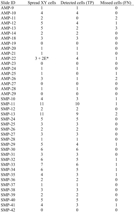
Table 2 Results obtained by manual scanning of rare cellular events hybridized by FISH technique. ... 63
CHAPTER II: article 2 Table 1 Interpretation of detected cellular events by automatic scanning according to the concordance of FISH or PRINS images with those previously taken with Giemsa. ... 83
Table 2 Results obtained by FISH technique. ... 85
Table 3 Results obtained by PRINS technique. ... 89
Table 4 Results obtained from counting of Fetal cells in maternal blood by FISH ... 90
CHAPTER III: article 3 Table 1 Results obtained from normal pregnancy cases (cases 1-6) normalized to 10 ml maternal blood. ... 112
Table 2 Results obtained from aneuploid pregnancy cases (cases 7-12) normalized to 10 ml maternal blood. ... 113
CHAPTER IV: Article 4 Table 1 Polymorphic markers used in the Primer Mix 10X ... 134
Table 2 Polymorphic markers used in the chromosome-specific primer mix. ... 135
Table 3 Results obtained by QF-PCR of amplified and extracted DNA in comparison to standard chromosomal analysis of fetal karyotype. ... 140
Table 4 Cumulative results obtained from QF-PCR analysis of amplified DNA from single cells for determination of fetal sex and major chromosomal aneuploidies. ... 143 CHAPTER IV: DISCUSSION
CHAPTER IV: CONCLUSION REFERENCES
LIST OF FIGURES
INTRODUCTION
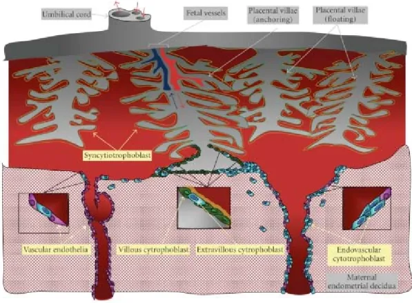
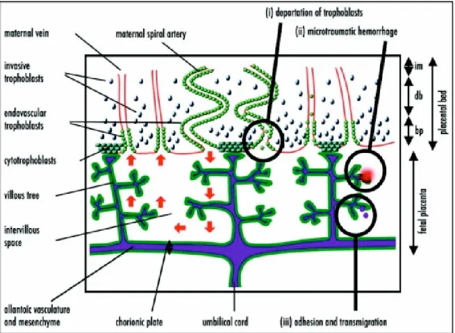
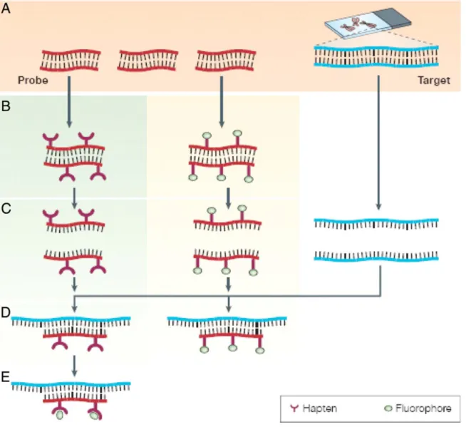
Figure 1 Diagrammatic presentation of the human maternal-fetal interface ... 25 Figure 2 Simplified diagram of the hypothesized mechanisms of fetomaternal cell trafficking ... 27 Figure 3 Diagrammatic presentation of the principles of the FISH technique ... 38 RESULTS
CHAPTER I: Article 1
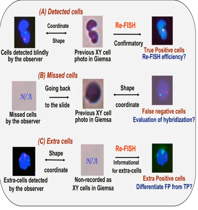
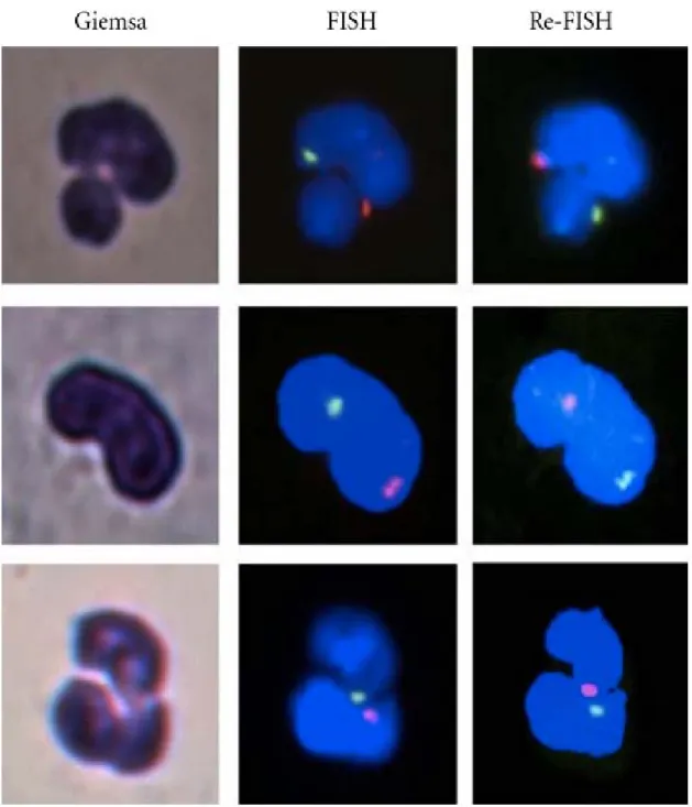
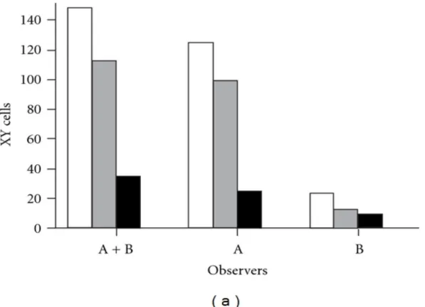
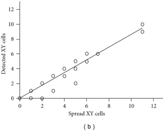
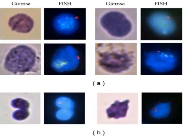
Figure 1 Schematic management of detected (A), missed (B), and extra cells (C). ... 60 Figure 2 Example of Giemsa, FISH and re-FISH images of three detected events. ... 61 Figure 3 (a) Comparison between detected cells and real number of XY cells on the slides. Analysis of detection efficiencies of the two observers by comparing detected cells to real number of XY cells. ... 63 Figure 3 (b) Comparison between detected cells and real number of XY cells on the slides. Regression analyses represent correlation between detected cells by manual scanning and real number of XY cells on the slides. ... 65 Figure 4 Giemsa and corresponding FISH photos of missed events due to inadequate hybridization (a) or non-hybridization (b). ... 66 CHAPTER II: Article 2
Figure 1 Schematic presentation of the custom-made detection algorithm used for detection of rare XY cells from pure population of XX cells. ... 82 Figure 2 Regression analyses represent correlation between detected cells by FISH with automatic scanning and real number of XY cells on the slides. ... 84 Figure 3 FISH and corresponding Giemsa images of missed events both due to
non-hybridization (Panel A) or inadequate non-hybridization (Panel B). ... 87 Figure 4 Regression analyses represent correlation between detected cells by PRINS with automatic scanning and real number of XY cells on the slides. ... 88 Figure 5 Assessment of selected events by re-FISH. ... 91 Figure 6 Comparison of average number of detected fetal cells in the first versus second trimester and in normal versus aneuploid pregnancies. ... 92 CHAPTER III: Article 3
Figure 1 Fetal cells detected by FISH (images A, C and E) and confirmed by reverse color FISH (images B, D and F) respectively ... 111 Figure 2 Histogram comparing average and standard deviation (SD) of total and fetal cell loss. ... 114 CHAPTER IV: Article 4
Figure 1 Flow chart of the entire methodology. ... 137 Figure 2 Comparison of DNA yield obtained by whole genome amplification of serial numbers of single cells. ... 138 Figure 3 The process of Laser-capture microdissection of single cells on the slides. ... 139 Figure 4 The QF-PCR profile of a male trisomy 21 from amplified DNA from 5
microdissected cells. ... 141 Figure 5 The QF-PCR profile of a female triploidy case from amplified DNA from 5 microdissected cells. ... 142
CHAPTER IV: DISCUSSION
Figure 1 Summary of the results of manual scanning using FISH technique for detection of rare cellular events. ... 163 Figure 2 Summary of the results of automatic scanning using FISH for detection of rare cellular events. ... 167
CHAPITER IV: CONCLUSION
LIST OF ABREVIATIONS, SIGNS AND SYMBOLS
AFP Alpha-fetoproteinACOG American Congress of Obstetricians and Gynecologists
aCGH Array comparative genomic hybridization
AuSc Automatic scanning
BAC Bacterial artificial chromosome
bp Basepair
CIHR Canadian Institute for Health Research
cffDNA Cell-free fetal DNA
°C Celsius temperature
CHUS Centre Hospitalier Universitaire de Sherbrooke
CEP Centromeric enumeration probe
CEP Centromeric enumeration probes
CVS Chorionic villous sampling
Ch Chromosome
CD Cluster differentiation
CD Cluster differentiation
CCMG College of Medical Geneticists
CGH Comparative genomic hybridization
C.C Correlation coefficient
Cy Cyanine dye
Cy3 Cyanine dye 3
Cy5 Cyanine dye 5
CK Cytokeratins
DAPI 4’,6’-diamidino-2-phénylindole
DOP-PCR Degenerate oligonucleotide-primed-polymerase chain reaction DGC Density gradient centrifugation
DNA Deoxyribonucleic acid
ε Epsilon
NRBC Erythroblasts or nucleated red blood cells
FN False negative
FBS Fetal bovine serum
FC Fetal cell
FCs Fetal cells
FITC fluorescein isothiocyanate
FACS Fluorescence activated cell sorting
FISH Fluorescence in situ hybridization
FSH Follicle-stimulating hormone
γ Gamma
gDNA Genomic desoxyribonucleic acid
g Gram
Hb Haemoglobin
HBSS Hank’s balanced salt solution
HASH-2 Human Achaete-Scute Homologue 2
hCG Human chorionic gonadotropin
HLA Human leucocytes antigen
HPL Human placental lactogen
ID Identification code
ISIS In situ imaging system
kb Kilobase
LCM Laser capture microdissection
LSI Locus specific probe
MACS Magnetic Activated Cell Sorting
MnSc Manual scanning MB Maternal blood MB Maternal blood µg Microgram µl Microlitre mg Milligram ml Millilitre mM Millimole min Minute M Mole
M-FISH Multicolour - Fluorescence in situ hybridization
MoM Multiple of median
NIFTY National Institutes of Health Fetal Cell Study
NIPD Non-invasive prenatal diagnosis
N/A Not acquired
p Short arm of chromosome
PBS Phosphate-buffered saline solution
PAC Plasmid P1-derived artificial chromosomes
PCR Polymerase chain reaction
KCl Potassium chloride
PAPP-A Pregnancy associated plasma protein A
PGD Preimplantation genetic diagnosis
PRINS PRImed In Situ labelling
PEP Primer extension pre-amplification
P Probability value
q Long arm of chromosome
QF-PCR Quantitative fluorescence - Polymerase chain reaction
RhD Rhesus D antigen
RNA Ribonucleic acid
RT Room temperature
SSC saline-sodium citrate buffer
SCs-WGA Single-cells whole genome amplification STRs Sort tandem repeat
SKY Spectral karyotyping
SAS Statistical Analysis Software
SSPS Statistical Package for the Social Sciences
TRITC Tetramethyl Rhodamine Iso-Thiocyanate
TCs the total number of cells
TP True positive
UV Ultraviolet
uE3 Unconjugated estriol
W Weeks
WCP Whole chromosome painting probes
WGA Whole genome amplification
YAC Yeast artificial chromosome
INTRODUCTION
1. General Information
The burden of genetic disorders is heavy in all parts of the world, particularly in under-resourced settings, which lack specialized health and social services to care for affected individuals. Large numbers of infants with genetic disorders are born each year from families in underserved populations as a result of the high birth rate, consanguinity and late procreation. As a result, most pregnant women would wish to be reassured that their unborn babies are healthy. Access to safe, accurate and affordable screening and diagnostic tests at a time that avails the mother an option of pregnancy termination is therefore essential.
Prenatal diagnosis of aneuploidies is usually performed by collecting fetal samples through either amniocentesis or chorionic villous sampling (CVS). These procedures are invasive and are associated with a significant risk of miscarriage. Therefore, in recent years, considerable effort has been made to develop non-invasive prenatal diagnostic procedures (Finegan et al., 1990).
One potential non-invasive approach is to utilize the fetal cells (FCs) within the maternal circulation. Cell’s trafficking between the fetus and its mother provides indirect clues as to the underlying pathophysiology during pregnancy. It also provides a source of fetal materials for non-invasive prenatal diagnosis. Many questions remain about the feasibility of using FCs from maternal blood (MB) for prenatal diagnosis.
Although the retrieval of FCs from maternal blood (MB) is an attractive concept, many questions remain regarding the feasibility of using FCs from maternal blood (MB) for prenatal diagnosis. In fact, the very low abundance of FC in MB is a technical challenge (Bianchi et al., 2002; Ariga et al., 2001; Krabchi et al., 2001) and there is currently no application for these methods in clinical practice. Additional work is needed to isolate FC early in pregnancy in order to provide, if necessary, pregnancy termination options to the parents.
2. Historical background
The presence of the FCs in maternal circulation during the pregnancy has been known for a long time. Indeed, the existence of FCs in MB was shown more than one century ago. Many authors in this field recognize the work of Georg Schmorl, a German pathologist, which was published in Leipzig in 1893,, to document the first description of feto-maternal cellular trafficking, as well as the first clues for the presence of retained FCs in maternal body organs (Schmorl, 1893). His elegant and pioneer work has been recently translated in English and critically re-evaluated from a 21st century perspective (Lapaire et al., 2007). Schmorl had noted for the first time the presence of multinucleated syncytial giant cells sequestered in the thin capillaries of the pulmonary parenchyma in the autopsies of 14 of 17 women who died of eclampsia. Schmorl assumed that the only source of the multinucleated cells could be the decidua or the placenta. Furthermore, he was the first physician to emphasize the importance of the placenta in the etiology of pregnancy complications such as eclampsia and preeclampsia (Schmorl, 1893). More than 60 years later, the placental origin of these trophoblastic cells was established. Other authors reported the presence of these cells in other tissues including the uterine veins and peripheral venous blood in both normal and complicated pregnancies (Wagner et al., 1964; Douglas et al., 1959).
Many investigators, in late 1950s, began to demonstrate that fetal erythrocytes were also present in the maternal peripheral circulation. Kleihauer found FCs in the blood of pregnant women by using a specific stain for fetal haemoglobin (Kleihauer et al., 1957). Clayton et al. noted that the proportion of fetal red cells was higher in women with pregnancies complicated with preeclampsia or vaginal bleeding, and after abortion or amniocentesis (Clayton et al., 1964).
The first cytogenetic study to identify FCs in maternal circulation was done in 1969 by Walknowska et al (Walknowska et al., 1969). These authors treated MB with a lymphocyte mitogen and then examined the metaphase cells. They clearly showed the presence of XY metaphases in maternal peripheral blood of 19 out of 21 pregnancies with
male fetus. De Grouchy and Trebuchet (de Grouchy and Trebuchet, 1971) as well as other groups confirmed these results (Schindler and Martin-du-Pan, 1972; Takahara et al., 1972).
Cell sorting techniques became available in late 1970s which allowed isolation and characterization of cells based on their antigenic characteristics. In 1979, Herzenberg et al added, to the step of analysis of Y chromatin of male FCs, a preliminary technique of enrichment using a flow cytometer (FACS: fluorescence-activated cell sorting) on the basis of cellular class I major histocompatibility antigens. They exploited the human leucocytes antigenic differences (HLA) between the mother and her fetus. Using blood from HLA-A2 negative women whose partners were HLA-A2 positive, cells were sorted based on the presence of the HLA-A2 antigen. The sorted cells, most likely lymphocytes, were then analyzed for the presence of Y-chromatin fluorescence. Y-chromatin positive cells were found more frequently in women carrying male fetuses than in those carrying female fetuses. Five out of twelve pregnancies with male fetuses presented cells with Y chromosome (0.3 to 1.6% of the sorted cells). F o r t he other seven, t h e r e w a s i n f a c t no cells with Y chromosome (Herzenberg et al., 1979). Iverson and co-workers (Iverson et al., 1981) used for the first time this technique to enrich FC in order to determine the sex of the foetus. The heterochromatin of the long arm of the Y chromosome was used as independent marker in pregnancies as early as 15 weeks of gestation. Y chromatin-containing cells were found among the sorted cells from prenatal MB specimens in eight pregnancies out of eight subsequently produced male infants. The presence of positive HLA-A2 with the heterochromatin of Y confirmed the fetal origin of the cells.
Molecular techniques became available in late 1980s, which allowed reliable and significant analysis of genetic markers to confirm the presence of the FCs in MB circulation. In 1989, Lo and co-workers (Lo et al., 1989) proposed the use of polymerase chain reaction (PCR) to identify Y-specific DNA sequences in MB. Y-chromosome specific sequence could be detected in MB as early as six weeks of gestation. Four years later, the same group reported performing PCR on unsorted first trimester MB and were able to correctly identify six of seven pregnancies with male fetuses and five of six pregnancies with female fetuses. The detection rate was at least as good in the first trimester as in the second or third. Serial
dilutions of male DNA were also performed to calculate that this method has the sensitivity to detect one male cell d i s p e r s e d in 300,000 female cells (Liou et al., 1994; Lo et al., 1993; Lo et al., 1990). These PCR results were also confirmed with the use of enrichment procedures of cellular sorting using either fluorescent monoclonal antibody by FACS (Bianchi et al., 1990; Mueller et al., 1990) or magnetic balls by Magnetic Activated Cell Sorting MACS (Wachtel et al., 1991). The recognition of FCs by specific antibodies and their isolation using cell sorter or magnetic beads were the subjects of intense search. It is however necessary to point out that the sensitivity and the specificity of these methods remain insufficient to allow a reliable prenatal diagnosis.
Following these reports, the potential use of FCs, for a non-invasive prenatal diagnosis (NIPD), was strongly reconsidered. Many research teams studied the nature of these FCs. These studies raised much controversy, in particular, on the origin of these cells, their lifespan and antigenic specificities that could distinguish them from maternal cells. Other questions regarding their time of appearance and relative frequency throughout the gestational age were also the subject of intense studies.
Several authors reported a quantification of the number of FCs (Krabchi et al., 2001; Bianchi, 1998). The frequency of FCs circulating in maternal peripheral blood in normal pregnancy is very low. In the pathological situations, such as for example in the preeclampsy or fetal aneuploidies, the fetomaternal cellular transfusion is apparently increased (Zhang et al., 2008; Krabchi et al., 2006b; Krabchi et al., 2006c).
Various categories of FCs have been proposed. The possible cell types that can be isolated from MB and used for prenatal diagnosis include trophoblasts, lymphocytes, erythroblasts, granulocytes, and thrombocytes (Goldberg, 1997). All these cells are nucleated except for the blood platelets (thrombocytes) which loss their genomics DNA during the process of differentiation. The presence of rare lymphoid progenitor cells of fetal origin in MB from former pregnancies has been also reported (Bianchi et al., 1996). Given the extreme scarcity of these events and their antigenic specificities, they are not regarded as major handicap for the development of NIPD. Many attempts of isolation of the FCs, for the sake of NIPD, focus mainly on the erythroblasts and cytotrophoblasts. Indeed, these two
types of cells have the advantage of being highly differentiated and are known to have a short life span and thus unlikely to persist in MB postpartum.
3. Aneuploidies and screening modalities
Prenatal care has existed for over 100 years as an approach to improve maternal and newborn outcomes. Screening tests are usually non-invasive and may help delineate which patients are at high risk and should be offered invasive testing. Diagnostic tests on the other hand have the benefit of providing a definite answer about the presence of a genetic disorder. Since these tests carry varying risks of pregnancy loss, they are usually reserved for high-risk women with positive screening.
3.1. Trisomy 21 and other aneuploidies
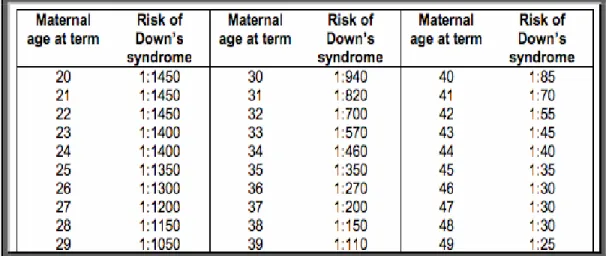
Trisomy 21, also known as Down syndrome, is the leading cause of prenatal chromosome abnormalities, accounting for more than half of all reported chromosomal aneuploidies (Hook et al., 1983). The incidence of trisomy 21 in the general population is 1.3 per 1000 live births (1/770). Incidence rate increases significantly with maternal age (Table1) (Huether et al., 1998).
Table 1 Maternal age and risk of Down's syndrome
Table 1 shows that risk of Down's syndrome increase with advancing maternal age. J Med Genet. Jun 1998; 35(6): 482–490
Down syndrome individuals have a distinct phenotype and show various degree of intellectual disability (Sherman et al., 2007). Patients with trisomy 21 have a slightly reduced life expectancy (Thorpe et al., 2012; Baird and Sadovnick, 1990). Indeed, they are often victims of medical complications and developmental disorders (Tenenbaum et al., 2012; Hayes and Batshaw, 1993; Carey, 1992). They account for 20 to 30% of the subjects with moderate to severe mental retardation. More than 15% of the adult subjects will develop Alzheimer's disease around the age of forty (Strydom et al., 2013).
In Canada, there are approximately 400,000 births with more than 600 new cases of trisomy 21 per year. A prenatal screening program has been created in Quebec in 1976 (Okun et al., 2008; Forest et al., 1995; Baird and Sadovnick, 1990). The majority of mothers with affected fetuses, after being informed of the prognosis and postnatal therapeutic options, choose medical interruption of the pregnancy (Grant and Flint, 2007; Fletcher, 1981).
Other trisomies, such as trisomy 18 (Edward syndrome) and trisomy 13 (Patau syndrome), are associated with fatal congenital malformations. Survival is very poor with approximately 50% miscarriages prior to birth and, most liveborns die within the first month of life (Hutaff-Lee et al., 2013).
3.2. Screening modalities
Screening is conventionally described as the evaluation of asymptomatic people to detect unsuspected disease or risk in order to improve health outcome in a defined population (Henry and Bronson, 1996).
Reproductive genetic screening is performed to assist reproductive decision-making and to give parents the opportunity to avoid the birth of an affected child. Because the decision to terminate a pregnancy is highly personal, prenatal screening is considered optional, in sharp contrast to the mandatory newborn screening after birth. In keeping with this difference, genetic professionals have developed a counselling approach to assist couples to determine their own preferred course of action (Mahowald et al., 1998).
3.2.1. Genetic counselling
Genetic counselling begins with a thorough medical and family history of the patient and her partner to identify high-risk patients who may benefit from diagnostic or therapeutic procedures. Historically, the first and most important risk factor for trisomy 21 is advanced maternal age. Incidence increases gradually up to the age of 35 years then rises very sharply. The reason for this is not entirely known but may have to do with abnormal function of the meiotic spindle during female meiosis, resulting in nondisjunction (Zournatzi et al., 2008).
Because, at the age of 35, the risk of having a newborn with chromosomal aneuploidy approximately equals the risk of pregnancy loss with invasive testing (0.5%, or 1/200), it is standard of practice to offer invasive testing to women who will be 35 years or older at the time of delivery. However, this strategy is not very efficient because two third of the trisomic 21 children are born from women having less than 35 years old (Zournatzi et al., 2008). For this reason, different screening modalities were developed and implemented in order to re-evaluate the risk and precisely identify those that would benefit from invasive diagnostic tests.
3.2.2. First trimester ultrasound
Anatomical ultrasound has been used since the 1980s for detection of major structural abnormalities. With advances in technology, prenatal ultrasound has expanded to include detection of soft markers more commonly found in fetuses with chromosomal abnormalities (Getz and Kirkengen, 2003). During the first trimester, between 10 and 14 weeks, thickening of an area behind the fetal neck (nuchal translucency) is associated with an increased risk of Down syndrome. This fluid-filled area of the posterior neck normally resolves by the second trimester (Snijders et al., 1998).
Other soft markers linked to Down syndrome include echogenic bowel, renal pelvic dilation, absence of the nasal bone, ventriculomegaly, clinodactyly, and sandal gap toe (Cicero et al., 2001). The presence of one of these markers increases the risk by two-fold while three or more markers increases the risk by 100-fold (Benacerraf, 2010; Nyberg and
Souter, 2001). While these criteria help in determining relative risk, they remain limited by the quality of the ultrasound and the expertise of the sonographer.
3.2.3. Maternal serum biomarkers
Maternal serum biomarkers are substances secreted by the fetus or placenta during pregnancy and that can be measured in the MB. The level of these markers can be useful to predict congenital anomalies and chromosomal abnormalities, particularly trisomy 21. The expected amount of these substances found normally in the mother's bloodstream changes weekly during pregnancy, so it is important to to accurately determine the gestational age, usually using ultrasound at 10-12 weeks.
The level of each marker is expressed as multiples of the median (MoM), obtained by dividing the serum concentration at a particular gestational age by the population median concentration at the same gestational age (Spector et al., 2005). Combined values of different markers provide a risk estimate rather than a definitive diagnosis.
Alpha-fetoprotein (AFP) is produced by the fetal liver, but is transported to the MB across the placenta. High level of AFP is frequently associated with neural tube defects. Other possible causes of a high AFP include incorrect dates, multiple pregnancies, fetomaternal bleed, and other fetal malformations, such as defects of the abdominal wall. This may be due to leaky placental barrier or placental dysfunction associated with these pathologies. Low levels of AFP are associated with Down syndrome. Any abnormal AFP measurement should be followed by a detailed fetal ultrasound (Guibaud et al., 1998; Rose and Mennuti, 1993).
Human chorionic gonadotropin (hCG) is also known as the ‘pregnancy hormone’. It is produced by the placenta very early in pregnancy. It is made of α and β chains. The rate of synthesis of the total hCG is dependent on the rate of synthesis of the free β -hCG fraction. This hormone peaks early in pregnancy at 8–10 weeks. After that peak, it progressively declines to reach a plateau at 18 to 20 weeks of gestation. Levels are increased in Down syndrome, and decreased in trisomy 18. Elevated mid-trimester levels
have been associated with congenital abnormalities, placental dysfunction and adverse pregnancy outcome (Rose and Mennuti, 1993; Bogart et al., 1987).
Unconjugated estriol (uE3) is the dominant form of estrogen during pregnancy. This hormone is derived from precursors from the fetal adrenal and liver that are processed in the placenta. Low estriol may be associated with Down syndrome and anencephaly, the most severe neural tube defect. Other syndromes associated with low estriol include congenital adrenal hypoplasia, and X-linked icthyosis (Guibaud et al., 1998; Canfield and O'Connor, 1991).
Inhibins are placental hormones that inhibit the secretion of follicle-stimulating hormone (FSH). There are two forms: inhibin-A and inhibin-B; however, only the former is found in pregnant women. Inhibin-A has been found to be increased in Down-syndrome pregnancies, and has most recently been added as a fourth serum marker for second trimester screening (Gagnon et al., 2008; Lockwood et al., 1997).
Pregnancy associated plasma protein A (PAPP-A) is produced by the placenta and thought to have several functions, including prevention of recognition of the fetus by the maternal immune system. A PAPP-A level was found to be low in pregnancies with Down syndrome and other chromosomal defects. Recent studies support an association between low PAPP-A levels in first trimester and risk for adverse pregnancy outcomes as prematurity and growth retardation (Smith et al., 2002).
3.3. Routine prenatal screening 3.3.1. Second trimester screening
Several studies showed that measurements of biochemical markers in the maternal serum between 15 and 17 weeks of gestation could be useful to identify complicated pregnancies. The possibility of prenatal screening, using maternal serum markers, was reported for the first time in 1984 by Merkatz et al. (Merkatz et al., 1984). AFP was the earliest serum marker used to detect open neural tube defects and abdominal wall defects
and with time it was extended to screen for Down syndrome (Cuckle et al., 1984; Wald et al., 1977).
In 1987, Bogart and co-workers (Bogart et al., 1987) showed that serum concentrations of hCG are higher than normal in the pregnancies affected by trisomy 21. Continued advancements in research resulted in the introduction of a multiple markers screening panel, or the” triple test”, in 1991 (MacDonald et al., 1991). In addition to AFP, the panel included uE3 and total hCG. The triple screen was widely employed in obstetrical practice to detect neural tube defects and chromosomal aneuploidies. The detection rate for Down syndrome varies from 30% to approximately 69%.
The quadruple test was introduced in 2000, when inhibin-A was added to the triple test panel (Hackshaw and Wald, 2001; Haddow et al., 1998b; Aitken et al., 1996). The introduction of the quadruple test has significantly increased the detection rate. By combining maternal age with the quad screen, the detection rate is roughly 75% for Down syndrome in women younger than 35 years and 80% in women 35 years and older with an approximately 5% false positive rate (Benn et al., 2001).
3.3.2. First trimester screening
It was not until the late 1990s that first trimester screening was introduced as an earlier screening option for the detection of Down syndrome. First trimester screening incorporates maternal age, nuchal translucency, and measurement of specific serum markers. A Collection of blood for biochemical analysis and ultrasound assessment is typically performed between 11 and 14 weeks. The most effective first-trimester biochemical markers are PAPP-A and free hCG in maternal serum (Biagiotti et al., 1998; Haddow et al., 1998a).
However, there is no single marker can detect all the pathological pregnancies. The echographic signs and the serum markers together can increase the detection rate. First trimester biochemical markers alone have only 60% sensitivity (Cuckle and van Lith, 1999). Combined with nuchal translucency, the detection rate is around 80%, with a 5% false– positive rate (Krantz et al., 2000).
Large collaborative, prospective studies have validated the clinical application of first-trimester screening, and showed that it could be superior to second-trimester screening (Nicolaides et al., 2005). In addition, it reduces both physical and psychological trauma related to late interruption of pregnancy as well as therapeutic costs.
3.3.3. Combined first and second trimester screening
Several investigators studied different ways of incorporating the results of first and second trimester serum screening to obtain the most accurate estimation of Down syndrome risk. Many modalities have been created to help maximize the sensitivity, while maintaining a low false-positive rate.
Integrated screening in which a patient’s first trimester screening results are not disclosed until second trimester screening is performed and a combined risk can be calculated, has been ethically debated. This option precludes patients who are at high risk based on first-trimester screening from being offered early CVS or other available options (Knight et al., 2005).
Independent sequential first and second-trimester screenings, with separate individualized risk assessments increases the detection rate from 80% to 90% but it also increases the false-positive rate from 5% to 11%. In contrast, stepwise sequential screening in which only patients who screen negative in the first trimester are offered second-trimester screening, increases the detection rate to more than 90% while still maintaining a low false-positive rate of 6% (Aagaard-Tillery et al., 2009; Platt et al., 2004).
Contingent screening method is similar to stepwise sequential screening. However, the contingent screening uses the first-trimester results to classify patients into three groups, i.e., screen-positive, screen-negative, and borderline. Second-trimester screening is only offered to patients who fall into either the screen-negative or the borderline group. The detection rate for this method is 95%, with a 5% false-positive rate (Palomaki et al., 2006; Wright et al., 2004).
4. Prenatal Diagnosis
In most developed countries the option of having prenatal diagnosis is discussed as part of routine antenatal care. Testing strategies, guidelines, and diagnostic options have expanded from their conception in the 1970s. At that time, any woman aged 35 years or older was considered to be of advanced maternal age, and this was the sole criterion used by the American Congress of Obstetricians and Gynecologists (ACOG) to define pregnancies that should be offered amniocentesis or CVS.
As of 2007, the ACOG has extended the definition of a “high-risk” pregnancy that justifies prenatal cytogenetic diagnosis to include advanced maternal age, parental chromosome rearrangements, previous pregnancy with autosomal anomaly, abnormal fetal ultrasound findings during the current pregnancy and increased risk calculated from non-invasive screening (ACOG, 2007b).
However, current ACOG guidelines stated unequivocally that neither age 35 years nor any specific age should be used as a threshold for invasive testing: ‘All women, regardless of age, should have the option of invasive testing’. The guidelines specifically elaborate that ‘patients informed of the risks, especially those at increased risk of having an aneuploid fetus, may elect to have diagnostic testing without first having screening’ (ACOG, 2007a). Younger women may elect an invasive procedure because they wish to achieve the near 100% detection, possible only with an invasive procedure; detection by an invasive procedure exceeds by 10–15% that of any non-invasive screening protocol.
4.1. Routine prenatal diagnosis 4.1.1. Invasive diagnostic procedures
The Prenatal diagnosis of the chromosomal anomalies generally requires collection of fetal tissue and chromosomal analysis of FCs. Fetal tissues can be obtained by either CVS, amniocentesis or less commonly cordocentesis through puncture of the umbilical cord. Valenti and co-workers, in1969, (Valenti et al., 1969) reported the first prenatal diagnosis of Down's syndrome three years after the achievement of amniotic cell growth by
Steele and Berg (1966) (Steele and Breg, 1966). Amniocentesis is usually performed through puncture of amniotic sac to obtain amniotic fluid for karyotyping and other biochemical tests at 16-18 weeks of gestation. Amniocentesis performed before 15 weeks is referred to as ‘early amniocentesis’. Early amniocentesis is not a safe alternative to second-trimester amniocentesis because of increased pregnancy loss, limb reduction defects and clubfoot (CEMAT, 1998).
The development of CVS in the early 1980's has allowed anticipation of diagnosis in the first trimester (Brambati and Simoni, 1983). CVS is usually performed between 9 and 13 weeks of gestation and involves aspiration or biopsy of placental villi. CVS can be performed using either a transabdominal or a transcervical approach. Several randomised trials show almost identical miscarriage rates after transcervical CVS compared with the transabdominal approach (Jackson et al., 1992; Brambati et al., 1991). Only one trial demonstrated the transabdominal approach to be significantly safer (Jackson et al., 1992).
Hundreds of thousands of amniocentesis and CVS after 10 weeks of gestation have been done without causing any complications or an increase in fetal malformations. However, both procedures are sometimes difficult and associated with some risks mainly to the pregnancy, but in certain circumstances to both the mother and the fetus. They also require the expertise of a specialized medical team and present a risk of iatrogenic fetal loss estimated between 0.5 and 2% (Tabor et al., 1986). The clear advantage of an early procedure like CVS over amniocentesis is the avoidance of a prolonged period of uncertainty and the availability of less stressful options in cases in which termination of pregnancy is desired after an abnormal result (Bindra et al., 2002). However, the disadvantage is the increased risk of miscarriage after first trimester CVS. Some authors even reported higher rates of limb reduction abnormalities and subsequent development of preeclampsia with CVS carried out at 9 weeks or earlier (Grobman et al., 2009; Philip et al., 2004). Other fetal risks include intrauterine fetal death and premature birth (Vigliani, 2009). Severe sepsis, including maternal death, has been reported following invasive prenatal procedures. The level of risk cannot be quantified as case report literature does not provide denominator information but the risk of severe sepsis is likely to be less than 1/1000
procedure (Bodner et al., 2011). Infection can be caused by inadvertent puncture of the bowel, skin contaminants or organisms present on the ultrasound probe or gel. The procedures also increase the risk of maternal isoimmunization provoked by fetomaternal hemorrhage. Therefore, maternal RhD status should be obtained and prophylaxis with anti-D immunoglobulin must be offered following each procedure to Rhanti-D negative women in line with international recommendations (ACOG, 2006).
Currently, these invasive procedures are offered only to small group of women who are in a higher risk of having an offspring with a chromosomal defect in comparison to the general population. This high-risk group constituted less than 5% of the pregnant population. In addition, only one out of 20 procedures performed will reveal aneuploidy (Crossley et al., 2002). Development of non-invasive methods would obviate this risk and change the risk-benefit ratio of prenatal diagnosis. Such a change would make it likely that more women presently eligible for prenatal diagnosis would choose to undergo testing. In addition, genetic testing could even be offered to women who are not considered at high risk. One of the most promising non-invasive sources of fetal genetic materials is the peripheral MB. In this view, analysis of FC represents a major objective of many researches. 4.1.2. Routine fetal karyotype
With invasive tests such as amniocentesis or CVS, FCs are obtained for culture. For routine fetal karyotype, culture of FCs is essential prior to analysis as chromosomes are only visible in dividing cells. The application of strategies for improving cell culture and chromosome banding has expanded the number of laboratories that may perform successfully fetal chromosome analysis (Cheung et al., 1987; Brackertz et al., 1983; Porreco et al., 1980). The standard analysis implies the study of the number and structure of the 23 chromosome pairs. The most common chromosome anomalies, are related to non-disjunctional errors, result in an extra copy or loss of one chromosome. Trisomy 21 is by far the single most common cause of aneuploidies. Other identified abnormalities involve trisomies of chromosomes 13, 18, or sex chromosomes.
Although high resolution banding could allow diagnosis of small structural anomalies, these anomalies are relatively uncommon, accounting for less than 1% of all chromosomal abnormalities. Furthermore, balanced translocations and inversions, which are the commonest identified structural anomalies, are clinically irrelevant for the current pregnancies (Warburton, 1984; Jensen et al., 1982). However, there is a general consensus among cytogeneticists and physicians that the extra knowledge provided by a full karyotype is beneficial and thus, a full fetal karyotype is the gold standard of prenatal diagnosis.
4.1.3. Rapid diagnostic techniques of fetal chromosomal anomalies
The time needed to culture FCs and complete the analysis ranges from 10 to 21 days, which is generally considered to be a psychological burden and results in late terminations following a pathological diagnosis. In the early 1990s, FISH (Fluorescence In Situ Hybridization) (Kuo et al., 1991) and, more recently, QF-PCR (quantitative fluorescence polymerase chain reaction) (von Eggeling et al., 1993) entered the field of prenatal diagnosis to overcome the need to culture FCs, and hence allowed a rapid diagnosis of some selected chromosomal anomalies. FISH and QF-PCR provide a rapid diagnosis of aneuploidies within 24–48 hours. Although both techniques could be applied to identify all chromosomes, only chromosomes 13, 18 and 21, as well as the sex chromosomes, are routinely tested (Divane et al., 1994). The result was, and still is, sufficient to take action if a chromosome anomaly is thus identified, but is usually considered only a preliminary step while awaiting the result of full karyotype.
Array comparative genomic hybridization (CGH) has been proposed as a genome-wide assessment approach for prenatal diagnosis of chromosomal abnormalities. Array CGH is a molecular cytogenetic method for analysing copy number variations relative to ploidy level in the DNA of a test sample compared to a reference sample, without the need for culturing cells. Many reports have demonstrated the sensitivity, specificity and accuracy of this methodology detecting large and small-size imbalances (Pickering et al., 2008; Shaffer et al., 2008; Shaffer et al., 2007). Although different types of chromosomal abnormalities have been successfully identified by array CGH, the CGH traditionally is costly and requires advanced equipment (Lao et al., 2008). Another disadvantage of an
array CGH system is the time required for analysis and interpretation of the results, especially with the many incidental findings of unknown clinical significance, which creates an ethical dilemma and raises the maternal anxiety (Keren et al., 2010). However, array CGH has particular importance in investigating cases with strong history of intellectual disability or congenital abnormalities despite a normal conventional karyotype.
Although these techniques hasten the process of prenatal diagnosis, they did not overcome the risk associated with invasive sampling of fetal tissues, which, therefore, limits offering prenatal diagnosis only to women with high risk pregnancies, as estimated by increased maternal age, abnormal biochemical markers and ultrasonographic findings. 4.2. Non-invasive prenatal diagnosis
The long-term goal of modern prenatal genetics is the development of definitive NIPD. That is, an analysis of MB that can detects fetal aneuploidy and other disorders without the need for an invasive procedure. Currently, prenatal diagnosis safety is limited by the need of invasive means to obtain fetal tissues. This limits its application to only small group of high-risk patients, which constitute less than 5% of all pregnancies. However a fairly good non-invasive method to obtain fetal tissues would obviate this risk and extend prenatal testing to include wider portion of pregnant population. It is currently agreed that genetic fetal material including both cells and cell-free fetal DNA (cffDNA) pass into maternal circulation during pregnancy. Non-invasive fetal diagnosis could therefore be possible from their isolation and analysis from peripheral MB. Although the cffDNA is increasingly used with massively parallel sequencing (Jensen et al., 2013; Liang et al., 2013; Liao et al., 2012; Ashworth, 1869) or targeted deep sequencing (Nicolaides et al., 2013; Zimmermann et al., 2012) to test for aneuploidy and single-gene disorders, it is still consider as a screening test and its application in clinical practice is very cumbersome and expensive. Although, cffDNA is currently a topic of great interest, this review will focus on the discussion of intact FCs in MB.
Intact FCs have considerable advantage over cffDNA as the whole fetal genomic DNA can be purely recovered without maternal contamination and consequently simplify
the analysis and enables more women to undergo prenatal diagnosis without a significant increase in health expenditure. Analysis of pure fetal DNA from intact FCs would allow not only readily diagnosis of aneuploidy but also other small structural genetic defects concurrently. Even if one or few FCs were recovered, the approach would merely be analogous to that already routinely carried out on blastomeres or polar bodies in pre-implantation genetic diagnosis. Analysis of intact FCs rather than cffDNA should facilitate provision of information about Mendelian disorder, and other chromosomal abnormalities.
An array allows comparative genomic hybridization (CGH) detection of aneuploidy for all chromosomes on a single cell is already available. This approach is proposed for analysis of a polar body or blastomere. The same approach could be applied for analysis of FCs. Furthermore; genetic diseases where the mother does not have the genetic alteration can be diagnosed by analyzing cffDNA in the maternal plasma. However, plasma analysis cannot be used for prenatal diagnosis of maternally inherited genetic diseases.
However, even if several diagnostic perspectives could theoretically be realized, the use of these FCs for non-invasive prenatal diagnosis is currently far from being achievable in routine. Many interrogations persist on the types of FCs, physiology of trans-placental passage to the maternal circulation and the feasibility of their use in NIPD. Original efforts involved recovering intact FCs from MB. Various types of FCs in MB were recovered, primarily trophoblastic, erythroblastic and leucocytic cells. Each cell type has its own unique cellular characteristics and antigenic specificity. Consequently, various types of enrichments and cellular sorting have been tried. Given the fragility and rarity of these cells in MB (1 fetal cell for 106 to 108 maternal cells), translation into clinical utility remained elusive. Adapted techniques and new technologies are required to enable cellular recovery and accurate single cell analysis.
Clearly, these newly developed techniques that could retrieve FCs from MB MB need additional improvements and clinical validation before being proposed in clinic for aneuploidy detection. Admittedly, though the harmlessness of these techniques seems attractive, but this does not mean they should necessary lead to NIPD.
5. Types of fetal cells in maternal blood
The possible cell types that can be isolated from MB and used for prenatal diagnosis include erythroblasts, trophoblasts and lymphocytes.
5.1. Erythroblasts
Fetal erythroblasts or nucleated red blood (NRBC) appear to be the ideal candidates for detection and enrichment of FCs in MB. For multiple reasons, they are the cellular category most commonly studied and, probably, well characterized in MB. Fetal erythroblasts are abundant in fetal blood especially in early gestation, comprising approximately 10% of the red blood cells in the 11-week fetus. This proportion declines as pregnancy progresses reaching approximately 0.5% at the 19 weeks of gestation.
The trafficking of fetal red blood cells into the maternal circulation is clinically evident by cases of ‘silent’ rhesus isoimmunisation in rhesus negative women (MacKenzie et al., 1999). They have a nucleus with full complement of genetic information and a limited lifespan of approximately 90 days. In contrast to lymphocytes, they are unlikely to persist more than few days’ post-partum and are rare in the peripheral blood of a normal adult (except in clinical circumstances of increased haematopoiesis such as pregnancy). Fetal NRBC can be recognised in a number of different ways and many groups have convincingly sorted this fetal cell type using a variety of strategies. These cells were identified by their morphological characteristics and positive coloration for fetal hemoglobin (Clayton et al., 1964).
Nucleated erythrocytes express several unique antigens, such as the transferrin receptor, which is recognize by the AntiCD71 antibody, which recognizes the transferrin receptor (Ganshirt-Ahlert et al., 1992). Unfortunately, NRBC in the maternal circulation are not exclusively fetal, as was previously thought. AntiCD71 antibody recognizes also other nucleated fetal and maternal cells. Slunga-Tallberg has demonstrated that a definite population of maternal NRBC can be found during pregnancy and others have shown that only half of the erythroblasts in MB are of fetal origin (Troeger et al., 1999a; Slunga-Tallberg et al., 1995).