Abstract
Patients with Alzheimer’s disease (AD) and semantic variant primary progressive aphasia (svPPA) can present with similar language impairments, mainly in naming. It has been hypothesized that these deficits are associated with different brain mechanisms in each disease, but no previous study has used a network approach to explore this hypothesis. The aim of this study was to compare resting-state functional magnetic resonance imaging (rs-fMRI) language network in AD, svPPA patients, and cognitively unimpaired elderly adults (CTRL). Therefore, 10 AD patients, 12 svPPA patients and 11 CTRL underwent rs-fMRI. Seed-based functional connectivity analyses were conducted using regions of interest in the left anterior temporal lobe (ATL), left posterior middle temporal gyrus (pMTG) and left inferior frontal gyrus (IFG), applying a voxelwise correction for gray matter volume. In AD patients, the left pMTG was the only key language region showing functional connectivity changes, mainly a reduced interhemispheric functional connectivity with its right-hemisphere counterpart, in comparison to CTRL. In svPPA patients, we observed a functional isolation of the left ATL, both decreases and increases in functional connectivity from the left pMTG and increased functional connectivity form the left IFG. Post-hoc analyses showed that naming impairments were overall associated with the functional disconnections observed across the language network. In conclusion, AD and svPPA patients present distinct language network functional connectivity profiles. In AD patients, functional connectivity changes were restricted to the left pMTG and were overall less severe in comparison to svPPA patients. Results in svPPA patients suggest decreased functional connectivity along the ventral language pathway and increased functional connectivity along the dorsal language pathway. Finally, the observed connectivity patterns are overall consistent with previously reported structural connectivity and language profiles in these patients.
Keywords: Alzheimer’s disease, semantic variant primary progressive aphasia, language
Introduction
The typical presentation of Alzheimer’s disease (AD) is most often characterized by an insidious and progressive decline in episodic memory. Nevertheless, language is also frequently impaired in AD patients (Verma & Howard, 2012). While word-finding difficulties are recognized as the most prominent language deficits (McKhann et al., 2011), verbal fluency (J.R. Hodges & Patterson, 1995; Huff, Corkin, & Growdon, 1986; Verma & Howard, 2012), and semantics (Joubert et al., 2010) are frequently impaired in AD as well. These symptoms are also the clinical hallmark of the semantic variant of primary progressive aphasia (svPPA). svPPA is characterized by impaired naming, impaired word comprehension, impaired object knowledge, and surface dyslexia/dysgraphia (Gorno-Tempini et al., 2011). A partial overlap of language symptoms, especially in language production, can therefore be observed in AD and svPPA. However, these impairments are not present in all AD patients and are overall less severe in comparison to svPPA patients (Montembeault et al., 2017; Reilly, Peelle, Antonucci, & Grossman, 2011; Rogers & Friedman, 2008; Sajjadi, Patterson, Tomek, & Nestor, 2012).
Classic neuroanatomical models of language, mainly based on post-stroke aphasic patients, postulate that language is sustained by two main language centers, namely Broca’s area (left pars triangularis/pars opercularis in the inferior frontal gyrus; IFG) and Wernicke’s area (left posterior middle temporal gyrus, pMTG) (Geschwind, 1970). Although the precise function and localization of these regions have been the object of a longstanding debate, their role within the language network is widely accepted (Dronkers, Ivanova, & Baldo, 2017; Tremblay & Dick, 2016). Further studies, mainly based on observations on svPPA patients, have however revealed the critical role of the left ATL within the language network, mainly in conceptual knowledge (Chedid et al., 2016; Heilman, 1972; J. R. Hodges, Patterson, Oxbury, & Funnell, 1992; Snowden, Goulding, & Neary, 1989; M. A. Wilson et al., 2012). Although it was initially omitted from the classic language model, there is now extensive support for its inclusion as a main language center in addition to the left IFG and the left pMTG (Damasio, Tranel, Grabowski, Adolphs, & Damasio, 2004; Ferstl, Neumann, Bogler, & von Cramon, 2008;
Hurley, Bonakdarpour, Wang, & Mesulam, 2015; Mesulam et al., 2013; Schwartz et al., 2009; Ueno, Saito, Rogers, & Lambon Ralph, 2011).
Consistently, the development of language symptoms in AD and svPPA is associated with a dysfunction in one or more of these three key language regions. Neuroimaging studies in AD patients have indeed shown that language impairments are associated with changes in functional activity or hypometabolism in the left IFG (Melrose et al., 2009; Teipel et al., 2006), the left pMTG (Nelissen et al., 2011; Nelissen et al., 2007; Vandenbulcke, Peeters, Dupont, Van Hecke, & Vandenberghe, 2007) and the left ATL (Hirono et al., 2001; Lars, Timo, Michael, & Philipp, 2016; Zahn et al., 2004). In svPPA, studies have more consistently attributed naming and semantic impairments to dysfunction of the left ATL (Acosta-Cabronero et al., 2011; Desgranges et al., 2007; Diehl et al., 2004; Wilson et al., 2012).
However, in addition to dysfunction in specific and isolated brain regions, it is now recognized that functional disconnection within brain networks can underlie the cognitive impairments observed in neurodegenerative disorders such as AD and svPPA (Guo et al., 2013; Seeley, Crawford, Zhou, Miller, & Greicius, 2009). Resting-state functional magnetic resonance imaging (rs-fMRI) is one of the neuroimaging techniques that allows the investigation of functional brain networks. This task-free fMRI method examines the interactions between brain regions through correlated changes in blood-oxygen-level dependent (BOLD) signal. In recent years, the language network has been studied and successfully characterized in healthy controls using this technique (Hurley et al., 2015; Tomasi & Volkow, 2012). In cognitively unimpaired elderly adults, Hurley and colleagues have confirmed that the left IFG, the left pMTG and the left ATL are functionally interconnected and form the rs-fMRI language network (Hurley et al., 2015). These key regions are also connected with the left angular gyrus and the left superior frontal gyrus (see visual representation of the rs-fMRI language network, based on Hurley et al. 2015, presented in Figure 1). This suggests that rs-fMRI could be a tool of choice to better understand network-level brain alterations underlying language symptoms in clinical populations such as AD and svPPA. To our knowledge, only a very few studies have assessed functional connectivity in the language network in these populations and none has directly compared AD and svPPA patients.
In AD patients, the majority of rs-fMRI studies have focused on the default-mode network (DMN) (Buckner et al., 2005). These studies have shown a disconnection between the regions of the DMN, most frequently the hippocampus, the precuneus and the posterior cingulate cortex (for a recent review, see Badhwar et al., 2017). More recently, studies observed functional connectivity alterations even beyond the DMN in AD patients. This extended interest to other brain networks such as the language network (Mascali et al., 2018; Weiler et al., 2014; Whitwell et al., 2015). The few rs-fMRI studies investigating the language network in AD patients have consistently reported lower resting-state functional connectivity in posterior temporal language regions (such as the left pMTG) (Mascali et al., 2018; Weiler et al., 2014; Whitwell et al., 2015). Anterior frontal language regions (such as the left IFG) have yielded less consistent results, studies showing preserved (Weiler et al., 2014) or altered (Mascali et al., 2018) functional connectivity. Nonetheless, in most of these studies, language was not the main focus. Furthermore, none of them has investigated functional connectivity in the left ATL, even though recent rs-fMRI studies support its inclusion in the language network of healthy individuals (Hurley et al., 2015). Given the role of this region in language impairments in AD patients (Apostolova et al., 2008; Brambati et al., 2015; Brambati et al., 2006; Brambati et al., 2009; Domoto-Reilly, Sapolsky, Brickhouse, & Dickerson, 2012; Grossman et al., 2004; Lars et al., 2011), it would be necessary to investigate its functional connectivity.
In svPPA patients, only two studies have investigated the language rs-fMRI network. Both studies demonstrated the functional isolation of the left ATL (Agosta et al., 2014; Guo et al., 2013). More specifically, this key region has been shown to be disconnected from several primary and associative cortical regions, and its reduced functional connectivity correlates with naming deficits in svPPA patients (Guo et al., 2013). Nonetheless, the functional connectivity in other parts of the language network remains unclear in these patients.
In this study, we aim to directly compare the rs-fMRI language network in AD patients, svPPA patients and cognitively unimpaired elderly adults (CTRL). In order to provide a full picture of the language network in these patients and to fill the gaps in the previous literature on this topic, regions of interest will be placed in the left IFG, left
pMTG and left ATL. Given that language impairments are present in both populations, we hypothesize that significant alterations in the rs-fMRI language network will be observed in both AD and svPPA patients. In AD patients, although language impairments have been associated with isolated functional/metabolic changes in all key regions of the language network, we aim to determine if functional connectivity is also disrupted in these regions in AD patients. Based on the few previous rs-fMRI studies in this population (Mascali et al., 2018; Weiler et al., 2014; Whitwell et al., 2015), we hypothesize that functional disconnection will be predominant from the left pMTG. In svPPA, based on the few previous rs-fMRI studies (Agosta et al., 2014; Guo et al., 2013), we hypothesize that functional disconnection will be predominant from the left ATL, but we also aim to explore if functional connectivity changes are also observed in other key regions of the language network. The results of this study could support the notion that language impairments in AD and svPPA are sustained by distinct disconnection patterns across the language network. They could also contribute to the understanding of the nature of language impairments in these two patient populations.
2. Methods 2.1 Participants
This study included ten patients with a clinical diagnosis of AD, twelve patients diagnosed with svPPA and eleven CTRL. These three groups were matched for age, gender, and education. Demographics of participants are presented in Table 1. The AD and svPPA patients were recruited through the Clinique interdisciplinaire de Mémoire du
Centre hospitalier universitaire (CHU) de Québec and referred by a behavioral
neurologist with expertise in neurodegenerative diseases and cognition (R.J.L.). Diagnosis of AD was made based on the criteria of the National Institute on Aging and the Alzheimer’s Association workgroup (McKhann et al., 2011). svPPA patients were diagnosed according to currently accepted criteria (Gorno-Tempini et al., 2011). General exclusion criteria were determined prior to conducting the study and were as follows: native tongue other than French, left-handedness, developmental learning disabilities, past psychiatric disorder, history of traumatic brain injury, incompatibility with magnetic resonance imaging (MRI) scanner and uncorrected hearing and/or vision problems. The
study was approved by the research ethics committee of the CHU de Québec (Project #2015-1909) and written informed consent was obtained from all participants. No part of the study procedures was pre-registered prior to the research being conducted.
2.2 Neuropsychological and language assessment
All participants completed a battery of standard neuropsychological tests (previously described in Montembeault et al., 2017) to assess general cognitive status (Mini-Mental State Examination; Folstein, Folstein, & McHugh, 1975), as well as a number of cognitive domains. These domains include nonverbal and verbal episodic memory (Immediate and delayed recall of the Rey Complex Figure Test (Meyers & Meyers, 1995; Osterrieth, 1944); Rey Auditory Verbal Learning Test (Rey, 1964)); language (Boston Naming Test (Kaplan, Goodglass, & Weintraub, 1983); Free fluency, orthographic and semantic fluency (Joanette, Ska, & Côté, 2004)), semantic associations (Pyramids and Palm Trees Test (Howard & Patterson, 1992)), verbal abstraction (Similarities subtest, WAIS-III (Wechsler, 1997)), working memory (Forward and Backward Digit-span (Wechsler, 1997)), visual perception (Benton Line Orientation test (Benton, Hamsher, Varney, & Spreen, 1983; Qualls, Bliwise, & Stringer, 2000); Benton Facial Recognition test (Benton et al., 1983)), visuoconstructional skills (copy of the Rey Complex Figure Test (Meyers & Meyers, 1995; Osterrieth, 1944); Clock-drawing Test (Rouleau, Salmon, Butters, Kennedy, & McGuire, 1992)); and executive functioning (Trail making test A&B (Tombaugh, 2004); Stroop-Victoria Test (Regard, 1981).
2.3 Neuroimaging 2.3.1 Image acquisition
All participants underwent an MRI protocol including a high-definition T1 and resting-state fMRI brain images. The scans were obtained with a 3T Philips Achieva TX scanner at IRM Québec-Mailloux in Quebec City. First, a volumetric magnetization prepared rapid gradient echo (MP-RAGE) sequence was used to acquire a high-resolution T1 3D structural image (TR = 8.2 ms, TE = 3.7 ms, FoV= 250 mm, flip angle = 8◦, 256×256 matrix, 180 slices/volume, slice thickness = 1mm, no gap). Secondly, a 9-minute resting-state echo-planar imaging (EPI) scan (TR = 2110 ms, TE = 30 ms,
FoV=224 mm, flip angle = 70◦, 64x64 matrix, 40 transverse slices/volume, slice thickness=3.5 mm, no gap, 300 volumes) was acquired for each participant. Data was acquired in a dark room so that participants did not receive visual stimulation. Participants were instructed to rest quietly with their eyes opened, to think of nothing, and to remain awake.
2.3.2 Resting-state fMRI preprocessing
The functional images were pre-processed using SPM12 (http://www.fil.ion.ucl.ac.uk/spm/software/spm12/) ran on MATLAB 7.14.0.739. (Mathworks, Natick, MA). After discarding the first ten volumes of each run, functional images were corrected for slice timing and realigned in order to account for minor head motion. Structural T1 images were coregistered to the mean of realigned functional images obtained during the previous realignment step. Coregistered structural images of all participants were then segmented. The DARTEL (Diffeomorphic Anatomical Registration Through Exponentiated Lie Algebra) imported gray and white matter images obtained during the segmentation step were used to create a DARTEL template using the images of all participants. The flow field images obtained during the DARTEL template creation was used to warp all realigned functional images and the coregistered structural images into the MNI (Montreal Neurological Institute) space. Warped functional images were then smoothed with an 8-mm Gaussian kernel (FWHM).
2.3.3 Statistical analyses
Smoothed normalized images were entered in the CONN toolbox (v.17.f.; www.nitrc.org/projects/conn, RRID:SCR_009550) (Whitfield-Gabrieli & Nieto-Castanon, 2012). The ART-based functional outlier detection option in CONN for scrubbing was used to regress out outlier scans as nuisance covariates in the first-level analysis. In order to remove unwanted motion, physiological and other artifactual effects from the BOLD signal, bandpass filtering (0.008 Hz < f < 0.15 Hz) and anatomical component-based noise correction method (aCompCor) were applied on the images (Behzadi, Restom, Liau, & Liu, 2007). More precisely, BOLD signal from the white matter and cerebrospinal fluid, as well as the six motion correction parameters and their first temporal derivatives were included as regressors. This method has shown to improve
the specificity of functional connectivity estimates (Muschelli et al., 2014). Three seed ROIs, consisting of 4 mm radius spheres centered on MNI coordinates, were used. They were located in the left ATL 39, 15, -33), the left pMTG 66, -42, 3) and the left IFG (-54, 24, 3) and were based on previous studies investigating language in cognitively unimpaired individuals (Binney, Embleton, Jefferies, Parker, & Lambon Ralph, 2010; Gesierich et al., 2011; Hurley et al., 2015). A visual representation of the location of the ROIs is presented in Figure 1.
Seed-to-voxel analyses were conducted using CONN. Pearson’s correlation coefficient was extracted between the time course in the seed region and the time course in all other voxels in the brain. The correlation coefficients were then converted to normally distributed scores using Fisher’s transformation in order to allow for second-level General Linear Model analysis (Whitfield-Gabrieli & Nieto-Castanon, 2012). First, group-level connectivity maps were derived by performing one-sample t-tests in order to detect voxels in which the time course was positively correlated with the time course in the seed ROI. Group-specific rs-fMRI network with a statistical threshold of p ≤ .001 uncorrected at height voxel-level and p ≤ .05 false discovery rate (FDR) will be presented for display. Second, between groups comparisons were conducted in order to identify, for each ROI, voxels that expressed differences in correlation between each pair of groups. Age and gender were entered as control covariates in the model. We also applied a voxelwise correction for gray matter volume using the VoxelStats toolbox (https://github.com/sulantha2006/VoxelStats) (Mathotaarachchi et al., 2016). This toolbox allows multimodal neuroimaging analyses, using the rs-fMRI maps obtained in the first step as the primary data and corresponding voxel-based morphometry data as imaging covariates. All analyses were conducted within GM voxels and with a statistical threshold of p ≤ .05 Random Field Theory (RFT) cluster corrected.
3. Results
3.1 Neuropsychological and language measures
Scores of each group and between-group differences in neuropsychological and language measures are presented in Table 1. Overall, both patient groups presented
significant language impairments in comparison to CTRL, and svPPA patients showed more severe language impairments in comparison with AD.
More specifically, in comparison to CTRL, AD patients were significantly impaired in naming and verbal fluency (free and semantic conditions). In comparison to CTRL, svPPA patients were impaired on all language measures, namely naming, verbal fluency, semantic associations and verbal abstraction. Finally, a significantly lower performance was observed in naming, semantic associations and verbal abstraction in svPPA patients in comparison to AD patients.
3.2 rs-fMRI networks
3.2.1 AD language network functional connectivity profile
From the left ATL seed, no difference in functional connectivity was observed between CTRL and AD patients (Figure 2).
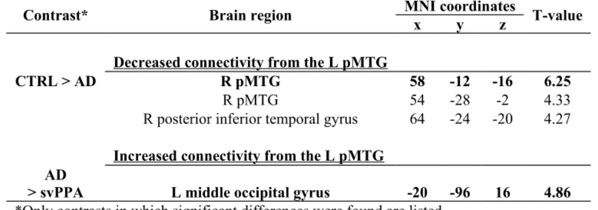
From the left pMTG seed, decreased functional connectivity was observed in AD patients in comparison to CTRL, more precisely with a cluster including the right MTG (Table 2 and Figure 3). In comparison to svPPA patients, AD patients also presented an increased correlation between the left pMTG and a cluster in the left middle occipital gyrus. This result was not significant in the comparison to CTRL, but there was a statistical trend.
From the left IFG seed, no difference in functional connectivity was observed between CTRL and AD patients (Figure 4).
3.2.2 svPPA language network functional connectivity profile
From the left ATL seed, svPPA patients presented decreased functional connectivity in comparison to both groups (Table 3 and Figure 2) but did not present any areas of increased functional connectivity. In comparison to CTRL, svPPA patients presented decreased functional connectivity between the left ATL and a cluster in the left pMTG, a cluster including the right medial orbitofrontal cortex and the right medial superior frontal gyrus, a cluster including the left superior and medial superior frontal gyri and finally, a cluster including the right postcentral and precentral gyri. In comparison to AD patients, svPPA patients showed a significant decrease in functional connectivity between the left ATL and a cluster in the right thalamus as well as a cluster in the left angular gyrus.
From the left pMTG seed, svPPA patients presented both areas of decreased and areas of increased functional connectivity in comparison to CTRL (Table 3 and Figure 3). In comparison to the CTRL, a decreased functional connectivity was observed between the left pMTG and a cluster in the right ATL. svPPA patients also presented increased functional connectivity between the left pMTG and a cluster including the left postcentral gyrus and the left inferior parietal lobule.
From the left IFG seed, svPPA patients presented increased functional connectivity from the left IFG seed in comparison to both CTRL and AD patients (Table 3 and Figure 4) but no decreased connectivity. In comparison to CTRL, svPPA patients presented increased functional connectivity between the left IFG and bilateral middle frontal gyri, the right precuneus as well as in a cluster including the left inferior parietal lobule and the left angular gyrus. In comparison to AD patients, svPPA patients presented increased functional connectivity between the left IFG and the left precuneus.
3.3 Post-hoc analyses: Brain-behavior relationships 3.3.1. Description of the statistical analyses
Our results have raised the question whether the observed functional disconnection across the language network in AD and svPPA patients is significantly associated with the presence of language symptoms. As reported in literature and as observed in our sample of AD and svPPA patients, the main overlapping language symptom in these two populations is a naming impairment (Montembeault et al., 2017; Reilly et al., 2011; Rogers & Friedman, 2008; Sajjadi et al., 2012). Therefore, we investigated if each of our seed region’s connectivity strength, with regards to other brain voxels, were correlated with the Boston Naming Test (BNT) performance across all participants. Three separate analyses were conducted (one for each seed region). First, scores on the BNT were entered as covariate of interest in a multiple regression statistical model, with sex and age as nuisance covariates. A contrast was set to identify voxels in which connectivity strength with the seed ROI correlated with BNT scores. The correlation was tested using a [1] t-contrast, assuming that decreased naming scores would be associated with decreased functional connectivity. A statistical threshold of p < .05 FDR cluster-corrected for multiple comparisons was used.
3.3.2. Results of the correlation between language network functional connectivity and naming abilities
The results showed that the BNT correlated with the functional connectivity between the left ATL and regions showing a functional disconnection with the left ATL in svPPA patients, namely the left and right superior medial frontal gyrus, the left pMTG and the right post- and precentral gyri (p≤.05 FDR cluster-corrected; Figure 5). Performance on the BNT also correlated with the connectivity strength between the left ATL and the right ATL, the right orbitofrontal cortex and the cingulate cortex (p≤.05 FDR cluster-corrected; Figure 5; Supplementary Figure 1).
Performance on the BNT also correlated with the functional connectivity between the left pMTG and the left ATL as well as the left orbitofrontal cortex (p≤.05 FDR cluster-corrected; Figure 5; Supplementary Figure 1). Lowering the statistical threshold, performance on the BNT also correlated with the region showing a lower connectivity strength to the left pMTG in AD (the right pMTG; x=52 y=-4 z=-30; p≤.001 uncorrected) and in svPPA (the right ATL; x=56 y=20 z=-18; p≤.001 uncorrected).
No correlation was observed with the BNT using the left IFG as a seed region.
4. Discussion
In this study, we directly compared the rs-fMRI language network in AD patients, svPPA patients and cognitively unimpaired elderly adults (CTRL). Overall, language impairments and language functional network alterations were observed in both populations. However, some differences were observed in the language profiles between AD and svPPA patients, and functional connectivity changes did not target the same regions in these two groups. In AD patients, the left pMTG was the only key language region showing functional connectivity changes, mainly a reduced interhemispheric functional connectivity with its homotopic counterpart, in comparison to CTRL. In svPPA patients, we observed a functional isolation of the left ATL, both decreases and increases in functional connectivity from the left pMTG and increased functional connectivity from the left IFG. These results will be interpreted within the ventral/dorsal language pathways framework (G. Hickok, 2001; G. Hickok & Poeppel, 2000, 2004; Saur et al., 2008). Our post-hoc analyses also suggest that naming impairments are
associated with the functional disconnections observed across the language network in AD and svPPA patients. Altogether, this study suggests that language impairments in AD and svPPA are associated with distinct functional connectivity patterns across the language network. These distinct profiles have significant implications for our understanding of these two diseases as well as of the neurobiology of language.
4.1 In AD, the left pMTG is the only key language region presenting functional connectivity changes.
Our results in the AD group first support the notion that functional connectivity alterations are not only present within the default-mode network (DMN), but also in the language network (Mascali et al., 2018; Weiler et al., 2014; Whitwell et al., 2015). Consistently with previous studies, we observed lower resting-state functional connectivity in posterior temporal language regions, i.e. the left pMTG (Mascali et al., 2018; Weiler et al., 2014; Whitwell et al., 2015). In the present study, this region was found to be functionally disconnected from its contralateral homologous region, the right MTG. Although not a key language region, this region is consistently reported as functionally connected to key regions of the language network (Hurley et al., 2015). Interestingly, it is not the first time that the role of the dynamics between left- and right-hemisphere posterior temporal regions is noted in the study of language in AD patients. First, this result has also been reported in previous rs-fMRI studies of AD patients (Weiler et al., 2014; Whitwell et al., 2015). Secondly, in a previous study by Nelissen et al. (2007), it was shown that the left posterior temporal regions had a decreased activation during an associative-semantic task, while the homologous right-hemisphere regions showed an increased activation. This right-hemisphere activity correlated positively with accuracy in a naming task and was therefore interpreted as functional reorganisation in AD patients. The brain regions obtained by these authors (51 -9 -15; 54 -24 0) lay very close to those of our study (58 -12 -16; 54, -28, -2). The fact that opposite patterns of activation (i.e. increased versus decreased) are observed in these two homotopic regions during the same associative-semantic task is highly coherent with the altered interhemispheric functional connectivity between these two regions demonstrated in our study.
Another significant change in functional connectivity from the left pMTG in AD patients was observed as an increased connectivity with the left middle occipital gyrus (significant in comparison to svPPA patients, statistical trend in comparison to CTRL). Overall, this intrahemispheric increased functional connectivity could be interpreted as a compensation mechanism accompanying the interhemispheric decreased functional connectivity from the left pMTG in AD patients. A previous rs-fMRI study in AD patients have also found increased functional connectivity in the middle occipital lobe (in relationship with the default-mode and executive control networks) in AD patients with high performance on an executive/language task (De Marco, Duzzi, Meneghello, & Venneri, 2017). These authors suggested that the middle occipital lobe, which is relatively preserved in AD patients, might be prone to brain plasticity and might support other brain networks.
Nonetheless, the interhemispheric decreased functional connectivity appears specific to the left pMTG seed region in AD patients and is not observed in the other two seed regions, in which derived functional connectivity did not differ from CTRL in AD patients. Consistently with a previous rs-fMRI study (Weiler et al., 2014), functional connectivity in anterior language regions, i.e. the left IFG, was preserved in AD patients. One of the novelties of our study is the investigation of the functional network derived from the left ATL, which had never been studied in AD patients. Our study shows that in AD patients, the left ATL remains functionally connected to the remaining part of the language network.
4.2 In svPPA, there is decreased functional connectivity along the ventral language pathway and an increased functional connectivity along the dorsal language pathway.
In the present study, svPPA patients showed a striking functional disconnection of the left ATL with other regions within and outside the language network (bilateral superior medial frontal, left pMTG and right post- and precentral gyri), even taking into account the major atrophy in the left ATL in svPPA patients. This result is consistent with previous rs-fMRI studies (Agosta et al., 2014; Guo et al., 2013) that support the role of the left ATL as the disease-specific epicenter of svPPA (Seeley et al., 2009). In addition to being observed in comparison to CTRL, reduced functional connectivity in
the left ATL was also observed in the direct comparison between svPPA and AD patients (in the right thalamus and left angular gyrus). This suggests that beyond GM atrophy, functional connectivity in the left ATL allow for the discrimination of svPPA and AD patients.
The originality of our study is the investigation of functional connectivity in other language key regions such as the the left pMTG and the left IFG. Even though svPPA generally affects the left ATL to a greater degree, the right ATL also showed decrease functional connectivity with the left pMTG. Nonetheless, in these two key seed regions, we mainly observed an increased functional connectivity in svPPA patients, in comparison to both CTRL and AD patients. More precisely, the left pMTG showed increased connectivity with the left postcentral gyrus and the left IFG showed increased connectivity with the middle frontal gyrus, the precuneus, the left inferior parietal lobule and the left angular gyrus. Only one preliminary rs-fMRI study has also reported increased connectivity in similar regions in svPPA patients, namely between the left IFG and the superior portion of the angular gyrus (Battistella et al., 2018).
Overall, the results observed in the svPPA group can be interpreted within the framework of the ventral/dorsal language pathways (G. Hickok, 2001; G. Hickok & Poeppel, 2000, 2004; Saur et al., 2008). According to this model, linguistic processing of sound to meaning would be subserved by a ventral pathway, connecting the middle temporal lobe to the ventrolateral prefrontal cortex. Sensory-motor mapping of sound to articulation, on the other hand, would be subserved by a dorsal pathway connecting the superior temporal lobe and premotor cortices in the frontal lobe. In the present study, decreases in functional connectivity were observed in the ventral language pathway, whereas increases in functional connectivity were observed in the dorsal language pathway. These observations have significant clinical implications, for example in the understanding of symptoms presented by svPPA patients (see section 4.4).
4.3 Language network functional connectivity alterations in svPPA and AD are generally consistent with white matter fiber damage.
Brain regions that present correlated changes in BOLD signal during rest are also structurally connected via white matter bundles (Greicius, Supekar, Menon, & Dougherty, 2009; Lemaire et al., 2013; Morgan, Mishra, Newton, Gore, & Ding, 2009;
Turken & Dronkers, 2011). Combining the present rs-fMRI results with previously obtained diffusion imaging results can enrich our understanding of the language brain network in AD and svPPA. Overall, these observations suggest a good agreement between our study using rs-fMRI and previous diffusion imaging studies.
On one hand, a previous meta-analysis of diffusion imaging studies in AD has shown that white matter (WM) fiber alterations are widespread (Sexton, Kalu, Filippini, Mackay, & Ebmeier, 2011). These authors reported large effect sizes (reduced fractional anisotropy and increased mean diffusivity) in the uncinate fasciculus, the superior longitudinal fasciculus and the posterior cingulum, and medium effect sizes in the splenium of the corpus callosum, as well as in temporal and parietal white matter (Sexton et al., 2011). In our study, the interhemispheric functional disconnection between homotopic pMTG appears coherent with the reduced structural connectivity within the splenium of the corpus callosum, which connects bilateral temporo-parietal regions. Future studies should investigate the association between language symptoms and WM damage in the splenium in AD patients.
On the other hand, WM damage in svPPA patients is predominantly observed in the ventral tracts that pass through the temporal lobe, more precisely the inferior longitudinal fasciculus, the uncinate fasciculus and the temporal segment of the arcuate fasciculus (Acosta-Cabronero et al., 2011; Agosta et al., 2013; Agosta et al., 2010; Galantucci et al., 2011). Consistent with these observations, in our study, a functional disconnection was observed between the left ATL and the left pMTG, two regions that are located along the inferior longitudinal fasciculus. Conversely, the dorsal frontoparietal white matter tract that do not involve the temporal lobes are relatively spared in svPPA (Acosta-Cabronero et al., 2011; Agosta et al., 2013; Agosta et al., 2010; Galantucci et al., 2011). Nonetheless, no structural connectivity study has previously found an increased structural connectivity in the dorsal pathway, similar to what we observed in our functional connectivity results. Future studies using multimodal neuroimaging techniques should be conducted to better understand these results.
4.4 Language network functional connectivity alterations in svPPA and AD are correlated with naming impairments in these patients.
Amongst the language impairments presented by AD and svPPA patients, the main overlapping symptom is a naming impairment. For this reason, we correlated naming performance with functional connectivity from our three key language seed regions in our sample. Our results have showed that naming impairments are indeed associated with the functional disconnections observed across the language network in AD and svPPA patients.
In addition, the functional connectivity profile observed in each population is theoretically consistent with the observed language profile. First, AD patients presented significant impairments in naming and verbal fluency tasks. This result is consistent with the reduced functional connectivity from the left pMTG seed region in our study. Indeed, the role of this brain region in lexico-semantic retrieval has been demonstrated in previous neuroimaging studies (Davey et al., 2016; Gold et al., 2006; Gregory Hickok & Poeppel, 2007; Noppeney, Phillips, & Price, 2004). In this regard, there has been a longstanding debate in recent years concerning the nature of language impairments (and more specifically naming impairments) in AD patients. While some authors argue that naming deficits in AD are caused by a semantic impairment (stored information is lost), others have suggested that they are caused by an impaired lexico-semantic access (access to stored information is dysfunctional) (Lambon Ralph, 2014). In the present study, the functional disconnection of the left pMTG and its correlation with poor naming performance support the notion that naming impairments are associated, at least in part, by a lexico-semantic access impairment in AD patients (Joubert et al., 2010; Montembeault et al., 2017; Rogers & Friedman, 2008).
Second, svPPA patients showed significant impairments on all language and semantic memory tasks, and these impairments were overall more severe than in AD patients. This is consistent with the functional disconnection of the ventral language pathway in svPPA patients. These results highlight the fact that the mechanisms underlying language impairments are different in svPPA and AD. The profile that we observed in svPPA patients is consistent with a core semantic impairment, in which stored conceptual information is lost, which is highly consistent with previous studies (Gorno-Tempini et al., 2011; Joyal et al., 2017; Montembeault et al., 2017; Reilly et al., 2011; Rogers & Friedman, 2008; Sajjadi et al., 2012). Furthermore, the
preserved/increased functional connectivity in the remaining parts of the language network (dorsal language pathway) is consistent with the relative sparing of other language functions sustained by these regions, such as motor speech, phonology and speech rate (Gorno-Tempini et al., 2011; S. M. Wilson et al., 2010).
Future studies with a larger sample and additional language tasks assessing the domains associated with the dorsal language pathway could further clarify the relationships between functional connectivity and language function in these patients’ populations. Yet, our post-hoc analyses represent a preliminary confirmation that the changes in the connectivity of the language network are implicated in language impairments in these patients.
4.5 Limitations
This study could be considered a preliminary report with limited sample size. Replication with a larger sample are required to confirm these results. Nonetheless, despite the low sample size, significant between-group differences were highlighted in the present study, and there is a very limited chance that these results represent false-positive results given that stringent motion correction, voxelwise gray matter volume correction and statistical analyses corrected for multiple comparisons were used in this study. Nonetheless, false negatives might have occurred given the low statistical power that is associated with such a low sample size.
There are also significant challenges and limitations inherent to comparing different neurodegenerative populations, especially svPPA and AD. We believe that there are no perfect control variables related to the disease stage in the study of svPPA vs AD, given the fact that these diseases are different on many aspects. Some authors have previously used disease duration (at the time of the study) as a control variable, but a reliable estimate of disease duration was not available for all patients included in the present study. Nonetheless, svPPA and AD are characterized by largely different mean age of onset (lower in svPPA) and mean total disease duration (longer in svPPA), which suggest different disease progression (J. R. Hodges et al., 2010; Kertesz, Blair, McMonagle, & Munoz, 2007; Kertesz, Jesso, Harciarek, Blair, & McMonagle, 2010; Tom et al., 2015; Waring, Doody, Pavlik, Massman, & Chan, 2005). Disease duration (at the time of the study) is therefore not equivalent nor comparable between these two
patients’ populations. In the present study, we decided to control for mean age. Although this is also not a perfect control variable in the study of these two populations, age is independent of the type of disease and remains an important variable to control for. Furthermore, the two patient groups presented equivalent scores on the general cognition test (MMSE) and we conducted an in-depth neuropsychological assessment in order to extensively characterize their clinical profiles.
4.6 Conclusion
In conclusion, the findings reported here contribute to our understanding of the functional connectivity changes that take place in AD and svPPA. This is the first study to provide a full picture of the functional connectivity in all key regions of the language network simultaneously, and to directly compare these two populations of patients. The results have significant clinical implications, highlighting the fact that functional connectivity in the language network allows for the discrimination of these two patient population, even controlling for gray matter atrophy. Using a network-based approach, we were also able to highlight the fact that similar language impairments in AD and svPPA patients are associated with different brain mechanisms in each disease.
Acknowledgements
The present work is supported by the Alzheimer Society of Canada. MM is supported by Alzheimer Society of Canada and Fonds de Recherche du Québec Santé (FRQ-S) doctoral awards. MB is supported by Canadian Institutes of Health Research (CIHR) and FRQ-S doctoral awards. MAW is supported by the Réseau Québécois de Recherche sur le Vieillissement (RQRV). RL is supported by Fondation du CHU de Québec and Société Alzheimer de Québec. SMB is supported by a Chercheur-boursier Junior 2 FRQ-S award. We would like to thank all participants for taking part in this research.
References
Acosta-Cabronero, J., Patterson, K., Fryer, T. D., Hodges, J. R., Pengas, G., Williams, G. B., & Nestor, P. J. (2011). Atrophy, hypometabolism and white matter abnormalities in semantic dementia tell a coherent story. Brain, 134(7), 2025-2035. doi: 10.1093/brain/awr119
Agosta, F., Galantucci, S., Canu, E., Cappa, S. F., Magnani, G., Franceschi, M., . . . Filippi, M. (2013). Disruption of structural connectivity along the dorsal and ventral language pathways in patients with nonfluent and semantic variant primary progressive aphasia: a DT MRI study and a literature review. Brain
Lang, 127(2), 157-166. doi: 10.1016/j.bandl.2013.06.003
Agosta, F., Galantucci, S., Valsasina, P., Canu, E., Meani, A., Marcone, A., . . . Filippi, M. (2014). Disrupted brain connectome in semantic variant of primary progressive aphasia. Neurobiol Aging, 35(11), 2646-2655. doi: 10.1016/j.neurobiolaging.2014.05.017
Agosta, F., Henry, R. G., Migliaccio, R., Neuhaus, J., Miller, B. L., Dronkers, N. F., . . . Gorno-Tempini, M. L. (2010). Language networks in semantic dementia. Brain,
133(1), 286-299. doi: 10.1093/brain/awp233
Apostolova, L. G., Lu, P., Rogers, S., Dutton, R. A., Hayashi, K. M., Toga, A. W., . . . Thompson, P. M. (2008). 3D mapping of language networks in clinical and pre-clinical Alzheimer’s disease. Brain and Language, 104(1), 33-41. doi: 10.1016/j.bandl.2007.03.008
Badhwar, A., Tam, A., Dansereau, C., Orban, P., Hoffstaedter, F., & Bellec, P. (2017). Resting-state network dysfunction in Alzheimer's disease: A systematic review and meta-analysis. Alzheimer's & Dementia: Diagnosis, Assessment & Disease
Monitoring, 8, 73-85. doi: 10.1016/j.dadm.2017.03.007
Battistella, G., Henry, M., Gesierich, B., Welch, A., Shwe, W., Miller, Z., . . . Gorno-Tempini, M. L. (2018). Connectivity changes of the inferior parietal lobule in
semantic primary progressive aphasia. Paper presented at the Organization for
Human Brain Mapping, Singapore.
Behzadi, Y., Restom, K., Liau, J., & Liu, T. T. (2007). A Component Based Noise Correction Method (CompCor) for BOLD and Perfusion Based fMRI.
NeuroImage, 37(1), 90-101. doi: 10.1016/j.neuroimage.2007.04.042
Benton, A. L., Hamsher, K. D., Varney, N. R., & Spreen, O. (1983). Contributions to
neuropsychological assessment: Tests. New York, NY: Oxford University Press.
Binney, R. J., Embleton, K. V., Jefferies, E., Parker, G. J. M., & Lambon Ralph, M. A. (2010). The Ventral and Inferolateral Aspects of the Anterior Temporal Lobe Are Crucial in Semantic Memory: Evidence from a Novel Direct Comparison of Distortion-Corrected fMRI, rTMS, and Semantic Dementia. Cerebral Cortex,
20(11), 2728-2738. doi: 10.1093/cercor/bhq019
Brambati, S. M., Amici, S., Racine, C. A., Neuhaus, J., Miller, Z. A., Ogar, J. M., . . . Gorno-Tempini, M. L. (2015). Longitudinal gray matter contraction in three variants of primary progressive aphasia: A tenser-based morphometry study.
Brambati, S. M., Myers, D., Wilson, A., Rankin, K. P., Allison, S. C., Rosen, H. J., . . . Gorno-Tempini, M. L. (2006). The Anatomy of Category-specific Object Naming in Neurodegenerative Diseases. Journal of Cognitive Neuroscience, 18(10), 1644-1653. doi: 10.1162/jocn.2006.18.10.1644
Brambati, S. M., Rankin, K. P., Narvid, J., Seeley, W. W., Dean, D., Rosen, H. J., . . . Gorno-Tempini, M. L. (2009). Atrophy progression in semantic dementia with asymmetric temporal involvement: a tensor-based morphometry study. Neurobiol
Aging, 30(1), 103-111. doi: 10.1016/j.neurobiolaging.2007.05.014
Buckner, R. L., Snyder, A. Z., Shannon, B. J., LaRossa, G., Sachs, R., Fotenos, A. F., . . . Mintun, M. A. (2005). Molecular, structural, and functional characterization of Alzheimer's disease: evidence for a relationship between default activity, amyloid, and memory. J Neurosci, 25(34), 7709-7717. doi: 10.1523/jneurosci.2177-05.2005
Chedid, G., Wilson, M. A., Provost, J.-S., Joubert, S., Rouleau, I., & Brambati, S. M. (2016). Differential Involvement of the Anterior Temporal Lobes in Famous People Semantics. [Original Research]. Frontiers in Psychology, 7(1333). doi: 10.3389/fpsyg.2016.01333
Damasio, H., Tranel, D., Grabowski, T., Adolphs, R., & Damasio, A. (2004). Neural systems behind word and concept retrieval. Cognition, 92(1), 179-229. doi: 10.1016/j.cognition.2002.07.001
Davey, J., Thompson, H. E., Hallam, G., Karapanagiotidis, T., Murphy, C., De Caso, I., . . . Jefferies, E. (2016). Exploring the role of the posterior middle temporal gyrus in semantic cognition: Integration of anterior temporal lobe with executive
processes. NeuroImage, 137, 165-177. doi:
https://doi.org/10.1016/j.neuroimage.2016.05.051
De Marco, M., Duzzi, D., Meneghello, F., & Venneri, A. (2017). Cognitive Efficiency in Alzheimer's Disease is Associated with Increased Occipital Connectivity. J
Alzheimers Dis, 57(2), 541-556. doi: 10.3233/jad-161164
Desgranges, B., Matuszewski, V., Piolino, P., Chételat, G., Mézenge, F., Landeau, B., . . . Eustache, F. (2007). Anatomical and functional alterations in semantic dementia: A voxel-based MRI and PET study. Neurobiology of Aging, 28(12), 1904-1913. doi: 10.1016/j.neurobiolaging.2006.08.006
Diehl, J., Grimmer, T., Drzezga, A., Riemenschneider, M., Förstl, H., & Kurz, A. (2004). Cerebral metabolic patterns at early stages of frontotemporal dementia and semantic dementia. A PET study. Neurobiology of Aging, 25(8), 1051-1056. doi: 10.1016/j.neurobiolaging.2003.10.007
Domoto-Reilly, K., Sapolsky, D., Brickhouse, M., & Dickerson, B. C. (2012). Naming impairment in Alzheimer's disease is associated with left anterior temporal lobe atrophy. Neuroimage, 63(1), 348-355. doi: 10.1016/j.neuroimage.2012.06.018 Dronkers, N. F., Ivanova, M. V., & Baldo, J. V. (2017). What Do Language Disorders
Reveal about Brain-Language Relationships? From Classic Models to Network Approaches. J Int Neuropsychol Soc, 23(9-10), 741-754. doi: 10.1017/s1355617717001126
Ferstl, E. C., Neumann, J., Bogler, C., & von Cramon, D. Y. (2008). The extended language network: a meta-analysis of neuroimaging studies on text comprehension. Hum Brain Mapp, 29(5), 581-593. doi: 10.1002/hbm.20422
Folstein, M. F., Folstein, S. E., & McHugh, P. R. (1975). "Mini-mental state". A practical method for grading the cognitive state of patients for the clinician. J Psychiatr
Res, 12(3), 189-198.
Galantucci, S., Tartaglia, M. C., Wilson, S. M., Henry, M. L., Filippi, M., Agosta, F., . . . Gorno-Tempini, M. L. (2011). White matter damage in primary progressive aphasias: a diffusion tensor tractography study. Brain, 134(Pt 10), 3011-3029. doi: 10.1093/brain/awr099
Geschwind, N. (1970). The Organization of Language and the Brain. Science, 170(3961), 940-944. doi: 10.1126/science.170.3961.940
Gesierich, B., Jovicich, J., Riello, M., Adriani, M., Monti, A., Brentari, V., . . . Gorno-Tempini, M. L. (2011). Distinct Neural Substrates for Semantic Knowledge and Naming in the Temporoparietal Network. Cerebral Cortex, 22(10). doi: 10.1093/cercor/bhr286
Gold, B. T., Balota, D. A., Jones, S. J., Powell, D. K., Smith, C. D., & Andersen, A. H. (2006). Dissociation of automatic and strategic lexical-semantics: functional magnetic resonance imaging evidence for differing roles of multiple frontotemporal regions. J Neurosci, 26(24), 6523-6532. doi: 10.1523/jneurosci.0808-06.2006
Gorno-Tempini, M. L., Hillis, A. E., Weintraub, S., Kertesz, A., Mendez, M., Cappa, S. F., . . . Grossman, M. (2011). Classification of primary progressive aphasia and its variants. Neurology, 76(11), 1006-1014. doi: 10.1212/WNL.0b013e31821103e6 Greicius, M. D., Supekar, K., Menon, V., & Dougherty, R. F. (2009). Resting-State
Functional Connectivity Reflects Structural Connectivity in the Default Mode Network. Cerebral Cortex, 19(1), 72-78. doi: 10.1093/cercor/bhn059
Grossman, M., McMillan, C., Moore, P., Ding, L., Glosser, G., Work, M., & Gee, J. (2004). What’s in a name: voxel based morphometric analyses of MRI and‐ naming difficulty in Alzheimer’s disease, frontotemporal dementia and corticobasal degeneration. Brain, 127(3), 628-649. doi: 10.1093/brain/awh075 Guo, C. C., Gorno-Tempini, M. L., Gesierich, B., Henry, M., Trujillo, A., Shany-Ur,
T., . . . Seeley, W. W. (2013). Anterior temporal lobe degeneration produces widespread network-driven dysfunction. Brain, 136(Pt 10), 2979-2991. doi: 10.1093/brain/awt222
Heilman, K. (1972). Anomic aphasia following anterior temporal lobectomy. Trans Am
Neurol Assoc, 97, 291-293.
Hickok, G. (2001). Functional anatomy of speech perception and speech production: psycholinguistic implications. J Psycholinguist Res, 30(3), 225-235.
Hickok, G., & Poeppel, D. (2000). Towards a functional neuroanatomy of speech perception. Trends Cogn Sci, 4(4), 131-138.
Hickok, G., & Poeppel, D. (2004). Dorsal and ventral streams: a framework for understanding aspects of the functional anatomy of language. Cognition, 92(1-2), 67-99. doi: 10.1016/j.cognition.2003.10.011
Hickok, G., & Poeppel, D. (2007). The cortical organization of speech processing. [Perspective]. Nature Reviews Neuroscience, 8, 393. doi: 10.1038/nrn2113
Hirono, N., Mori, E., Ishii, K., Imamura, T., Tanimukai, S., Kazui, H., . . . Sasaki, M. (2001). Neuronal Substrates for Semantic Memory: A Positron Emission
Tomography Study in Alzheimer’s Disease. Dementia and Geriatric Cognitive
Disorders, 12(1), 15-21. doi: 10.1159/000051231
Hodges, J. R., Mitchell, J., Dawson, K., Spillantini, M. G., Xuereb, J. H., McMonagle, P., . . . Patterson, K. (2010). Semantic dementia: demography, familial factors and survival in a consecutive series of 100 cases. Brain, 133(Pt 1), 300-306. doi: 10.1093/brain/awp248
Hodges, J. R., & Patterson, K. (1995). Is semantic memory consistently impaired early in the course of Alzheimer's disease? Neuroanatomical and diagnostic implications.
Neuropsychologia, 33(4), 441-459. doi: http://dx.doi.org/10.1016/0028-3932(94)00127-B
Hodges, J. R., Patterson, K., Oxbury, S., & Funnell, E. (1992). Semantic dementia: Progressive fluent aphasia with temporal lobe atrophy. Brain, 115(6), 1783-1806. doi: 10.1093/brain/115.6.1783
Howard, D., & Patterson, K. (1992). Pyramids and palm trees: A test of semantic access
from pictures and words. Bury St. Edmunds, England: Thames Valley Test
Company.
Huff, F. J., Corkin, S., & Growdon, J. H. (1986). Semantic impairment and anomia in Alzheimer's disease. Brain and Language, 28(2), 235-249. doi: 10.1016/0093-934X(86)90103-3
Hurley, R. S., Bonakdarpour, B., Wang, X., & Mesulam, M. M. (2015). Asymmetric Connectivity between the Anterior Temporal Lobe and the Language Network.
Journal of cognitive neuroscience, 27(3), 464-473. doi: 10.1162/jocn_a_00722
Joanette, Y., Ska, B., & Côté, H. (2004). Protocole Montreal d’Evaluation de la
Communication MEC. Isbergues, France: Ortho Editions.
Joubert, S., Brambati, S. M., Ansado, J., Barbeau, E. J., Felician, O., Didic, M., . . . Kergoat, M. J. (2010). The cognitive and neural expression of semantic memory impairment in mild cognitive impairment and early Alzheimer's disease.
Neuropsychologia, 48(4), 978-988. doi: 10.1016/j.neuropsychologia.2009.11.019
Joyal, M., Brambati, S. M., Laforce, R. J., Montembeault, M., Boukadi, M., Rouleau, I., . . . Wilson, M. A. (2017). The Role of the Left Anterior Temporal Lobe for Unpredictable and Complex Mappings in Word Reading. Frontiers in
Psychology, 8, 517. doi: 10.3389/fpsyg.2017.00517
Kaplan, E., Goodglass, H., & Weintraub, S. (1983). The Boston Naming Test. . Philadelphia: Lea and Febiger.
Kertesz, A., Blair, M., McMonagle, P., & Munoz, D. G. (2007). The diagnosis and course of frontotemporal dementia. Alzheimer Dis Assoc Disord, 21(2), 155-163. doi: 10.1097/WAD.0b013e31806547eb
Kertesz, A., Jesso, S., Harciarek, M., Blair, M., & McMonagle, P. (2010). What is semantic dementia?: a cohort study of diagnostic features and clinical boundaries.
Arch Neurol, 67(4), 483-489. doi: 10.1001/archneurol.2010.55
Lambon Ralph, M. A. (2014). Neurocognitive insights on conceptual knowledge and its breakdown. Philosophical Transactions of the Royal Society of London B:
Biological Sciences, 369(1634). doi: 10.1098/rstb.2012.0392
Lars, F., Stefan, K., Stefan, T., Oliver, P., Lutz, F., Johannes, P., . . . Michael, H. (2011). Left Anterior Temporal Lobe Sustains Naming in Alzheimers Dementia and Mild
Cognitive Impairment. Current Alzheimer Research, 8(8), 893-901. doi: 10.2174/156720511798192673
Lars, F., Timo, S. S., Michael, H., & Philipp, T. M. (2016). Left Anterior Temporal Glucose Metabolism and not Amyloid-beta Load Predicts Naming Impairment in Alzheimer’s Disease. Current Alzheimer Research, 13(6), 678-681. doi: 10.2174/1567205013666160322141955
Lemaire, J. J., Golby, A., Wells, W. M., 3rd, Pujol, S., Tie, Y., Rigolo, L., . . . Kikinis, R. (2013). Extended Broca's area in the functional connectome of language in adults: combined cortical and subcortical single-subject analysis using fMRI and DTI tractography. Brain Topogr, 26(3), 428-441. doi: 10.1007/s10548-012-0257-7 Mascali, D., DiNuzzo, M., Serra, L., Mangia, S., Maraviglia, B., Bozzali, M., & Giove, F.
(2018). Disruption of Semantic Network in Mild Alzheimer's Disease Revealed by Resting-State fMRI. Neuroscience, 371, 38-48. doi: 10.1016/j.neuroscience.2017.11.030
Mathotaarachchi, S., Wang, S., Shin, M., Pascoal, T. A., Benedet, A. L., Kang, M. S., . . . Rosa-Neto, P. (2016). VoxelStats: A MATLAB Package for Multi-Modal Voxel-Wise Brain Image Analysis. [Methods]. Frontiers in Neuroinformatics, 10(20). doi: 10.3389/fninf.2016.00020
McKhann, G. M., Knopman, D. S., Chertkow, H., Hyman, B. T., Jack, C. R., Jr., Kawas, C. H., . . . Phelps, C. H. (2011). The diagnosis of dementia due to Alzheimer's disease: recommendations from the National Institute on Aging-Alzheimer's Association workgroups on diagnostic guidelines for Alzheimer's disease.
Alzheimers & Dementia, 7(3), 263-269. doi: 10.1016/j.jalz.2011.03.005
Melrose, R. J., Campa, O. M., Harwood, D. G., Osato, S., Mandelkern, M. A., & Sultzer, D. L. (2009). The neural correlates of naming and fluency deficits in Alzheimer's disease: an FDG-PET study. International Journal of Geriatric Psychiatry, 24(8), 885-893. doi: 10.1002/gps.2229
Mesulam, M. M., Wieneke, C., Hurley, R., Rademaker, A., Thompson, C. K., Weintraub, S., & Rogalski, E. J. (2013). Words and objects at the tip of the left temporal lobe in primary progressive aphasia. Brain, 136(2), 601-618. doi: 10.1093/brain/aws336
Meyers, J. E., & Meyers, K. R. (1995). Rey complex figure test and recognition trial.:
Professional manual: Psychological Assessment Resource.
Montembeault, M., Brambati, S. M., Joubert, S., Boukadi, M., Chapleau, M., Laforce, R. J., . . . Rouleau, I. (2017). Naming unique entities in the semantic variant of primary progressive aphasia and Alzheimer's disease: Towards a better understanding of the semantic impairment. Neuropsychologia, 95, 11-20. doi: 10.1016/j.neuropsychologia.2016.12.009
Morgan, V. L., Mishra, A., Newton, A. T., Gore, J. C., & Ding, Z. (2009). Integrating functional and diffusion magnetic resonance imaging for analysis of structure-function relationship in the human language network. PLoS One, 4(8), e6660. doi: 10.1371/journal.pone.0006660
Muschelli, J., Nebel, M. B., Caffo, B. S., Barber, A. D., Pekar, J. J., & Mostofsky, S. H. (2014). Reduction of motion-related artifacts in resting state fMRI using aCompCor. Neuroimage, 96, 22-35. doi: 10.1016/j.neuroimage.2014.03.028
Nelissen, N., Dupont, P., Vandenbulcke, M., Tousseyn, T., Peeters, R., & Vandenberghe, R. (2011). Right hemisphere recruitment during language processing in frontotemporal lobar degeneration and Alzheimer's disease. J Mol Neurosci,
45(3), 637-647. doi: 10.1007/s12031-011-9603-6
Nelissen, N., Vandenbulcke, M., Fannes, K., Verbruggen, A., Peeters, R., Dupont, P., . . . Vandenberghe, R. (2007). Aβ amyloid deposition in the language system and how the brain responds. Brain, 130(8), 2055-2069. doi: 10.1093/brain/awm133
Noppeney, U., Phillips, J., & Price, C. (2004). The neural areas that control the retrieval and selection of semantics. Neuropsychologia, 42(9), 1269-1280. doi:
https://doi.org/10.1016/j.neuropsychologia.2003.12.014
Osterrieth, P. A. (1944). Le test de copie d’une figure complexe; contribution à l'étude de la perception et de la mémoire. [Test of copying a complex figure; contribution to the study of perception and memory.]. Archives de Psychologie, 30, 206-356. Qualls, C. E., Bliwise, N. G., & Stringer, A. Y. (2000). Short Forms of The Benton
Judgment of Line Orientation Test: Development and Psychometric Properties.
Archives of Clinical Neuropsychology, 15(2), 159-163. doi: http://dx.doi.org/10.1016/S0887-6177(98)00043-2
Regard, M. (1981). Cognitive Rigidity and Flexibility: A Neuropsychological Study: University of Victoria (B.C.).
Reilly, J., Peelle, J. E., Antonucci, S. M., & Grossman, M. (2011). Anomia as a marker of distinct semantic memory impairments in Alzheimer's disease and semantic dementia. Neuropsychology, 25(4), 413-426. doi: 10.1037/a0022738
Rey, A. (1964). L’examen clinique en psychologie. Paris: Presses Universitaires de France.
Rogers, S. L., & Friedman, R. B. (2008). The underlying mechanisms of semantic memory loss in Alzheimer's disease and semantic dementia. Neuropsychologia,
46(1), 12-21. doi: http://dx.doi.org/10.1016/j.neuropsychologia.2007.08.010
Rouleau, I., Salmon, D. P., Butters, N., Kennedy, C., & McGuire, K. (1992). Quantitative and qualitative analyses of clock drawings in Alzheimer's and Huntington's disease. Brain and Cognition, 18(1), 70-87.
Sajjadi, S. A., Patterson, K., Tomek, M., & Nestor, P. J. (2012). Abnormalities of connected speech in semantic dementia vs Alzheimer's disease. Aphasiology,
26(6), 847-866. doi: 10.1080/02687038.2012.654933
Saur, D., Kreher, B. W., Schnell, S., Kümmerer, D., Kellmeyer, P., Vry, M.-S., . . . Weiller, C. (2008). Ventral and dorsal pathways for language. Proceedings of the
National Academy of Sciences, 105(46), 18035-18040. doi:
10.1073/pnas.0805234105
Schwartz, M. F., Kimberg, D. Y., Walker, G. M., Faseyitan, O., Brecher, A., Dell, G. S., & Coslett, H. B. (2009). Anterior temporal involvement in semantic word retrieval: voxel-based lesion-symptom mapping evidence from aphasia. Brain,
132(12), 3411-3427. doi: 10.1093/brain/awp284
Seeley, W. W., Crawford, R. K., Zhou, J., Miller, B. L., & Greicius, M. D. (2009). Neurodegenerative diseases target large-scale human brain networks. Neuron,
62(1), 42-52. doi: 10.1016/j.neuron.2009.03.024
Sexton, C. E., Kalu, U. G., Filippini, N., Mackay, C. E., & Ebmeier, K. P. (2011). A meta-analysis of diffusion tensor imaging in mild cognitive impairment and
Alzheimer's disease. Neurobiol Aging, 32(12), 2322.e2325-2318. doi: 10.1016/j.neurobiolaging.2010.05.019
Snowden, J. S., Goulding, P. J., & Neary, D. (1989). Semantic dementia: A form of circumscribed cerebral atrophy. Behavioural Neurology.
Teipel, S. J., Willoch, F., Ishii, K., Bürger, K., Drzezga, A., Engel, R., . . . Hampel, H. (2006). Resting state glucose utilization and the CERAD cognitive battery in patients with Alzheimer's disease. Neurobiology of Aging, 27(5), 681-690. doi: 10.1016/j.neurobiolaging.2005.03.015
Tom, S. E., Hubbard, R. A., Crane, P. K., Haneuse, S. J., Bowen, J., McCormick, W. C., . . . Larson, E. B. (2015). Characterization of dementia and Alzheimer's disease in an older population: updated incidence and life expectancy with and without dementia. Am J Public Health, 105(2), 408-413. doi: 10.2105/ajph.2014.301935 Tomasi, D., & Volkow, N. D. (2012). Resting functional connectivity of language
networks: characterization and reproducibility. Mol Psychiatry, 17(8), 841-854. doi: 10.1038/mp.2011.177
Tombaugh, T. N. (2004). Trail Making Test A and B: Normative data stratified by age and education. Archives of Clinical Neuropsychology, 19(2), 203-214. doi:
http://dx.doi.org/10.1016/S0887-6177(03)00039-8
Tremblay, P., & Dick, A. S. (2016). Broca and Wernicke are dead, or moving past the classic model of language neurobiology. Brain Lang, 162, 60-71. doi: 10.1016/j.bandl.2016.08.004
Turken, A. U., & Dronkers, N. F. (2011). The neural architecture of the language comprehension network: converging evidence from lesion and connectivity analyses. Frontiers Systems Neuroscience, 5, 1. doi: 10.3389/fnsys.2011.00001 Ueno, T., Saito, S., Rogers, Timothy T., & Lambon Ralph, Matthew A. (2011).
Lichtheim 2: Synthesizing Aphasia and the Neural Basis of Language in a Neurocomputational Model of the Dual Dorsal-Ventral Language Pathways.
Neuron, 72(2), 385-396. doi: 10.1016/j.neuron.2011.09.013
Vandenbulcke, M., Peeters, R., Dupont, P., Van Hecke, P., & Vandenberghe, R. (2007). Word Reading and Posterior Temporal Dysfunction in Amnestic Mild Cognitive Impairment. Cerebral Cortex, 17(3), 542-551. doi: 10.1093/cercor/bhj179
Verma, M., & Howard, R. J. (2012). Semantic memory and language dysfunction in early Alzheimer's disease: a review. Int J Geriatr Psychiatry, 27(12), 1209-1217. doi: 10.1002/gps.3766
Waring, S. C., Doody, R. S., Pavlik, V. N., Massman, P. J., & Chan, W. (2005). Survival among patients with dementia from a large multi-ethnic population. Alzheimer
Dis Assoc Disord, 19(4), 178-183.
Wechsler, D. (1997). WAIS-III administration and scoring manual. San Antonio, Texas: The Psychological Corporation.
Weiler, M., Fukuda, A., Massabki, L. H., Lopes, T. M., Franco, A. R., Damasceno, B. P., . . . Balthazar, M. L. (2014). Default mode, executive function, and language functional connectivity networks are compromised in mild Alzheimer's disease.
Current Alzheimer Research, 11(3), 274-282. doi:
Whitfield-Gabrieli, S., & Nieto-Castanon, A. (2012). Conn: a functional connectivity toolbox for correlated and anticorrelated brain networks. Brain Connect, 2(3), 125-141. doi: 10.1089/brain.2012.0073
Whitwell, J. L., Jones, D. T., Duffy, J. R., Strand, E. A., Machulda, M. M., Przybelski, S. A., . . . Josephs, K. A. (2015). Working memory and language network dysfunction in logopenic aphasia: a task-free fMRI comparison to Alzheimer’s dementia. Neurobiology of aging, 36(3), 1245-1252. doi: 10.1016/j.neurobiolaging.2014.12.013
Wilson, M. A., Joubert, S., Ferre, P., Belleville, S., Ansaldo, A. I., Joanette, Y., . . . Brambati, S. M. (2012). The role of the left anterior temporal lobe in exception word reading: reconciling patient and neuroimaging findings. Neuroimage, 60(4), 2000-2007. doi: 10.1016/j.neuroimage.2012.02.009
Wilson, S. M., Henry, M. L., Besbris, M., Ogar, J. M., Dronkers, N. F., Jarrold, W., . . . Gorno-Tempini, M. L. (2010). Connected speech production in three variants of primary progressive aphasia. Brain, 133(Pt 7), 2069-2088. doi: 10.1093/brain/awq129
Zahn, R., Juengling, F., Bubrowski, P., Jost, E., Dykierek, P., Talazko, J., & Huell, M. (2004). Hemispheric asymmetries of hypometabolism associated with semantic memory impairment in Alzheimer's disease: a study using positron emission tomography with fluorodeoxyglucose-F18. Psychiatry Research: Neuroimaging,
Table 1 : Demographic and neuropsychological/language characteristics of CTRL, AD
patients and svPPA patients.
CTRLs
(n = 11) (n = 10)AD (n = 12)svPPA valuep comparisonGroup Demographics
Gender (F/M) 4/7 5/5 4/8 NA NA
Age (in years) 65.7 (8.1) 69.8 (8.5) 65.9
(10.8) = .528 CTRL = AD =svPPA Education (in years) 16.5 (3.1) 15.7 (3.0) 16.8
(4.3) = .784 CTRL = AD =svPPA
Neuropsychological assessment Global cognitive status
MMSE 28.9 (0.7) 24.9 (3.1) 25.1 (2.6) < .001 CTRL > AD =svPPA Episodic memory RCFT (Immediate recall) 19.2 (4.3) 5.5 (3.5) 10.1 (6.0) < .001 CTRL > AD = svPPA RCFT (Delayed recall) 20.3 (4.6) 4.8 (4.1) 9.8 (6.5) < .001 CTRL > AD = svPPA RAVLT (Trials 1-5) 52.9 (7.4) 29.0 (6.3) 29.5 (7.8) < .001 CTRL > AD =svPPA RAVLT (Immediate recall) 11.1 (2.6) 2.9 (2.6) 4.8 (2.3) < .001 CTRL > AD =
svPPA RAVLT (Delayed recall) 10.8 (2.7) 2.1 (3.2) 4.7 (2.7) < .001 CTRL > AD =
svPPA RAVLT (Recognition) 47.2 (1.6) 30.4 (7.3) 41.0
(5.7) < .001 CTRL = svPPA> AD
Language and semantic memory
Boston Naming Test 49.9 (4.4) 42.9 (9.8) 12.4
(7.8) < .001 CTRL > AD >svPPA Pyramids and Palm Trees Test 50.2 (1.5) 47.7 (2.4) 32.0
(11.2) < .001 CTRL = AD >svPPA Free Fluency 67.6 (17.3) 39.0 (14.9) 30.7 (11.2) < .001 CTRL > AD = svPPA Letter Fluency - P 27.0 (8.8) 19.3 (7.9) (5.9)12.6 < .001 CTRL > svPPACTRL = AD AD = svPPA Semantic Fluency - Clothing 26.3 (4.4) 13.5 (7.3) 9.3 (7.0) < .001 CTRL > AD =
svPPA Similarities subtest - WAIS-III 18.2 (3.6) 15.1 (3.7) 5.8 (2.9) < .001 CTRL = AD >
svPPA
Visual perception
Benton Line Orientation test 27.5 (2.2) 23.8 (7.4) 26.6
(2.4) = .199 CTRL = AD =svPPA Benton facial recognition test 48.0 (2.9) 45.4 (3.5) 44.4 = .052 CTRL = AD =
(3.4) svPPA Visuoconstruction RCFT (copy) 32.3 (2.7) 26.6 (8.5) 29.8 (4.8) = .094 CTRL = AD =svPPA Clock-drawing test 9.4 (1.0) 7.5 (2.6) 7.8 (1.9) = .067 CTRL = AD = svPPA Clock-copy test 9.8 (0.5) 9.4 (0.8) 9.7 (0.4) = .310 CTRL = AD = svPPA
Executive functions / working memory
Trail making test A (s) 29.5 (5.6) 81.4
(96.3) (14.2)50.5 = .108 CTRL = AD =svPPA Trail making test B (s) 60.6
(20.8) (120.2)240.1 (63.3)117.2 < .001 CTRL = svPPA> AD SVT Word-color interference
task (32.6)127.4 (113.4)237.8 (49.0)151.3 < .01 CTRL = svPPA> AD Digit span (total) 18.2 (4.3) 15.3 (2.6) 14.5
(3.3) = .051
CTRL = AD = svPPA Abbreviations: MMSE = Mini-Mental State Examination; RCFT: Rey Complex Figure Test; RAVLT: Rey Auditory Verbal Learning Test; SVT = Stroop-Victoria Test
Table 2 : Between-group differences in the rs-fMRI networks of AD patients (p ≤ .05
RFT cluster-corrected).
Contrast* Brain region MNI coordinatesx y z T-value
Decreased connectivity from the L pMTG
CTRL > AD R pMTG 58 -12 -16 6.25
R pMTG 54 -28 -2 4.33
R posterior inferior temporal gyrus 64 -24 -20 4.27
Increased connectivity from the L pMTG AD
> svPPA L middle occipital gyrus -20 -96 16 4.86
*Only contrasts in which significant differences were found are listed. Abbreviations: L = left; R = right; pMTG = posterior middle temporal gyrus.


![L'IMMIGRATION ET L'EUROPE [Immigration and Europe]](data:image/gif;base64,R0lGODlhAQABAIAAAP///wAAACH5BAEAAAAALAAAAAABAAEAAAICRAEAOw==)