HAL Id: tel-00937287
https://tel.archives-ouvertes.fr/tel-00937287
Submitted on 28 Jan 2014HAL is a multi-disciplinary open access
archive for the deposit and dissemination of sci-entific research documents, whether they are pub-lished or not. The documents may come from teaching and research institutions in France or abroad, or from public or private research centers.
L’archive ouverte pluridisciplinaire HAL, est destinée au dépôt et à la diffusion de documents scientifiques de niveau recherche, publiés ou non, émanant des établissements d’enseignement et de recherche français ou étrangers, des laboratoires publics ou privés.
Primary T cell immunodeficiencies associated with
disturbed proximal T cell receptor signalling caused by
human autosomal recessive LCK, ZAP-70 and
ITK-mutations
Fabian Hauck
To cite this version:
Fabian Hauck. Primary T cell immunodeficiencies associated with disturbed proximal T cell receptor signalling caused by human autosomal recessive LCK, ZAP-70 and ITK-mutations. Human health and pathology. Université René Descartes - Paris V, 2013. English. �NNT : 2013PA05T031�. �tel-00937287�
DOCTORAL THESIS
UNIVERSITY PARIS RENÉ DESCARTES
DOCTORAL SCHOOL GC2IDG
GENETICS,CELLS,IMMUNOLOGY,INFECTIOLOGY,DEVELOPMENT
SPECIALITY:IMMUNOLOGY
Presented by FABIAN HAUCK
To obtain the PhD degree from the University Paris René Descartes
PRIMARY T CELL IMMUNODEFICIENCIES ASSOCIATED WITH
DISTURBED PROXIMAL T CELL RECEPTOR SIGNALLING CAUSED BY
HUMAN AUTOSOMAL RECESSIVE LCK, ZAP-70 AND ITK-MUTATIONS
Thesis defended on 12.11.2013
DR ALAIN TRAUTMANN PRÉSIDENT
DR SYLVAIN LATOUR DIRECTEUR DE THÈSE
PR ORESTE ACUTO RAPPORTEUR
DR NAOMI TAYLOR RAPPORTEUR
Preface
Was ist das Schwerste von allem? Was dir das Leichteste dünket: Mit den Augen zu sehn, was vor den Augen dir lieget.
Johann Wolfgang von Goethe Deutscher Dichter (1749 - 1832)
What is hardest of all? That which seems most simple: to see with your eyes what is before your eyes.
Johann Wolfgang von Goethe German poet (1749 - 1832)
L'acte le plus difficile est celui que l'on croit le plus simple: percevoir, d'un regard en éveil, les choses qui se présentent à nos yeux.
Johann Wolfgang von Goethe Poète allemand (1749 - 1832)
Summary
T lymphocytes express either a preTCR, or a clonotypic γδ TCR or αβ TCR together with the CD3-complex and the associated ζ-chain. TCR:CD3:ζ-signalling is crucial for T cell development and antigen-specific activation including proliferation, differentiation, effector functions and apoptosis of mature T cells. Protein tyrosine kinase (PTK) cascades lie at the heart of proximal TCR:CD3:ζ-signalling. The CSK-, SRC-, SYK- and TEC-family members C-terminal SRC kinase (CSK), lymphocyte-specific protein tyrosine kinase (LCK), ζ-chain associated protein tyrosine kinase of 70 kDa (ZAP-70) and interleukin-2-inducible T cell kinase (ITK), respectively, are the major T cell players. After TCR:CD3:ζ-complex triggering, activation of PTKs results in tyrosine phosphorylation signals. These include phosphorylation of immunoreceptor tyrosine-based activation motifs (ITAMs) of the CD3 and ζ-chains, and adaptor proteins that nucleate the proximal LAT:SLP-76-signalosome controlling almost all TCR:CD3:ζ-induced signalling events. These events initiate Ca2+-flux, activation of mitogen-activated protein kinases (MAPKs), activation of nuclear factor of kappa light polypeptide gene enhancer in B-cells (NF-κB), activation of nuclear factor of activated T cells (NFAT) and activator protein 1 (AP-1) as well as actin reorganization, cell-adhesion and motility.
Throughout the last five decades, the immune system has been extensively investigated in vitro and in animal models such as the murine system. Additionally, studying and taking care of human primary immunodeficiency diseases (PIDs) has been seminal for our understanding of the human immune system as animal models not always recapitulate the subtleties found in men.
In my doctoral thesis I report the first case of autosomal recessive human LCK-deficiency, a novel autosomal recessive mutation leading to human ZAP-70-deficiency and a novel autosomal recessive mutation leading to human ITK-deficiency. I provide detailed clinical, immunological and biochemical analyses especially of TCR:CD3:ζ-signalling and compare my findings to the well-established Lck-/-, Zap-70-/- and Itk-/- murine models.
Résumé
Les lymphocytes T sont caractérisés par l’expression d’un récepteur à l’antigène des cellules T (TCR), soit le preTCR, soit le γδ TCR et le αβ TCR clonotypique, associé à un complexe de transduction formé du CD3 et de la chaîne ζ. La signalisation du complexe TCR:CD3:ζ est cruciale pour le développement des cellules T et pour leur activation spécifique par l'antigène. Ces signaux déclenchent la prolifération, la différentation, les fonctions effectrices et l’apoptose des cellules T. Les évènements proximaux de la signalisation du TCR:CD3:ζ impliquent des protéines tyrosine kinases (PTK) des familles CSK, SRC, SYK et TEC dont les acteurs principaux sont CSK (C-terminal SRC kinase), LCK (lymphocyte-specific protein tyrosine kinase), ZAP-70 (ζ chain-associated protein tyrosine kinase of 70 kDa) et ITK (interleukin-2-inducible T cell kinase). Après stimulation du complexe TCR:CD3:ζ, les PTKs sont activées et déclenchent une cascade de phosphorylation sur tyrosine dont la phosphorylation des motifs activateurs ITAM (immunoreceptor tyrosine-based activation motif) du CD3 et de la chaîne ζ, et la phosphorylation des protéines adaptrices. Celles-ci conduisent à l’assemblage du signalosome LAT:SLP-76, lequel contrôle en grande partie la diversité des signaux associés au complex TCR:CD3:ζ, dont le flux calcique, l’activation de la cascade des MAP kinases, l’activation des facteurs de transcription NF-κB, NFAT et AP-1 ainsi que la réorganisation du cytosquellette d’actine, l’adhésion cellulaire et la motilité.
Pendant les cinq dernières décennies, le système immunitaire a été analysé extensivement in vitro et à l’aide de modèles animaux comme la souris. Par ailleurs, l’étude des déficits immunitaires héréditaires chez l’homme a permis aussi des avancées importantes et novatrices dans la compréhension du système immunitaire humain que les modèles animaux ne permettaient pas d’appréhender.
Dans ma thèse de doctorat je rapporte le premier cas de déficit humain en LCK de transmission autosomique récessive et l’identification de nouvelles mutations autosomiques récessives provoquant un défaut humain de ZAP-70 et un défaut humain d’ITK. Je rends compte des phénotypes cliniques et immunologiques associés à ces immunodéficiences et je caractérise les défauts biochimiques de la signalisation TCR:CD3:ζ associés à ces déficits. Enfin, je compare mes observations avec les modèles murins déficients Lck-/-, Zap-70-/- et Itk -/-déjà bien établis.
Ce travail de thèse a été effectué dans le laboratoire suivant : INSERM U768
Développement Normal et Pathologique du Système Immunitaire Hôpital Necker-Enfants Malades
149 rue de Sèvres 75015 Paris France
Europe
Les illustrations de l’introduction et de la discussion ont été en partie réalisées par Jean-Pierre Laigneau, IRNEM IFR 94.
Table of contents
1
Introduction
20
1.1 The immune system 20
1.2 TCR:CD3:ζ-signalling 22
1.2.1 The TCR:CD3:ζ-complex 22
1.2.2 The receptor layer 26
1.2.2.1 The T cell synapse 27
1.2.2.2 CD4 and CD8 28
1.2.2.3 CD45 29
1.2.2.4 Co-stimulation 29
1.2.2.5 Adhesion molecules 30
1.2.3 The signalling layer 30
1.2.3.1 LCK 30 1.2.3.2 ZAP-70 32 1.2.3.3 LAT:SLP-76-signalosome 34 1.2.3.4 ITK 37 1.2.3.5 PLCγ-1 38 1.2.3.6 PKCθ 38 1.2.3.7 NF-κB 39 1.2.3.8 NFAT 40 1.2.3.9 MAPKs 41
1.2.4 Ion and lipid second messengers 42
1.2.5 The cytoskeletal layer 45
1.3 T cell development 46
1.3.1 Haematopoïetic stem cells and precursors 46
1.3.2 Thymopoïesis 47 1.3.3 V(D)J-recombination 48 1.3.3.1 TRD@ 53 1.3.3.2 TRG@ 54 1.3.3.3 TRB@ 54 1.3.3.4 TRA@ 54
1.4 The T cell immune response 58
1.4.1 Naïve αβ T cell homeostasis 58
1.4.2 Immune expansion and effector αβ T cells 60
1.4.3 Immune contraction and memory αβ T cells 62
1.4.4 Innate-like γδ T cells 63
1.4.5 Innate-like natural killer T cells 64
1.4.6 Innate-like mucosa-associated invariant T cells 65
1.5 (Severe) combined immunodeficiency 65
1.5.1 The human experience 65
2
Objective of the doctoral thesis
69
3
Results
70
3.1 Identification of the first LCK mutation causing CID with immunodysregulation 70
3.1.1 Clinical phenotype 70
3.1.2 Immunological phenotype 72
3.1.3 Gene defect 81
3.1.4 Analysis of TCR:CD3:ζ-signalling in LCK-deficiency 92
3.1.5 Complementation 96
3.2 Identification of a novel null mutation in ZAP-70 causing SCID 99
3.2.1 Clinical phenotype 99
3.2.2 Immunological phenotype 100
3.2.3 Gene defect 106
3.2.4 Analysis of TCR:CD3:ζ-signalling in ZAP-70-deficiency 109
3.3 Identification of a novel null mutation in ITK causing CID with susceptibility to
EBV-infection 115
3.3.1 Clinical phenotype 115
3.3.2 Immunological phenotype 118
3.3.3 Gene defect 126
3.3.4 Analysis of TCR:CD3:ζ-signalling in ITK-deficiency 129
4.3 ITK-deficiency 149
Table of figures
Figure 1. TCR:CD3:ζ-complex. 23
Figure 2. T cell synapse and proximal TCR:CD3:ζ-signalling. 26 Figure 3. T cell receptor, signalling and cytoskeletal layers. 27 Figure 4. Inactive and active lymphocyte-specific protein tyrosine kinase (LCK). 31 Figure 5. Domain structure and scheme of inactive and active ζ-chain associated protein
tyrosine kinase of 70 kDa (ZAP-70). 33
Figure 6. Domain structure of interleukin-2-inducible T cell kinase (ITK). 37
Figure 7. Ca2+- and Mg2+-signalling in T cells. 43
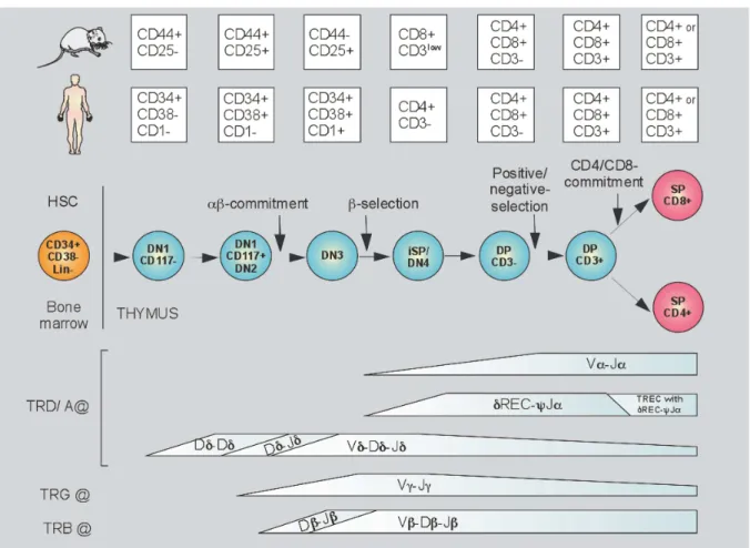
Figure 8. Human thymopoïesis. 48
Figure 9. Human T cell receptor delta, gamma and alpha loci (TRD/G/A@s). 49
Figure 10. Human T cell receptor beta locus (TRB@). 50
Figure 11. Thymocyte developmental sequence of V(D)J-recombination. 51
Figure 12. V(D)J-recombination mechanism. 52
Figure 13. Thymocyte selection processes. 55
Figure 14. Kinetic signalling model. 57
Figure 15. Naïve αβ T cell homeostasis. 59
Figure 16. Immune contraction and memory αβ T cells. 63
Figure 17. Severe combined immunodeficiency (SCID). 66
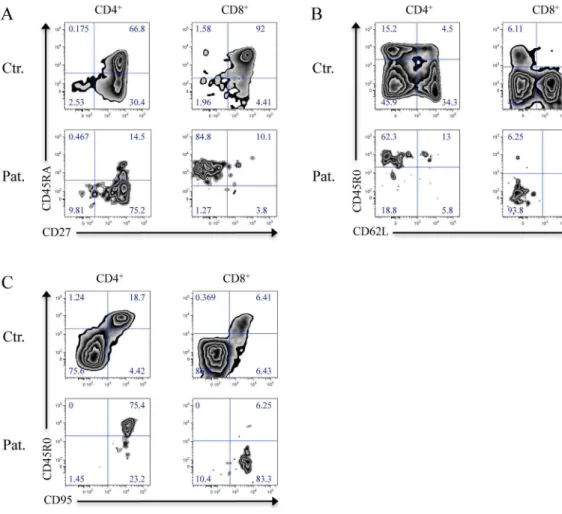
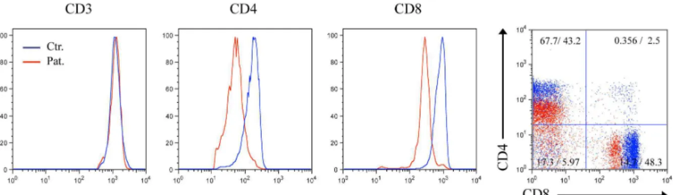
Figure 18. Macroscopic and microscopic aspects of sterile skin and joint inflammation. 71 Figure 19. Immunophenotype of LCK-deficient peripheral CD4+ and CD8+ T cells. 74 Figure 20. Immunophenotype of LCK-deficient peripheral TReg. 75
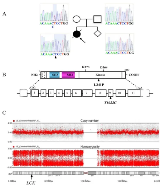
Figure 21. Analysis of LCK-deficient αβ TCR repertoire by spectratyping. 76 Figure 22. Analysis of LCK-deficient αβ TCR repertoire by electropherogram. 77 Figure 23. Analysis of LCK-deficient γδ TCR repertoire by spectratyping. 78 Figure 24. Immunophenotype of Vβ- and Vδ-expressing LCK-deficient PBMCs. 80 Figure 25. Expression of CD3, CD4 and CD8 on LCK-deficient PBMCs and T cell blasts. 81 Figure 26. Genetic analysis of the LCK-deficient patient and her family. 83 Figure 27. Sequence alignment of LCK orthologs and tyrosine kinase superfamily members.
85
Figure 28. Three-dimensional LCK protein structure. 86
Figure 31. Kinase activity of LCK variants. 90
Figure 32. Kinase activity of titrated LCK variants. 91
Figure 33. Impaired protein tyrosine phosphorylation in LCK-deficient T cell blasts. 93 Figure 34. Impaired Ca2+-flux in LCK-deficient primary T cells and T cell blasts. 94 Figure 35. Disturbed downstream signalling in LCK-deficient T cell blasts. 95
Figure 36. Disturbed AICD in LCK-deficient T cell blasts. 95
Figure 37. Expression of LCK variants in Jurkat and transduced JCam1.6 cells. 97 Figure 38. Complementation of protein tyrosine phosphorylation in JCam1.6 cells. 98 Figure 39. Complementation of Ca2+-flux in JCam1.6 cells. 99 Figure 40. Immunophenotype of ZAP-70-deficient primary T cells. 102 Figure 41. Analysis of the ZAP-70-deficient αβ TCR repertoire by spectratyping and
electropherogram. 104
Figure 42. Analysis of the ZAP-70-deficient γδ TCR repertoire by spectratyping and
electropherogram. 105
Figure 43. Genetic analysis of the ZAP-70-deficient patient and his family. 108 Figure 44. Impaired protein tyrosine phosphorylation in ZAP-70-deficient T cell blasts. 110 Figure 45. Expression of signalling molecules in ZAP-70-deficient T cell blasts. 111 Figure 46. Impaired proximal TCR:CD3:ζ- signalling in ZAP-70-deficient T cell blasts. 112 Figure 47. Impaired Ca2+-flux in ZAP-70-deficient primary T cells. 113 Figure 48. Impaired distal TCR:CD3:ζ- signalling in ZAP-70-deficient T cell blasts. 114 Figure 49. Disturbed AICD in ZAP-70-deficient T cell blasts. 115
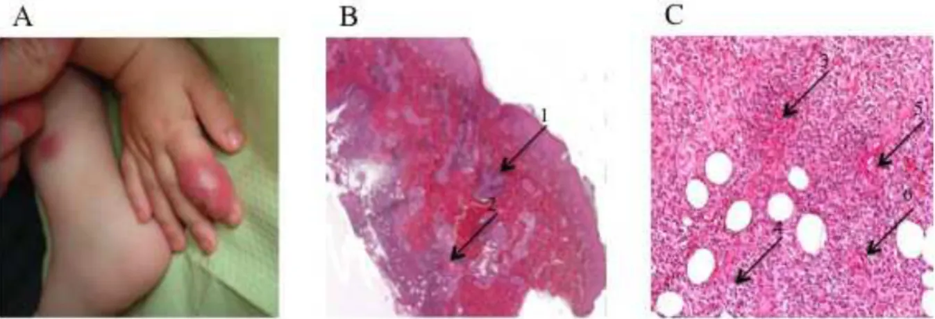
Figure 50. Chest radiography of the miliary infiltrate. 117
Figure 51. Immunohistochemical work-up of the pulmonary infiltrate. 118
Figure 52. Immunophenotype of ITK-deficient PBMCs. 122
Figure 53. Proliferation of T cell blasts of the newborn ITK-deficient brother. 123 Figure 54. Analysis of the ITK-deficient patient’s TCRVβ repertoire by flow cytometry. 124 Figure 55. Genetic analysis of the ITK-deficient patient and his family. 129 Figure 56. Impaired protein tyrosine phosphorylation in ITK-deficient T cell blasts. 130 Figure 57. Impaired PKCθ phosphorylation in ITK-deficient T cell blasts. 131 Figure 58. Impaired Ca2+-flux in ITK-deficient T cell blasts. 133 Figure 59. Impaired distal TCR:CD3:ζ-signalling in ITK-deficient T cell blasts. 134 Figure 60. Impaired migration of ITK-deficient T cell blasts. 135
Figure 61. Scheme of wildtype LCK and LCK ΔEx7. 142
Figure 63. Semiqualitative model of CD4 and CD8 lineage determination. 146
Table of tables
Table 1. Gene defects associated with combined immunodeficiencies (CIDs) in humans. 67 Table 2. Immunological features of the LCK-deficient patient. 72
Table 3. CDR3 sequencing of 74 clones of TRAV9-TRAC. 79
Table 4. CDR3 sequencing of 90 clones of TRBV6a-TRBC. 79
Table 5. Genomic LCK sequencing primers. 82
Table 6. Immunological features of the ZAP-70-deficient patient. 101 Table 7. Genomic and complementary ZAP-70 sequencing primers. 106 Table 8. Immunological features of the ITK-deficient patient and the newborn brother. 119
Table 9. TCRVβ repertoire of the ITK-deficient patient. 125
Table 10. Genomic ITK sequencing primers. 126
Table 11. Known PID-causing genes on chromosome 1. 137
Table 12. Known human ZAP-70 mutations. 144
Abbreviations
ADAP: Adhesion and degranulation-promoting adaptor protein AICD: Activation-induced cell death
AIRE: Autoimmune regulator
A-loop: Activation loop
AP-1: Activator protein 1 APC: Antigen presenting cell ARP-2/3: Actin related protein 2/3
ATF-2: Activating transcription factor 2 BCL-2/6/10: B cell lymphoma 2, 6 and 10 BCR: B cell antigen receptor
BH: BTK homology
BM: Bone marrow
C: Constant
[Ca2+]e: Extracellular calcium-ion concentration
[Ca2+]er: Endoplasmic reticulum calcium-ion concentration
[Ca2+]i: Intracellular calcium-ion concentration
CARD: Caspase recruitment domain
CARMA-1: CARD-containing MAGUK protein 1 CBL: Casitas B-lineage lymphoma
CCR-7: Chemokine C-C motif receptor 7 CD: Cluster of differentiation
CDC-37: Cell division cycle 37
CD62L: CD62 ligand
CDC-42: Cell division cycle 42
cDNA: Complementary DNA
CDK: Cyclin-dependent kinase
CDR: Complementarity determining region CGH: Comparative genomic hybridization CIP: Cytokine-induced proliferation
CMP: Common myeloïd precursor
CRAC: Ca2+-release-activated Ca2+-channel CRKII: CT10 regulator of kinase II
CsA: Cyclosporin A
CSK: C-terminal SRC kinase
cSMAC: Central supramolecular activation cluster CTLA-4: Cytotoxic T lymphocyte antigen 4 CVID: Common variable immunodeficiency CWID: Cytokine withdrawal-induced cell death CXCR-5: Chemokine (C-X-C motif) receptor 5
D: Diversity
DAG: Diacylglycerol
DAMP-R: Danger-associated molecular pattern-receptor
DN: Double-negative
DNA: Deoxyribonucleic acid DOK-1/2: Docking protein 1 and 2
DP: Double-positive
DPC: Distal pole complex
DYRK-2: Dual-specificity tyrosine-phosphorylation regulated kinase 2 dSMAC: Distal supramolecular activation cluster
EBER-1: EBV-encoded small RNA 1
EBV: Epstein-Barr virus
EDP: Early double positive
ER: Endoplasmic reticulum
ERCA: Endoplasmic reticulum Ca2+-ATPase
ERK-1/2: Extracellular signal-regulated kinase 1 and 2 ETP: Early thymic progenitor
FACS: Fluorescence-activated cell sorting F-actin: Filamentous-actin
FAS-L: FAS ligand
FOX-P3: Forkhead box P3
FRC: Fibroblastic reticular cell
FYN: FYN oncogene related to SRC, FGR, YES
GATA-3: GATA binding protein 3
γc: Common γ-chain
GEF: Guanine nucleotide exchange factor GFP: Green fluorescent protein
GRB-2: Growth factor receptor-bound protein 2 GSK-3: Glycogen synthase kinase 3
HLA: Human leukocyte antigen
HPK-1: Haematopoïetic progenitor kinase 1 HSCGT: Haematopoïetic stem cell gene therapy HSCTx: Haematopoïetic stem cell transplantation HSP-90: Heat shock protein of 90 kDa
ICAM-1: Intercellular adhesion molecule 1 ICOS: Inducible T-cell co-stimulator iDC: Immature dendritic cell
IFN-γ: Interferon gamma
Ig: Immunoglobuline
Igβ: Immunoglobulin-associated beta chain
IgC: Immunoglobuline constant
IgV: Immunoglobuline variable
IκB: Inhibitor of NF-κB
IKK: IκB kinase
IL: Interleukin
IL-R: Interleukin-receptor
IMGT: ImMunoGeneTics
iNKT cell: Invariant natural killer T cell IP3: Inositol 1,4,5-trisphosphate
IP3-R: Inositol 1,4,5-trisphosphate-receptor
IPEX: Immune dysregulation, polyendocrinopathy, enteropathy, X-linked
iSP: Immature single positive
ITAM: Immunoreceptor tyrosine-based activation motif ITK: Interleukin-2-inducible T cell kinase
JNK: JUN N-terminal kinase
KREC: Kappa-deleting recombination excision circle LAT: Linker for activation of T cells
LCK: Lymphocyte-specific protein tyrosine kinase LFA-1: Leukocyte function-associated antigen 1 LIP: Lymphopenia-induced proliferation
LPS: Lipopolysaccharide
LT-β: Lymphotoxin β
LTRC: Long-term repopulating cell
mAb: Monoclonal antibody
MAGT-1: Magnesium transporter 1
MAGUK: Membrane-associated guanylate kinase MAIT cell: Mucosa-associated invariant T cell MALT-1: Mucosa-associated lymphoïd tissue 1 MAPK: Mitogen-activated protein kinase
MAP3K: MAPK kinase kinase
MAP2K: MAPK kinase
MCU: Mitochondrial Ca2+-uniporter
[Mg2+]e: Extracellular magnesium-ion concentration
[Mg2+]i: Intracellular magnesium-ion concentration
MHC: Major histocompatibility complex mDC: Myeloïd dendritic cell
mDC: Mature dendritic cell
MEK-1/2: Mitogen-activated protein kinase 1 and 2 MEP: Megakaryocyte/erythrocyte precursor
MR1: Major histocompatibility complex class I-related MTOC: Microtubule-organizing center
mTOR: Mammalian target of rapamycin NCK: Non-catalytic region of tyrosine kinase NFAT: Nuclear factor of activated T cells
NF-κB: Nuclear factor of kappa light polypeptide gene enhancer in B cells NHEJ: Non-homologous end joining
NHR: NFAT-homology region
NES: Nuclear export signal
NLR: Nucleotide-binding oligomerization domain (NOD)-like receptor NLS: Nuclear localization signal
nTReg: Natural TReg
ORAI-1: ORAI calcium release-activated calcium modulator 1 pAB: Polyclonal antibody
PAG-1: Phosphoprotein associated with glycosphingolipid microdomains 1 PAMP-R: Pathogen-associated molecular pattern-receptor
PAX-5: Paired box protein 5
PBMC: Peripheral blood mononuclear cell PCR: Polymerase chain reaction
PD: Programmed cell death
pDC: Plasmacytoïd dendritic cell PDPK-1: PI3K-dependent protein kinase 1
PEP: PEST-domain phosphatase
PH: Pleckstrin homology
PHA: Phytohaemagglutinin
PI3K: Phosphatidylinositol-4,5-bisphosphate 3-kinase PID: Primary immunodeficiency disease
PIP2: Phosphatidylinositol 4,5-bisphosphate
PIP3: Phosphatidylinositol 3,4,5-trisphosphate
PKCθ: Protein kinase Cθ
PLCγ-1: Phospholipase Cγ 1
PMA: Phorbol 12-myristate 13-acetate PMCA: Plasma membrane Ca2+-ATPase
pMHC: Peptide:major histocompatibility complex PRR: Proline rich region
pSMAC: Peripheral supramolecular activation cluster pTα: Pre T cell antigen receptor α
PTEN: Phosphatase and tensin homolog PTK: Protein tyrosine kinase
qPCR: Quantitative real-time PCR
RAC-1: RAS-related C3 botulinum toxin substrate 1 RACK: Receptor for avtivated C kinase
RAG-1/2: Recombination activating gene 1 and 2 RASGRP: RAS guanyl releasing protein
RHO: RAS-homolog
RHR: REL-homology region
RICD: Restimulation-induced cell death RLK: Resting lymphocyte kinase
RNA: Ribonucleic acid
RORγt: Retinoic acid receptor-related orphan receptor γt RSS: Recombination signal sequence
RSV: Respiratory syncytial virus
RT-PCR: Reverse transcription polymerase chain reaction RUNX-3: Runt-related transcription factor 3
SAP: Signalling lymphocyte activation molecule-associated protein SCID Severe combined immunodeficiency
SDF-1: Stromal cell-derived factor 1 SDS: Standard deviation score
SDS-PAGE: Sodium dodecyl sulfate polyacrylamide gel electrophoresis SHC: Src homology 2 domain containing protein
SHIP-1: SH2 domain-containing 5’-inositol phosphatase 1 SHP-1: SH2 domain-containing phosphatase 1
SLAM: Signalling lymphocytic activation molecule
SLP-76: SH2 domain-containing leukocyte protein of 76 kDa SMAC: Supramolecular activation cluster
SNP: Single nucleotide polymorphism SOCE: Store-operated Ca2+-entry
SOCS-1: Suppressor of cytokine signalling 1 SOS-1: Son of sevenless homolog 1
SP: Single-positive
SP: Ser-Pro-X-X repeat
SRR: Serine rich region
STK39: Serine threonine kinase 39
STIM-1/2: Stromal interaction molecule 1 and 2 SWAP-70: Switch-associated protein 70 kDa SYK: Spleen tyrosine kinase
TAD: Transcription activation domain
TCM Central memory T cell
TCR: T cell antigen receptor
TCS: T cell synapse
TCyt: Cytotoxic T cell
TEA: T-early α
TEC: Thymic epithelial cell
TEM Effector memory T cell
TGF-β: Transforming growth factor β TFH: Follicular helper T cell
TH1/2/17: Helper T cell type 1, 2 and 17
TH-POK: T-helper-inducing POZ/Kruppel-like factor TLR: Toll-like receptor
TMem: Memory T cell
TNF-α: Tumour necrosis factor alpha
TOX: Thymus high-mobility group box protein TRA/B/G/D@: T cell receptor alpha/beta/gamma/delta locus TRAF-2/6: TNF receptor-associated factor 2 and 6
TRA/B/C/DC: TRA@/TRB@/TRG@/TRD@ constant segment TRA/B/C/DV: TRA@/TRB@/TRG@/TRD@ variable segment TREC: T cell receptor excision circle
TReg: Regulatory T cell
TSAD: T cell specific adaptor protein TSLP: Thymic stromal lymphopoietin TSP: Thymus seeding progenitor
V: Variable
WASP: Wiskott-Aldrich syndrome protein XIAP: X-linked inhibitor of apoptosis
[Zn2+]e: Extracellular zinc-ion concentration
1 Introduction
1.1 The immune system
Human beings are confronted with physical, chemical and (micro-) biological insults throughout their entire lifespan. To preserve their structural and functional integrity, they have evolved a specialized system, the immune system. The main function of the immune system is to recognize self, altered-self and non-self antigens and to decide whether to reject them by initiating a immune response or to accept them by inducing immune tolerance.1
Historically, the immune system has been divided into an innate and adaptive branche, even thought there is important cross-talk at their interface. The primary lymphoïd organs, i.e. bone marrow, thymus and spleen, and the secondary lymphoïd organs, i.e. lymph nodes, tonsils and organ-associated lymphoïd tissues, constitute its principal anatomical compartments.1 Furthermore, in the context of chronic inflammation, the immune system is able to establish tertiary lymphoïd tissues at almost every site of the body.2
Besides an array of secreted molecules, i.e. lipid mediators, interferons, cytokines and chemokines, and their cognate receptors, that are partially shared by both branches, the innate and adaptive immune system comprise particular receptor families, cell populations and their individual effector molecules.1
Briefly, the innate immune system is build up by epithelial barriers, antimicrobial peptides, danger-associated molecular and pathogen-associated molecular pattern-recognizing molecules and receptors (DAMP-Rs and PAMP-Rs), i.e. pentraxins, complement, innate antibodies, nucleotide-binding oligomerization domain (NOD)-like (NLRs) and Toll-like receptors (TLRs), mast cells, monocyte-macrophages, neutrophil/eosinophil/basophil granulocytes, natural killer (NK) cells and antigen presenting cells (APCs), i.e. plasmacytoïd and myeloïd dendritic cells (pDCs and mDCs) and Langerhans cells.1
Innate immune responses are triggered by the sensing of DAMPs and PAMPs and initially lead to inflammation, increased effector function and consequently antigen clearance. Thereafter, the innate immune system downmodulates its inflammatory response and initiates tissue repair.3 Importantly, the innate immune system triggers and finetunes the adaptive immune response by establishing particular cytokine microenvironments and by processing
The adaptive immune system comprises T cells, B cells and specific antibodies.1 A central feature of T cells and B cells is the expression of the T cell antigen receptor (TCR) and the B cell antigen receptor (BCR), respectively, that theoretically endow them with the ability to recognize all possible antigens. While the TCR and the BCR are membrane bound, specific antibodies corespond to secreted forms of the BCR.1
T cells are pivotal to the adaptive immune system as they co-ordinate immune tolerance and efficient adaptive immune responses, aquire antigen-specific effector functions and build up antigen-specific memory. A variety of T cell subpopulations exist, i.e. natural killer T cells (NKTs), mucosa-associated invariant T cells (MAITs), innate-like γδ T cells, and conventional αβ T cells. The latter can be further subdivided into CD4+ helper T cells (TH), CD4+CD25+ forkhead box P3+ (FOX-P3+)regulatory T cells (TReg) and CD8+ cytotoxic
T cells (TCyt).
B cells contribute to the adaptive immune response by acquiring APC function and by differentiating into specific antibody-secreting plasma cells.1
To obtain the ability of antigen-specific immune recognition and response, T and B cells pass through unique maturation programms that take place in the thymus and the bone marrow, respectively.5
The DAMP-Rs and PAMP-Rs of the innate immune system are encoded by entire genes and can be directly expressed in a non-clonal manner. As they have been evolutionarily selected, they recognize a limited array of conserved molecular patterns, e.g. lipopolysaccharide (LPS), single-strand or double-strand ribonucleic acid (RNA) and deoxyribonucleic acid (DNA), and efficiently discriminate self, altered-self and non-self. Consequently, they can rapidyl initiate immune responses to a narrow antigenic spectrum.6
The TCRs and BCRs, however, are encoded in huge genetic loci by particular gene segments and their somatic rearrangement is necessary to express a clonally restricted but highly variable receptor repertoire. As the individual TCRs and BCRs have been selected in somatic cells, they recognize particular epitopes of protein and carbohydrate antigens and their ability to discriminate self, altered-self and non-self is imperfect. Consequently, they can initiate a delayed immune response and immunological memory to virtually all possible antigenic structures.6
Throughout the last five decades, the immune system has been extensively investigated in vitro and in animal models such as the murine system. Additionally, studying and taking care of human primary immunodeficiency diseases (PIDs) has been seminal for our understanding of the human immune system’s developement, homeostasis, and function
(see chapter 1.5.1).7 Currently, more than 150 PIDs have been classified by the International Union of Immunological Societies Expert Committee for Primary Immunodeficiency.8
This immunobiological thesis is aimed to contribute to our knowledge of the human immune system by analyzing disturbed proximal TCR:CD3:ζ-signalling caused by autosomal recessive lymphocyte-specific protein tyrosine kinase (LCK)-, ζ-chain associated protein tyrosine kinase of 70 kDa (ZAP-70)- and interleukin-2-inducible T cell kinase (ITK)- deficiency.
1.2 TCR:CD3:ζ-signalling
On their cell surface T cells express either a preTCR, or an (invariant) γδ TCR or a clonotypic αβ TCR together with the cluster of differentiation (CD) 3-complex and the associated ζ-chain.9-12 TCR:CD3:ζ-signalling is crucial for T cell development and antigen-specific activation, proliferation, differentiation, effector function and apoptosis of mature T cells.5, 13, 14 Basically, APCs assimilate and process protein antigens and present antigenic peptide fragments bound to major histocompatibility complex (MHC) molecules to T cells. T cells recognize and bind peptide:MHCs (pMHC) with their TCR and signal with their CD3:ζ-complex at a molecular interface termed T cell synapse (TCS).15
The TCS can be devided into different anatomical and functional layers, i.e. the receptor layer, the signalling layer, the ion channel and transporter system, and the cytoskeletal layer.16
The nature of the presented antigenic fragment and the spatiotemporal and molecular composition of the TCS are pivotal for the functional outcome of TCR:CD3:ζ-signalling.17
1.2.1 The TCR:CD3:ζ-complex
The mature TCR is build up by a disulfide-linked heterodimer of either one TCRα- and one TCRβ- or one TCRγ- and one TCRδ-glycoprotein. The TCRα- has sequence homology to the TCRδ- and the TCRβ- to the TCRγ-chain, respectively.18, 19 Additionally, during T cell development thymocytes transiently express the preTCR consisting of a TCRβ-chain and the invariant pre T cell antigen receptor α (pTα) -TCRβ-chain, that serves as a surrogate for the TCRα-chain (Fig. 1A).5, 20, 21
Figure 1. TCR:CD3:ζ-complex.
(A) Schematic representation of the preTCR-, the αβ TCR- and the γδ TCR-heterodimer with the extracellular variable (V) and constant (C) immunoglobulin-like domains, the transmembrane domains containing the basic amino acid residues arginine (R) and lysine (L) and the signalling or non-signalling intracellular domains of the pTα- and the TCR α-, β-, γ- and δ-glycoproteins, respectively.
(B) Schematic representation of the αβ TCR-heterodimer as in (A) and the CD3δε-, CD3γε- and ζζ-heterodimers with the extracellular domains, the transmembrane domains containing the acidic amino acid residues aspartic acid (D) and glutamic acid (E) and/or the signalling intracellular immunoreceptor tyrosine-based activation motif (ITAM)-encompassing domains.
(C) Schematic representation of the canonical TCRαβ:CD3γε:CD3δε:ζζ-complex composed of the heterodimers indicated in (A) and (B).
Figure adapted from Call ME, Wucherpfennig KW,Annu Rev Immunol, 2005.22
Each TCR-chain contains an extracellular domain, a transmembrane region and a short intracellular domain. The extracellular domain comprises a variable (V) and constant (C) immunoglobuline (Ig)-like domain, the transmembrane region contains three conserved basic amino acid residues, i.e. one arginine (R) and two lysines (K), and the short intracellular domain lacks intrinsic signalling activity.10 The pTα-chain comprises a single extracellular Ig-like domain and a transmembrane region, both of which resemble that of the TCRα-chain, and an intracellular domain. In contrast to the TCRα-chain, the intracellular domain of the
pTα-chain possesses two phosphorylation sites, suggesting a CD3:ζ-complex independent signalling capacity of the preTCR.23, 24
The prototypical and clonotypic αβ TCR is expressed by conventional T cells, such as CD4+ TH, CD4+CD25+FOX-P3+ TReg and CD8+ TCyt, and recognizes antigenic peptides, that
are presented by classical and polymorphic MHC class I or class II molecules.25 The αβ TCR IgV-like domain binds diagonally over the entire pMHC.26 Each αβ TCR IgV-like domain consists of three hypervariable complementarity determining regions (CDRs). CDR1 and CDR2 are determined by germline V gene segments while CDR3 is determined by germline diversity (D) and joining (J) gene segments and junctional diversity (see chapter 1.3.3).27 The CDR3α and CDR3β loops interact with the central part and the CDR1α and CDR1β with the amino-terminal and carboxy-terminal part of the presented peptide, respectively. The MHC molecule principally is bound by CDR1αβ and CDR2αβ loops but CDR3αβ loops as well can form minor contacts.28
Semi-invariant αβ TCRs are expressed by invariant NKT (iNKT) cells and bind lipid antigens presented by MHC class I like CD1 molecules by only partially contacting the binding groove and parallel to its long axis. The NKT cell TCR Vα24Jα18 binds the glycosyl head group of α-galactosylceramide with CDR1α and CDR3α that are germline-encoded by Vα24 and Jα18, respectively.29, 30
Mucosa-associated invariant T cells (MAITs) express a semi-invariant TCR Vα7.2Jα33 that is restricted by the evolutionarily conserved major histocompatibility complex class I-related (MR1) molecule and recognize bacterially infected cells in a MR1-dependent manner.31, 32 The lipid antigen α-mannosylceramide has been proposed as the specific MR1-ligand but in silico modelling favours a hydrophilic compound.33, 34
Invariant γδ TCRs are expressed by γδ T cells with an innate-like phenotype and bind as yet poorly defined bacterial phosphoantigens presented by non-polymorphic MHC-like molecules. This interaction differs strongly from that of TCRαβ:pHMC as the γδ TCR binds sideways to the nonclassical MHC-like molecule and only one CDR, namely the CDR3δ, contacts the binding groove.35, 36
Thus, polymorphic αβ TCRs, semi-invariant αβ TCRs and (invariant) γδ TCRs are MHC or MHC-like restricted and this is due to germline encoded sequence and structure homologies that have co-evolved between the MHC molecules and the particular TCR V Ig-like domains.37, 38
but responsible for signal transduction. The stoichiometry of the TCRαβ:CD3:ζ-complex is TCRαβ:CD3γε:CD3δε:ζζ while that of the TCRγδ:CD3:ζ-complex is TCRγδ:CD3γε:CD3δε:ζζ.39-43 The definitive stoichiometry of the human preTCR has not yet been resolved (Fig. 1 B and C).44
For correct assembly and surface expression the TCRαβ-, the CD3γε-, CD3δε-and the ζζ-chains are necessary and sufficient.41, 42 The TM regions of the TCRαβ-heterodimer contain three conserved basic amino acid residues and the TM regions of the CD3γε-,CD3δε- and ζζ-dimers two conserved acidic amino acid residues, i.e. aspartic and glutamic acid. Probably by forming pairwise ionic interactions, these putatively charged residues are crucial for correct TCR:CD3:ζ-complex association.45
The CD3- and ζ-chains have cytoplasmic tails containing immunoreceptor tyrosine-based activation motifs (ITAMs) that are phosphorylated upon receptor triggering and that recruit further signalling molecules.46-49 The entire TCRαβ:CD3:ζ-complex contains ten ITAMs and each ITAM contains two conserved tyrosine, one acidic and two aliphatic amino acid residues. The ITAM consensus sequence is D/Ex0-2YXXL/IX6-8YXXL/I (with D
denominating aspartic acid, E glutamic acid, Y tyrosine and X any amino acid residue with the lower case number indicating their variable frequencies) and the CD3γ-, CD3δ- and CD3ε-chains each contain one ITAM while the ζ-chain contains three tandem ITAMs (Fig. 2).46
The cytoplasmic tails of the CD3ε- and the ζ-chain interact with acidic phospholipids of the inner plasma membrane leaflet and it has been proposed that in an untriggered TCRαβ:CD3:ζ-complex the ITAMs might remain in a lipid-bound stage being inaccesible for activating protein tyrosine phosphorylation.50, 51
Even though there are different models of how initial TCR triggering is transduced into intracellular signalling, the TCR might be a mechanotransductor and TCR:CD3:ζ-complex interaction with pMHC might induce conformational changes that render the ITAMs accessible for protein tyrosine phosphorylation.52
Once the TCR:CD3:ζ-complex has been triggered by pMHC, possible mechanisms of signal termination are dephosphorylation, internalisation and ubiquitin-mediated degradation in the lysosomal compartment.52, 53
Figure 2. T cell synapse and proximal TCR:CD3:ζ-signalling.
Schematic representation of the membrane interface of an antigen presenting cell (APC) and a T cell forming the T cell synapse (TCS) and initiating proximal TCR:CD3:ζ-signalling. Initially, the peptide:major histocompatibility complex class II (pMHC II) is recognized by its cognate αβ TCR. This enables the co-receptor CD4 to bind the pMHC II and recruits and activates the membrane-anchored and CD4-bound protein tyrosine kinase (PTK) lymphocyte-specific protein tyrosine kinase (LCK). Next, LCK phosphorylates the ITAMs - the ITAM consensus sequence is indicated in parentheses - of the ζζ-heterodimer and creates the docking site for the PTK ζ-chain associated protein tyrosine kinase of 70 kDa (ZAP-70). ZAP-70 binds to the phosphorylated ITAMs, in turn is phosphorylated and activated by LCK and amplifies and diversifies the TCR:CD3:ζ-signalling by phosphorylating downstream adaptor proteins including the linker for activation of T cells (LAT) and the SH2 domain-containing leukocyte protein of 76 kDa (SLP-76).
Figure adapted from Wang H, et al, Cold Spring Harb Perspect Biol, 2010.54
1.2.2 The receptor layer
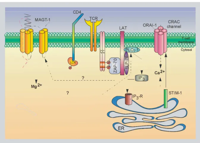
The receptor layer is the cell membrane compartment where antigen:receptor- and receptor:receptor-interactions take place. It contains the TCR:CD3:ζ-complex and co-receptors as well as co-stimulatory, co-inhibitory and adhesions molecules.16 Additionally, TCR:CD3:ζ-signalling is mediated by a network of ion channels and transporters partially
Figure 3. T cell receptor, signalling and cytoskeletal layers.
Once the T cell synapse (TCS) has formed, T cell signalling takes place in three major cellular compartments, i.e. the receptor, signalling and cytoskeletal layer. The receptor layer is comprised of the TCR:CD3:ζ-complex itself, activating co-receptors such as CD4 and CD28 and inhibitory co-receptors such as CD45 and CD148. Furthermore, membrane-bound and -derived phospholipids such as phosphatidylinositol 4,5-bisphosphate (PIP2), phosphatidylinositol 3,4,5-trisphosphate (PIP3), inositol 1,4,5-trisphosphate (IP3) and diacylglycerol (DAG) are important anchor molecules and second messengers, respectively. Ion-channels such as the Mg2+- and the Ca2+ -prevalent magnesium transporter 1 (MAGT1) and Ca2+-release-activated Ca2+-channel (CRAC) are also located in the cell membrane. The signalling layer contains a vast array of signalling molecules and intermediates that either are attached directly to the recptor layer or are nucleated basically by the adaptor proteins linker for activation of T cells (LAT) and SH2 domain-containing leukocyte protein of 76 kDa (SLP-76). The so-called proximal TCR-signalosome amplifies and couples the initial TCR:CD3:ζ-signal to various downstream singalling pathways, such as the mitogen-activated protein kinase (MAPK)-, nuclear factor of kappa light polypeptide gene enhancer in B cells (NF-κB)-, nuclear factor of activated T cells (NFAT)- and activator protein 1 (AP-1)-pathway. Additionally, the TCR-signalosome governs cellular processes such as adhesion and cytoskeletal reorganisation predominantly mediated by the cytoskeletal layer.
Figure adapted from Dustin ML, Depoil D, Nat Rev Immunol, 2012, and Balagopalan L, et al, Cold Spring Harb Perspect Biol, 2010.16, 56
1.2.2.1 The T cell synapse
The TCS, also termed immunological synapse, lies at the heart of the receptor layer and anatomically is organized in concentric supramolecular activation clusters (SMACs), i.e.
the inner central SMAC (cSMAC), the intermediate peripheral SMAC (pSMAC) and the outer distal SMAC (dSMAC). Basically, SMAC formation is due to lateral segregation of particular molecules.57 Upon pMHC-recognition, 11-17-valent TCR:CD3:ζ-microclusters form in the pSMAC and move centripetally towards the cSMAC.58 Additionally, the cSMAC contains microclusters of the co-recptors CD4 and CD8 and of co-stimulators such as CD28.58, 59 Adhesion molecules, such as the integrin leukocyte function-associated antigen 1 (LFA-1), form microclusters as well but localize to the pSMAC.60 Inhibitory proteins, such as the protein tyrosine phosphatase (PTP) receptor type C (PTPRC, CD45) and the PTP receptor type J (PTPRJ, CD148), are excluded from the cSMAC and the pSMAC and locate to the dSMAC.57
How precisely lateral segregation is achieved, remains a matter of debate. The size of the separated molecules seems to be important as the intercellular APC:T cell-distance spanned by a pMHC:TCR:CD3:ζ-complex is about 15 nm while that of a LFA-1:intercellular adhesion molecule 1 (ICAM-1)-complex is about 40 nm.61 Moreover, once the TCR:CD3:ζ-microclusters have reached the cSMAC, they loose their interactions with the actin cytoskeleton while the integrin-microclusters of the pSMAC maintain these interactions, that are necessary for their stabilization. It has therefore been proposed, that the cSMAC functions as a drop-off basin that includes microclusters with the right size that rest assembled without stabilizing contacts to the cytoskeleton.60, 62
The classical view of TCR:CD3:ζ-signalling being located to the cSMAC has recently been challenged by the observations that TCR:CD3:ζ-micorclusters already signal in the pSMAC and that the cSMAC might instead be the place of signal termination via receptor internalisation.63, 64
1.2.2.2 CD4 and CD8
The most important TCR:CD3:ζ-co-receptors are the single-pass type I membrane proteins CD4 and CD8 that phenotypically define CD4+ TH and CD8+ TCyt cells,
respectively.65 CD4 is composed of two extracellular IgV-like domains, two IgC-like domains, one TM region and one intracellular domain.66 The first IgV-like domain interacts with the β2-domain of MHC class II.67
CD8 is a heterodimer generally composed of CD8α and CD8β that are linked by two disulfide bonds. Each CD8 molecule consists of an extracellular IgV-like domain, a TM
domain of MHC class I.68, 69 Thus, CD4 and CD8 stabilize the interaction of the TCR:CD3:ζ-complex with either MHC class II or MHC class I molecules, respectively.
Moreover, the intracellular domains of CD4 and CD8 bind the protein tyrosine kinase (PTK) LCK and recruit it to the TCR:CD3:ζ-complex, thus crucially contributing to the initiation of TCR:CD3:ζ-signalling (Fig. 2).70
1.2.2.3 CD45
The PTPRC CD45 is a single-pass type I membrane protein that is expressed in all nucleated haematopoïetic cells.71 CD45 contains an extracellular portion that is composed of a N-terminal region and two fibronectin-type III repeats, a TM region and an intracellular portion that contains a wedge-like region and two tandem protein tyrosine phosphatase domains (PTPs).72 The first PTP domain (D1) is catalytically active while the second one (D2) appears inactive and might endow substrate specificity to CD45.73 The wedge-like region has been found to inhibit D1 of homodimerized CD45 in trans and contributes to negative regulation.74
Up to eight CD45 isoforms exist and in T cells the most prominent ones are CD45RA, CD45RB and CD45R0. The extracellular N-terminal regions of the CD45 isoforms vary in size and glycosylation pattern as a consequence of alternative splicing and post-translational modification. CD45RA is the longest isoform and is predominantly expressed by naïve T cells while CD45R0 is the shortest isoform and is expressed by activated and memory T cells.75 CD45 can inhibit TCR:CD3:ζ-complex activation but upon TCR triggering is excluded rapidly from the cSMAC as a function of its size.76 Importantly, CD45 dephosphorylates the SRC kinase LCK at its inhibitory tyrosine Y505 and therefore is a positive regulator of TCR:CD3:ζ-signalling.77 Additionally, CD45 dephophorylates the activating tyrosine Y394 at the activation loop (A-loop) of LCK with a lower affinity than that for Y505 and this might again be a negative feedback mechanism.78 CD45 spontaneously homodimerizes and this inhibits its catalytic activity. The autoinhibitory efficacy of homodimerization is inversely correlated with the size of the N-terminal regions, thus the smallest isoform CD45R0 is less efficient in catalyzing TCR:CD3:ζ-signalling than the longest isoform CD45RA.74
1.2.2.4 Co-stimulation
Co-stimulatory and co-inhibitory molecules modulate TCR signalling strenght or duration and importantly fine-tune activation, proliferation, differentiation and effector function.16 The co-stimulatory CD28 and inducible T-cell co-stimulator (ICOS, CD278) and
the co-inhibitory cytotoxic T lymphocyte antigen 4 (CTLA-4) interact with CD80 and CD86 expressed by APCs and are the best studied examples (Fig. 3).79 Numerous other co-stimulatory and co-inhibitory receptor families, such as the signalling lymphocytic activation molecule (SLAM)- or the programmed cell death (PD)-receptors exist.80, 81 Co-stimulation, as illustrated by pMHC:TCR- and CD80/CD86:CD28-interactions, is important for productive T cell activation, as T cells lacking proper co-stimulation enter a state of unresponsiveness designated anergy, an important means of peripheral tolerance.82, 83
1.2.2.5 Adhesion molecules
Adhesion molecules function in an antigen-independent manner and predominantly intensify the interaction of APCs with T cells as illustrated by the CD2:CD58- and the LFA-1:ICAM-1-interaction.61 Adhesion receptors further stabilize and couple the TCS to the cytoskeleton and are involved in cell motility.84
1.2.3 The signalling layer
Protein tyrosine kinase (PTK) cascades lie at the heart of TCR:CD3:ζ-signalling. In T cells, the PTKs of the CSK-, SRC-, SYK- and TEC-family are the major players and the most important family members are CSK, LCK, ZAP-70 and ITK, respectively.54, 85, 86 Globally, these four classes of PTKs act sequentially to propagate protein tyrosine phosphorylation signals. The first class CSK inhibits, the second class LCK initiates, the third class ZAP-70 amplifies and the fourth class ITK diversifies protein tyrosine phosphorylation signals. T cells express a variety of additional PTKs of the SRC-, SYK- and TEC-family, such as the FYN oncogene related to SRC, FGR, YES (FYN), the spleen tyrosine kinase (SYK) related to ZAP-70 and the resting lymphocyte kinase (RLK) related to ITK, respectively.54, 85, 86
1.2.3.1 LCK
The PTK LCK comprises 509 amino acid residues, has a molecular weight of 56 kDa and is expressed almost exclusively in T cells. LCK consists of a N-terminal unique SRC homology 3 (SH3) domain, followed by a SH2 and a C-terminal SH1 or kinase domain (Fig. 4). The SH3 and SH2 domains infere intra- and inter-protein interactions by binding to polyproline and phosphotyrosine motifs, respectively.87 The LCK N-terminus is myristoylated and palmitoylated and is mediating constitutive membrane anchoring.88 The N-terminus contains a di-cysteine motif that together with a di-cysteine motif in the intracellular tails of
CD8α and TCR:CD4-co-stimulation generates stronger LCK-mediated signals than TCR:CD8-co-stimulation does.91 The kinase domain contains the ATP binding lysine residue K273 and the catalytic proton acceptor aspartic acid residue D364 that conduct protein tyrosine phosphorylation of the ITAMs of the TCR:CD3:ζ-complex.92 Furthermore, LCK functions as an adaptor protein via its SH2 and SH3 domains.93, 94
Figure 4. Inactive and active lymphocyte-specific protein tyrosine kinase (LCK).
(A) The inactive conformation of LCK is induced by C-terminal SRC kinase (CSK)-mediated phosphorylation of the inhibitory tyrosine 505 (pY505) that binds to the LCK SH2 domain. The restrained conformation is further stabilized by the interaction of a polyproline motif located in the SH2-kinase linker domain with the SH3 domain.
(B) The active conformation of LCK is induced by CD45-mediated dephosphorylation of the inhibitory Y505. As a consequence the A-loop opens up and the activating Y394 is phosphorylated by LCK auto-/transphosphorylation endowing LCK with full catalytic activity. The active conformation is further stabilized by protein:protein interactions mediated by the SH3 and SH2 domains of LCK.
The kinase function of LCK is tightly regulated by conformational changes caused by ligand binding of the SH3 and the SH2 domains and by phosphorylation and dephosphorylation of pivotal tyrosine residues.85 When the inhibitory C-terminal tyrosine Y505 is phosphorylated by CSK, that is recruited by the adaptor phosphoprotein associated with glycosphingolipid microdomains 1 (PAG-1), it binds to the LCK SH2 domain and induces a closed conformation.95-97 The restrained conformation is further stabilized by the interaction of a polyproline motif located in the SH2-kinase linker domain with the SH3 domain (Fig. 4A).98 The PTPRC CD45 can dephosphorylate the inhibitory tyrosine Y505 and induces an open and basally active LCK conformation.77 The activating tyrosine Y394 is located in the A-loop of the kinase and when dephosphorylated the A-loop aquires an α-helical conformation blocking the catalytic center. When tyrosine Y505 is dephosphorylated and LCK opens up, the A-loop is displaced from the catalytic center.99 LCK then performs auto-/transphosphorylation of tyrosine Y394 and aquires full catalytic activity.100 Additionally, LCK becomes phosphorylated at the serine residue S59 by the extracellular signal-regulated kinase 1/2 (ERK-1/2) and this and further protein:protein-interactions stabilize the active conformation (Fig 4B).101
The overall activating modifications lead to an increase of the molecular weight of LCK from 56 to 60 kDa.102
By dephosphorylation events LCK activity is kept in check and several PTPs such as CD45, SH2 domain-containing phosphatase 1 (SHP-1) and PEST-domain phosphatase (PEP) rapidyl dephosphorylate the activating tyrosine Y394.103-105
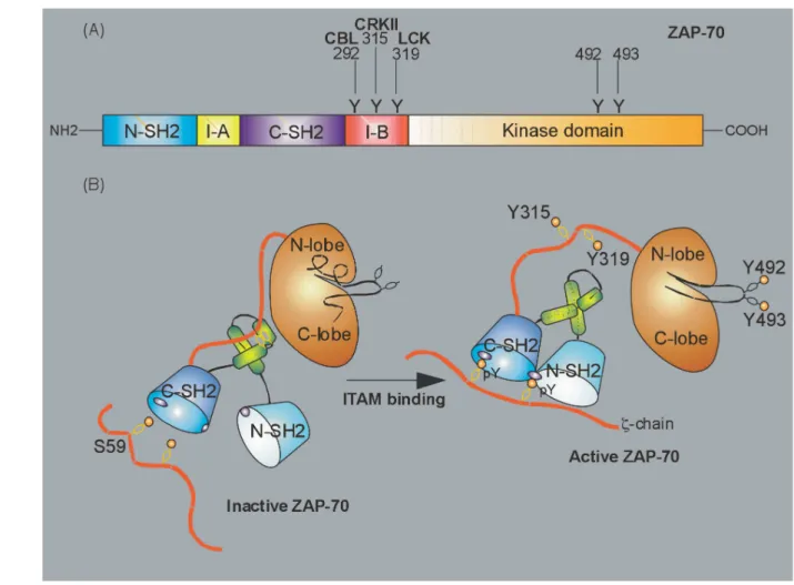
1.2.3.2 ZAP-70
The PTK ZAP-70 comprises 619 amino acid residues, has a molecular weight of 70 kDa and is mainly expressed in T cells. On its N-terminus, ZAP-70 consists of two tandem SH2-domains denominated N-terminal and C-terminal SH2-domain, respectively, that are separated by an interdomain A and followed by an interdomain B and one C-terminal SH1 or kinase domain (Fig. 5A).106
In resting T cells ZAP-70 is located in the cytoplasm, but upon TCR:CD3:ζ-signalling rapidly is recruited to the cSMAC plasma membrane.107, 108 The tandem SH2-domains bind with high affinity and specificity to double phosphorylated ITAMs of the ζ-chain.109 While the C-terminal phosphotyrosine binding pocket is formed by the C-terminal SH2-domain, the
of ZAP-70 necessary for ITAM-binding are stabilized by a coiled-coil domain of the interdomain A (Fig. 5B).110, 111
Figure 5. Domain structure and scheme of inactive and active ζ-chain associated protein tyrosine kinase of 70 kDa (ZAP-70).
(A) The domain structure of ZAP-70 is shown with the N-terminal and C-terminal tandem SH2-domains, the interdomains A and B and the kinase domain. The tyrosine residues Y292, Y315 and Y319 of the interdomain B that are targeted by CBL, CRKII and LCK, respectively, and Y492 and Y493 of the A-loop that are trans/autophosphorylated by LCK and ZAP-70 are indicated.
(B) On the left the inactive conformation of ZAP-70 is shown with Y315 and Y319 masked by the coiled-coil domain of the interdomain A and the A-loop bound to the catalytic cleft of the kinase domain. On the right the active conformation of ZAP-70 is shown with the tandem SH2-domains bound to the phosphorylated ITAMs of the ζ-chain. The active confirmation is further stabilized by the coiled-coil domain and the phosphorylated Y315 and Y319 that are now available for protein:protein-interactions. Due to activating phosphorylation of Y492 and Y493 the A-loop has opend up and is endowing ZAP-70 with full catalytic activity.
Figure adapted from Wang H, et al, Cold Spring Harb Perspect Biol, 2010.54
Similar to LCK, the A-loop of the ZAP-70 kinase domain becomes phosphorylated predominantly by LCK and to some extend is autophosphorylated by ZAP-70 at tyrosine residues Y492 and Y493.112, 113 This removes the A-loop from the catalytic center and leads to
full kinase activity.112 The interdomain B contains the regulatory tyrosine residues Y292, Y315 and Y319. Upon ZAP-70 activation they are bound and phosphorylated by LCK and this further stabilizes the activated conformation by impeeding autoinhibition (Fig. 5B).114 Furthermore, the regulatory tyrosine residues serve as docking sites for upstream and downstream adaptor and signalling molecules and e.g. in the case of the adaptor protein CT10 regulator of kinase II (CRKII) this links ZAP-70 to the actin cytoskeleton.115-117
The negative regulation of activated ZAP-70 is not well established. However, the E3 ubiquitin protein ligase Casitas B-lineage lymphoma (CBL) has been implicated in negative regulation of TCR:CD3:ζ- and ZAP-70-signalling. It has been proposed that by binding phosphorylated tyrosine Y292, CBL uses ZAP-70 as an adaptor to ubiquitinate and target the ζ-chain and ZAP-70 itself for proteosomal degradation.118, 119 Additionally, the SH2 domain-containing 5’-inositol phosphatase 1 (SHIP-1)-associated docking proteins 1 and 2 (DOK-1/2) are considered negative regulators of ZAP-70.120
ZAP-70 phosphorylates the adaptor proteins linker for activation of T cells (LAT) and SH2 domain-containing leukocyte protein of 76 kDa (SLP-76) and thus amplifies and diversifies the initial TCR:CD3:ζ-complex signalling.121, 122
1.2.3.3 LAT:SLP-76-signalosome
The adaptor proteins LAT and SLP-76 lack enzymatic activity but are central in TCR:CD3:ζ-signalling as they assemble or exclude important signalling intermediates in a spatial and temporal manner. It is important to understand that the LAT:SLP-76-signalosome is composed of a variety of proteins that undergo reversible post-translational modifications and that the complex is stabilized, modulated and dissolved by cooperative multiprotein-interactions (Fig. 3).56, 123
After TCR:CD3:ζ-complex triggering, ITAM phosphorylation and PTK activation, adaptor proteins nucleate the proximal LAT:SLP-76-signalosome controlling almost all TCR:CD3:ζ-induced signalling events such as Mg2+- and Ca2+-flux, activation of mitogen-activated protein kinases (MAPKs), activation of nuclear factor of kappa light polypeptide gene enhancer in B-cells (NF-κB), activation of nuclear factor of activated T cells (NFAT) and activator protein 1 (AP-1, i.e. FOS:JUN-heterodimer) as well as actin reorganization, cell-adhesion and motility (Fig. 3).56, 123
and to post-translational modifications, respectively.56 Lacking a signal sequence, LAT is a single-pass type III membrane protein with a small N-terminal extracellular region, a transmembrane region and a C-terminal large intracellular region with no apparent classical protein:protein-interaction domains (Fig. 3).125
LAT contains nine conserved tyrosine residues, Y36, Y45, Y64, Y110, Y127, Y132, Y171, Y191 and Y226, two conserved cysteine residues, C26 and C29, and two conserved lysine residues, K52 and K204, that are phosphorylated, palmitoylated and ubiquitylated, respectively. Phosphorylation is central for the binding of a variety of SH2 domains, palmitoylation is required for membrane targeting and localization to lipid rafts and ubiquitylation might modulate internalization and protein turnover.126-128
Upon TCR:CD3:ζ-signalling, LAT is primarily phosphorylated by ZAP-70 and to a lesser extend by LCK and ITK.129-131 LAT is constitutively anchored in glycolipid-enriched membrane microdomains and after TCR:CD3:ζ-signalling recruits its binding partners, such as SLP-76, from the cytosol to the membrane anchored LAT:SLP-76-signalosome.132, 133 For signal termination, LAT is dephosphorylated by the PTPRJ CD148 and by a variety of not clearly defined PTPs.134
LAT binds the N-terminal SH2 domain of the phospholipase Cγ 1 (PLCγ-1) with high affinity via the phosphorylated tyrosine residue Y132 and the phosphorylated tyrosine residues Y171, Y191 and Y226 contribute with lower binding affinities.129, 135, 136 For full catalytic activity of PLCγ-1, cooperative assembly of the entire LAT:SLP-76-signalosome is necessary as additional binding partners such as SLP-76, the adaptor protein GRB2-related adaptor protein downstream of SHC (GADS), the PTK ITK, the guanine nucleotide exchange factor (GEF) VAV, and all three SH domains of PLCγ-1 are required (Fig. 3).56, 136-138
The adaptor protein GADS contains a N-terminal SH3 domain, a central SH2 domain followed by a unique glutamine and proline riche domain and a C-terminal SH3 domain. GADS binds the PRR of SLP-76 via high-affinity Arg-X-X-Lys-motifs of its SH3 domains and phosphorylated LAT (pY191) via its SH2 domain and thus stabilizes the LAT:SLP-76-signalosome.56, 139, 140
The adaptor protein growth factor receptor-bound protein 2 (GRB-2) comprises a N-terminal and a C-N-terminal SH3 domain enclosing a central SH2 domain.141 Via its SH2 domain GRB-2 binds pairs of phosphorylated tyrosine residues of LAT, such as Y171/Y191, Y171/Y226 and Y191/Y226, and recruits the GEF son of sevenless homolog 1 (SOS-1).136, 142 This interaction couples the LAT:SLP-76-signalosome via the MAPKs RAS and RAF to the MAPK-pathway, finally resulting in activation of the extracellular signal-regulated kinase 1
and 2 (ERK-1/2) (Fig. 3).143 Furthermore, the GRB-2:SOS-1-intraction can mediate LAT-clustering contributing to higher order complex formation.144
CBL constitutively binds to GRB-2 and after TCR:CD3:ζ-signalling is recruited to LAT.145 However, CBL also interacts with other proteins of the LAT:SLP-76-signalosome such as non-catalytic region of tyrosine kinase (NCK), the regulatory p85 subunit of the phosphatidylinositol-4,5-bisphosphate 3-kinase (PI3K) and PLCγ-1, that are associated with SLP-76.145-147 Thus, CBL appears to have a dual function by stabilizing the LAT:SLP-76-signalosome and by targeting CD3δ, the ζ-chain and ZAP-70 for proteosomal degradation.148
SLP-76 comprises 533 amino acids, has a molecular weight of 76 kDa and consists of a N-terminal acidic region, followed by a central proline rich region (PRR) and a C-terminal SH2 domain.149 The acidic region contains three tyrosines, Y113, Y128 and Y145, that are phosphorylated by ZAP-70 and bind to VAV, NCK, PI3K and ITK.150-152 The PRR contains a SH3 domain binding motif mediating the constitutive binding to the SH3 domain of GADS and of PLCγ-1. Via its SH2 domain GADS binds to phosphorylated LAT and the LAT:SLP76-interaction is further stabilized by PLCγ-1.136, 139, 153
The C-terminal SH2 domain of SLP-76 can bind the adaptor protein adhesion and degranulation-promoting adaptor protein (ADAP) that is associated with the haematopoïetic progenitor kinase 1 (HPK-1).154, 155 ADAP links the LAT:SLP-76-signalosome to the integrin inside-out signalling pathway and mediates firm cell adhesion (Fig. 3).156 However, HPK-1 appears to be a negative regulator of SLP-76.157
VAV binding to SLP-76 stabilizes the VAV:NCK:ITK-complex and activates RAS-homolog (RHO)-family GTPases such as cell division cycle 42 (CDC-42) (Fig. 3).158 NCK recruits the Wiskott-Aldrich syndrome protein (WASP) and VAV-activated CDC-42 activates WASP.159 WASP triggers actin related protein 2/3 (ARP-2/3) mediated actin polymerization and links the LAT:SLP-76-signalosome to the cytoskeletal layer.160
The inositol-kinase PI3K is composed of a regulatory 85 kDa and a catalytic 110 kDa subunit and phosphorylates phosphatidylinositol 4,5-bisphosphate (PIP2) to produce
membrane bound phosphatidylinositol 3,4,5-trisphosphate (PIP3).161, 162 The membrane
recruitment of proteins of the LAT:SLP-76-signalosome containing pleckstrin homology (PH) domains such as PLCγ-1, ITK, VAV and SOS-1 is dependent on PIP3 and thus enhanced by
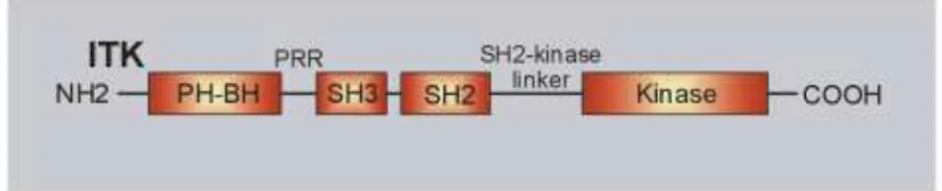
1.2.3.4 ITK
The PTK ITK comprises 620 amino acid residues, has a molecular weight of 72 kDa and is predominantly expressed in T cells.163, 164 ITK contains a N-terminal PH domain, followed by a Zn2+-binding BTK homology (BH) motif, a PRR, a SH3 domain, a SH2 domain, a SH2-kinase linker region and a C-terminal SH1 or kinase domain (Fig. 6).86, 165 Following TCR:CD3:ζ-induced and LAT:SLP-76-mediated activation of PI3K and consecutive accumulation of PIP3 in the plasma membrane, cytosolic ITK is recruited to the
LAT:SLP-76-signalosome.166 Via its SH3 and SH2 domains, ITK cooperatively interacts with a PRR (amino acid residues 184-195) and the phophorylated tyrosine residue Y145 of SLP-76, respectively, and via its PH domain additionally anchors to membrane bound PIP3.152, 167
Figure 6. Domain structure of interleukin-2-inducible T cell kinase (ITK).
The domain structure of ITK is shown with the N-terminal PH-domain containing the BH-motif, the PRR, the SH3-domain, the SH2-domain, the SH2-kinase linker and the C-terminal kinase domain.
Figure adapted from Andreotti AH, et al, Cold Spring Harb Perspect Biol, 2010.86
Only SLP-76-bound ITK is activated by LCK-mediated phosphorylation of the tyrosine residue Y511 of its A-loop and consequently performs autophosphorylation of the tyrosines residue Y180 of its SH3 domain that is thought to further stabilize ITK binding to the LAT:SLP-76-signalosome.168, 169 The main target of ITK is PLCγ-1 that binds to ITK via its second SH2 domain and that is directly activated by ITK via phosphorylation of its tyrosine residue Y783. Thus, ITK is important for the generation of the second messengers inositol 1,4,5-trisphosphate (IP3) and diacylglycerol (DAG).170, 171
Additionally, ITK functions as an adaptor stabilizing the SLP-76:VAV:NCK-complex and thus is involved in actin reorganization (Fig. 3).172, 173
The ITK PRR has been proposed to interact with PLCγ-1, VAV, GRB-2 and FYN.152,
171
How precisely the structure of ITK is involved in protein:protein-interactions and kinase-dependent or kinase-inkinase-dependent functions is not definitively established as ITK has proven resistent to crystalization up to now86 While the isolated kinase-domain of SRC kinases retain their function, the isolated ITK-kinase domain shows only residual activity indicating that the
additonal domains and cooperative interactions with the LAT:SLP-76-signalosome are essential for ITK-kinase-activity.174, 175 In that context, the tryptophan residue W335 in the SH2-kinase linker region appears to be an important activating residue of ITK, while the comparable tryptophan residue in SRC kinases behaves as an inhibitory one.174, 175
ITK is negatively regulated by the lipid phosphatase and tensin homolog (PTEN) that cleaves PIP3 and thus interferes with ITK membrane recruitment.176 Taking into account
available comparative structural data, it has been proposed that an extended ITK configuration with reduced interdomain contacts might lead to ITK-homodimerization thus impeeding association with the LAT:SLP-76-signalosome and infering further negative regulation of adaptor- and kinase-function.177-179
1.2.3.5 PLCγ-1
PLCγ-1 is composed of a N-terminal PH domain that binds to membrane bound PIP3,
a Ca2+-binding EF-hand, two SH2 domains, one SH3 domain for protein:protein-interactions and one C-terminal catalytic PI-PLC X-box. PLCγ-1 hydrolyzes PIP2 to liberate the second
messengers IP3 and DAG.180 DAG activates the serine/threonine protein kinase Cθ (PKCθ)
and the GEF RAS guanyl releasing protein (RASGRP).181 IP3 binds to IP3-receptors (IP3-Rs)
expressed on the endoplasmic reticulum.55 Thus, ITK-mediated PLCγ-1 activation contributes to Ca2+-, MAPK- and NF-κB-signalling (Fig. 3).182
1.2.3.6 PKCθ
PKCs are serine/threonine protein kinases that can be devided into conventional (cPKC), novel (nPKC) and atypical (aPKC) subfamilies activated by Ca2+ and DAG, DAG alone or neither Ca2+ nor DAG, respectively.183 The nPKC PKCθ comprises 706 amino acid residues, has a molecular weight of 82 kDa and is mainly expressed in T cells where it has non-redundant functions in TCR:CD3:ζ- and CD28-(co-)signalling.183, 184
PKCθ consists of a N-terminal C2-like domain, followed by a pseudosubstrate, a C1, a V3 and a C-terminal kinase domain.183 Unlike conventional C2 domains, the regulatory C2-like domain is not able to bind Ca2+ but instead mediates phospholipid- and protein:protein-interactions. Furthermore, it contains the regulatory tyrosine residue Y90 that is phosphorylated by LCK.185 The structure of the pseudosubstrate domain resembles that of PKC substrates, but the serine residue targeted by the kinase domain is replaced by an alanine residue. Thus, the pseudosubstrate domain binds to the kinase domain in cis and induces a