Pour l'obtention du grade de
DOCTEUR DE L'UNIVERSITÉ DE POITIERS Faculté des sciences du sport
Unité de recherche Mobilité, Vieillissement, Exercice (Poitiers) (Diplôme National - Arrêté du 25 mai 2016)
École doctorale : Sciences Biologiques et Santé (Limoges)
Secteur de recherche : Physiologie, biologie des organismes, populations, interactions
Présentée par : Roman Goenarjo
Effect of age, vascular parameters, physical activity, and cardiorespiratory fitness on executive performances : role of
cerebral oxygenation
Directeur(s) de Thèse : Laurent Bosquet, Olivier Dupuy
Soutenue le 06 février 2020 devant le jury
Jury :
Président Benoît Dugué Professeur, MOVE, Université de Poitiers
Rapporteur Cédric Albinet Maître de conférences, INUC, Albi
Rapporteur Fabrice Prieur Maître de conférences, Université d'Orléans
Membre Laurent Bosquet Professeur, MOVE, Université de Poitiers
Membre Olivier Dupuy Maître de conférences, MOVE, Université de Poitiers
Membre Pramita Dwipoerwantoro Professeur, Universitas Indonesia, Depok, Jakarta
Membre Anaïck Perrochon Maître de conférences, Université de Limoges
Membre Karen Davranche Chargée de recherche, Université d'Aix-Marseille
Pour citer cette thèse :
Roman Goenarjo. Effect of age, vascular parameters, physical activity, and cardiorespiratory fitness on executive performances : role of cerebral oxygenation [En ligne]. Thèse Physiologie, biologie des organismes, populations, interactions. Poitiers : Université de Poitiers, 2020. Disponible sur Internet <http://theses.univ-poitiers.fr>
Pour l’obtention du Grade de
DOCTEUR DE L’UNIVERSITE DE POITIERS (Faculté des Sciences du Sport)
(Diplôme National - Arrêté du 25 mai 2016) Ecole Doctorale « Sciences Biologiques & Santé »
Secteur de Recherche : Physiologie, Biologie des Organismes, Populations, Interactions
Présentée par :
Roman Ardian GOENARJO ************************
EFFET DE L’AGE, DU PROFIL TENSIONNEL, ET DES NIVEAUX D’ACTIVITE PHYSIQUE ET DE CONDITION PHYSIQUE SUR LES PERFORMANCES EXECUTIVES : ROLE DE L’OXYGENATION CEREBRALE
************************ Directeur de Thèse : Laurent Bosquet, PR ************************
Soutenue le 6 Février 2020 devant la Commission d’Examen
************************
JURY
Pr. Cédric Albinet Institut National Universitaire Champollion (Rapporteur) Pr. Laurent Bosquet Université de Poitiers (Directeur)
Dr. Karen Davranche Université de Marseille (Membre externe) Dr. Benoit Dugue Université de Poitiers (Membre interne) Dr. Olivier Dupuy Université de Poitiers (Codirecteur) Dr. Pramita Dwipoerwantoro Universitas Indonesia (Membre externe) Dr. Anaick Perrochon Université de Limoges (Codirecteur) Dr. Fabrice Prieur Université d’Orléans (Rapporteur)
i
Resume
De nombreuses études ont montré que l’activité physique régulière et le niveau de condition physique étaient associés à la performance cognitive et plus particulièrement aux fonctions exécutives. Parmi les nombreux mécanismes physiologiques qui sous-tendent cette association, l’oxygénation du cortex préfrontal semble avoir un rôle majeur. Il est bien établi que l'impact spécifique de l'oxygénation préfrontale sur le lien entre l’activité physique et la cognition est influencé par certains facteurs comme le sexe, l’âge, et la santé cardio-vasculaire.
Par conséquent, l'objectif de cette thèse était d'étudier l'impact de l'activité physique et du niveau de condition physique sur les fonctions exécutives au cours du vieillissement chez des hommes sains. L'influence de l'oxygénation du cortex préfrontal et de la santé cardiovasculaire sur la relation « niveau de condition physique – fonctions exécutives » a également été étudiée. A partir de la littérature existante, nous avons formulé l’hypothèse que l’activité physique et le profil cardiorespiratoire auraient des effets bénéfiques sur les fonctions exécutives chez les hommes jeunes et plus âgés, et que pour ces deux populations, l’oxygénation du cortex préfrontal et la santé cardiovasculaire seraient impliquées dans cette relation. Une revue de littérature et quatre études transversales ont été réalisées pour vérifier ces hypothèses.
Notre revue de littérature a indiqué que :
Chez les personnes âgées, un niveau de condition physique plus élevé est associé à de meilleures performances dans certains tests de fonction exécutive. Même si le nombre limité d’études disponibles ne permet pas de tirer des conclusions définitives.
Un niveau de condition physique plus élevé chez les personnes âgées est associée à une moindre rigidité artérielle, à une réactivité vasculaire plus élevée, et à une plus grande amplitude de l'oxygénation cérébrale au cours de l'exercice ou d’une tâche cognitive.
Un plus grand volume de matière grise et une plus grande d’intégrité de la substance blanche sont liés àu profil cardiorespiratoire, mais moins systématiquement associés à l’activité physique.
Au moins de 12 semaines d'un programme d'exercice aérobie sont nécessaires pour obtenir un effet significatif sur les fonctions cognitives
Nos études expérimentales nous ont permis de conclure que :
Chez les jeunes hommes:
Les individus actifs obtenaient de meilleures performances dans les tâches exécutives que les inactifs et avaient un changement important dans l'oxygénation du cortex préfrontal lors des conditions les plus complexes de la tâche de Stroop.
Un niveau de condition physique plus élevé était liée à une meilleure performance en double tâche et une plus grande oxygénation des deux côtés du cortex préfrontal.
ii Chez les hommes âgés:
Le niveau de condition physique n’est pas lié aux performances de Stroop ni à l’oxygénation du cortex préfrontal.
Un niveau de condition physique plus élevé est lié à une meilleure performance et une plus grande oxygénation de cortex préfrontal droit au cours d’une tâche de Stroop dans le groupe de 61+ ans mais pas dans le groupe de 55-60 ans, ce qui suggère l'importance de la classification de groupe d'âge pour évaluer l'effet du niveau de condition physique sur les fonctions exécutives chez les hommes âgés.
Les hommes âgés ont des relations plus fortes entre plusieurs paramètres cardiovasculaires et la performance des taches de Stroop que les jeunes hommes.
Ce projet de recherche montre que l'activité physique et le niveau de condition physique ont des effets positifs sur les fonctions exécutives chez les jeunes hommes et âgés, en particulier dans la tâche la plus complexes. L’oxygénation du cortex préfrontal et la santé cardiovasculaire modulent la relation entre l'activité physique et la cognition. Nous concluons que d’être physiquement actif ou d'avoir un meilleur niveau de condition physique donne des effets avantageux pour la santé vasculaire, l’oxygénation du cortex préfrontal, et les fonctions exécutives non seulement chez les hommes âgés, mais aussi chez les jeunes hommes.
Mots-clés: activité physique, niveau de condition physique, cortex préfrontal, oxygénation cérébrale, spectroscopie infrarouge, pression artérielle, vasculaire, tâche de Stroop, double-tâche, tâche de n-back, jeunes hommes, hommes âgés, athlète master
iii
Abstract
Many studies have reported that regular physical activity and cardiorespiratory fitness were associated with cognitive performance and more selectively with executive functions. Among numerous physiological mechanisms that may underlie the association between them, prefrontal cortex oxygenation seems to play a major role. However, the specific impact of prefrontal oxygenation on the link between physical activity and cognition is influenced by several factors, such as gender, age, or cardiovascular health. Therefore, the aim of this thesis was to investigate the impact of physical activity and cardiorespiratory fitness on executive functions across the adults' age span in healthy males, as well as the influence of prefrontal cortex oxygenation and cardiovascular health. To obtain those objectives, we conducted a review of the effect of physical activity on the brain in older adults and four cross-sectional studies.
From our review, we highlighted that:
In older adults, higher fitness level is associated with better performance in several executive function tests. Even though the limited number of studies available makes it difficult to draw definitive conclusions.
Better cardiovascular fitness in older adults is associated with improve arterial stiffness, higher vascular reactivity, and greater amplitude of cerebral oxygenation during
exercise or cognitive tasks.
Greater gray matter volume and white matter integrity were related to the cardiorespiratory fitness but less consistently related to physical activity.
At least 12 weeks of an aerobic exercise program are required to give advantageous effects to the brain
And our experimental works show that:
In young males :
The active individuals performed better in executive tasks than their inactive counterparts and had a larger change in prefrontal cortex oxygenation during the most complex conditions of Stroop task.
High cardiorespiratory fitness was related to a better performance in dual-task and greater oxygenation on both sides of the prefrontal cortex.
In older males :
Cardiorespiratory fitness is not related to Stroop task performance nor prefrontal cortex oxygenation in overall older males.
Higher cardiorespiratory fitness was related to a better performance and greater right prefrontal cortex oxygenation during a Stroop task in 61+ years old group but not in
55-iv
60 years old group, suggesting the importance of age-group classification to evaluate the effect of cardiorespiratory fitness on executive function in older male subjects.
Older males have stronger relationships between several vascular parameters and Stroop task performance than young males
This work shows the relationship between physical activity and cardiorespiratory fitness on executive functions in young and older males. The potential neurophysiological mechanisms that underlie that relationship, especially prefrontal oxygenation and vascular health, are presented.
Keywords: physical activity, cardiorespiratory fitness, prefrontal cortex, cerebral oxygenation, functional near-infrared spectroscopy, blood pressure, vascular, Stroop task, dual-task, n-back task, young males, older males, master athlete
v
Table of contents
Resume ... i
Abstract ... iii
Table of contents ... v
List of publication ... ix
List of tables ... xi
List of figures ... xii
INTRODUCTION ... xv
LITERATURE REVIEW ... 2
1
Executive Function... 2
1.1 Executive Function ... 2
1.2 Components of Executive Function ... 2
1.2.1 Inhibitory Control ... 3
1.2.2 Working Memory ... 3
1.2.3 Cognitive Flexibility ... 4
1.2.4 Resume ... 4
1.3 Brain Organization of Executive Function ... 4
1.3.1 Frontal Lobe ... 5
1.3.2 Parietal Lobe ... 6
1.3.3 Temporal Lobe ... 6
1.3.4 Occipital Lobe ... 7
1.3.5 Resume ... 7
1.4 Effect of Executive Function on The Health-Promoting Behaviors ... 8
1.4.1 Resume ... 9
1.5 Summary of Section ... 10
2
Physiological Basis of Brain Function ... 12
2.1 Brain Cells and Their Functions ... 12
2.1.1 Neurons ... 12
2.1.2 Glia ... 13
vi
2.2 Brain Activation ... 15
2.2.1 Neuronal Charged Membranes ... 15
2.2.2 Resting Membrane Potential ... 16
2.2.3 Action Potential ... 17
2.2.4 Resume ... 18
2.3 Energy Demands for Brain Activation ... 18
2.3.1 Resume ... 20
2.4 Brain Metabolism ... 21
2.4.1 Model of Glucose Metabolism ... 21
2.4.2 The Cellular Specificity of Energy Metabolism in Neuron and Astrocyte... 23
2.4.3 Neurometabolic Coupling between Astrocytes and Neurons ... 25
2.4.4 Role of Oxygen in Neurovascular Signaling ... 27
2.4.5 Integration of Neurovascular Coupling during Neuronal Activity ... 28
2.4.6 Resume ... 30
2.5 Cerebral Blood Flow (CBF) ... 31
2.5.1 Effect of CBF on Executive Function ... 31
2.5.2 Main Determinants of CBF ... 31
2.5.3 Resume ... 34
2.6 Summary of Section ... 34
3
Aging and Executive Function ... 36
3.4 Physiological Implication of Aging on Body Organs ... 36
3.5 Theories/Hypothesis of Cognitive Aging ... 37
3.5.1 Frontal Hypothesis... 37
3.5.2 Processing Speed and Inhibition Theory ... 38
3.5.3 Cognitive Reserve Hypothesis ... 39
3.5.4 Resume ... 39
3.6 Aging and Neuro-Cardiovascular Parameters ... 39
3.6.1 Aging and Angiogenesis ... 39
3.6.2 Resume ... 41
3.6.3 Aging and Cerebral Blood Flow ... 41
3.6.4 Resume ... 44
3.7 Aging on Brain Structures and Functions ... 44
3.7.1 Effect of Aging on Grey Matter ... 44
vii
3.7.3 Resume ... 46
3.8 Aging and Executive Function ... 46
3.8.1 Working Memory ... 47
3.8.2 Selective Attention and Inhibition Control ... 48
3.8.3 Cognitive Flexibility ... 48
3.8.4 Resume ... 50
3.9 Summary of Section ... 50
4
Physical Activity and Executive Function ... 52
4.1 Physical Activity and Neuro-Cardiovascular Parameters ... 52
4.1.1 Physical Activity and Angiogenesis ... 52
4.1.2 Resume ... 54
4.1.3 Physical Activity and Cerebral Blood Flow ... 54
4.1.4 Resume ... 57
4.2 Physical Activity on Brain Structures and Functions ... 57
4.2.1 Grey Matter ... 57
4.2.2 White Matter ... 58
4.2.3 Resume ... 60
4.3 Physical Activity and Executive Function ... 60
4.3.1 Working Memory ... 60
4.3.2 Cognitive Flexibility ... 62
4.3.3 Resume ... 63
4.4 Summary of Section ... 63
5
Summary of the Literature Review ... 64
6
Objective and Hypotheses ... 65
6.1 Objective ... 65
6.1.1 Studies of the Association between Physical Activity, Cerebral Oxygenation, and Executive Function in Young Males (Study 1 and 2) ... 65
6.1.2 Studies of the Association between Physical Activity, Cerebral Oxygenation, and Executive Function in Older Males (Study 3 and 4) ... 65
6.1.3 Studies of the Influence of Vascular Health on the Interaction between Age, Physical Activity, and Executive Function (Study 5) ... 66
6.2 Hypotheses ... 66
7
Materials and Methods ... 67
viii
7.1.1 Participants ... 67
7.1.2 General Protocols ... 67
7.1.3 Physical Activity Measurement ... 70
7.1.4 Physiological Measurement ... 71
7.1.5 Psycho-Cognitive Measurement ... 75
STUDIES OF THE THESIS ... 79
1
Study 1 ... 79
1.1 Resume ... 79 1.2 Article ... 812
Study 2 ... 98
2.1 Resume ... 98 2.2 Article ... 1003
Study 3 ... 123
3.1 Resume ... 123 3.2 Article ... 1244
Study 4 ... 138
4.1 Resume ... 138 4.2 Article ... 1405
Study 5 ... 154
5.1 Resume ... 154 5.2 Article ... 156GENERAL DISCUSSION ... 168
GENERAL CONCLUSION ... 175
REFERENCES ... 177
ix
List of publication
List of publications on this thesis :1. Roman Goenarjo, Laurent Bosquet, Sarah Fraser, Anaick Perrochon, Nicolas Berryman, Valentine Metier, Olivier Dupuy. Prefrontal oxygenation reserve: The relationship between physical activity level and the cognitive load during a Stroop task in healthy young males. In revision process in Frontiers in Behavioral Neuroscience.
2. Roman Goenarjo, Olivier Dupuy, Sarah Fraser, Anaick Perrochon, Nicolas Berryman, Laurent Bosquet. Cardiorespiratory fitness, blood pressure, and cerebral oxygenation during a dual-task in healthy young males. Published in Behavioral Brain Research. DOI: 10.1016/j.bbr.2019.112422.
3. Olivier Dupuy, Roman Goenarjo, Sarah Anne Fraser, Louis Bherer, Laurent Bosquet. (2019) Master athlete and cognitive performance: What are the potential explanatory neurophysiological mechanism? Published in Movement and Sports Sciences. DOI: 10.1051/sm/2019023.
4. Roman Goenarjo, Olivier Dupuy, Sarah Fraser, Anaick Perrochon, Nicolas Berryman, Laurent Bosquet. The relationship between cardiorespiratory fitness and prefrontal oxygenation during a Stroop task in healthy older males. In preparation to be submitted. 5. Roman Goenarjo, Olivier Dupuy, Sarah Fraser, Anaick Perrochon, Nicolas Berryman, Laurent Bosquet. The correlation between vascular parameters and Stroop task performance in young and older males. In preparation to be submitted.
List of communications on this thesis : Poster communications :
1. Roman Goenarjo, Olivier Dupuy, Sarah Fraser, Anaick Perrochon, Nicolas Berryman, Laurent Bosquet. (March 2017). Interactions of blood pressure regulation, brain oxygenation, and executive functions during dual task walking in older men. Was presented in le séminaire thématique de l’école doctorale Biosante, Université de Poitiers, Poitiers, France.
2. Roman Goenarjo, Olivier Dupuy, Sarah Fraser, Anaick Perrochon, Nicolas Berryman, Laurent Bosquet. (Juin 2018). Interactions of physical activity, cerebral oxygenation and executive functions in older males. Was presented in the 10th Joint Working Group Indonesia-France Cooperation 2018 in Higher
Education and Research, Poitiers, France. Oral communications :
1. Roman Goenarjo, Olivier Dupuy, Sarah Fraser, Anaick Perrochon, Nicolas Berryman, Laurent Bosquet. (Mars 2018). Interactions of blood pressure regulation, brain oxygenation, and executive functions during dual task walking in young and older men. Was presented in le séminaire thématique de l’école doctorale Biosante, Université de Poitiers, Poitiers, France.
x
2. Roman Goenarjo (Juin 2018). Interactions cœur-cerveau: de la rigidité artérielle, de la sensibilité baroréflexe et de l'oxygénation cérébrale sur les fonctions exécutives pendant une double tâche. Was presented in the competition of “Ma These en 180 secondes (MT180)” in the 10th Joint Working Group
Indonesia-France Cooperation 2018, Poitiers, France.
3. Roman Goenarjo, Olivier Dupuy, Anaick Perrochon, Nicolas Berryman, Sarah Fraser, Laurent Bosquet. (Juillet 2018). The effect of aerobic fitness and blood pressure on brain activation during a dual task in healthy young adults. Was presented in the 23ème annual congress of European Congress of Sport Science
(ECSS), Dublin, Ireland.
4. Roman Goenarjo, Olivier Dupuy, Anaick Perrochon, Nicolas Berryman, Sarah Fraser, Laurent Bosquet. (Juillet 2019). Prefrontal oxygenation reserve: link between physical activity level and executive functioning during computerized stroop task in healthy young males. Was presented in the 23ème annual congress of European Congress of Sport Science (ECSS), Prague, Czech République and was participated in the competition of Young Investigator Award (YIA) ECSS 2019.
xi
List of tables
Table 1. Relevancy of Executive Function to Some Aspects of Life ... 3
Table 2. Relation of Health Behaviors with Executive Function ... 9
Table 3. Extracellular and Intracellular ion concentrations during resting membrane potential ... 16
Table 4. Relative oxygen consumption in adult human organs ... 19
Table 5. Percentage of Energy Consumption in Neurons ... 20
Table 6. Description of Cognitive Aging Theories or Hypotheses ... 38
Tabel 7. General and specific switch costs between young-aged, middle-aged, and old aged ... 49
xii
List of figures
Figure 1. Resume of components of executive function and its relevance to some aspects
of life ... 5
Figure 2. The lobes of the brain ... 5
Figure 3. The Lobes of the brain and its association with executive function ... 8
Figure 4. The reciprocal relationship between executive function, healthy behaviors, and health benefits ... 8
Figure 5. Schematic integration of components and brain organization of executive function in their relationship with healthy behaviors and neurovascular components ... 10
Figure 6. Cartoon illustration of neuron and glia in the brain ... 12
Figure 7. Cartoon illustration of a neuron within the nervous system ... 13
Figure 8. Cartoon illustration of glial cells within the neurovascular system ... 14
Figure 9. A sequence of action of a voltage-gated ion channel ... 15
Figure 10. Interaction of ions, channels, and Na+/K+ ATPase during resting membrane potential in a neuron ... 16
Figure 11. Events involving ions, channels, and Na+/K+ ATPase that lead to depolarization ... 17
Figure 12. Events involving ions, channels, and Na+/K+ ATPase that lead to hyperpolarization ... 18
Figure 13. Distribution of energy use in the brain; specified into the basic non-signaling processes and the specialized physiological signaling processes ... 19
Figure 14. Transport and metabolism of glucose into the neurons ... 21
Figure 15. Schematic representation of four glucose metabolism pathways in the brain .. 22
Figure 16 Comparative energy metabolism pathways of glucose between astrocyte and neuron. ... 23
Figure 17. Schematic illustration of the Astrocyte-neuron lactate shuttle (ANLS) model . 26 Figure 18. Neurovascular signaling responses at a decreased concentration of oxygen ... 27
Figure 19. Hemodynamics response curves, showing changes in HbO2, HHb, tHb concentration during brain activation ... 28
xiii
Figure 20. Multiscale integration of neurovascular coupling and brain activation ... 30
Figure 21. The conceptual framework of the integrated regulation of brain perfusion ... 33
Figure 22. Schematic integration of brain activity, neurovascular coupling, and cerebral blood flow (CBF) ... 34
Figure 23. Changes in maximal physiological body system function as a function of age . 36 Figure 24. Chemical structure of dopamine ... 37
Figure 25. Cartoon representation of the x-ray structure of chemically synthesized VEGF ... 39
Figure 26. Regulation of angiogenesis ... 40
Figure 27. Decade-by-decade alterations in prefrontal CVR and CBF ... 41
Figure 28. Secretion rates of adrenaline and noradrenaline at various age groups of Wistar rats ... 43
Figure 29. The Hemispheric Asymmetry Reduction in Older Age model (HAROLD) ... 46
Figure 30. Summary of effects of aging on neuro-cardiovascular parameters, brain structures and functions, and executive functions ... 50
Figure 31. Exercise-induced activation of transcription factors in angiogenesis ... 53
Figure 32. Risk of progression from prehypertension to hypertension group on fitness level categories ... 55
Figure 33. Cardiac output in relation to systemic ̇VO2 in untrained and trained subjects and elite athletes ... 56
Figure 34. Summary of effects of physical activity on neuro-cardiovascular parameters, brain structures and functions, and executive functions ... 63
Figure 35. Integrated-algorithm of physical activity and aging effects on neuro-cardiovascular parameters, brain structures and functions, and executive functions ... 64
Figure 36. Schematic illustration of relations of each study to research topics ... 66
Figure 37. Study design for Study 1 ... 68
Figure 38. Study design for Study 2 and Study 5 ... 68
Figure 39. Study design for Study 4 ... 69
xiv
Figure 41. The French version of the Global Physical Activity Questionnaire (GPAQ) .... 71
Figure 42. Image of Leicester Height Measure ... 71
Figure 43. Image of Tanita® BC-418 ... 71
Figure 44. Image of the SphygmoCor® system ... 72
Figure 45. Image of Mobil-O-Graph 24-hour Ambulatory Blood Pressure Monitor (AMBP) ... 72
Figure 46. Image of Finapres® NOVA ... 73
Figure 47. Image of motorized treadmill Valiant 2 sport ... 73
Figure 48. Illustration of the Modified Balke walking protocol ... 73
Figure 49. Image of PortaLite fNIRS system ... 74
Figure 50. Image of the GAITRite® system ... 74
Figure 51. Image of the form of the French version of Beck’s Depression Impression (BDI) ... 75
Figure 52. Image of the form of the French version of Montreal Cognitive Assessment (MoCA) ... 76
Figure 53. Image of form Trail Making Test B (TMT B) ... 76
Figure 54. Paradigms of Stroop task in naming, inhibition, and switching (flexibility) conditions ... 77
xv
INTRODUCTION
Executive function refers to a collection of cognitive abilities such as planning, set maintenance, impulse control, working memory, and attentional control” (Roberts and Pennington 1996). In general, executive function refers to an ability to maintain an appropriate problem-solving set for the accomplishment of a future goal (Yogev-Seligmann, Hausdorff, and Giladi 2008; Welsh and Pennington 1988). Executive function is essential for the quality of life, as executive functions are associated with the mental and physical aspects of health, also success in school and life. As human goes older, executive function is one of the main contributors to cognitive decline (Glisky 2006). Interestingly, when older adults receive supporting stimuli, executive function performance is better (Kirova, Bays, and Lagalwar 2015). Those facts reflect that executive function is a malleable cognitive function and could be influenced by various factors.
Aging is a factor that influences the executive functions. Several hypotheses, such as the hemispheric asymmetry reduction in older age model (HAROLD), the compensation-related utilization of neural circuits hypothesis (CRUNCH), and the astrocyte-neuron lactate shuttle (ANLS) model put forward a physiological basis of the effect of aging in the brain as a functional unit (Cabeza 2002; Reuter-Lorenz and Cappell 2008; Pierre J. Magistretti and Allaman 2015). Hypothetically, this relationship is mediated by the cerebrovascular system (Pierre J. Magistretti and Allaman 2015). The age-related change and dysfunction in the cerebrovascular system differentiate the executive function performances in young and older adults.
On the other side, physical activity has been known to give benefit to the cognitive function and to resist the deteriorating effect of aging (Mandolesi et al. 2018). More specifically, physical activity has been reported to improve executive function performance and cerebrovascular control. However, cardiovascular control is regulated differently in young and older adults (Uchino, Birmingham, and Berg 2010; Strait and Lakatta 2012). The benefit of physical activity, as well as the effect of aging on executive function, may be mediated differently between young and older adults (Strait and Lakatta 2012). The variety of methods that are used in the topic regarding the measure of physical activity and executive function give the challenge to draw a conclusion between results obtained. Standardization of the methods of research in this topic remains an issue that needs to be considered. Therefore, a comprehensive study that integrates components on the axis of the cardio-neurovascular-cognition in young and older subjects is required to give a better illustration of the relationship between physical activity and executive function performance.
We hope that we could shed some light on the effect of age and physical activity on executive function in males with this thesis. Therefore, we have two objectives: (1) To understand the impact of age and physical activity on cerebral oxygenation and executive function in males. (2)
xvi
To observe the influence of age on the correlation between vascular health and executive function. To obtain those objectives, five studies were conducted in this thesis project.
The first objective is defined by four studies, a literature review study and three experimental studies. The review study will summarize the effect of physical activity on brain structure, brain function, and executive function performance in older adults. Therefore, we could grasp the general view of the effect of physical activity on the brain. Hence, three cross-sectional studies will be directed to observe the impact of aging and the effect of physical activity on the brain with an emphasis on cerebral oxygenation and executive functions. Last, a study to identify the relation of vascular health variables to executive function will be conducted to fulfill the second objective. We will compare several vascular parameters and executive function performance on young and older male groups to observe the correlation between parameters and performances and also whether these groups modified the relationship in a different manner.
1
__________________________________________________________________________________
“On Wednesday, 13 September 1848, a railway foreman named Phineas Gage had a work accident in which an explosion launched a tamping iron out of the borehole and through the left side of his skull
(Haas 2001). It entered from under his left cheekbone, exiting through the top of his head, destroyed some
of his brain (Baars and Gage 2010). Amazingly, Phineas Gage is still alive after that accident, he even could walk, speak, and had normal awareness (Haas 2001; Baars and Gage 2010). However, He had changed from a capable foreman with a well-balanced mind and shrewd business sense to a fitful, irrelevant, grossly profane, impatient, capricious, and vacillating individual (Haas 2001). He was also incapable to proceed with any plans. He was “no longer Gage” according to his friends (Haas 2001). After his dead, the skull and the tamping iron were donated to the Warren Museum at Harvard University School of Medicine (Haas 2001). Computer reconstruction on the skull found that the areas of the damaged brain corresponded to the frontal lobe (Baars and Gage 2010). This series of events have inspired research in the field of cognitive neuroscience about the relationship between frontal lobe and executive function.”
2
LITERATURE REVIEW
1 Executive Function
This section discusses the definition and components of executive function, brain organization of executive function, and the physiological basis of executive functioning in the brain. It presents the implications of executive impairment in various aspects. This chapter also explains the recent brain organization of executive function.
1.1 Executive Function
Executive function is one of the cognitive domains according to the fifth edition of Diagnostic and Statistical Manual of Mental Disorders (DSM-5) along with complex attention, learning and memory, language, perceptual-motor function, and social cognition (Sachdev et al. 2014). It was Pribram in 1973, one of the first to use the term “executive” to describe the function of the frontal cortex (Pribram 1973). After that, Lezak (1982) defines executive function as a mental capacity necessary for setting goals, planning how to achieve them, and accomplishing these plans effectively (Lezak 1982). Then, Welsh and Pennington (1988) added inhibition as a part of executive function and describe it as “the ability to maintain set for future-oriented and goal-directed activity” (Welsh and Pennington 1988). This description includes the capacity to inhibit or defer a response, to plan strategically future actions, and to maintain a mental representation of the desired goal (Welsh and Pennington 1988). Then in 1996, Roberts and Pennington tried to cluster the components of executive function as they stated: “Executive function refers to a collection of related but somewhat distinct abilities such as planning, set maintenance, impulse control, working memory, and attentional control” (Roberts and Pennington 1996). In general, executive function refers to an ability to maintain an appropriate problem-solving set for the accomplishment of a future goal (Yogev-Seligmann, Hausdorff, and Giladi 2008; Welsh and Pennington 1988).
Modification of information by executive function is essential for the quality of life. With EF, skills such as reasoning, planning, and problem-solving are developed to overcome problems. Indeed, EFs are relevance to the mental and physical aspects of health, also success in school and life (see Table 1).
1.2 Components of Executive Function
There are three core of executive function (Diamond 2013; Miyake et al. 2000; Miyake and Friedman 2012): inhibition [inhibitory control, including self-control (behavioral inhibition) and interference control (selective attention and cognitive inhibition)], working memory, and cognitive flexibility (also called set-shifting, mental flexibility, or mental set shifting and closely linked to creativity). From these, the more complex executive functions are built, such as reasoning, problem-solving, and planning (Collins and Koechlin 2012).
3 1.2.1 Inhibitory Control
Inhibitory control is described as control of attention, behavior, thoughts, and emotions to outweigh an internal drift or external lure, and instead, do what is more appropriate or needed (Diamond 2013). Inhibitory control enables us to focus attention on a stimulus and suppressing attention to other stimuli. Besides known as selective or focused attention, this has been called attentional control or attentional inhibition, endogenous, top-down, active, goal-driven, voluntary, volitional, or executive attention (Theeuwes 2010). Self-control is an aspect of inhibitory control of behavior and emotion. This aspect is about resisting temptations and not acting impulsively (Diamond 2013). Stroop test is the most frequently used test in the research measuring inhibitory control. (Faria, Alves, and Charchat-Fichman 2015)
Table 1. Relevancy of Executive Function to Some Aspects of Life
Aspects Relevancy of Executive Function
Mental health Executive functions are impaired in mental disorders, including addictions, attention deficit hyperactivity (ADHD), conduct disorder, depression, obsessive-compulsive disorder (OCD), schizophrenia (Baler and Volkow 2006; Lui and Tannock 2007; Fairchild et al. 2009; Taylor Tavares et al. 2007; Penadés et al. 2007; Barch 2005) Healthy lifestyle Better executive functions are associated with higher participation in regular exercise
classes, less obesity, overeating, and substance abuse (Will Crescioni et al. 2011; H. V. Miller, Barnes, and Beaver 2011; Riggs et al. 2010; Hall, Elias, et al. 2008)
School readiness Executive functions are more critical for school readiness than IQ or reading or math score (Blair and Razza 2007; Duncan et al. 2007)
School success Executive functions predict both math and reading competence throughout the school years (Borella, Carretti, and Pelegrina 2010; Duncan et al. 2007)
Job success Better executive functions are associated with higher productivity and lower difficulty to find and keep a job (Bailey 2007)
Marital harmony Partners with better executive functions are less difficult to live with, more responsible, and less likely to act impulsively (Eakin et al. 2004)
Public safety Better executive functions are associated with less reckless behavior, violence, and crime (Broidy et al. 2003; Denson et al. 2011)
Risk of fall Better executive functions are associated with decreased risk of falls (Mirelman et al. 2012)
Quality of life Better executive functions are associated with a better quality of life (Davis et al. 2010)
1.2.2 Working Memory
Working memory is defined as the capability to hold information in mind and to manipulate or to mentally work with it (Baddeley 1998; Smith and Jonidesl 1999). Working memory is essential for making sense of information that develops over time. Many activities require holding in mind what happened previously and relating to what comes later, such as making sense of written or spoken language, doing a calculation, reordering items, and translating instruction into action plans. Working memory is also crucial to the ability to see the connection between things (Diamond 2013). Digit Forward and Backward subtests (WAIS-R or WAIS-III) and N-back test are two frequently used tests in this component. (Faria, Alves, and Charchat-Fichman 2015)
4 1.2.3 Cognitive Flexibility
Cognitive flexibility is termed as the ability to switch flexibly back and forth between tasks, operations, or mental sets (Miyake et al. 2000). Cognitive flexibility is constructed on inhibitory control and working memory (Davidson et al. 2006). In cognitive flexibility, switching between tasks needs a capacity to change perspectives. To change perspectives, inhibition of the previous perspective and activation of another perspective are needed (Diamond 2013). In this logic, cognitive flexibility involves inhibitory control and working memory. Another aspect of cognitive flexibility is creative thinking or to change a way to solve a problem (Diamond 2013). Trail Making Test (TMT) form B, Wisconsin Card Sorting Test (WCST), and Stroop Test are the most frequently used tests to assess cognitive flexibility (Faria, Alves, and Charchat-Fichman 2015).
1.2.4 Resume
Figure 1. Resume of components of executive function and its relevance to some aspects of life
1.3 Brain Organization of Executive Function
The underlying organization of executive function has historically been attributed to the frontal lobes, Firstly based on work by Luria (1969), who reported that patients with frontal lobe damage failed to modify their behavior appropriately on various mental tasks (Munro et al. 2018).
Executive function is defined as the ability to maintain an appropriate problem-solving set for the accomplishment of a future goal.
The executive function consists of three core components: inhibition, working memory, and flexibility.
Executive function is related to various aspects of life, such as mental health, unhealthy lifestyle, school readiness and success, job success, marital harmony, public safety, risk of fall, and quality of life.
5
Shallice (1982) and Goldman-Rakic (1987) declared that the prefrontal cortex has an important role in executive function (Shallice 1982; Goldman-Rakic 1987). However, Some of the investigators in this topic, such as Fletcher (1996) argued that the underlying physiological organization of executive function is complex and likely involves integrated neural networks from various brain regions (J. M. Fletcher 1996). In the following part, according to their lobes, as presented in Figure 2, brain lobes associated with tasks of executive functions in a healthy subject are described.
1.3.1 Frontal Lobe
The frontal brain lobe, in general, was implicated in all executive functions’ measures across all studies. Based on tasks, specifically, the N-back working memory task indicated activation in similar areas. Specifically, results from Drobyshevsky, Baumann, and Schneider (2006) showed activation in the dorsolateral prefrontal cortex and the left precentral cortex (Drobyshevsky, Baumann, and Schneider 2006). Moreover, this study reported decreased activation in the left dorsolateral prefrontal cortex, inferior frontal cortex, and the precentral gyrus with increased age. Satterthwaite et al. (2013) reported increased activation in the dorsolateral prefrontal cortex due to an increased working memory load from the N-back task (Satterthwaite et al. 2013). Also, Ball et al. (2011) reported increased activation in the lateral prefrontal cortex, as well as the left inferior and middle frontal gyrus (Ball et al. 2011).
Figure 2. The lobes of the brain (Taken from (Lumen 2019b))
Secondly, fMRI results while performing the go/no-go task indicated increased activation in the right middle and inferior frontal gyrus (Fuentes-Claramonte et al. 2016; Mulligan et al. 2014; Ball et al. 2011), as well as bilateral activation in the anterior prefrontal, and inferior and middle frontal lobes (Dodds, Morein-Zamir, and Robbins 2011; Mulligan et al. 2014). The frontal brain lobe increased activation was found to be associated with inhibition (Ball et al. 2011; Dodds, Morein-Zamir, and Robbins 2011; Mulligan et al. 2014).
6
Several fMRI studies that used the Stop-signal task (SST) reported increased activation in the frontal gyrus, and prefrontal and frontal cortices (Chikazoe et al. 2009; Jimura et al. 2014). Specifically, Ide and Li (2011) reported the adult participants of their study showed increased activation in the ventrolateral prefrontal cortex during behavior adjustment as a response to errors (J. S. Ide and Li 2011). Similarly, Hu and Li (2012) indicated activation in the right prefrontal cortex during reparatory motor inhibition (Hu and Li 2012). Also, adult participants with shorter stop-signal reaction times showed greater activity in the right orbital frontal cortex during preparatory inhibition (Hu and Li 2012). White et al. (2014) found increased right frontal pole and left middle frontal gyrus activation was correlated with quicker processing of the Go stimulus in Stop and Go trials and the right inferior frontal gyrus, the medial frontal gyrus, and basal ganglia were correlated with the stimulus processing speed during Stop-trials (White et al. 2014).
Lastly, shifting tasks were found to increase in activation within the frontal brain lobe (Munro et al. 2018). Specifically, Witt and Stevens (2012) found activity in frontal lobes during the switching of a task (Witt and Stevens 2012). Another study indicated that adults with an average IQ showed greater activation in the prefrontal lobe via fMRI than did a comparison group of high IQ individuals (Graham et al. 2010). Besides, a study with youth participants who underwent DTI imaging while completing the Stroop task found improved task switching was related to greater integrity of the precentral gyrus WM and superior corona radiate in the frontal cortex (Seghete, Herting, and Nagel 2013).
1.3.2 Parietal Lobe
The parietal brain lobe is activated during a range of executive function tasks, including the N-back task, go/no-go task, and SST (Munro et al. 2018). Regarding the N-N-back task, fMRI results found to significantly activate the precuneus, as well as the superior parietal cortex (Satterthwaite et al. 2013). With the go/no-go task, a study indicated that the attention-shifting requirements within this task were associated with the increased activation in the left inferior parietal cortex of the adult participants (Dodds, Morein-Zamir, and Robbins 2011). Similarly, the SST was also found increased activity in the precuneus among a group of healthy adult participants (Chikazoe et al. 2009; S. Zhang and Li 2012).
1.3.3 Temporal Lobe
In the healthy subjects, no study was found an association between executive functioning and activation in the temporal lobe. However, in subjects with ADHD or dementia, four studies indicated brain activation in the temporal lobe (Banich et al. 2009; Dibbets et al. 2010; Spinelli et al. 2011; Eslinger et al. 2011). Specifically, those studies reported activation in the temporal gyrus based on fMRI imaging, among adult, children, and adolescent participants with ADHD and in patients with dementia. The executive functions’ measures consisted of the Stroop task (Eslinger et al. 2011; Banich et al. 2009), a switch task (Dibbets et al. 2010), and the go/no-go task (Spinelli et al. 2011).
7 1.3.4 Occipital Lobe
Same as in the temporal lobe, no study was found an association between executive functioning and activation in the occipital lobe in healthy subjects. In studies among participants with ADHD, results in the occipital brain lobe varied across three studies, with each study using a different executive function measure. Specifically, Dibbets et al. (2010) found increased activation in the lingual gyrus of the occipital lobe from administration of the switch task (Dibbets et al. 2010), while two other studies indicated reduced activation in the cortico-subcortical networks of the bilateral occipital lobe from the N-back task (Massat et al. 2012) and the right occipital cortex from the go/no-go task (Suskauer et al. 2008).
1.3.5 Resume
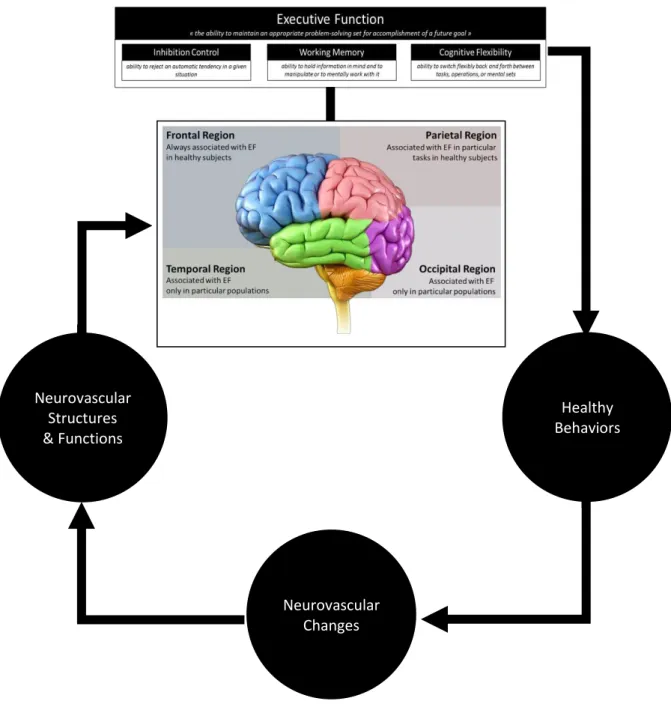
Figure 3. The Lobes of the brain and its association with executive function
The frontal lobe was reported in all studies that relate brain lobe activation with executive function tasks.
The parietal lobe was also associated with executive functions in several studies that used go/no-go task, Stop-signal task, and N-back task.
The temporal and occipital lobes were involved during executive function tasks in subjects with ADHD or dementia but not in healthy subjects.
Studies of brain organization of executive function showed that the frontal lobe is the main lobe of executive functions with the involvement of the parietal lobe in several tasks. Temporal and occipital lobes seem activated only in a particular population of subjects.
8
1.4 Effect of Executive Function on The Health-Promoting Behaviors
In addition to being affected by physical activity level and cardiorespiratory fitness, executive function is also likely to be necessary for the initiation and maintenance of physical activity and cardiorespiratory fitness by promoting healthy behaviors. Healthy behaviors typically involve the pursuit of longer-term benefits at the expense of short-term gain (Chapman 2005). Important outcomes such as weight loss, fitness, and health are all achieved by effortfully and consistently changing behavior in the present (e.g., by doing exercise, by eating healthy foods, by resisting sedentary activities, by not smoking, or by drinking less alcohol). Therefore, it is reasonable that good EF— an ability to maintain an appropriate problem-solving set for the accomplishment of a future goal (Yogev-Seligmann, Hausdorff, and Giladi 2008; Welsh and Pennington 1988). — should improve the chances that people will be able to initiate and maintain healthy behaviors to obtain health benefits. This reciprocal relationship of executive function, healthy behaviors, health benefits (specifically on the brain and neurovascular function) outlined in figure 4.
Figure 4. The reciprocal relationship between executive function, healthy behaviors, and health benefits
A cohort study from Peeters et al. (2015) followed up over two years, 534 young adults who yet to drink alcohol. Executive function performance at the beginning of the study significantly predicts when they begin drinking alcohol and whether or not they experience an over-drinking (Peeters et al. 2015). The same study also reported that in the next 6-month period, Executive function also significantly predicts an increase in alcohol consumption (Peeters et al. 2015). Studies of transcranial magnetic stimulation (TMS), a technique where the activity in specific areas of the cortex can be reduced using external electromagnetic pulses, further revealed the role of executive function on health behaviors. Lowe et al. (2014) used TMS focused at the left dorsolateral prefrontal cortex (DLPFC) to decrease its activation temporarily [338]. They reported that the subjects had a decrease in performance on executive function tests and an increase of self-reported snack food cravings and consumption after left DLPFC stimulation (Lowe, Hall, and Staines 2014). The result is in line with fMRI studies, which report that left DLPFC activation is associated with the self-control of cravings for unhealthy foods and cigarettes (Hare, Camerer, and Rangel 2009; Kober et al. 2010).
9
Executive function is essential for healthy behaviors. With executive function, skills such as reasoning, planning, and problem-solving are developed to initiate and maintain healthy behaviors. Indeed, executive functions are related to participation and maintenance of a wide range of health behaviors (see Table 2).
Table 2. Relation of Health Behaviors with Executive Function
Aspects Relation with Executive Function
Alcohol consumption
Individuals with better executive functions having less alcohol temptation and less likely to drink to excess or develop problems with alcohol (Hofmann et al. 2012; Fernie et al. 2013)
Food consumptions
Individuals with better executive functions are more likely to stick to their stated dietary intentions, less likely to consume fatty foods, have less unintentional chocolate consumption (Allan, Johnston, and Campbell 2011; Allan, McMinn, and Daly 2016; Hall 2012)
Medication more likely to correctly adhere to medication regimes (Stilley et al. 2010; Panos et al. 2014)
Physical activity
Better executive functions predict the realization of physical activity intentions, participation in regular exercise classes, related to a closer correspondence between intended and actual physical activity, and associated with physical activity behaviors (Hall, Fong, et al. 2008; Hall, Elias, et al. 2008; McAuley et al. 2011; Sheeran 2002)
Smoking
Smokers with greater brain activation in the right inferior frontal gyrus, pre-supplementary motor area, and basal ganglia during a response inhibition task are less likely to smoke in response to cravings than others; and better executive function smokers are more likely to quit smoking, successfully (Brega et al. 2008; Nestor et al. 2011).
Weight loss program
Dieters with better response inhibition are more likely to resist temptation successfully and to lose weight than the ones with weak response inhibition (Hofmann et al. 2014) Attachment to
general health behaviors
Executive functions moderate the strength of the association between intentions and health behaviors (Hall, Fong, et al. 2008)
1.4.1 Resume
In addition to being affected by physical activity level and cardiorespiratory fitness, executive function is also likely to be necessary for the initiation and maintenance of physical activity and cardiorespiratory fitness by promoting healthy behaviors. Executive functions are related to participation and maintenance of a wide range of
health behaviors
The link of executive function, healthy behaviors, and health benefits is likely a reciprocal relationship.
10
1.5 Summary of Section
____________________________________________________________________
Figure 5. Schematic integration of components and brain organization of executive function in their relationship with healthy behaviors and neurovascular components
Neurovascular Structures & Functions Neurovascular Changes Healthy Behaviors
11
Having its first annual meeting in 1971, cognitive neuroscience, a relatively new multi-disciplinary science that is concerned with the biological processes and aspects that underlie cognition, began to be known publicly (“Cognitive Neuroscience” 2019; Baars and Gage 2010). At that time, research in the field of cognitive neuroscience was constrained by the unavailability of conducting empirical study at physiological level (Posner and DiGirolamo 2000). The discoveries and developments of various brain imaging tools, such as EEG, fMRI, and fNIRS, helped to unveil mysteries of human brain and opened various opportunities for cognitive neuroscientists to investigate and determine brain-cognition mechanisms, from the development of the brain to the aging process of the brain, including the understanding of executive function and its underlying physiological processes
12
2 Physiological Basis of Brain Function
This section discusses the primary cells that made the brain (neuron and glia), how brain cells activated, and how the energy generated during the activation. The last part of this chapter discussed neurovascular coupling and the role of cerebral blood flow (CBF) on neurovascular coupling.
2.1 Brain Cells and Their Functions
The central nervous system (CNS) includes the brain and spinal cord. It consists of two basic types of cells: neurons and glia. Neurons are the primary cells for the cognitive processes in the brain as they transmit signals between them, and glia provides a supporting system to the function of neurons. Neurons and glia are illustrated in Figure 6.
Figure 6. Cartoon illustration of neuron and glia in the brain (modified from (Freepik 2019; Designua 2019))
2.1.1 Neurons
Neurons are information messengers that control various behaviors from basic reflexes to more complicated behaviors like executive functioning. This function based on the ability of neurons to communicate with cells or other neurons. Neurons use electrical impulses and chemical signals to transmit information to the target cells (L. R. Squire et al. 2012).
Similar to other cells, each neuron has a cell body that comprises a nucleus, mitochondria, Golgi apparatus, smooth and rough endoplasmic reticulum, and other cellular structures (L. R. Squire
13
et al. 2012). Neurons also have dendrites for receiving the electrical signals that start neuronal communication. Dendrites are structures that extend away from the cell body to collect messages from other neurons at specialized junctions called synapses (L. R. Squire et al. 2012). Some neurons have multiple dendrites, while other neurons do not have any dendrites (L. R. Squire et al. 2012). Dendritic spines are small protrusions that increase surface area for possible synaptic connections (L. R. Squire et al. 2012).
After a signal is received by the dendrite, it will travel to the cell body. The cell body has a specialized structure, the axon hillock, that integrates signals from multiple dendrites and serves as a connector between the cell body and an axon (Costanzo 2014; Bloom 2012). An axon is a structure that propagates the integrated signal from the axon hillock to specialized endings of the axon called axon terminal (L. R. Squire et al. 2012). These terminals form synapse on other neurons, muscles, or other target organs. Chemicals released from axon terminals to the synapse allow signals to be communicated. Neurons mostly have one or two axons, but some neurons do not have any axons (Costanzo 2014). Axons are covered with myelin, a lipid substance that minimizes the dissipation of the traveling electrical signal along the axons, increasing the velocity of signal conduction (Costanzo 2014). Along the axon, there are gaps in between the myelin sheaths (L. R. Squire et al. 2012; Costanzo 2014). These gaps are called nodes of Ranvier, sites where ions diffuse along the axon (L. R. Squire et al. 2012). The myelin is not part of the neuron. Myelin is produced by glial cells, which will be explained in the following section (Bloom 2012). All of those neurons' structures are illustrated in Figure 7 (M. Miller, Kibiuk, and Stuart 2008).
2.1.2 Glia
While glia is often thought of as the supporting cast of the nervous system, the number of glial cells in the brain 1.4 times the number of neurons (Hilgetag and Barbas 2009). Glial cells have a
Figure 7. Cartoon illustration of a neuron within the nervous system. Inset, bottom left corner: illustration of a synapse, a specialized junction where the end of axon terminal of one neuron contacts with the dendrite of another neuron or other structures (Modified from [M. Miller, Kibiuk, and Stuart 2008]).
Nodes of Ranvier
14 vital role in supporting the function of
neurons. Glial cells buffer ions and chemicals that could harm neurons, provide myelin sheaths around axons, and guide developing neurons to their destinations (Bloom 2012; Costanzo 2014; L. R. Squire et al. 2012). Scientists have discovered that glial cells also play a role in responding to nerve activity and modulating communication between nerve cells (Bloom 2012).
There are several different types of glia, three of which are shown in Figure 8 (Allen and Barres 2009). They provide structural support for synapses, control the concentrations of ions and chemicals in the extracellular fluid, and deliver nutrients and other
substances to neurons (Bloom 2012). Astrocytes connect both capillaries and neurons in the CNS to synchronize neurotransmission and metabolism of neurons (Bloom 2012; Allen and Barres 2009). Astrocytes also form the blood-brain barrier to blocks the entrance of toxic substances into the brain (Bloom 2012; Costanzo 2014; Allen and Barres 2009). Astrocytes regulate nerve activity, transmit calcium waves between astrocytes, and modulate the activity of surrounding synapses (Bloom 2012). Microglia scavenge dead cells and protect the brain from invading microorganisms (Bloom 2012). Last, oligodendrocytes produce myelin sheaths around axons in the CNS to speed up the conduction velocity of the action potential (Bloom 2012; L. R. Squire et al. 2012; Allen and Barres 2009). One oligodendrocyte can produce myelin for multiple neurons, but also one axon can be myelinated by several oligodendrocytes (Bloom 2012).
2.1.3 Resume
Neurons are the main cells in the brain for cognitive function by stimulating electrical impulses and secreting chemical signals to relay information between them.
Glia is the supporting cells of nervous systems by buffer harmful ions and chemicals and modulates oxygen and nutritional supply to neurons.
Several types of glia cells: Astrocyte, microglia, and oligodendrocyte.
Figure 8. Cartoon illustration of glial cells within the neurovascular system (Taken from [Allen and Barres 2009]).
15
2.2 Brain Activation
Brain activation is related to the function of the brain to generate electrical potentials and release chemical signals (Roland 1994). All functions performed by the brain, from a simple motor reflex to more advanced functions like making a memory or a decision, need neurons to communicate with other neurons. While humans use words and body language, neurons use electrical and chemical signals to communicate (L. R. Squire et al. 2012). Neurons usually receive and integrate signals from multiple other neurons before they send signals to other neurons. For the nervous system to be well-functioning, neurons must be able to receive and send signals. These actions can occur because neuron has a charged cellular membrane (an electrical potential difference between the inside and outside of the membrane), and it can change in response to neurotransmitter released from other neurons (Costanzo 2014). To understand how neurons communicate, it is important to know the resting membrane potential and how an action potential is generated.
2.2.1 Neuronal Charged Membranes
The lipid bilayer that constructs the membrane of a neuron is impermeable to charged ions (Costanzo 2014; L. R. Squire et al. 2012). Charged Ions must pass through ion channels located in the membrane to enter or exit the neuron. Ion channels have different configurations: closed, open, and inactive, as illustrated in Figure 9 (Lumen 2019a). To allow charged ions to pass inside or outside of the cell, some ion channels need to be activated (L. R. Squire et al. 2012). These types of ion channels are sensitive to the environment changes and can change their configuration accordingly (L. R. Squire et al. 2012). These Ion channels that change their configuration in response to voltage changes are called voltage-gated channels (Costanzo 2014). Voltage-gated channels regulate the relative concentrations of ions between the inside and outside the cell. The difference in total electrical charge between the inside and outside of the cell is called the membrane potential (Costanzo 2014).
Figure 9. A sequence of action of a voltage-gated ion channel. A. Closed, B. Open, C. Inactivated (modified from (Lumen 2019a))
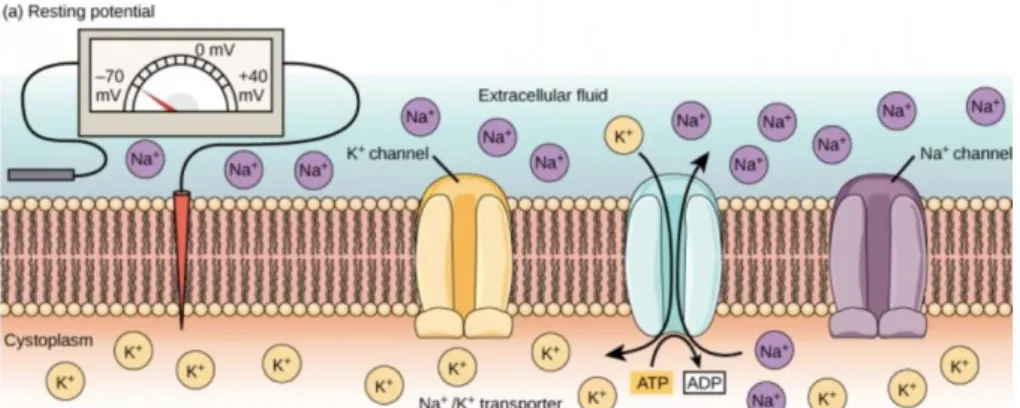
16 2.2.2 Resting Membrane Potential
Neurons at rest have a negative charge: generally, the inside of a cell is 70 millivolts more negative than the outside (Costanzo 2014). This state is called the resting membrane potential; it is caused by differences in the concentrations of ions inside and outside the cell at rest (Costanzo 2014; L. R. Squire et al. 2012). If the membrane were evenly permeable to all ions, the system would reach equilibrium. However, because ions cannot cross the cell membrane freely, there are different concentrations of ions inside and outside the cell, as presented in Table 2 (Costanzo 2014; Bloom 2012).
Table 3. Extracellular and Intracellular ion concentrations during resting membrane potential (modified from (Costanzo 2014))
Substance Extracellular Fluid (mEq/L) Intracellular Fluid (mEq/L) Na+ 140 14 K+ 4 120 Ca2+ 2.5 1x10⁻⁴ Cl- 105 10 HCO3- 24 10
Na+, sodium ions; K+, potassium ions; Ca2+, calcium ions; Cl-, chloride ions; HCO3-, bicarbonate ions; mEq/L, milliequivalents per litre
The difference in the quantity of positively charged potassium ions (K+) between the inside and outside of the cell determines the resting membrane potential (Costanzo 2014; L. R. Squire et al. 2012). The negative charge when the cell at rest is generated by the cell membrane greater permeable to K+ movement than the sodium ion (Na+) movement (Costanzo 2014; L. R. Squire et al. 2012). In neurons, K+ is maintained at high concentrations within the cell, while Na+ is maintained at high concentrations outside of the cell (Costanzo 2014; L. R. Squire et al. 2012). The cell possesses K+ and Na+ leakage channels that allow the two cations to diffuse down their concentration gradient. However, the neurons have far more K+ leakage channels than Na+ leakage channels (L. R. Squire et al. 2012). Therefore, K+ diffuses out of the cell at a much faster rate than leaks in of Na+. Because more K+ are leaving the cell than Na+ are entering, negative charge is generated inside the cell. Once established, the actions of the Na+/K+ ATPase pump help to preserve the resting potential. The action of Na+/K+ ATPase is an energy-consuming event for bringing two K+ ions into the cell while removing three Na+ ions, one molecule of adenosine triphosphate (ATP) is consumed (Costanzo 2014; L. R. Squire et al. 2012). These interactions of events displayed in Figure 10 (Lumen 2019a).
17
Figure 10. Interaction of ions, channels, and Na+/K+ ATPase during resting membrane potential in a neuron
(Taken from (Lumen 2019a))
2.2.3 Action Potential
A neuron receives stimuli from other neurons and sends the signal to another neuron. Transmission of a signal between neurons is carried by a chemical called neurotransmitters (Bloom 2012; L. R. Squire et al. 2012; Costanzo 2014). Transmission of a signal along the neuron is carried by a sudden change of the resting membrane potential called an action potential (Costanzo 2014; Bloom 2012; L. R. Squire et al. 2012). Neurotransmitters bind to receptors on dendrites then opened ion channels (Bloom 2012; L. R. Squire et al. 2012; Costanzo 2014). At excitatory synapses, this opening of the ion channels allows sodium ions to enter the neuron and depolarize the membrane. Depolarization marked by a decrease in the voltage difference between the inside and outside of the neuron (L. R. Squire et al. 2012). If the depolarization of the target neuron reaches its threshold potential (-55 mV) (L. R. Squire et al. 2012), Na+ channels in the axon hillock open, permitting positive ions to enter the cell (Lumen 2019a). After the sodium channels open, the neuron will completely depolarize to a membrane potential of about +40 mV (Costanzo 2014; L. R. Squire et al. 2012). Action potentials are an “all or none” event, that once the threshold potential is reached, the neuron always completely depolarizes (Costanzo 2014; Bloom 2012; L. R. Squire et al. 2012).
Figure 11. Events involving ions, channels, and Na+/K+ ATPase that lead to depolarization (Taken from (Lumen 2019a))
Once depolarization is complete, the cell must reset the membrane voltage back to the resting potential. To return to the resting potential, the sodium (Na+) channels will close and cannot be opened (Costanzo 2014; L. R. Squire et al. 2012). Concurrently, voltage-gated potassium (K+) channels start to open and allow K+ to leave the cell (Costanzo 2014; L. R. Squire et al. 2012).
18
As K+ leaves the cell, the membrane potential starts to decrease and will become negative. The diffusion of K+ to the outside of the cell continues until it hyperpolarizes the membrane cell. At this point, the Na+ channels already return to their resting state and able to open again if the membrane potential exceeds their threshold potential (Costanzo 2014). Eventually, the extra K+ diffuses out of the cell through the K+ leakage channels, changing the cell from its hyperpolarized state to the resting membrane potential (Costanzo 2014; L. R. Squire et al. 2012).
Figure 12. Events involving ions, channels, and Na+/K+ ATPase that lead to hyperpolarization (Taken from (Lumen 2019a))
2.2.4 Resume
2.3 Energy Demands for Brain Activation
Although the human brain is 2% of the body’s weight, it accounts for 20% of relative oxygen consumption (Attwell et al. 2010) Compare to other organs, the brain is one of the greatest users of oxygen in humans (Table 3) (Dienel 2018). The fact that the brain has a minimal oxygen reserve and neuron cannot store glycogen, making the sustainability of energy in the brain has
Brain activation is related to the basic function of the brain to transmit information between neurons by generating electrical potentials and releasing chemical signals, which consists of preserving resting membrane potential and forming action potential. The numbers of potassium ions (K+) and sodium ion (Na+) across the cell membrane
determine the resting membrane potential.
The movements of potassium ions (K+) and sodium ion (Na+) across the cell membrane through its channels or pumps generate an action potential.
The formation of an action potential can be separated into five steps: o Depolarization of target cell by stimuli from a sensory cell or another neuron. o Opening of all the Na+ channels, If the threshold of excitation is reached.
o Opening of K+ channels at the peak action potential. K+ begins to leave the cell, and at the same time, Na+ channels close.
o Hyperpolarization of the membrane as K+ ions continue to leave the cell. The hyperpolarized membrane is a refractory period and then cannot fire another action potential.
19
important implications. Deficits in the brain’s energy supply make it susceptible to decrease cognitive performance to brain damage during anoxia or ischemia, and knowing the demands made by different neural mechanisms may help understand the importance of the brain’s energy metabolism to maintain brain functions.
Table 4. Relative oxygen consumption in adult human organs (Modified from (Dienel 2018))
Organ % Body Mass % Oxygen Utilization (%O2use) %O2use/%Body Mass Liver 2 17 8.5 Gastrointestinal tract 2 10 5 Kidney 0.5 6 12 Heart 0.4 11 27.5 Brain (adult) 2 20 10 Brain (5-years-old) 6 50 8.3
% body mass, percentage of organ mass per total body mass; %O2use, percentage of organ oxygen use per total oxygen use; %O2use/%body mass, (percentage of organ oxygen use per total oxygen use) per (percentage of organ mass per total body mass)
Based on the energy-requiring processes in neurons, the estimation of the relative demands of energy compared to the total energy is presented in Table 4. Conceptually, energy demands on neurons distinguish between the energy used for basic vegetative (non-signaling) processes and the energy used for specialized physiological signaling processes (Figure 13) (Engl and Attwell 2015; Ames 2000). Specialized physiological signaling processes seem to consume the majority of energy metabolism in CNS as glucose utilization by brain regions increased several times in response to physiological stimulation that affects physiological activity (Hibbard et al., n.d.). Inhibition of sodium-potassium pumps (Na+/K+ ATPase) with its specific inhibitors resulted in a reduction of energy usage around 50% of total energy consumption in the brain (Astrup 1982).
Figure 13. Distribution of energy use in the brain; specified into the basic non-signaling processes and the specialized physiological signaling processes (Engl and Attwell 2015)


![Figure 8. Cartoon illustration of glial cells within the neurovascular system (Taken from [Allen and Barres 2009])](https://thumb-eu.123doks.com/thumbv2/123doknet/7786277.259355/33.892.409.794.110.508/figure-cartoon-illustration-glial-neurovascular-taken-allen-barres.webp)