HAL Id: dumas-01206454
https://dumas.ccsd.cnrs.fr/dumas-01206454
Submitted on 29 Sep 2015HAL is a multi-disciplinary open access
archive for the deposit and dissemination of sci-entific research documents, whether they are pub-lished or not. The documents may come from teaching and research institutions in France or abroad, or from public or private research centers.
L’archive ouverte pluridisciplinaire HAL, est destinée au dépôt et à la diffusion de documents scientifiques de niveau recherche, publiés ou non, émanant des établissements d’enseignement et de recherche français ou étrangers, des laboratoires publics ou privés.
Sclérodermie systémique : description de la cohorte
bordelaise : corrélation phénotypique avec le dosage de
la TSLP
Anne Bertrand
To cite this version:
Anne Bertrand. Sclérodermie systémique : description de la cohorte bordelaise : corrélation phéno-typique avec le dosage de la TSLP. Médecine humaine et pathologie. 2015. �dumas-01206454�
Université de Bordeaux
U.F.R. DES SCIENCES MEDICALES Année 2015 – N°3059
Thèse d’exercice pour l’obtention du
DIPLOME D’ETAT DE DOCTEUR EN MEDECINE Présentée et soutenue publiquement par
Anne Bertrand
Née le 30 juin 1987 à Bordeaux
Le 14 septembre 2015
Sclérodermie systémique
Description de la cohorte bordelaise
Corrélation phénotypique avec le dosage de la TSLP
Directeur de la thèse Docteur M-E. Truchetet
Jury Président
Monsieur T. Schaeverbeke, Professeur Juges
Madame E. Lazaro, Professeur
Madame C. Contin-Bordes, Maître de conférence Madame M-E. Truchetet, Docteur
Monsieur C. Richez, Professeur
Rapporteur de la thèse
REMERCIEMENTS
A mes juges et mon rapporteur
Monsieur le Professeur Schaeverbeke, je vous remercie de me faire l’honneur de présider ce jury de thèse. C’était un plaisir de travailler comme interne dans votre service. Vous m’avez appris (toujours dans la bonne humeur ;)) de nombreuses choses sur cette belle spécialité tout au long de mon cursus médical. Merci pour votre disponibilité, votre pédagogie et votre bienveillance. Merci pour cette belle aventure sur les anti CTLA4.
Madame le Docteur Truchetet, merci de m’avoir proposé ce travail de thèse avec toi et de l’avoir dirigé. Ca m’aurait presque donné envie de faire un M2. Trois semestres à tes côtés, un séjour à Boston ;) Merci pour toutes ces images qui aident à communiquer avec les patients : les différentes famille de tomates chez le maraîcher pour les AAN, le sirop dans l’eau pour les dilutions d’AAN, les mutineries du système immunitaire pour les maladies auto immunes, les articulaires postérieures et les anses de tasse à café ;) Merci pour tout ce que tu m’as apporté pendant mon internat, tant sur le plan médical qu’humain.
Monsieur le Professeur Richez, je te remercie d’avoir accepté de faire partie de mon jury de thèse. Tu es toujours disponible, toujours soucieux d’améliorer notre formation. Merci pour tous tes bons conseils et ton écoute tout au long de mon internat. J’ai eu grand plaisir à travailler avec toi.
Madame le Professeur Lazaro, j’ai eu la chance d’être une de vos externes en D4 et je me souviens encore de ces visites si instructives à vos côtés. Je vous remercie de me faire l’honneur d’avoir accepté d’être membre du jury de ma thèse.
Madame le Docteur Contin-Bordes, je vous remercie pour votre accueil dans le laboratoire d’immunologie, m’ayant permis de réaliser ce travail. Merci d’avoir accepté avec un si grand sourire de faire partie des membres du jury.
Monsieur le Professeur Seneschal, je voulais te remercier d’avoir accepté d’être le rapporteur de ma thèse. Merci pour l’attention que tu as porté à ce travail.
A mes stages d’internat
La Rhumatologie à Pellegrin Une deuxième maison! Trois stages d’internat et 2 d’externat!
Monsieur le Professeur Bannwarth, je vous remercie pour toutes ces agréables visites où j’ai appris bien plus que la Rhumatologie. Merci pour votre gentillesse et votre bienveillance. A Nadia, merci pour tout ces bons moments en hôpital de jour avec toi.
A Nicolas, merci pour tout ce que tu m’as appris! Toujours disponible, pédagogue, à l’écoute et bienveillant! C’était un plaisir de faire tous ces stages avec toi. La seule chose que tu peux améliorer c’est ton jeu au ping-pong ;)
A Steeve avec sa bonne humeur communicative. A Thomas, pour ton aide précieuse tout au long de l’internat et ces réponses rapides aux mails intitulés « Help »!
A mes co-internes, Alexia (Jumelle astrale et coéquipière de consultation), Clém (Pichou râleuse préférée), Stéphanie (Pétillante toujours happy), Laeti, Margaux, Fanny, Alice, Pauline, Amandine… Une mention spéciale pour Marie, partenaire des anti CTLA4 qui nous auront fait suer jusqu’au bout et passionnée comme moi par l’ophtalmologie…
Merci à toute l’équipe du 12ème
qui est parfaite.
Merci pour ces 3 semestres dans le service à vos côtés que je n’oublierai jamais.
La Rhumatologie au Bouscat Une troisième maison! Deux stages d’internat et d’externat!
Pierre, vous m’avez transmis l’envie de choisir cette spécialité dès mon premier stage d’externat, en partageant votre passion. Quand on me demandait comment je voyais ma façon d’exercer plus tard la Rhumatologie, je répondais : “J’aimerais bien faire comme M. Germain”. Grâce à vous, j’ai la chance de prolonger ma formation pendant 2 ans à l’hôpital du Bouscat. Merci d’y avoir cru sans relâche et de m’avoir toujours soutenue et rassurée. Merci pour les visites pleines de bonne humeur et pour votre gentillesse.
Maud, merci pour ta bienveillance et ton soutien permanents. J’ai démarré et je termine mon internat avec toi avec toujours autant de plaisir. Je suis ravie de pouvoir continuer à travailler à tes côtés ainsi qu’avec toute l’équipe du 1er.
Merci aussi au Dr Roux et Sophie pour leur accueil en Gastro-Entérologie pendant ces 2 mois d’été. J’ai appris beaucoup de choses avec vous tant sur le plan humain que médical.
A mes co internes: Marie, Adeline, Romain, Guillaume. La Radiologie à Pellegrin
A mes co-internes François, Gautier, Alexis.
A Damien, un de mes meilleurs chefs de clinique mais aussi un ami. Merci pour tout ce que tu m’as appris.
Merci à Olivier Hauger pour ses cours d’ostéo articulaire très intéressants. Merci à Claire, pour son soutien féminin. Aux manips adorables.
Merci à tous pour ces 6 mois que j’ai adoré! La Dermatologie à Haut Lévêque
Merci aux Professeurs Doutre et Bellot-Barry pour cette initiation à la Dermatologie. Merci à mes deux chefs de clinique, Emilie et Anne Lien.
A Noémie, ce stage a permis le début d’une belle amitié. La Médecine interne à Dax
A mes co internes de choc (ou pare-choc) Martin (« le bouclier de la visite du lundi matin ») et Jennifer, ma co-interne, ma coloc et mon amie. Merci pour ton soutien et ton courage sans faille. J’ai énormément appris à tes côtés.
A Catherine ma préférée. A Isa, pour ses pastis landais, ses madeleines, ses expressions landaises et surtout son oreille attentive dans les moments difficiles.
A mes ami(e)s
A Isa (depuis et pour toujours!), Véro (sous-colleuse d’exception, que de bons souvenirs de ses années avec toi, je te voudrais plus près qu’aux Antilles), Camille (aux oreilles attentives et aux mots toujours attentionnés) et Val (depuis bébé, je pense et j’espère que l’on continuera à partager tous nos moments importants)
A toute la bonne clic et tous mes amis depuis la P1: Dadoo, Antoine, Edouard Mag et Chasseuil, Valentin, Laura, Aude, Gab, Hugo, Séb, Emilie, Peg, Jean Séb, Marc, Sylvain… Aux plus anciens: Jabi, Alban, Gaby, Sarah, Guilhem (et Ju)…
Au labo d’immunologie et surtout à Jorge. Merci pour ton aide précieuse dans la réalisation de ce travail ; pour ta patience avec mes nombreux ELISA ratés. Merci pour ces bons moments au laboratoire passés ensemble.
Les meilleurs pour la fin…
Un grand merci à Quentin
L’homme de ma vie. Merci pour tout, vraiment tout… Un grand merci à ma famille
A mes parents, pour m’avoir toujours soutenue (dans les bons moments comme les moins bons ;)). A Papa, pour m’avoir transmis sa passion et m’avoir donné l’envie de devenir rhumatologue. A mon Paulo (et Juju) et ma petite Marie. Je vous aime.
A Alex, Mano et Henri (j’aurais aimé que vous soyez là ++), Mimi et Paul.
A Mathilde, marraine la bonne fée, et son destrier Sapper qui m’a permis de me fracturer cette belle vertèbre et d’écrire plus vite cette thèse :)
A ma « belle famille » : Karine et Jean Paul, Gautier et Giovanna et Hugues. A Mamily, Paps et Mutty.
Table of contents
1. TITLE………6
2. ABSTRACT………..7
3. INTRODUCTION………9
a. Systemic sclerosis and generalities………..………...9
b. Systemic sclerosis and biomarkers………. 11
c. Systemic sclerosis and immune dysregulation………..12
d. Description of TSLP………13
e. Systemic sclerosis and TSLP………... 14
4. PATIENTS AND METHODS………16
a. Clinical data………...16
b. Variables definitions………..17
c. Measurement of serum TSLP concentrations………...18
d. Statistical analysis………...19
5. RESULTS……….20
a. Description of the VISS Cohort……....……….20
b. Correlation of TSLP with phenotypic characteristics………...25
6. DISCUSSION………..29
7. CONCLUSION………34
8. SUPPLEMENTARY CONTENT……….35
Systemic sclerosis
Description of a French cohort in Bordeaux hospital center
Clinical associations with the serum level of TSLP
ABSTRACT
BACKGROUND Systemic sclerosis (SSc) is characterized by endothelial dysfunction, fibrosis and immunological dysregulation. The pro-fibrotic cytokines Th2 (IL-13 and IL-4) induce excessive production of extracellular matrix but the mechanisms skewing Th2 polarization remain unknown. Recent data converge on TSLP (Thymic Stromal LymphoPoietin), a central cytokine in the Th2 polarization produced by keratinocytes, fibroblasts and endothelial cells.
Our objective was to describe the cohort of patients with SSc followed in Bordeaux hospital center and to perform clinical associations with their blood TSLP dosage.
METHODS Patients with limited SSc (lcSSc) or diffuse SSc (dcSSc) were included in the VISS (Vasculopathy and Inflammation in SSc) cohort. The serum level of TSLP was correlated with phenotypic data. TSLP was measured in 22 healthy controls.
RESULTS 203 patients (65.5% lcSSc and 34.4% dcSSc) including 151 women (sex ratio 2.9:1) were enrolled between March 2012 and January 2015. We identified 97% of patients with a Raynaud’s phenomenon, 7.9% with pulmonary arterial hypertension, 4.9% with scleroderma renal crisis, 19.8% with pulmonary fibrosis, 49.2% with digital ulcers, 69.4% with gastrointestinal involvement and 59.1% with articular involvement. Immunosuppressive drugs were more frequently prescribed in dcSSc than in lcSSc (65.7% and 13.5% respectively; p <0.001). 50.5% of patients received corticosteroids with a median dose of 12.5mg [7.5-30]. 23.3% SSc sera were positive for TSLP. All controls were negative. The dosage of TSLP was correlated with the presence or a history of digital ulcers (p = 0.03).
CONCLUSION The analysis of the characteristics and therapeutic strategies in the VISS cohort allow us to have an overview of the management of these patients in the CHU of Bordeaux. Moreover, we demonstrate that TSLP is associated to SSc, probably early in its pathogenesis, and can constitute a new biomarker for the development of the vasculopathy.
CONTEXTE La sclérodermie systémique (ScS) est caractérisée par la dysfonction endothéliale (DE), la fibrose et la dérégulation du système immunitaire. Les cytokines pro- fibrotiques Th2 (IL-13 et IL-4) induisent une production excessive de matrice extra cellulaire mais les mécanismes reliant la DE à la polarisation Th2 restent inconnus. Des données récentes convergent vers la Thymic Stromal LymphoPoietin (TSLP) qui est une cytokine centrale dans la polarisation Th2 et produites par les cellules endothéliales, les fibroblastes et les kératinocytes.
Notre objectif était de décrire la cohorte bordelaise de ScS et réaliser des corrélations phénotypiques avec le dosage de la TSLP.
METHODES Les patients ayant une ScS limitée (lScS) ou diffuse (dScS) ont été inclus dans la cohorte VISS (Vasculopathie et Inflammation dans le ScS). Le dosage de la TSLP a été corrélé aux données phénotypiques. La TSLP a été dosée chez 22 témoins sains.
RESULTATS 203 patients (65.5% lScS et 34.4% dScS) dont 151 femmes (sexe ratio 2.9 :1) ont été inclus entre mars 2012 et janvier 2015. Nous avons identifié 97% de Raynaud, 7.9% d’hypertension artérielle pulmonaire, 4.9% de crises rénales, 19.8% de fibrose pulmonaire, 49.2% d’ulcères digitaux, 69.4% d’atteinte digestive et 59.1% d’atteinte articulaire. Les immunosuppresseurs étaient plus fréquemment prescrits dans la dScS que dans la lScS (65.7% et 13.5% respectivement, p<0.001). 50.5% des patients ont reçu des corticoïdes avec une dose médiane de 12.5mg [7.5-30]. 23.3% des 201 dosages pour la TSLP étaient positifs. Aucun témoin n’était positif. Le dosage de la TSLP était corrélé à la présence ou l’antécédent d’ulcères digitaux (p=0.03).
CONCLUSION Les caractéristiques cliniques des patients et les différentes stratégies thérapeutiques dans la cohorte VISS permettent d’avoir une vue d’ensemble sur la prise en charge de ces patients. La TSLP est un nouveau biomarker de développement de la vasculopathie.
MOTS CLES – Sclérodermie systémique, vasculopathie, fibrose, TSLP, OX40-L, Réponse immune Th2, IL-13, ulcères digitaux.
INTRODUCTION
Systemic sclerosis and generalities
Systemic sclerosis (SSc) is a heterogeneous and rare autoimmune disorder with a complex pathogenesis associating immune dysregulation, vasculopathy and skin and/or internal organs fibrosis. SSc epidemiology has been reported divergently with prevalence ranging from 7 to 1580 per million inhabitants. In France (in the Parisian region), the prevalence of SSc has been estimated at 158 per million inhabitants (1). It affects typically the middle-aged women with an average sex ratio around 3:1 (2).
The two major clinical subsets are limited cutaneous form (lcSSc) and diffuse cutaneous form (dcSSc). Clinical involvements refer to vascular dysfunction (Raynaud’s phenomenon (RP), pulmonary hypertension (PAH), scleroderma renal crisis (SRC)) and fibrosis (skin thickness, pulmonary fibrosis, gastro intestinal dysmotility and malabsorption, heart conduction blocks and friction rubs) (3).
SSc is regarded as an identifiable disease entity that can be easily recognised by experienced physicians if fully developed. The diagnosis of SSc in clinical practice generally relies on physician opinion. Classification criteria (for use in clinical studies but not for diagnosis) were developed in 1980 (‘Preliminary criteria for the classification of systemic sclerosis (scleroderma)’, 1980) (4) (table 1).
Table 1- Preliminary criteria for the classification of systemic sclerosis, 1980.
Sclerodactily Digital.ulcers.or.pitting.scars The.major.criterium.or.2.of.the.minors.=.systemic.sclerosis Major.criterium:. 1980.ACR.Preliminary.criteria.for.the.classification.of.SSc proximal.scleroderma Minor.criteria:. Pulmonary.fibrosis.(chest.XHRay)
Then, LeRoy et al. proposed in 1988 (5) to distinguish SSc in two subsets (lcSSc and dcSSc) (table 2). These criteria classified most SSc patients correctly, except those with early SSc or a limited form of SSc. To include these later patients, new classification criteria were proposed in 2001 by Leroy and Medsger (table 3), taking advantage of recently developed autoantibody testing techniques and methods to detect micro-vasculopathy in the nailbed. These criteria brought interesting arguments for the diagnosis of early and limited
scleroderma leading to a better identification of these patients in daily practice (6). Table 2 – Limited and diffuse cutaneous systemic scleroderma – LeRoy 1988.
Limited'cutaneous'SSc Diffuse'cutaneous'SSc Skin'involvement'restricted'to'hands,'face,' forearms'and'feet 'History'of'Raynaud's'with'onset'within'1 year Delayed'but'often'severe'onset'of'pulmonary' arterial'hypertension Skin'sclerosis'extending'proximal'to'the'elbow;'may'involve'truncal'areas Ectopic'calcinosis,'telangiectasias Tendon'friction'rubs'may'occur Anticentromere'antibodies'common'but' antitopoisomerase 1'very'rare Early'onset'of'pulmonary,'renal'and'diffuse'gastrointestinal'involvement Dilated'nailfold'caps'seen'but'no'capillary' destruction Rarely'anticentromere'antibodies'but'often'antitopoisomerase'1'antibodies Nailfold'capillary'destruction Limited'vs'Diffuse'cutaneous'systemic'sclerosis
Table 3 – Early SSc classification criteria – LeRoy and Medsger 2001.
limited'scleroderma RP's'objective (lSc) AND'any'one SSc'type'nailfold'capillay'pattern SSc'selective'auto'antibodies OR RP's'subjective AND'both SSc'type'nailfold'capillay'pattern SSc'selective'auto'antibodies lcSSc citeria'for'lSSc AND Distal'cutaneous'changes dcSSc criteria'for'lSSc AND Proximal'cutaneous'changes Early'SSc'classification'criteria
These criteria were revised with a new classification: 2013 ACR-EULAR classification criteria (7) (figure 1).
Figure 1 – The 2013 ACR-EULAR classification criteria.
Systemic sclerosis and biomarkers
In analogy with the management of rheumatoid arthritis, it has been proposed that the window of opportunity to prevent tissue fibrosis and irreversible damages in SSc occurs during the early stage of the disease. Four clinical signs have been identified for the Very Early Diagnosis Of Systemic Sclerosis (VEDOSS program): RP, puffy swollen digits turning into sclerodactyly, antinuclear antibodies and specific SSc antibodies (centromere and anti-Scl70 antibodies) and abnormal capillaroscopy with scleroderma pattern (8). These patients are the targets of a specific follow-up to diagnose complications as soon as possible with the proposal of an early and an appropriate treatment before irreversible damages.
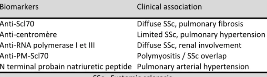
Biomarkers are needed in SSc to help in achieve the early identification process and the life-threatening complications detection. Many candidate molecules have been identified in the past two decades, reflecting the three main pathogenic mechanisms in SSc (9,10). However, validated biomarkers are still missing and very few of them have a real impact in daily practice (table 4).
Table 4 – Validated biomarkers in SSc used in daily practice. Anti%Scl70 Diffuse0SSc,0pulmonary0fibrosis Anti%centromère Limited0SSc,0pulmonary0hypertension Anti%RNA0polymerase0I0et0III Diffuse0SSc,0renal0involvement Anti%PM%Scl70 Polymyositis0/0SSc0overlap N0terminal0probain0natriuretic0peptide Pulmonary0arterial0hypertension SSc=0Systemic0sclerosis Biomarkers Clinical0association
The research for biomarkers has also permitted to identify new therapeutic targets as endothelin-1, a potent endogenous vasoconstrictor. Bosentan, an endothelin-1 receptor antagonist, is now daily used in the treatment of PAH in SSc with evidence of improved survival (11). It is also safe and efficient in the prevention of new digital ulcers, a common vascular complication in SSc (12).
Although there have been progresses over the years in therapeutic options for SSc with vascular targeted treatments, biologic agents and autologous stem cell transplantation, the mainstays of treatment are organ-based and primarily focused on symptom management (13) and severe forms of SSc still have a poor prognosis with significant morbidity and mortality (14). Unfortunately, there is no single agent or therapeutic combination with a clear impact on the disease process, partly due to the fact that the fundamental mechanisms linking endothelial dysfunction, fibrosis and immune dysregulation are poorly understood.
Systemic sclerosis and immune dysregulation
SSc is primarily a vascular disease mediated by autoimmunity, evolving into tissue fibrosis (15). Immune dysregulation is an important contributor to the pathogenesis, initiating and/or maintaining the mechanisms resulting in fibrosis (16). More precisely, it has been established that T helper (Th) 2 polarization of T-cell is dominant in SSc and plays a crucial role, leading to a pro-fibrotic environment with secretion of type 2 pro-fibrotic cytokines such as interleukin (IL) 13 and IL-4 (17,18). Hence, IL-13 is considered as a master regulator of fibrosis. It leads to fibrosis through the induction of TGF β1 production by macrophages and also through TGF β-independent mechanism (direct stimulation of fibroblasts) (19). However, the mechanisms by which Th2-biaised immune responses are initiated in scleroderma remain unknown.
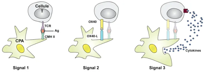
Th polarization requires a series of interactions between dendritic cells (DC), naive Th cells, and the delivery of antigenic (first), co-stimulatory (second) and cytokinic (third) signals (figure 2).
Figure 2 - Three signals in the communication between dendritic cells and T cells.
Firstly, antigen (Ag) is presented by major histocompatibility complex II (MHC II) on DC and recognized by the T cell receptor (TCR) of T-cell. Secondly a co-stimulation signal occurs in response to binding of OX40 ligand (OX40-L) on DC with OX40 receptor on T-cell. Thirdly cytokines are produced by DC and constitute a polarizing Th2 microenvironment.
OX40-L, as a second signal, has been identified as a fundamental requirement for optimal induction of Th2 responses (20–22). It has been involved in allergic process (23) and cancer (24). More recently OX40L expression has been found increased in SSc skin in our laboratory. Interestingly, OX40L expression is induced on DC under different stimulatory conditions and one of them is the thymic stromal lymphopoietin (TSLP).
Description of TSLP
TSLP is an IL-7 cytokine family member which role in the promotion of Th2 response has been widely studied in allergic diseases (25,26). TSLP has effects on multiple lineages and specially on DC (26) with the capacity to guide T-cell to a DC-mediated Th2 phenotype through OX40-L inducing (27).
TSLP was demonstrated to be highly produced by epithelial cells located at barrier surfaces (skin, lung and gut) of patients with atopic diseases(25). Other cell types can produce TSLP, such as stroma cells, mast cells and DC (28–30). TSLP regulates barrier immunity, being one
of the first molecules produced after disturbances of homeostasis (31) secondary to stimuli such as microorganisms presence (bacterial and viral products), physical injury, inflammatory cytokines and toll like receptors (TLR) ligands (32,33). In atopic dermatitis, TSLP is overexpressed in acute and chronic skin lesions (26) and in allergic asthma, TSLP is increased in the airways and correlates with disease severity and Th2 activity (34). Overexpression of TSLP in the mouse lung induces the spontaneous progressive airway inflammation (35) and expression in the skin can induce atopic dermatitis–like lesions.
It is increasingly clear that TSLP impacts a growing number of different Th2-dependant disorders, beyond allergic diseases. In nearly all cancers (both solid tumours and leukaemia), Th2 response is preponderant and supports tumour development. Thus, TSLP has been identified as an important regulator, produced by cancer cells and cancer associated fibroblasts, able to promote tumour cells growth and survival by manipulating the immune system (36). TSLP has also been implicated in autoimmune diseases such as rheumatoid arthritis. In mice models, administration of TSLP significantly exacerbated the severity of collagen-induced arthritis and the joint damage. Furthermore, TSLPR (-/-) mice had less severe arthritis (37). Recently, and very logically because of its involvement in Th2 pathway, TSLP has been studied in fibrotic diseases as idiopathic pulmonary fibrosis. It has been reported that fibroblasts represent both a cellular source and a target for TSLP as they express a functional TSLP Receptor complex that could be activated in response to TSLP (38). Systemic sclerosis and TSLP
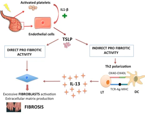
In SSc, two recent studies converge for a potential role of TSLP. Christman et al. (39) and Usategui et al.(40) have demonstrated that TSLP is strongly expressed in the skin of patients with SSc, mainly by keratinocytes and by immune cells in perivascular areas. These data suggest that TSLP may have an indirect pro-fibrotic activity by skewing Th2 polarization in SSc. A potential direct pro-fibrotic activity has been proved with proliferation and activation of fibroblasts by TSLP (unpublished data from the laboratory). Moreover, endothelial cells are able to produce TSLP secondary to IL1-β stimulation through activated platelets (unpublished data from the laboratory).
These findings shed a light on the role of TSLP in the pathogenesis of scleroderma (figure 2).
Thus, TSLP might be a strong link between endothelial dysfunction and Th2 response in SSc,
Figure 2 – Schematic overview of the supposing role of TSLP in SSc.
Abbreviations: TSLP=Thymic Stromal Lymphopoietin, IL=InterLeukin, LT=T-cell, DC=Dendritic Cell, Ag=Antigen, TCR=T-Cell-Receptor.
In the present study, we described for the first time the VISS (Vasculopathy and Inflammation in SSc) cohort constituted of patients with SSc followed at the Bordeaux University Hospital Centre. Furthermore, we determined serum level of TSLP and its clinical association in SSc patients.
PATIENTS AND METHODS
VISS is a biomedical research project founded in 2012 with the aim of compiling all patients with SSc followed at the Bordeaux University Hospital center (CHU de Bordeaux) and advancing in the understanding of disease pathogenesis.
We prospectively enrolled patients with SSc (lcSSc or dcSSc) between March 2012 and January 2015 at 7 clinical departments from the Bordeaux hospital (two departments of Dermatology, one department of Rheumatology, two departments of Internal Medicine, one department of Cardiology, one department of Pneumology and one department of Vascular Medicine). We recruited patients diagnosed with scleroderma by their clinician in charge. All patients included were screened for the classification criteria proposed by the ACR-EULAR in 2013 (7) (figure 1).
The biomedical project of the cohort was approved by the Bordeaux hospital and ethics committee (CPP, 2012-A00081-42, Aquitaine) and allowed us to begin a translational project through the collection of serum, blood cells and skin biopsies from SSc patients. This project was conducted in accordance with the ethical principles originating from the Declaration of Helsinki. All participants gave written informed consent before their inclusion.
Clinical data
A disease and organ specific questionnaire was filled out by the clinician in charge of the patient and then centralized by the two investigators (AB and MET). The investigators performed data extraction from Dx Care® database (the hospitals’ electronic healthcare database, where patient medical data are recorded) independently (supplementary content,
annex 1). Clinical features (scleroderma form, sex, age at RP onset, age at non-RP onset,
duration of the disease, symptoms of skin, articular, heart, lung, kidney, gastrointestinal involvement), immunologic test results (antinuclear antibodies, Scl70 antibodies, anti-centromere antibodies (ACA) and anti-RNA polymerase (RNAP) III antibodies), imaging (thorax CT scans, respiratory functions tests, capillaroscopy, cardiac ultrasonography and right heart catheterization) and treatments received by these patients were retrospectively recorded. We chose to keep the worst values from patient’s medical history.
Variables definitions
Skin involvement was evaluated using the modified Rodnan skin score (mRSS) that assesses the skin thickness by clinical palpation of 17 body areas on a scale of 0–3 (supplementary content, annex 2) (41). Pigmentation disorders were also recorded.
Vascular involvement - Peripheral vascular manifestations were defined by the presence of RP, with or without digital ulcerations. A suspected PAH was defined as increased right ventricular systolic pressure RVSP > 35mmHg on the echocardiogram. A confirmed PAH was defined as increased mean pulmonary arterial pressure mPAP > 25mmHg on the right heart catheterization (RHC).
Cardiac involvement was defined as one of the following: left ventricular ejection fraction (LEVF) dysfunction (LEVF was considered reduced if it was fewer than 55%), abnormal diastolic function on the echocardiogram, increased BNP levels, palpitations, pericarditis or conduction block.
Pulmonary involvement was defined as one of the following: dyspnea grade III of the NYHA classification (New York Heart Association) and upwards, reduced diffusing capacity of carbon monoxide DLCO < 80%, restrictive pulmonary abnormalities on pulmonary function tests with forced vital capacity FVC < 80% and interstitial lung disease or lung fibrosis confirmed by high-resolution CT scans.
Renal involvement was defined as a history of scleroderma renal crisis.
Gastrointestinal involvement was defined as eso-gastro-intestinal motility dysfunction with esophageal (dysphagia and reflux), stomach (epigastralgia) and intestinal (constipation, diarrhea, malabsorption) symptoms.
Musculoskeletal involvement was defined as the existence of synovitis, joint contractures, tendon friction rubs, muscle weakness or atrophy. CPK levels were recorded.
Investigators re-read together the patients’ record in the event of any discrepancy. When data was not available, efforts were made to contact the patient and/or the clinician in charge.
Measurement of serum TSLP concentrations
Serum samples were obtained from patients with SSc at the date of inclusion in the VISS study. We also analyzed serum of 22 healthy controls matched for sex and age. Recombinant human TSLP used for standard curve was obtained from R&D Systems. Serum TSLP levels were measured with a specific enzyme-linked immunosorbent assay (ELISA) kit (Human TSLP ELISA Ready-SET-Go! ® R&D Systems). Anti-TSLP antibody (capture antibody) was pre-coated onto microtiter wells (figure 3-1). Aliquots of serum were added to each well (figure 3-2), with 2 hours of incubation period, followed by biotin-conjugated anti-TSLP antibody (detection antibody) (figure 3-3). Color was developed with Avidin-HRP (detection enzyme) (figure 3-4) and tetramethylbenzidine substrate solution (figure 3-5). A 2N H2SO4 solution was used to stop the enzymatic reaction and sample absorbances were measured at 450 nm. The concentration of TSLP in each sample was determined in pg/mL by interpolation from a standard curve.
Statistical analysis
The statistical analysis was carried out using PRISM® (6.0 version). All values are expressed as mean ± standard deviation or median ± interquartile range (continuous variables) or as counts and percentages (categorical variables). A Kolmogorov-Smirnov normality test was performed to check the normality of data. Statistical evaluation was performed using contingency table tests (χ2-test or Fisher’s exact t-test) or Student t test to describe significant differences or associations in the cohort. Contingency table tests were used to describe associations or differences between TSLP (positivity or nullity) and multiple categorical parameters (form, sex, digital ulcers, respiratory involvement, fibrotic involvement, vascular involvement). Unpaired t tests were used to describe associations or differences between TSLP dosage and multiple categorical parameters as mentioned above. Pearson's correlation coefficient was calculated to describe the linear correlation between TSLP dosage and multiple continuous parameters (age, years since RP, years since non RP symptoms, years between RP onset and non RP symptoms onset, Rodnan, DLCO, FVC, RVSP, mPAP). P values less than 0.05 were considered significant.
RESULTS
Description of the VISS cohort
Patient and disease characteristics
By January 2015, 203 SSc patients were consecutively recruited on the VISS study. Ninety-five percent of them (100% in dcSSc and 92.4% in lcSSc) fulfilled the classification criteria proposed by the ACR-EULAR in 2013. The remaining 5% concerned patients with a limited form. They fulfilled a mean of 6.1 criteria and may correspond to pre-systemic sclerosis. LcSSc was the most frequent subset, present in 65.5% of the cohort, followed by dcSSc in 34.4%. The proportion of male patients was higher in the diffuse form than in the limited form (42.9% vs 16.6% respectively; p<0.0001) even if the sex ratio remains shifted towards female patients in both subsets (2.9:1 for all subsets; 5:1 for lcSSc and 1.3:1 for dcSSc). A summary of the clinical profile of VISS cohort can be found in table 5.
The occurrence of RP was very high in all forms of SSc (97%). The average age at onset of RP was statistically different between each subset (47.4±12.4 years in dcSSc and 42±12.9 years in lcSSc, p=0.006) whereas the average age at onset of non-RP symptoms was not (50.8±12 years in dcSSC and 50.2±11.5 years in lcSSc, p=0.73).The onset of non-RP symptoms of the disease followed the onset of RP in patients with dcSSc on average in 3.18±4.01 years and in lcSSc in 6.65±7.8 years (p=0.001).
Anti-nuclear antibodies were positive in 92.6% of the patients (90.2% in lcSSc and 97.1% in dcSSc, p=0.052). Positivity of anti-Scl70 antibodies was more frequent in dcSSc (68.5%), whereas positivity of ACA was more frequent in lcSSc (62.4%), but both were also seen in each subset. One patient (0.49%) had positivity of both anti-Scl70 and ACA. Only 28 patients (13.7%) had anti-RNAP III antibodies status available, including 6 anti-RNAP III antibodies positives (21,4%) and 22 anti-RNAP III antibodies negatives. One patient (0.49%) had positivity of both anti-Scl70 and anti-RNAP III antibodies. Anti-RNAP III antibodies status was available for 6 patients with SRC (60%) with two positive results.
Overlap syndrome was observed in 16 patients (12%) with lcSSc (5 rheumatoid arthritis, 4 primary biliary cirrhosis, 3 Gougerot-Sjögren syndrome, 2 myositis and 2 Sharp syndrome) and 10 patients with dcSSc (14.2%) (5 rheumatoid arthritis, 1 Gougerot-Sjögren syndrome, 1 myositis, 2 anti-synthetase syndrome and 1 autoimmune cholangitis).
Table 5- Comparison of clinical features of limited and diffuse cutaneous form of SSc patients included in the VISS cohort.
Limited'cutaneous'Systemic'Sclerosis Diffuse'cutaneous'Systemic'Sclerosis p* Clinical'features Female&sex&)&no&(%) <0.0001 Age&at&onset&(Raynaud)&)&mean&±&SD&years 0.006 Age&at&onset&(other&symptoms)&)&mean&±&SD&years 0.73 Disease&duration&(since&Raynaud)&)&mean&±&SD&years 0.001 Disease&duration&(since&other&symptoms)&)&mean&±&SD&years 0.24 Positive&test&for&anti&nuclear&antibodies&)&no&(%) 0.052 &&&Anti)centromere&)&no&(%)& <0.0001 &&&Anti)Scl70&)&no&(%) <0.0001 Raynaud)&no&(%) 0.66 Digital&ulcers&)&no&(%) <0.0001 Rodnan&score&)&mean&±&SD&years <0.0001 RVSP>35mmHg&)&no&(%) 0.87 Pulmonary&arterial&hypertension&)&no&(%) 0.28 Interstitial&lung&disease&)&no&(%) <0.0001 Lung&fibrosis&)&no&(%) <0.0001 Renal&crisis&)&no&(%) 0.09 Digestive&involvement&)&no&(%) 0.08 Articular&involvement &&&Arthritis&)&no&(%) 0.04 &&&Joint&contracture&)&no&(%) 0.004 *&between&the&limited&cutaneous&form&and&the&diffuse&cutaneous&form.&Abbreviations:&no=number&and&SD=standard&deviation.
87&(65.4) 54&(77.1) 141&(69.4) 30&(22.5) 25&(35.71) 55(27) 69&(51.8) 51&(72.85) 120&(59.1)
16&(7.9)&(n=201) n&=&133 n&=&70 111&(83.4) 40&(57.1) 43.9±12.8&(n=183) 15±10.1&(n=183) 188&(92.6) 197&(97) 50.2±11.5&(n=130) 50.8±12&(n=69) 50.4±11.7&(n=199)
9.5±5.9&(n=130) 8.5±5.5&(n=69) 9.2±5.8&(n=199) 87&(42.8) 50&(24.6) All'subsets n&=&203 151&(74.3) 73&(37.2)&(n=196) 39&(19.8)&(n=196) 10&(4.9) 6&(8.5) 16.8±10.9&&(n=117) 120&(90.2) &4.7±2.6&(n=132) 34&(25.9)&(n=131) 10&(7.9)&(n=126) 4&(3) 11.8±7.8&(n=66) 68&(97.1) 17.78&±&9.54 25&(35.7) 29&(41.4) 128&(96.2) 69&(97.1) 8&(6.1) 8&(11.4) 23&(18.2)&(n=126) 50&(71.4) 42±12.9&(n=117) 47.4±12.4&(n=66) 52&(39) 48&(68.5) Disease&characteristics&of&203&patients&with&Systemic&Sclerosis 100&(49.2) &9.2±7.3&&(n=202) 59&(29.3)&(n=201) 83&(62.4) 2&(1.5) 4&(5.7) 48&(68.5) Organ involvement Skin involvement
As might be expected, the extent of cutaneous sclerosis measured by the mRSS was higher in the diffuse form (mean of 17.7± 9.54) than in the limited form (mean of 4.7± 2.6). Patients without cutaneous sclerosis were 8.9% (18/202) of our cohort. Results of pigmentation disorders are not shown because of the lack of data concerning these findings on DxCare® database.
Pulmonary involvement
Pulmonary involvement was observed in 66.5% of the patients (57.4% in lcSSc and 82.8% in dcSSc). Dyspnea was present in 20.7% of the patients either due to lung fibrosis (19.8%, 39/196), interstitial lung disease (37.2%, 73/196), PAH (7.9%, 16/201) or all of them. Lung fibrosis was strongly higher in the diffuse form (41.4%, p<0.001). Concomitantly, FVC values below 80% of expected value (ie restrictive effect) were also more frequently detected in patients with dcSSc (47.82%, 33/69) than in patients with lcSSc (15.2%, 19/125) (p<0.0001). Patients with restrictive effect had an average of FVC 66.53±9.06%. DLCO was reduced in patients with lung fibrosis (100%) and with PAH on echocardiogram (80%) and on RHC (87.5%). In addition, there were more patients with reduced DLCO in dcSSc (85.71%) than in lcSSc (57.7%, 71/123) (p<0.0001). Patients with decreased DLCO (n=131) had a
mean of DLCO measured at 53.55±14.36%.
Vascular involvement
Digitals ulcers were most frequently reported in the dcSSc subset (68.5%) than in lcSSc (39%) (p<0.0001). Patients with digital ulcers (49.2%) received iloprost infusions (40%), endothelin 1 inhibitors (50%), phosphodiesterase inhibitors (10%) and calcium channel blockers (79%).
PAH on echocardiogram was slightly higher in the diffuse form with no statistically difference (35.7% in dcSSc vs 25.9% in lcSSc, p=0.87). Fifty-nine patients had a PAH on echocardiogram with a mean RVSP 44.9±8.3 mmHg. Twenty-one out of 59 patients did not have a RHC. One of them could not undergo the procedure because of several comorbidities. For 19 of them (mean RVSP 37.4±1.8mmHg), the clinician in charge decided to control the echocardiogram within 6 to 12 months, which was found normal except for 2 patients. Unfortunately for these 2 patients, no RHC was performed. For another one, PAH on echocardiogram was previously known due to an inter-auricular communication diagnosis several years before.
A RHC was performed in the other 38 patients and additionally in 8 patients with no PAH on echocardiogram but suspected PAH clinical symptoms or decreased DLCO with no pulmonary involvement. Among these 46 patients, 16 had a PAH confirmed with a mean mPAP 36.2±5.9 mmHg (RHC).
Cardiac involvement
Heart involvement was observed in 21.5% of the patients (15.3% (n=130) in lcSSc and 32.8% in dcSSc). Eleven patients (4 with lcSSc and 7 with dcSSc) had pericarditis (5.4%). Palpitations were more frequent in dcSSc (25.7%) than in lcSSc (10.5%). LEVF was reduced in 3% in lcSSc (4/130) and in 8,57% in dcSSc (6/70). As for pigmentation disorders, there are lots of missing data concerning abnormal diastolic function on echocardiogram, conduction block on electrocardiography and BNP levels. For this reason results are not shown.
Gastro intestinal tract
Digestive tract involvement was observed in 69.4% of the patients. The esophagus was the most frequent site of digestive involvement, present in 71.4% of dcSSc patients and in 61.6%
of lcSSc patients. Stomach and intestinal symptoms were less observed in both groups.
Musculoskeletal involvement
Articular involvement such as arthritis or joint contractures was more frequent in the diffuse form (35.71% vs 22.5% and 72.85% vs 51.8% respectively; p<0.05). Results of muscle involvement are not shown because of lots of missing data.
Kidneys
Scleroderma renal crisis was more common in dcSSc than in lcSSc patients but did not reach the level of significance (8.5% vs 3%, p 0.09). Among the 10 patients with a SRC, 3 received more than 30mg per day of corticosteroids; none received corticosteroids between 16 and 30mg per day, 6 patients received corticosteroids < 15mg per day and 1 patient received corticosteroids with an unknown dosage. Among the 6 patients with corticosteroids < 15mg per day, one was RNAP III antibody positive, one had developed a rapidly progressive skin hardening and two had an inaugural SRC. For the two remaining patients, some information might be missing in their medical history, as they were followed-up in another centre when they developed SRC.
Treatments
The most commonly prescribed drugs were proton pump inhibitors (80.7%), calcium channel blockers (69.4%), prednisone (50.75%, median of the maximum dosage received by the patients is 12.5mg/day [7.5-30]; 101/199), angiotensin-converting enzyme inhibitors (28%), plaquenil (21.1%), methotrexate (28%), iloprost infusions (26.1%), endothelin receptor antagonist (32%) and phosphodiesterase inhibitor (5.4%). Immunosuppressant agents were given in 30.18%, mainly for dcSSC in 64.28%. Tumor necrosis factor (TNF) alpha blockage therapy was exclusively used in 4 patients with rheumatoid arthritis (1 with lcSSc and 3 with dcSSc). A summary of treatment management of patients in the VISS cohort can be found in
Table 6 – Comparison of treatment management of limited and diffuse cutaneous form of SSc patients included in the VISS cohort.
Limited'cutaneous'Systemic'Sclerosis Diffuse'cutaneous'Systemic'Sclerosis p* Treatments)*)no)(%) Steroids <0.0001 ---Dose,-median-[IQR]-Eq-prednisone-(mg) Immunosuppressive-therapy <0.0001 Methotrexate 0.006 Hydroxycholoroquine 0.97 Proton-pomp-inhibitors 0.01 Calcium-channel-blocker 0.07 AngiotensinMconverting-enzyme-inhibitors- 0.01 Iloprost 0.002 Endothelin-receptor-antagonist 0.0005 Phosphodiesterase-inhibitor 0.0509 Disease-characteristics-of-203-patients-with-Systemic-Sclerosis n-=-203 n-=-133 n-=-70 51-(82.6)-(n=69) 15-[10M30] 28-(21) 50-(38.1)-(n=131) 10-[6M30] 18-(13.5) 46-(65.7) 4-(3) 7-(1) 87-(65.4) 26-(19.5) 32-(24) 15-(21.4) 54-(77.1) *-between-the-limited-cutaneous-form-and-the-diffuse-cutaneous-form. 101-(75.9) 57-(28) 43-(21.1) 141-(69.4) 53-(26,1) 65-(32) All'subsets 101-(50.5)-(n=200) 12.5-[7.5M30] 64-(31.5) 11-(5.4) 27-(38.5) 33-(47.1) 29-(21.8) 28-(40) 63-(90) 164-(80.7) 30-(22.5) 27-(38.5) 57-(28)
Table 7 – Immunosuppressive agents prescribed in SSc patients included in the VISS cohort.
Immunosuppressive Limited/cutaneous/Systemic/Sclerosis Diffuse/cutaneous/Systemic/Sclerosis
therapy/8/n/(%) n=18 n=45 cyclophosphamide 9/(50) 29/(64.4) rituximab 3/(16.6) 16/(35.5) tocilizumab 2/(1.1) 4/(8.8) infliximab 1/(1.1) 1/(2.2) etanercept 1/(1.1) 3/(6.6) adalimumab 1/(1.1) 1/(2.2) imatinib 0 6/(13.3) azathioprine 4/(22.2) 10/(22.2) mycophenolate/mofetil 6/(33.3) 18/(40)
One patient with the diffuse form was treated with anti TGF beta therapy in a protocol, which was ineffective. Two patients with diffuse form were treated with autologous hematopoietic stem cell transplantation, which was also ineffective.
Corticosteroid therapy was given at the dosage upper or equal to 15mg per day in 46 patients. Median of the dosage per os was 40mg per day in lcSSc and 30mg per day in dcSSc. Eight patients had bolus of solumedrol. Among all of them, 6.5% (3/46 patients treated with corticosteroids for myeloma, autoimmune cytopenia and SSc) developed a SRC. However, we found neither significant association between SRC and doses equal or superior to 15mg per day of prednisone (p=1), nor significant difference between the number of SRC in patients with doses equal or superior to 15mg per day and the number of SRC in patients with doses under 15mg per day (p=0.39).
Reasons for given corticosteroids greater dosages were summarized in table 8.
of their SSc. In overlap syndrome, corticosteroids were given with a median dose of 25mg/day [20-50] (n=13) but none of these patients have developed a SRC.
Table 8 – Reasons of greater dosages of corticosteroids (equal or superior to 15mg per day).
Limited'cutaneous'Systemic'Sclerosis Diffuse'cutaneous'Systemic'Sclerosis PER$OS n=19 n=26 median'[IQR]'Eq'prednisone'(mg) 40'[20F52.5] 30'[18.5F60] REASONS$)$n$(%) Overlap'syndrom' 7'(36.8) 5'(19.2) Systemic'sclerosis 6'(31.5) 17'(65.3) Other'diseases 4'(21) 1'(3.8) Myeloma'(140mg)'F'n=1 Eosinophilia'(30mg)'F'n=1 Multiple'sclerosis'(120mg)'F'n=1 Eosinophilia'(20mg)'F'n=1 Mononeuritis'(60mg)'F'n=1 Other 2'(10.5) 3'(11.5) 'ENT'infections'(general'practitioner)'(50mg)'F'n=1 ENT'infections'(general'practitioner)'(60mg)'F'n=1 Polyarthralgia'(before'diagnosis)'(20mg)'F'n=1 Polyarthralgia'(before'diagnosis)'(20mg)'F'n=1 Systemic'sclerosis'(general'practitioner)'(110mg)'F'n=1 IV n=3 n=5 median'[IQR]'Eq'prednisone'(mg) 250'[173F325] 400'[400F400] REASONS$)$n$(%) Multiple'sclerosis'F'n=1'(33.3) Eosinophilia'F'n=1'(20) Myositis'F'n=1'(33.3) AutoFimmune'cholangitis'F'n=1(20) AutoFimmune'cytopenia'F'n=1'(33.3) Pulmonary'involvement'in'SSc'F'n=3'(60) abbrevations:'ENT=Ear,'Nose,'Throat
Correlation of TSLP with phenotypic characteristics
Serum TSLP levels in SSc
A total of 201 serum samples out of 203 patients from the cohort were analyzed because we could not obtain samples for two patients with the diffuse form. The control group was constituted of 15 women and 7 men, with a mean age at 38.14 years old. All serum TSLP levels were negative for these patients (p=0.005). We set the cut-off value for positivity of TSLP more than zero, as all dosages for controls were null.
TSLP was positive in 23.3% of patients with SSc (21.8% - 29/133 - in the lcSSc and 26.4% - 18/68 - in the dcSSc; p= 0.48; Contingency table test). Serum TSLP levels were elevated in patients with SSc 14.6 ± 22.8 pg/mL. Among all patients with SSc, there were no differences in serum TSLP levels between lcSSc and dcSSc (p=0.28, unpaired t-test) (figure 4).
We did not find a significant association between age (r=-0.02; p=0.69), disease duration since RP (r=0.001; p=0.98), disease duration since non RP symptoms (r=-0.007; p=0.91) and years between RP onset and non RP symptoms onset (r=0.018; p=0.81) with serum TSLP levels. Sex was not associated with serum TSLP levels (p=0.59, Unpaired t test) or positivity of TSLP (p=0.7, Contingency table test).
Figure 4 – Clinical subset and TSLP positivity or nullity (A) or serum levels (B).
As TSLP seems to be the link between endothelial dysfunction and fibrogenesis, we separate the results in two parts: fibrotic profile and vascular profile.
Fibrotic profile
We evaluated the correlation of parameters reflecting skin sclerosis and interstitial or fibrotic lung disease, such as Rodnan, FVC, and DLCO, with serum TSLP levels in SSc. Serum TSLP level correlated with none of these parameters (Rodnan (r=0.09; p=0.17), FVC (r=-0.007; p=0.91) and DLCO (r=0.04; p=0.53)). Positivity (Contingency table test) or serum TSLP levels (Unpaired t test) were not associated with Rodnan>14 or respiratory involvement (dyspnea, lung fibrosis, interstitial lung disease, restrictive effect and decreased DLCO value) (figure 5).
Fibrotic profile (Rodnan>14 and/or lung fibrosis) was not associated with TSLP positivity (p=0.8, Contingency table test) or its serum levels (p=0.7, Unpaired t test).
A" B" C"
P=0.36" P=0.38" P=0.85"
Figure 5 – Fibrotic involvement and serum TSLP levels.
Serum TSLP levels and Rodnan (A) – Serum TSLP levels and lung fibrosis (B) – Serum TSLP levels and fibrotic profile (C).
Vascular profile
We next examined the correlation of serum TSLP levels with vascular clinical symptoms in SSc. Serum TSLP levels were significantly increased in SSc patients with digital ulcers, in comparison with those without (p=0.03, Unpaired t test) (figure 6). The mean dosage of TSLP was 20.8±30.7 pg/mL in patients with digital ulcers whereas it was 8.6±14 pg/mL in patients without. However, no correlation was found between the value of RSVP on echocardiogram (r=0.01; p=0.88) and the value of mPAP on RHC (r=-0.1; p=0.52) with TSLP levels. TSLP positivity was not associated with digital ulcers, PAH on echocardiogram or RHC and SRC. There are more positive (p=0.2, Contingency table test) and increased serum TSLP levels (p=0.06 Unpaired t test) in vascular profile (digitals ulcers and/or SRC and/or PAH on RHC) but this observation did not reach a significant association.
A" B" C"
P=0.03" P=0.39" P=0.06"
Figure 6 – Vascular involvement and serum TSLP levels.
Serum TSLP levels and digital ulcers (A) – Serum TSLP levels and PAH on echocardiography (B) – Serum TSLP levels and vascular damage (C).
DISCUSSION
This is the first collection of data describing the clinical profile of patients with SSc in Bordeaux University Hospital Center. We found 65.5% lcSSc and 34.4% dcSSc among the 203 patients included. We identified 97% of patients with a Raynaud’s phenomenon, 7.9% with pulmonary arterial hypertension, 4.9% with scleroderma renal crisis, 19.8% with pulmonary fibrosis, 49.2% with digital ulcers, 69.4% with gastrointestinal involvement and 59.1% with articular involvement. Immunosuppressive drugs were more frequently prescribed in dcSSc than in lcSSc (65.7% and 13.5% respectively; p <0.001). 50.5% of patients received corticosteroids with a median dose of 12.5mg [7.5-30]. This study was also undertaken to investigate the clinical correlation of serum TSLP levels in SSc patients as an initial step to clarify the clinical significance of TSLP. We found that serum TSLP levels were correlated with the presence or a history of digital ulcers (p=0.03).
Ninety-five percent (100% in dcSSc and 92.4% in lcSSc) of the patients fulfill the 2013 ACR-EULAR classification criteria (7). These criteria were recently proposed to correct the lack of sensitivity of the 1980 ACR classification criteria (4). Indeed, the 1980 ACR classification criteria were developed in patients with longstanding SSc and were insufficient (as shown in other studies in table 9) to diagnose early lcSSc with a sensitivity of 75% and a specificity of 72%. Thus, about 30% of the patients with SSc did not meet these criteria (42). The advances in knowledge about SSc (RP, puffy fingers, nail fold capillary abnormalities, SSc specific auto antibodies) leaded to these new criteria in 2013 taking in account patients in early as well as late stages in the disease process and including vascular, fibrotic and immunologic manifestations. Sensitivity and specificity have increased to respectively 91% and 92%. In a recent study, Park et al. (43) have shown that 17 out of 64 patients (26.5%) presented RP, but did not fulfill the 1980 ACR classification criteria, were newly classified as SSc by the 2013 ACR-EULAR classification criteria.
Table 9 – Patients meeting the 1980 ACR classification criteria.
Trial Enrollment,size All,patients dcSSc lcSSc
le,Guern,et,al.,2004 104 64% 100% 69%
Simeon>Aznar,et,al.,2012 916 68.1% 100% 65.3%
Meier,et,al.,2012 9165 83.5% n/a n/a
ACR,1980
Prevalence of dcSSc and lcSSc may vary among different studies (table 10). The percentages in our study (65.5% for lcSSc and 34.4% for dcSSc) are within the ranges proposed by previous studies. The differences observed could be attributed to the fact that among the studies some consider more or less subsets of the disease (lcSSc, dcSSc, overlap syndrome, scleroderma sine scleroderma and undifferentiated scleroderma). In addition, scleroderma is a heterogeneous autoimmune disease with genetic and ethnic heterogeneity (44,45). Chinese (46) and Indian (47) patients had a higher proportion of the dcSSc subtype than Caucasian patients. This observation is similar in SSc patients of Choctaw Native American and African American descendants (45) and in the GENISOS study group (3 US ethnic groups: Hispanics, African Americans compared to White) (48). These findings suggest that SSc patients differ in their clinical manifestations according to various geographic origins.
Table 10 – Prevalence of the diffuse and the limited cutaneous form in systemic sclerosis.
Trial Enrollment,size dcSSc lcSSc Ethnies Meier,et,al.,2012 7655 37.1% 58.5% European,(EUSTAR) Hunzelmann,et,al.,2008 1483 32.7% 66.2% German SimeonHAznar,et,al.,2012 916 26.5% 61.8% Spanish Mayes,et,al.,2003 473 33.8% 66.2% United,States le,Guern,et,al.,2004 104 22.1% 61.5% French Wang,et,al.,2012 419 59.7% 40.3% Chinese Pradhan,et,al.,2014 110 40.9% 29.1% Indian
RP is almost constant (97%) as it is the first sign of endothelial vascular alteration. The time interval between the onset of the RP and non-RP symptoms varies significantly between disease subsets, being shortest for the dcSSc variant (3.18±4.01 years) and longer for the lcSSc disease variant (6.65±7.8 years). These data are consistent with previous studies (1.8±5.5 years in dcSSc and 5.1±9.1 years in lcSSc in EULAR Scleroderma Trials and Research (EUSTAR) cohort) (49). Determing a precise time for scleroderma onset was controversial in the litterature, but now it is accepted that patients with well-defined RP and abnormal nailfold capillaries in the presence of an SSc-specific autoantibody have SSc (15).
RP has probably a different meaning in the two forms of scleroderma. It is quickly or immediately integrated to the other symptoms in the diffuse form, which is well known to be more aggressive than the limited form.
The values for involvement of the lung (lung fibrosis 19.8%), as heart (21.5%), skin (digital ulcers 49.2%), kidney (SRC 4.9%) and the digestive tract (69.4%) complications in this cohort are also in the range of previous studies (49–51). PAH was found in 7.9% without
statistically difference between each subset which was similar in the German, EUSTAR and Spanish registries (49–51). However, there is some controversy comparing our data to a recent meta-analysis of 5 studies in European Caucasian (52) which found the limited cutaneous form as a risk factor of PAH. NT-pro-BNP dosage is not available in our study because of missing data. Increased NT-pro-BNP is now widely recognized to be an interesting predictor factor of PAH-SSc (53,54). It is recommended to perform NT-pro-BNP in initial evaluation of patients with SSc and to control it in case of any new signs or symptoms of PAH (55). Identifying early PAH is challenging, but essential in order to treat it as early as possible, reducing morbidity and mortality.
Patients have received greater doses of corticosteroids (10mg per day in lcSSc and 15mg per day in dcSSc) than patients in the EUSTAR cohort (7.5mg per day in lcSSc and 10mg per day in dcSSc) (49). In our cohort, 46 (22.6%) patients were treated with corticosteroids doses superior to 15 mg per day. It is now well known that corticotherapy upper than 15mg per day is a major risk factor of SRC. In a recent systematic review, 26 studies identified 500 scleroderma patients starting steroid therapy. Ten patients developed SRC. Among them, 8 have received high doses of corticosteroids (>30mg per day) and 2 received medium doses of corticosteroids (16–30 mg per day); none of the SRC patients were on low doses (<15mg per day) (56). On the contrary, in our study, 6 patients with SRC had received corticosteroids < 15mg per day. However, two of them had other risk factors for SRC (one with RNAP III antibody positive and one with rapidly progressive skin hardening) and two others had an inaugural SRC explaining a low dosage of corticosteroids in their medical history.
Some reasons are acceptable for using high doses of corticosteroids as comorbidities of poor prognosis as multiple sclerosis and myeloma, for instance. However, Ear/Nose/Throat infections or polyarthralgia are not included in this set. Clinicians must be aware of the association between glucocorticoids and SRC.
Using corticosteroids in diffuse SSc with severe internal organ involvement and in overlap syndrome may reflect that we have no efficient treatment in SSc. There are publications on the use of TNF-α antagonists in patients with SSc, indicating a wide-spread use of these agents (57–59). However, the results of efficacy and safety are controversial. Recently, EUSTAR expert consensus carried out an expertise on use of TNF-α antagonists. Among 14 centres, from 65 patients treated with TNF-α blockers, 48 reported some improvement. Fibrosis regressed in 6 patients whereas in 7 patients (11%), disease was reported to worsen
during treatment with a fibrosis progression. Although these observations suggest that SSc with severe arthritis might benefit from TNF-α blockers, further studies are needed to precise the unclear risk of potential pro-fibrotic effects (60).
Other biologic agents have been used in scleroderma with promising results. An observational case control cohort study from the EUSTAR group has produced new evidence suggesting that rituximab (RTX) infusions may be effective in reducing the progression of skin thickness and lung fibrosis disease in dcSSc patients. After a follow-up of 7 months, the mRSS in the RTX group decreased by 15% compared with baseline (p=0.008). For the most severe subgroup of 25 dcSSC patients (mRss > 16), there was a 24% reduction compared with baseline. The comparison between RTX-treated patients and controls revealed a significant difference in favour of RTX (p=0.03). In addition, in patients with interstitial lung disease, RTX prevented significantly the decline of FVC compared with controls (0.4% vs -7.7%, p=0.02) (61). Currently, a multicentre French clinical trial RECOVER (Rituximab in Systemic Sclerosis) is on-going, with the aim of evaluates RTX in SSc associated polyarthritis.
Tocilizumab (TCZ) have been demonstrated to be effective on joints in 13 out of 15 SSc polyarthritis with no significant change for skin or lung fibrosis (62). However in case series, some patients undergoing TCZ treatment were reported to soften their skin and to stop progression of their lung disease (63,64). Recently, results of a phase II double blind and placebo-controlled study (FASSCINATE) exploring the effect of blocking IL-6 in SSc were presented. Treatment with tocilizumab resulted in consistent, but not statistically significant, improvements in skin sclerosis (mRSS -3.9 in TCZ group vs -1.2 in control group; p=0.09 at week 24 and -6.3 vs -2.8; p=0.06 at week 48), in quality of life (HAQ-DI, patient global assessment VAS and FACIT-fatigue) and in pulmonary function at weeks 24 and 48 (65). A phase III double blind and placebo-controlled study is on-going.
Hence, both RTX and TCZ seem to be hopeful treatment strategies in systemic sclerosis. In analogy with the treatment of rheumatoid arthritis, they could be valuable options in the treatment of SSc polyarthritis in order to avoid corticosteroids therapy.
The frequency of auto-antibodies (42.8% of ACA and 24.6% of anti-Scl70 antibodies) corresponds with previous European data (1,49–51). Only 13.7% of the patients had their status anti-RNAP III antibodies available. ACA and anti-Scl70 antibodies have been
historically considered to be mutually exclusive (66) that could explain, in analogy, lesser dosages of anti-RNAP III antibodies in patients with positivity of ACA or anti-Scl70 antibodies. In this study, one patient (0.49%) had positivity of both ACA and anti-Scl70 antibodies and another one (0.49%) had positivity of both anti-Scl70 and anti-RNAP III antibodies that goes against this theory. Double positivity of ACA and anti-Scl70 has been frequently described in other studies: 0.6% in EUSTAR database (n= 4687 patients) (67) and 1.6% in patients of the German Network for systemic sclerosis (n= 863) (68). It is noteworthy that scleroderma specific antinuclear antibodies are associated with both clinical phenotype and survival (69). Thus, anti-RNAP III antibodies have prognostic implications in SRC and have been associating to its diagnosis in patients with SRC in 28 to 59% (70). It seems to be essential to check all anti-RNAP III antibodies status in patients with SSc to identify those with an excessive risk of SRC in order to monitor them carefully, as it is a potential fatal complication. In the EUSTAR database, among 11,399 SSc patients, 4,986 had anti-RNAP antibodies status available (43.7%) including 223 anti-RNAP antibodies positives (4.5%) and 4,763 anti-RNAP antibodies negatives. Anti-RNAP III antibody was found to be a marker available to stratify SSc patients in subsets with particular clinical features including rapid progression of skin involvement, SRC, fibrosis X ray, joint contractures, gastric antral vascular ectasia and an increased risk of diagnosis of cancer simultaneous to the onset of SSc (71). Even if anti-RNAP III antibody is a useful biomarker, it is currently very few checked in daily practice.
We found in our study that TSLP could be an interesting biomarker too. Indeed, serum TSLP levels in SSc differ from those in healthy control (14.6±22.8 pg/mL vs 0; p<0.005). For this reason, TSLP can be considered closely related to the disease pathogenesis.
The increase in serum TSLP levels appears to be associated with the development of vasculopathy in SSc. Hence, in patients with the presence or a history of digital ulcers, serum TSLP levels were more frequently positive than in those without any digital ulcers. This observation is plausible because endothelial cells produce TSLP (unpublished data of our laboratory). SSc is described by some investigators as a primarily vascular disease and digital ulcers are an early manifestation of vasculopathy occurring within 1 year following the first non-RP symptoms in 43% of cases and within 5 years in 73% of patients (72). This complication is very painful and cause impairment of hand function and daily activities, having a major impact on life quality (73). Additionally, digital ulcers have been identified as a sentinel sign for early internal organ involvement in very early systemic sclerosis as
oesophageal manometry alteration (p<0.01) (74). Mihai et al. have shown that a history of digital ulcers is predictive of active digital ulcers at prospective visits (p<0.001), for an elevated RVSP (p=0.032) and for death (p=0.003) (75). Thus, elevated serum TSLP level might be correlated with severity of the disease.
However, a cut off value for positivity for serum TSLP levels has to be determined. In this study, standard deviations are large and some values can overlap between patients with DU and those without. Moreover, further studies are needed to know if TSLP can predict digital ulcers (i.e. dosage of serum TSLP levels in patients with no digital ulcers and then following them up to check apparition of digital ulcers) but such statistical analyses are difficult because they require a high number of patients and SSc is a rare disease.
As we mentioned in introduction, endothelin-1 receptor antagonist (bosentan) is an efficient treatment in the prevention of new digital ulcers. Endothelin-1 was initially studied as a biomarker. Thus, higher endothelin-1 plasma levels were correlated with digital ulcers and nail fold capillaroscopy abnormalities (p<0.05) supporting its involvement in the progression of the microvascular SSc damage (76). Finally endothelin-1 is now a main new target for treatment. In the same way, modulation of TSLP may be a novel therapeutic strategy in early systemic sclerosis to be explored.
CONCLUSION
This interdisciplinary VISS cohort could improve the detection, the follow up and the management of the patients with SSc. Indeed, this cohort could be used to assist on the longitudinal observation of disease presentation, management, and outcome.
As first important findings, we correlated serum TSLP levels with clinical features of patients included in the VISS cohort. This cytokine is a biomarker of digital ulcers, reflecting its probably early involvement in the pathogenesis of SSc (endothelial dysfunction).
Acknowledgments
We are very grateful to the 7 clinical departments from the Bordeaux University Hospital Center for their help in including SSc patients in the VISS cohort.
SUPPLEMENTARY CONTENT
Annex 2 – Modified Rodnan Skin Score.
REFERENCES
1. Le Guern V et al. Prevalence of systemic sclerosis in a French multi-ethnic county. Rheumatol Oxf Engl. sept 2004;43(9).
2. Chifflot H et al. Incidence and prevalence of systemic sclerosis: a systematic literature review. Semin Arthritis Rheum. févr 2008;37(4).
3. Katsumoto TR et al. The pathogenesis of systemic sclerosis. Annu Rev Pathol. 2011;6. 4. Preliminary criteria for the classification of systemic sclerosis (scleroderma).
Subcommittee for scleroderma criteria of the American Rheumatism Association Diagnostic and Therapeutic Criteria Committee. Arthritis Rheum. mai 1980;23(5). 5. LeRoy EC et al. Scleroderma (systemic sclerosis): classification, subsets and
pathogenesis. J Rheumatol. févr 1988;15(2).
6. LeRoy EC, Medsger TA. Criteria for the classification of early systemic sclerosis. J Rheumatol. juill 2001;28(7).
7. Van den Hoogen F et al. 2013 Classification Criteria for Systemic Sclerosis: An American College of Rheumatology/European League Against Rheumatism
Collaborative Initiative: ACR/EULAR Classification Criteria for SSc. Arthritis Rheum. nov 2013;65(11).
8. Bellando-Randone S et al. Very early diagnosis of systemic sclerosis. Pol Arch Med Wewnętrznej. 2012;122 Suppl 1.
9. Abignano G et al. Biomarkers in the management of scleroderma: an update. Curr Rheumatol Rep. févr 2011;13(1).
10. Castro SV, Jimenez SA. Biomarkers in systemic sclerosis. Biomark Med. févr 2010;4(1).
11. Williams MH et al. Systemic sclerosis associated pulmonary hypertension: improved survival in the current era. Heart Br Card Soc. juill 2006;92(7).
12. Tingey T et al. Meta-Analysis of Healing and Prevention of Digital Ulcers in Systemic Sclerosis. Arthritis Care Res. 1 sept 2013;65(9).
13. Young A, Khanna D. Systemic sclerosis: a systematic review on therapeutic management from 2011 to 2014. Curr Opin Rheumatol. mai 2015;27(3).
14. Nikpour M, Baron M. Mortality in systemic sclerosis: lessons learned from population-based and observational cohort studies. Curr Opin Rheumatol. mars 2014;26(2). 15. Matucci-Cerinic M et al. Review: evidence that systemic sclerosis is a vascular disease.
Arthritis Rheum. août 2013;65(8).
16. Chizzolini C et al. Fibrosis and immune dysregulation in systemic sclerosis. Autoimmun Rev. mars 2011;10(5).