Endocrinology 1999 140: 595-602, doi: 10.1210/en.140.2.595
J. Bakker, B. S. Rubin and M. J. Baum
the Ferret
Ribonucleic Acid Levels Induced by Mating or Ovariectomy in a Reflex Ovulator,
Changes in Mediobasal Hypothalamic Gonadotropin-Releasing Hormone Messenger
Society please go to: http://endo.endojournals.org//subscriptions/
or any of the other journals published by The Endocrine Endocrinology
To subscribe to
Changes in Mediobasal Hypothalamic
Gonadotropin-Releasing Hormone Messenger Ribonucleic Acid Levels
Induced by Mating or Ovariectomy in a Reflex Ovulator,
the Ferret*
J. BAKKER, B. S. RUBIN, AND M. J. BAUM
Department of Biology, Boston University (J.B., M.J.B.), Boston, Massachusetts 02215; and the Department of Anatomy and Cell Biology, Tufts Medical School (B.S.R.), Boston, Massachusetts 02211
ABSTRACT
The ferret is a reflex-ovulating species in which receipt of an in-tromission induces a prolonged (612 h) preovulatory LH surge in the estrous female. This LH surge is probably stimulated by a large release of GnRH from the mediobasal hypothalamus (MBH). In Exp 1 we asked whether GnRH messenger RNA (mRNA) levels increase in response to mating so as to replenish the MBH GnRH stores needed to sustain the preovulatory LH surge. Estrous females were killed 0, 0.25, 0.5, 1, 3, 6, 14, or 24 h after the onset of a 10-min intromission from a male. Coronal brain sections ranging from the rostral preoptic area caudally to the posterior hypothalamus were processed for in situ hybridization using a35S-labeled oligoprobe complementary to the
human GnRH-coding region. We found no evidence of increased MBH GnRH mRNA levels during the ferret’s mating-induced preovulatory LH surge. Instead, the number of GnRH mRNA-expressing cells
dropped significantly in the arcuate region beginning 6 h after onset of intromission and remained low thereafter. Furthermore, cellular GnRH mRNA levels decreased in the arcuate region toward the end of the preovulatory LH surge. In Exp 2 we asked whether ovarian hormones regulate MBH GnRH mRNA levels in the female ferret. Ovariectomy of estrous females significantly reduced the number of GnRH mRNA-expressing cells in the arcuate region. This decrease was probably not due to the absence of circulating estradiol. Gonad-ally intact anestrous females had levels of MBH GnRH mRNA similar to those in estrous females even though plasma estradiol levels were equally low in anestrous females and ovariectomized females. Ovar-ian hormones other than estradiol may stimulate MBH GnRH mRNA levels in anestrous and estrous females. (Endocrinology 140: 595– 602, 1999)
I
N THE FERRET, a reflex ovulator, receipt of an intromis-sion induces a preovulatory LH surge in the estrous female (1, 2). This elevation in circulating LH begins around 1.5 h after the onset of intromission, peaks approximately 6 h later, and is sustained for at least 12 h (2). The preovulatory LH surge in the female ferret is probably stimulated by a large, sustained release of GnRH from the mediobasal hy-pothalamus (MBH) into the pituitary portal vessels. It was previously found that the in vitro release from perifused MBH slices and MBH tissue content of GnRH were signifi-cantly reduced in estrous females killed 0.25 h after receipt of an intromission (3). Also, fewer GnRH-immunoreactive perikarya were detected in the MBH of ovariectomized, es-tradiol-primed female ferrets killed 20 min after receiving mechanical vagino-cervical stimulation (4). In the vole, an-other reflex ovulating species, a similar depletion in hypo-thalamic GnRH content was found in females 5 min after mating (5). These findings suggest that in these species mat-ing induces a large release of GnRH from the MBH that initially depletes GnRH neuronal terminals of peptide. In-terestingly, no decrease in the MBH release of GnRH was observed in estrous female ferrets killed 1 or 2.6 h after thereceipt of an intromission (3), suggesting that releasable GnRH stores in the MBH are replenished as early as 1 h after mating. This replenishment could reflect a mating-induced increase in the biosynthesis of GnRH peptide as a result of increased GnRH gene expression. In Exp 1, we addressed this question by comparing GnRH messenger RNA (mRNA) lev-els in MBH neurons of estrous female ferrets killed at dif-ferent times during the course of the mating-induced pre-ovulatory LH surge.
In spontaneous ovulators such as rat, hamster, sheep, and human, estrogens exert both positive and negative feedback actions on the hypothalamus and/or pituitary gland to con-trol LH secretion. In the ferret, there is only evidence of a negative feedback action of estrogen (1). Female ferrets in estrus have high levels of circulating estrogen coupled with low or undetectable levels of LH (6). Ovariectomy caused a gradual rise in plasma LH in ferrets (6), which was sup-pressed by administering estradiol (7). One might expect that the hypersecretion of LH observed after ovariectomy is driven by increased GnRH release from the MBH. However, a body of evidence from the rat (reviewed in Ref. 8) suggests that GnRH release, measured in the MBH using either in vitro or in vivo methods, is actually diminished after ovariectomy. Likewise, ovariectomy of estrous ferrets caused a decrease in the in vitro release and content of GnRH peptide in the MBH (3). This decrease could reflect a decrease in the biosynthesis of GnRH peptide in response to a reduction in GnRH gene expression. In Exp 2, we addressed this question by
com-Received May 19, 1998.
Address all correspondence and requests for reprints to: Dr. Julie Bakker, Department of Biology, Boston University, 5 Cummington Street, Boston, Massachusetts 02215. E-mail: bakker@bio.bu.edu.
* This work was supported by Grants HD-21094 and MH-00392 (to M.J.B.) and P30-HD-28897.
Copyright © 1999 by The Endocrine Society
paring GnRH mRNA levels in MBH neurons of ovariecto-mized female ferrets as well as gonadally intact estrous and anestrous females. In both experiments, neuronal GnRH mRNA levels were measured using isotopic in situ hybridization.
Materials and Methods
Animals and experimental design
Adult, gonadally intact, European male and female ferrets in breed-ing condition were purchased from Marshall Farms (North Rose, NY). Subjects were housed individually in modified rabbit cages under a long day photoperiod (16 h of light, 8 h of darkness; lights on at 0700 h). All ferrets were fed moistened Purina ferret chow (Ralston Purina Co., St. Louis, MO) once a day. Water was available ad libitum.
In Exp 1, estrous females received a 10-min intromission from a male in breeding condition. This mating stimulus reliably provokes a pre-ovulatory LH surge (2). Mated females were killed 0.25, 0.5, 1, 3, 6, 14, or 24 h after the onset of intromission. Additional estrous females were taken directly from their home cage and killed (0 h; unmated controls). All estrous females had fully swollen vulvas, and all mated females showed high levels of behavioral receptivity. In Exp 2, estrous females were ovariectomized via a single midline incision and killed 22 days later when plasma LH levels were expected to be high (7). Additional gonadally intact females in estrus or anestrus were taken directly from their home cage and killed.
Blood and brain collection
Ferrets were quickly anesthetized using CO2and decapitated, and the
brains were removed and frozen in powdered dry ice before being stored at280 C. Trunk blood was collected in heparinized tubes. Blood samples were spun down, and plasma was collected and stored at220 C before being shipped elsewhere on dry ice for hormone assays.
Hormone assays
Plasma LH levels were quantified in duplicate in a RIA using the GDN 15 antiovine LH antiserum (6). The minimum detection level of the assay was 0.45 ng/ml. The LH assay was performed by Dr. Kathleen Ryan (Magee-Womens Research Institute, Pittsburgh, PA). Plasma es-tradiol levels were measured in duplicate using a double antibody RIA kit (Diagnostic Products Corp., Los Angeles, CA). The minimum de-tection level of the assay was 2 pg/ml. The estradiol assay was per-formed by Dr. Geralyn Messerlian Lambert (Womens and Infants Hos-pital, Providence, RI).
In situ hybridization for GnRH mRNA
Frozen brains were sectioned coronally at 14mm using a cryostat and mounted onto Vectabond-coated slides. Brain sections were collected beginning rostrally at the level of the organum vasculosum of the lamina terminalis and extending caudally to the posterior hypothalamus. Slides were stored in boxes containing desiccant at280 C until in situ hybrid-ization was performed.
Every fourth brain section was used for in situ hybridization, which was carried out at the Tufts University Center for Reproductive Research using a 48-base synthetic oligonucleotide probe complementary to the GnRH-coding region (bases 102–149) of the human complementary DNA (9). This oligoprobe has previously been used successfully in the rat (10) and ferret (11). An initial batch of the oligoprobe was provided by Dr. Cheryl Sisk of Michigan State University (East Lansing, MI). Then, additional amounts of the oligoprobe were synthesized at the Depart-ment of Physiology, Tufts Medical School (Boston, MA). The GnRH oligoprobe was 39-end labeled by incubation with [35S]deoxy-ATP (75
pmol; New England Nuclear, Boston, MA) and terminal deoxynucleo-tidyl transferase (25 U; Boehringer Mannheim, Indianapolis, IN) to a specific activity of approximately 106cpm/ml. The size and the relative
purity of the labeled oligoprobe were determined by gel electrophoresis (Phast system, Pharmacia, Uppsala, Sweden).
The hybridization protocol was modified slightly from the method used by Tang et al. (11). Prehybridization treatment consisted of
warm-ing the sections to room temperature, fixwarm-ing in 4% paraformaldehyde for 10 min, acetylating with 0.25% acetic anhydride, dehydrating through a series of ethanols (70%, 80%, 95%, 100%, and 95%), defatting in chlo-roform, and air-drying at room temperature for at least 1 h. The35
S-labeled GnRH oligoprobe was mixed with 23 SSC, 1 3 Denhardt’s solution, 10% dextran sulfate, 25mg/ml yeast transfer RNA, and 0.5 mg/ml salmon sperm DNA to a specific activity of 63 103cpm/ml. The
resulting hybridization solution was heated to 65 C, quenched in ice, and applied to the sections (20ml/section). Slides were coverslipped and placed in humid hybridization chambers overnight at 41 C. After hy-bridization, sections were desalted using decreasing concentrations of SSC (2, 1, and 0.53) containing 1 m dithiothreitol (DTT), followed by a 30-min wash in 0.13 SSC containing 1 m DTT at 41 C. After a final wash in 0.13 SSC containing 1 m DTT at room temperature, sections were dehydrated through a series of ethanols (50%, 60%, 95%, and 100%) and air-dried overnight in slide boxes. Slides were dipped into photo-graphic emulsion (Kodak NTB-3, Eastman Kodak Co., Rochester, NY; diluted 1:1 with distilled water) and exposed for 10 days at 4 C. Then slides were developed in Kodak D-19, fixed with Kodak general purpose fix, counterstained lightly with 0.1% toluidine O blue, and coverslipped using Permount (Fisher Scientific, Fairlawn, NJ). Addition of an excess of unlabeled probe to the hybridization solution completely abolished labeling.
Data analysis
Cell counts. Brain sections from 44 ferrets distributed over 9 in situ
hybridization runs were analyzed. Each run contained brain sections from an unmated estrous female and a subset of brain sections from different treatment groups. All slides were coded so that the treatment of the ferret was unknown to the investigator analyzing the slides. First, all hybridized cells detected in every section run for in situ hybridization were counted in each brain region (preoptic area, anterior hypothala-mus, arcuate region, and median eminence). Only cells with more than 5 times the number of silver grains in the adjacent background were considered labeled. Then the mean number of GnRH mRNA-expressing cells per section was computed for each of these 4 brain regions by dividing the total numbers of hybridized cells by the number of sections included in each brain region. Then, 4 anatomically matched brain sections (containing at least 2 hybridized cells/section) from the anterior hypothalamus and arcuate region and 3 anatomically matched brain sections (containing at least 1 hybridized cell/section) from the rostral preoptic area and the median eminence were selected for image analysis. GnRH mRNA-positive cells were identified by the presence of silver grains overlying a counterstained cell body.
Image analysis. All GnRH mRNA-positive neurons in the three or four
anatomically matched sections from four brain regions were digitized for image analysis. Digital images were taken at31000 magnification using a Zeiss Axioscope (Bellingham, MA) and a Hamamatsu charge-coupled device video camera together with an 8-bit (256 gray scale levels) frame grabber board controlled by Bioscan, Inc.’s OPTIMAS image analysis software. A threshold intensity was set at the level of the underlying counterstained cell body so that only the silver grains over-lying this cell were above this threshold. In addition, the same set of three GnRH mRNA-expressing cells from one animal was used as a standard to calibrate the system during each analysis session. For each hybridized cell, the cell body was circumscribed manually, and the total hybrid-ization area per cell was estimated by computing the sum of areas occupied by silver grains. All hybridized cells found in three or four matched sections per region were analyzed for each subject. The average hybridization area per cell was calculated for each brain region for each animal, and these values were used to determine the group mean and sem for each postcoital time point (Exp 1) or endocrine treatment (Exp 2).
Statistical analysis. Because of variability in the quantitative results, cell
numbers and cellular GnRH mRNA values were compared using non-parametric two-tailed Mann-Whitney U tests. LH and estradiol levels in plasma were also analyzed using these tests.
596 GnRH mRNA LEVELS IN A REFLEX OVULATOR, THE FERRET Endo• 1999 Vol 140• No 2
Results
Distribution of neurons containing GnRH mRNA
Cells expressing GnRH mRNA were recognized as a clus-ter of silver grains overlying a counclus-terstained cell. Examples of GnRH mRNA-positive neurons in the arcuate region from an unmated estrous female (A) and an ovariectomized fe-male (B) are shown in Fig. 1. GnRH mRNA-expressing cells were widely distributed across the MBH (Fig. 2). Only a few GnRH mRNA cells were detected in the rostral preoptic area and septal area. Larger numbers of GnRH mRNA cells were found near the base of the anterior hypothalamus and in the ventral arcuate region, and only a few GnRH mRNA cells were seen in the median eminence (Fig. 2). The distribution and number of cells hybridized for GnRH mRNA were sim-ilar to those reported in the male ferret (11) and those found to be immunoreactive for GnRH protein in ferrets of both sexes (12–14).
Effect of mating on GnRH mRNA cell numbers
The mean number of GnRH mRNA-expressing cells in the arcuate region decreased significantly during the course of the preovulatory LH surge (Fig. 3). Significantly fewer hy-bridized cells were detected in the arcuate region of mated females killed 6 or 14 h after onset of intromission compared with those in unmated females (P 5 0.03 and P 5 0.04, respectively). There was also a trend for cell numbers in the preoptic area (P5 0.06) in females killed 6 or 24 h after onset of intromission and in the anterior hypothalamus (P5 0.07) in females killed 6, 14, or 24 h after the onset of intromission
to decline over the course of the preovulatory LH surge (Fig. 3).
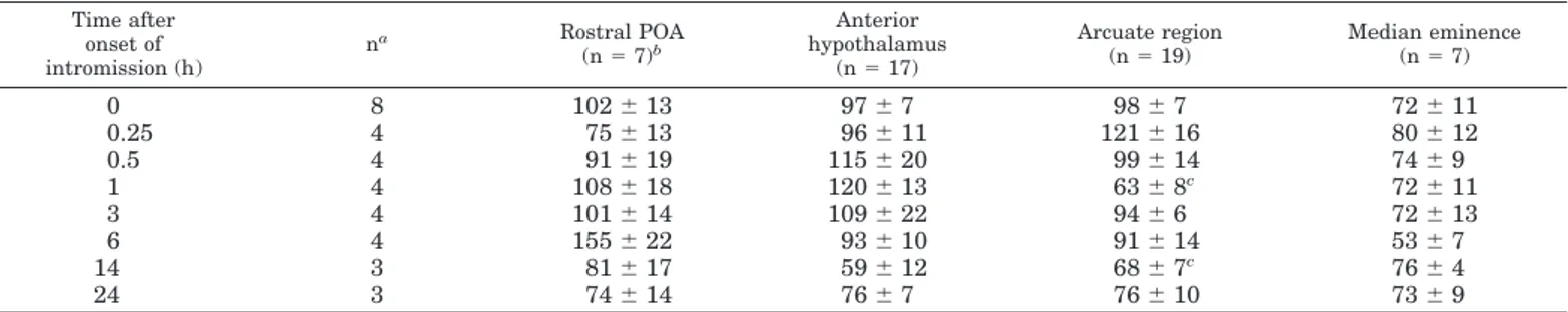
Effect of mating on cellular GnRH mRNA levels
Cellular GnRH mRNA levels decreased in the arcuate region over the course of the preovulatory LH surge (Table 1). Mean cellular levels of GnRH mRNA in hybridized cells of the arcuate region were significantly lower in mated es-trous females killed 1 or 14 h after the onset of intromission compared with those in unmated estrous controls (Table 1). There were no significant mating-induced changes in cellular GnRH mRNA levels in the preoptic area, anterior hypothal-amus, or median eminence (Table 1).
Effect of ovariectomy on GnRH mRNA levels
Ovariectomy significantly decreased the mean number of GnRH mRNA-expressing cells per section in the arcuate region compared with those in gonadally intact estrous fe-males (P5 0.03; Fig. 4). The mean number of GnRH
mRNA-FIG. 1. Photomicrographs of cells expressing GnRH mRNA in the arcuate region (Arc) and the median eminence (Me) of an unmated estrous female ferret (A) and an ovariectomized female ferret (B). The high magnification inset in B shows a labeled cell with silver grains over a counterstained cell body. Arrows indicate labeled cells.
FIG. 2. Camera lucida drawings of coronal sections through the
ros-tral preoptic area (15.0 mm anterior to the interaural line), anterior hypothalamus (13.4 mm), arcuate region (12.6 mm), and median eminence (11.9 mm) showing the distribution of GnRH mRNA-con-taining neurons ( ) in a representative, unmated, estrous female ferret. 3V, Third ventricle; AC, anterior commissure; Arc, arcuate nucleus; Me, median eminence; OC, optic chiasm; OT, optic tract.
expressing cells did not differ significantly between ovari-ectomized females and gonadally intact anestrous females. There was no effect of ovariectomy on cellular GnRH mRNA levels in any of the brain regions analyzed (Table 2). In addition, cellular GnRH mRNA levels did not differ between gonadally intact estrous and anestrous females.
Plasma LH and estradiol
There was evidence of a mating-induced preovulatory LH surge in estrous females (Fig. 5A). Mean plasma LH levels were significantly higher in mated females killed 0.25 h (P5 0.008), 0.5 h (P5 0.004), 1 h (P 5 0.02), 14 h (P 5 0.01), or 24 h
(P5 0.04) after the onset of intromission than in unmated estrous females (0 h). Ovariectomy increased plasma LH levels to those seen in mated, estrous females during the preovulatory LH surge (Fig. 5A). Anestrous females had plasma LH levels that were intermediate between those of unmated estrous and ovariectomized females. Both ovariec-tomized females (P 5 0.004) and anestrous females (P 5 0.005) had significantly higher plasma LH levels than un-mated estrous females, whereas ovariectomized females had higher plasma LH levels than anestrous females (P5 0.009). When the data from Exp 1 and 2 were combined, no signif-icant correlation was found between plasma LH levels and the number of GnRH mRNA-expressing cells or cellular GnRH mRNA levels in any of the four brain regions analyzed.
Plasma estradiol levels were significantly higher in estrous females (unmated; 0 h) than in anestrous (P5 0.05) or ovari-ectomized females (P5 0.01; Fig. 5B). Plasma estradiol levels did not vary significantly among groups of estrous females killed at different times during the mating-induced preovu-latory LH surge (Fig. 5B). Also, plasma estradiol levels were equally low in anestrous and ovariectomized females. When data from Exp 1 and 2 were combined, no significant cor-relation was found between plasma estradiol levels and the number of GnRH mRNA-expressing cells or cellular GnRH mRNA levels in any of the four brain regions analyzed.
Discussion
Relationship between GnRH mRNA levels and GnRH release after mating
We found no evidence of increased GnRH mRNA levels in the MBH during the ferret’s mating-induced preovulatory LH surge. Instead, GnRH mRNA levels actually decreased in some MBH regions over the course of the preovulatory LH surge. In a previous study (3), we reported that the in vitro release of GnRH from perifused MBH slices was significantly reduced in estrous female ferrets killed 0.25 h after the onset of intromission, but was restored to unmated levels in fe-males killed 1 or 2.6 h after the onset of intromission. These results suggest that there is a large release of GnRH imme-diately after mating that depletes releasable stores of the peptide in MBH nerve terminals. Replenishment of GnRH stores apparently occurs within 1 h after mating. We hy-pothesized that an increase in MBH GnRH gene expression contributes to this replenishment. However, in the present study MBH GnRH mRNA levels were not elevated during the first hour after the onset of intromission. The absence of any increase in GnRH mRNA levels suggests that posttran-scriptional events, such as increased GnRH mRNA transla-tion, increased conversion of the pro-GnRH peptide into the mature GnRH decapeptide, and/or increased transport of the peptide to the nerve terminals, contribute to the previ-ously observed (3) replenishment of GnRH stores in the MBH.
GnRH mRNA levels began to decrease in the MBH during the peak phase of the preovulatory LH surge. Specifically, the number of GnRH mRNA-expressing cells was lower in the arcuate region of estrous females killed 6, 14, or 24 h after the onset of intromission. This decrease probably reflects a
re-FIG. 3. Effect of receipt of an intromission from a male on the mean (6SEM) number of GnRH mRNA-expressing cells per brain section. The mean (6SEM) numbers of coronal brain sections within each region were: preoptic area (POA), 236 1; anterior hypothalamus (AH), 176 1; arcuate region (Arc), 26 6 1; and median eminence (Me), 266 1. *, P , 0.05, by two-tailed Mann-Whitney U comparisons with unmated (0 h) estrous females. The number of subjects per group is shown above the bars in the top panel.
598 GnRH mRNA LEVELS IN A REFLEX OVULATOR, THE FERRET Endo• 1999 Vol 140• No 2
duction in cellular mRNA content, so that cells expressing decreased levels of mRNA were no longer detected with the in situ hybridization method used. Indeed, cellular levels of GnRH mRNA in the arcuate region decreased significantly in estrous females killed 14 h after the onset of intromission. In addition, there was a trend in the preoptic area and an-terior hypothalamus for cellular GnRH mRNA levels and the number of GnRH mRNA-expressing cells to decline over the course of the preovulatory LH surge.
The decrease in MBH GnRH mRNA levels during the preovulatory LH surge may have been caused by reductions in GnRH gene transcription and/or GnRH mRNA stability. In rats, GnRH mRNA levels appear to be regulated across the estrous cycle at the level of gene transcription and mRNA stability (15, 16). For example, GnRH mRNA levels repeat-edly exhibit two significant peaks in cycling rats, one on diestrus and another on the afternoon of proestrus (15). The increase in GnRH mRNA levels on diestrus is probably due to an increase in mRNA stability, whereas the increase on proestrus is accompanied by an increase in gene transcrip-tion (15). However, the decrease in GnRH mRNA levels during the mating-induced preovulatory LH surge in estrous ferrets is most likely not the result of changes in ovarian steroid secretion. Plasma progesterone concentrations do not rise until 5 days after receipt of an intromission (17), and plasma estradiol concentrations did not fluctuate signifi-cantly during the mating-induced preovulatory LH surge (present study) and only began to drop 4 days after receipt of an intromission (17).
In the present study, mean plasma LH levels were signif-icantly elevated in most groups of mated estrous females, suggesting that they experienced a mating-induced preovu-latory LH surge. This confirms our previous observations that intromissions that last longer than 2 min always suffice to induce a preovulatory LH surge in estrous female ferrets (1–3). However, there was considerable variability in the data, with relatively low levels of plasma LH measured in females killed 1, 3, or 6 h after the onset of intromission. Carroll et al. (2) demonstrated that the mating-induced pre-ovulatory LH surge is characterized by increases in pulse frequency and amplitude. As we took a single trunk blood sample when subjects were killed for in situ hybridization, we probably measured LH in both peaks and valleys of these
FIG. 4. Effect of ovariectomy on the mean (6SEM) number of GnRH mRNA-expressing cells per brain section. The mean (6SEM) numbers of coronal brain sections within each region were: preoptic area (POA), 196 0; anterior hypothalamus (AH), 20 6 1; arcuate region (Arc), 27 6 1; and median eminence (Me), 276 1. *, P , 0.05, by two-tailed Mann-Whitney U comparisons with estrous females. The number of subjects per group is shown above the bars in the top panel.
TABLE 1. Effect of receipt of an intromission from a male on cellular GnRH mRNA levels in three or four anatomically matched sections
from the mediobasal hypothalamus of estrous female ferrets
Time after onset of intromission (h) na Rostral POA (n5 7)b Anterior hypothalamus (n5 17) Arcuate region (n5 19) Median eminence (n5 7) 0 8 1026 13 976 7 986 7 726 11 0.25 4 756 13 966 11 1216 16 806 12 0.5 4 916 19 1156 20 996 14 746 9 1 4 1086 18 1206 13 636 8c 726 11 3 4 1016 14 1096 22 946 6 726 13 6 4 1556 22 936 10 916 14 536 7 14 3 816 17 596 12 686 7c 766 4 24 3 746 14 766 7 766 10 736 9 Data are expressed as the mean6SEMhybridization area (square microns) per cell.
aNumber of subjects per group.
bMean number of hybridized cells analyzed per region in each subject. cSignificantly lower (P, 0.05) than unmated female value (0 h).
pulses. Wersinger and Baum (14) also found relatively low plasma LH levels in female ferrets killed 1.5 or 3 h after the onset of a 5-min intromission, suggesting that synchronized phases of reduced LH secretion occur during the ferret’s
preovulatory LH surge. Even so, Wersinger and Baum (14) found appreciable numbers of MBH Fos/LHRH double-la-beled neurons in all mated females, including those with low LH levels at the time of death. This suggests that MBH GnRH neurons were activated in all estrous females that received an intromission.
Ovarian regulation of GnRH mRNA levels
There appears to be a relationship between levels of GnRH mRNA in the ferret’s MBH (present study) and the in vitro release of GnRH peptide from the MBH (3). In estrous female ferrets, GnRH mRNA (present study) and basal in vitro re-lease and content of GnRH peptide (3) in the MBH are high. After ovariectomy, GnRH mRNA levels (present study) and basal in vitro release and content of GnRH peptide (3) in the MBH are reduced. These data suggest that ovarian hormones stimulate GnRH gene expression as well as GnRH release. This stimulatory action of ovarian hormones on GnRH gene expression and release are in contrast to the negative feed-back action of ovarian hormones on LH release from the pituitary (7, 18). There is evidence that estrogen promotes GnRH gene transcription in the rat (16). Estradiol, but not progesterone, treatment increased levels of GnRH RNA pri-mary transcript in the organum vasculosum of the lamina terminalis/rostral preoptic area of ovariectomized rats, which points to an estrogenic induction of GnRH gene tran-scription (16). Also, GnRH primary transcript levels were elevated on proestrus in cycling female rats, a finding con-sistent with an estrogen-induced increase in GnRH tran-scription (15). However, the present study provides no in-dication that estradiol is the ovarian hormone that promotes MBH GnRH mRNA levels in the female ferret. We found equivalent MBH GnRH mRNA levels in gonadally intact female ferrets that were in estrus or anestrus even though plasma estradiol levels were significantly higher in estrous females. Also, there was no positive correlation between plasma estradiol levels and GnRH mRNA levels in any brain region studied. The absence of significant differences in GnRH mRNA levels between estrous and anestrous females suggests that the reduced function of the hypothalamic-pi-tuitary-gonadal axis during anestrus might reflect a reduced release of GnRH from the MBH or reduced sensitivity of the pituitary to GnRH, as opposed to a down-regulation of MBH GnRH mRNA levels. The reduction in MBH GnRH mRNA levels after ovariectomy suggests that ovarian hormones other than estradiol may facilitate MBH GnRH mRNA levels in anestrous and estrous females.
TABLE 2. Effect of ovariectomy on cellular GnRH mRNA levels in three or four anatomically matched sections from the mediobasal
hypothalamus of female ferrets
Group na Rostral POA (n5 9)b Anterior hypothalamus (n5 18) Arcuate region (n5 20) Median eminence (n5 8) Estrous 3 1116 25 1006 12 1086 13 916 25 Anestrous 5 786 7 996 3 946 15 676 9 Ovariectomy 4 1116 11 1016 14 856 14 656 10 Data are expressed as the mean6SEMhybridization area (square microns) per cell.
aNumber of subjects per group.
bMean number of hybridized cells analyzed per region in each subject.
FIG. 5. LH (A) and estradiol (B) levels in trunk plasma taken when female ferrets were killed to obtain brains for GnRH mRNA in situ hybridization. Individual values (solid circles) and group means (bars) 6SEMare shown. Anest, Anestrous; ovx, ovariectomized. The number (N) of subjects per group is given at the bottom of B. Note that five of eight unmated estrous females had plasma LH levels that were below the detection limit of the assay (0.45 ng/ml) and that all ovariecto-mized females and three of four anestrous females had plasma es-tradiol levels that were below the detection limit of the assay (2 pg/ml).
600 GnRH mRNA LEVELS IN A REFLEX OVULATOR, THE FERRET Endo• 1999
Species comparisons
Our finding that the mating-induced preovulatory LH surge is accompanied by progressively decreasing levels of GnRH mRNA is comparable to earlier findings in sheep. In the ovariectomized ewe, the estrogen-induced LH surge was associated with decreased levels of GnRH mRNA in the preoptic area (19, 20). In the rat, however, in those studies in which changes were reported (reviewed in Ref. 21), increases in either cellular GnRH mRNA levels or GnRH mRNA cell numbers occurred in the organum vas-culosum of the lamina terminalis/rostral preoptic area during the estrogen-induced LH surge (10, 15, 22, 23). There is a species difference in the temporal relationship between the changes observed in GnRH mRNA levels and the LH surge. In the ferret, in which the preovulatory LH surge is induced by mating, MBH GnRH mRNA levels started to decrease during the peak phase of the surge and remained low until its end. In the sheep, GnRH mRNA levels in the preoptic area decreased in advance of the onset of the estrogen-induced LH surge (20). In the rat, GnRH mRNA levels in the organum vasculosum of the lamina terminalis/rostral preoptic area increased in ad-vance (15) or at the time of the LH surge (10, 22, 23). These species differences in the temporal relationship between changes in hypothalamic GnRH mRNA levels and LH release are one characteristic of the different mechanisms controlling the preovulatory LH surge in reflex vs. spon-taneous ovulators.
We found that ovariectomy decreased the GnRH mRNA cell number in the arcuate region of the ferret. A similar decrease in GnRH mRNA levels has been reported in the rat (24, 25), although this finding is controversial (re-viewed in Ref. 21). Other researchers found no effect (26) or an increase (27) in GnRH mRNA levels after ovariec-tomy in female rats. In the female ferret, estradiol alone appears not to stimulate MBH GnRH mRNA expression. However, estradiol may play a permissive role by increas-ing the sensitivity of MBH GnRH neurons to activation by somatosensory or olfactory stimuli associated with mating.
Our finding that anestrous and estrous female ferrets have equivalent levels of MBH GnRH mRNA agrees with earlier findings in another long day seasonal breeder, the hamster (28, 29). Male hamsters chronically exposed to either long or short photoperiods had similar numbers of hypothalamic GnRH mRNA-expressing neurons. This suggests that the reduced function of the hypothalamic-pituitary-gonadal axis in both sexually quiescent male hamsters and anestrous female ferrets is mediated pri-marily by reductions in GnRH release or pituitary sensi-tivity to GnRH, as opposed to reduced hypothalamic GnRH mRNA levels.
Acknowledgments
We thank Dr. Kathleen D. Ryan at Magee-Womens Research Institute (Pittsburgh, PA) for generously performing the LH assay, Dr. Geralyn Messerlian Lambert at Womens and Infants Hospital (Providence, RI) for generously performing the estradiol assay, Diagnostic Products Corp. (Los Angeles, CA) for providing the reagents for the estradiol assay, Dr. Cheryl L. Sisk of Michigan State University (East Lansing, MI)
for generously providing an initial batch of the oligoprobe, and the animal care staff at Boston University (Boston, MA) for their care of our ferrets. Finally, we thank Dr. Patricia F. Finn for critical reading of an earlier version of this manuscript.
References
1. Carroll RS, Erskine MS, Doherty PC, Lundell LA, Baum MJ 1985 Coital stimuli controlling luteinizing hormone secretion and ovulation in the female ferret. Biol Reprod 32:925–933
2. Carroll RS, Erskine MS, Baum MJ 1987 Sex difference in the effect of mating on the pulsatile secretion of luteinizing hormone in a reflex ovulator, the ferret. Endocrinology 121:1349 –1359
3. Lambert GM, Rubin BS, Baum MJ 1992 Sexual dimorphism in the effects of mating on the in vitro release of LHRH from the ferret mediobasal hypothal-amus. Physiol Behav 52:809 – 813
4. Bibeau CE, Tobet SA, Anthony ELP, Carroll RS, Baum MJ, King JC 1991 Vaginocervical stimulation of ferrets induces release of luteinizing hormone-releasing hormone. J Neuroendocrinol 3:29 –36
5. Versi E, Chiapp SA, Fink G, Charlton HM 1982 Effect of copulation on the hypothalamic content of gonadotrophic hormone-releasing hormone in the vole, Microtus agrestis. J Reprod Fertil 64:491– 494
6. Ryan KD, Siegel SF, Robinson SL 1985 Influence of day length and endocrine status on luteinizing hormone secretion in intact and ovariectomized adult ferrets. Biol Reprod 33:690 – 697
7. Carroll RS, Baum MJ 1989 Evidence that oestrogen exerts an equivalent negative feedback action on LH secretion in male and female ferrets. J Reprod Fertil 86:235–245
8. Kalra SP, Kalra PS 1989 Do testosterone and estradiol-17b enforce inhibition or stimulation of luteinizing hormone-releasing hormone secretion? Biol Re-prod 41:559 –570
9. Adelman JP, Mason AJ, Hayflick JS, Seeburg PH 1986 Isolation of the gene and hypothalamic cDNA for the common precursor of gonadotropin-releasing hormone and prolactin release-inhibiting factor in human and rat. Proc Natl Acad Sci USA 83:179 –183
10. Porkka-Heiskanen T, Urban JH, Turek FW, Levine JE 1994 Gene expression in a subpopulation of luteinizing hormone-releasing hormone (LHRH) neu-rons prior to the preovulatory gonadotropin surge. J Neurosci 14:5548 –5558 11. Tang YP, Kashon ML, Sisk CL 1997 Brain region-specific regulation of lu-teinizing hormone-releasing hormone messenger ribonucleic acid in the male ferret: interactions between pubertal maturation and testosterone. Endocri-nology 138:4740 – 4747
12. King JC, Anthony ELP 1984 LHRH neurons and their projections in humans and other mammals: species comparisons. Peptides 5:195–207
13. Tang YP, Sisk CL 1992 LHRH in the ferret: pubertal decrease in the number of immunopositive arcuate neurons. Peptides 13:241–247
14. Wersinger SR, Baum MJ 1996 The temporal pattern of mating-induced im-mediate-early gene product immunoreactivity in LHRH and non-LHRH neu-rons of the estrous ferret forebrain. J Neuroendocrinol 8:345–359
15. Gore AC, Roberts JL 1995 Regulation of Gonadotropin-releasing hormone gene expression in the rat during the luteinizing hormone surge. Endocrinol-ogy 136:889 – 896
16. Petersen SL, Gardner E, Adelman J, McCrone S 1996 Examination of steroid-induced changes in LHRH gene transcription using33P- and35S-labeled probes
specific for intron 2. Endocrinology 137:234 –239
17. Villars TA, Erskine MS, Lambert GM, Jacobson D, Weaver DE, Baum MJ 1990 Endocrine correlates of mating-induced reductions in estrous behavior in an induced ovulator, the ferret. Horm Behav 24:198 –214
18. Baum MJ, Gerlach JL, Krey LC, McEwen BS 1986 Biochemical and radioau-tographic analysis of estrogen-inducible progestin receptors in female ferret brain and pituitary: correlations with effects of progesterone on sexual be-havior and gonadotropin-releasing hormone-stimulated secretion of luteiniz-ing hormone. Brain Res 368:296 –309
19. Dhillon H, Dunn AM, Esquivel E, Hamernik DL, Wise ME 1997 The estra-diol-induced luteinizing hormone surge in the ewe is not associated with increased gonadotropin-releasing hormone messenger ribonucleic acid levels. Biol Reprod 57:107–111
20. Harris TG, Robinson JE, Evans NP, Skinner DC, Herbison AE 1998 Gona-dotropin-releasing hormone messenger ribonucleic acid expression changes before the onset of the estradiol-induced luteinizing hormone surge in the ewe. Endocrinology 139:57– 64
21. Sagrillo CA, Grattan DR, McCarthy MM, Selmanoff M 1996 Hormonal and neurotransmitter regulation of GnRH gene expression and related reproduc-tive behaviors. Behav Genet 26:241–277
22. Zoeller RT, Young III WS 1988 Changes in cellular levels of messenger ribonucleic acid encoding gonadotropin-releasing hormone in the anterior hypothalamus of female rats during the estrous cycle. Endocrinology 123:1688 –1689
23. Park O-K, Gugneja S, Mayo KE 1990 Gonadotropin-releasing hormone gene expression during the rat estrous cycle: effects of pentobarbital and ovarian steroids. Endocrinology 127:365–372
24. Kim K, Lee BJ, Park Y, Cho WK 1989 Progesterone increases messenger ribonucleic acid (mRNA) encoding luteinizing hormone-releasing hormone (LHRH) level in the hypothalamus of ovariectomized estradiol-primed rats. Mol Brain Res 6:151–158
25. Roberts JL, Dutlow CM, Jakubowski M, Blum M, Miller RP 1989 Estradiol stimulates preoptic area-anterior hypothalamic proGnRH-GAP gene expres-sion in ovariectomized rats. Mol Brain Res 6:127–134
26. Kelly MJ, Garret J, Bosch MA, Roselli CE, Douglass J, Adelman JP,
Ronnekleiv OK1989 Effects of ovariectomy on GnRH mRNA, proGnRH and GnRH levels in preoptic hypothalamus of the female rat. Neuroendocrinology 49:88 –97
27. Toranzo D, Dupont E, Simard J, Labrie C, Couet J, Labrie F, Pelletier G 1989 Regulation of pro-gonadotropin releasing hormone gene expression by sex steroids in the brain of male and female rats. Mol Endocrinol 3:1748 –1756
28. Ronchi E, Krey LC, Pfaff DW 1992 Steady state analysis of hypothalamic GnRH messenger RNA levels in male Syrian hamsters: influences of photo-period and androgen. Neuroendocrinology 55:146 –155
29. Porkka-Heiskanen T, Khoshaba N, Scarbrough K, Urban JH, Vitaterna MH,
Levine JE, Turek FW, Horton TH1997 Rapid photoperiod-induced increase in detectable GnRH mRNA-containing cells in Siberian hamster. Am J Physiol 273:R2032–R2039
5th International Symposium on Insulin-Like Growth Factors 31 October– 4 November, 1999, Brighton, UK
Conference Chairman: Professor Jeff Holly, University of Bristol
Programme Committee: R. Baxter (Aus), D. Clemmons, (US), C. Conover (US), P. Gluckman (NZ), N. Hizuka (Jap), J. Holly (UK), D. LeRoith (US), R. Rosenfield (US), J. Zapf (Switz). Abstracts deadline: 1 May 1999. Contact for further details: Janet Crompton, 29 North Road, St. Andrews, Bristol BS6 5AD, UK. Tel: 144 (0)117 924 8160; Fax: 144 (0)117 924 1208; E-mail: janetcrompton@compuserve.com.
602 GnRH mRNA LEVELS IN A REFLEX OVULATOR, THE FERRET Endo• 1999 Vol 140• No 2