Pferdeheilkunde 22 (2006) 5 (September/Oktober)
Stress echocardiography in horses – a review
Charlotte Sandersen and Hélène AmoryUniversity of Liege, Faculty of Veterinary Medicine, Department of Clinical Sciences, Liege, Belgium
Summary
The increasing number of publications in the field of equine stress echocardiography demonstrates the growing interest in this area. Stress echocardiography consists of B- and/or M-mode echocardiography under active or passive stimulation of the heart, where the pre-stimu-lation images are compared to those obtained during or immediately after stimupre-stimu-lation. In human medicine, stress echocardiography is mainly used as a routine tool in diagnosis and prognosis of coronary artery disease, but also in the evaluation of hypertrophied and dila-ted cardiomyopathy and valvular diseases. In horses, the principal indications include the detection of exercised-induced myocardial insuf-ficiency and refining the prognosis of low- and mid-grade valvular diseases. The two major cardiac stressors that can be used in horses are (1) physical exercise, which has the major disadvantage of a rapid decline in heart rate in the immediate post-exercise period and (2) pharmacological stimulation, which ideally consists of dobutamine infusion in combination of a parasympatholytic drug, in order to over-come the strong dobutamine-induced baroreceptor reflex in horses. Most stress echocardiographic studies performed in horses demon-strated a significant decrease of left ventricular length, diameter, area and volume in response to stimulation. Other studies also revealed a stimulation-induced increase in the interventricular septum, left ventricular free wall thickness, and left ventricular fractional. Some of the-se changes the-seemed to be more pronounced during pharmacological stimulation than after exercithe-se. One study described the application of a semi-quantitative wall motion scoring system in horses. Exercise as well as pharmacological stress echocardiography enhances the diagnostic possibilities in equine medicine, because it increases the chance to detect a problem that is not present at rest. Furthermore, this technique could help to investigate the relationship between valvular insufficiencies and ventricular dysfunction.
Keywords: exercise-induced myocardial dysfunction, poor performance, wall motion index, dobutamine, atropine Stressechokardiographie beim Pferd – eine Übersicht
In den vergangenen Jahren hat das Interesse an Stressechokardiographie beim Pferd deutlich zugenommen, was durch die steigende Anzahl der Publikationen zu dem Thema belegt wird. Die Technik basiert auf der Anwendung von Schnittbildechokardiographie in Kombination mit aktiver oder passiver Stressinduktion, wobei die unter Stimulation gewonnenen Bilder mit denen im Ruhezustand verglichen werden. In der Humanmedizin wird Stressechokardiographie hauptsächlich als Routinemittel in der Diagnose und Prognose von Koronarerkrankungen, und darüber hinaus in der Bewertung von hypertrophischen und dilatierten Kardiomyopathien und Herzklappenerkrankung eingesetzt. Bei Pferden ist die Untersuchung des Herzens im stimulierten Zustand vor allem zur Erkennung von belastungsinduzierter Myokardinsuffizienz und zur genaueren Prognosestellung von gering- und mittelgradigen Klappeninsuffizienzen von Nutzen. Ähnlich wie in der Humanmedizin stehen grundsätzlich zwei Formen der Stressinduktion zur Ve r f ü g u n g. Aktive Belastung durch Longen- oder La u f b a n d b e l a s t u n g, hat den Vorteil, dass sie leicht verfügbar und gut verträglich ist, hat allerdings den wesentlichen Nachteil, dass die Herzfrequenz rapide nach Ende der Belastung unter 100 Schläge/Minute absinkt und dass der Zeitraum der zur echokardiographischen Bildermittlung zur Verfügung steht, sehr kurz ist. Bei der pharmakologischen Stressinduktion hat sich eine Kombination aus einer Dauerinfusion des beta-Agonisten Dobutamin und dem Pa r a s y m-patholytikums Atropin bewehrt. Atropin hat die Aufgabe, den durch Dobutamin ausgelösten Barorezeptor- Reflex herabzusenken, und bewirkt damit eine Steigerung der Herzfrequenz. Diese Kombination hat sich gegenüber anderen Stressoren als vorteilhaft erwiesen, da sie gut ver-träglich ist, weniger kardiomyotoxisch ist und seltener Herzarrhythmien auslöst als z.B. alleiniges hochdosiertes Dobutamin oder Adrenalin. In den meisten Studien wurden bei den Pferden das linke Herz von rechts in B- und M-mode Technik dargestellt. In einigen Studien wurde dop-plerechokardiographisch der Pulmonararterien- und Aortenfluss gemessen und daraus das Schlagvolumen berechnet. Dabei belegen alle Stu-dien unter Stimulation eine Verminderung des Durchmessers, der Länge, des Volumens und des Querschnittsfläche des linken Ventrikels. Außer-dem wurde in einigen Studien eine Zunahme der der Wandstärke des Interventrikularseptums und der linksventrikulären Hinterwand festge-stellt. Die linksventrikuläre Kürzungsfraktion nimmt in der Regel unter Stimulation zu. Diese Veränderungen können sowohl nach aktiver als auch nach passiver Stressinduktion beobachtet werden; sie sind teilweise ausgeprägter nach pharmakologischer Stimulation. Darüber hinaus wurde in einer Studie an gesunden und herzkranken Pferden der in der Humanmedizin übliche semiquantitative Ansatz vor und nach einer Laufbandbelastung getestet. Dabei wurde eine Längsansicht des linken Ventrikels in sechs Segmente unterteilt und diese vor und nach der La u f-bandbelastung anhand ihres Bewegungsmusters mit den Graden 1 für Normokinesie, 2 für Hypokinesie, 3 für Akinesie und 4 für Dyskinesie beurteilt. Die Summe der Grade geteilt durch die Anzahl der Segmente ergab bei herzgesunden Pferden einen mittleren Wa n d b e w e g u n g s i n-dex von 1.1 vor und 1.12 nach Belastung und bei herzkranken Pferden einen mittleren Wandbewegungsinn-dex von 1.5 vor und 1.52 nach der B e l a s t u n g. Diese viel versprechende Methode zeigt, dass auch ein semiquantitativer Ansatz bei Pferden sinnvoll ist. Stressechokardiographie stellt eine Bereicherung der Diagnostik in der Pferdemedizin dar, da sie die Untersuchung des Herzens während oder sofort nach der Belastung ermöglicht. Dadurch können Veränderungen erkannt werden, die im Ruhezustand nicht zu erkennen sind. Außerdem könnte diese Methode eventuell helfen, die pathophysiologischen Zusammenhänge zwischen Ventrikeldysfunktion und Klappenkrankheiten zu erklären.
Schlüsselwörter: belastungsinduzierte Myokardinsuffizienz, Leistungsintoleranz, Wandbewegungsindex, Dobutamin, Atropin
Introduction
There is a high incidence of cardiac murmurs in athletic hor-ses (Glendenning 1972). A study including 545 racehorhor-ses reported cardiac murmurs in 68 % of 545 racehorses (Patte
-son and Cribbs 1993). Another study demonstrated cardiac murmurs in 81.1% of 846 Thoroughbreds (Kriz et al. 2000), but failed to demonstrate a correlation between presence of cardiac murmurs and race performance. Determining the
sig-nificance of cardiac murmurs in horses with poor performan-ce remains a difficult task (Mitten 1996) because, even if the majority of these murmurs are physiological, some of them are clinically significant.
The possibilities for objective evaluation of heart murmurs in horses have significantly increased with the introduction of echocardiography in equine medicine. Two-dimensional and M-mode echocardiography gives information on dimensions of the different cavities and walls of the heart, while Doppler echocardiography allows determination of the direction and of the velocity of the blood flow within the heart (Blissit and Bonagura 1995a, Blissit and Bonagura 1995b, Reef 1992). Regurgitant flow can be detected and a semi-quantitative measurement of the severity of the regurgitation can be made by measuring the spatial distribution of the regurgitant jet in the receiving chamber (jet area, length and width). However, quantifying the regurgitation does not always provide a pro-gnosis. Furthermore, determination of the prognosis of cardi-ac murmurs has been shown to be a crucial point in pre-pur-chase examination as demonstrated in a study by Verdegaal et al. (2002). The authors of this study found that from 56 horses with heart murmurs detected at pre-purchase exami-nation only 24 were sold for the expected price, while 12 were sold for a lower price and 20 were not sold at all. The-refore the question arises whether other techniques, like e.g. stress echocardiography, could provide additional informa-tion on the prognosis of valvular insufficiencies in horses. Apart from the prognostic evaluation of valvular insufficien-cies, stress echocardiography will be applicable in the dia-gnosis of exercise-induced myocardial dysfunction, which has been described as a cause of poor performance in horses. For instance, in a study including 348 cases with poor per-formance, exercised-induced myocardial dysfunction was claimed to be the aetiology in 5.5% of the horses studied (Martin et al. 2002). A slightly higher incidence was found by Reef et al. (1997), who suspected exercise-induced myocar-dial dysfunction in 8% of 250 horses examined for poor per-formance. The majority of these horses had no other abnor-mality detected during resting or treadmill examination, and the diagnosis of exercise-induced myocardial dysfunction was made on the basis of stress echocardiography.
Stress echocardiography in humans Indications
In human medicine, stress echocardiography was first descri-bed in 1977 as a combination of two-dimensional echocar-diography with physical, pharmacological or electrical stress applied to the heart (Autenrieth et al.). In human patients, stress echocardiography is most frequently used in the dia-gnosis and prodia-gnosis of coronary artery disease (Cohen et al. 1991). However, the indications for stress echocardiography have also expanded into risk evaluation of patients undergo-ing vascular surgery (Poldermans et. al. 1993, Poldermans et al. 1995), evaluation of chest pain (Geleijnse et al. 2000), assessment of myocardial viability (Smart et al. 1993, Wata -da et al. 1994, Previtali et al. 1993, Pierard et al. 1990), detection of occult pulmonary hypertension (Armstrong and Zoghbi 2005), assessment of mitral valvular disease (Wu et
al. 2004) and the evaluation of prosthetic mitral valve (Lea -vitt et al. 1991).
Exercise stress echocardiography
Both treadmill and bicycle exercise are used to perform exer-cise stress echocardiography. When a treadmill test is perfor-med, scanning during exercise is not feasible, so most proto-cols rely on post-exercise imaging within one minute after cessation of exercise (Armstrong and Zoghbi 2005). This technique assumes that exercise-induced wall motion abnor-malities persist during this period. However, due to rapid recovery of the wall motion abnormalities after exercise, fal-se-negative results occur (Picano 2004). The advantages of treadmill exercise echocardiography are widespread availabi-lity of treadmill systems and the wealth of clinical experience with this form of stress testing. Bicycle exercise echocardio-graphy is performed during either an upright or a recumbent posture. The patient pedals against an increasing workload while echocardiographic imaging is performed. The major advantage of bicycle exercise is the opportunity to obtain images during various levels of exercise rather than relying on post-exercise imaging. Exercise is the prototype of ischemic stress and the most commonly stressor used to perform stress echocardiography in humans (Picano 2004). However, it has been shown that 20% of the patients undergoing exercise stress echocardiography are unable to exercise, 20 % exerci-se submaximally, and 20% have an uninterpretable ECG (Picano 2004). In addition, exercise induces hyperventilation and excessive chest wall movement, which degrades image quality, increases inter-observer variability, and therefore lowers diagnostic accuracy. These factors explain the popula-rity of pharmacological stress induction.
Pharmacological stress echocardiography
Although different pharmacological stressors are used in human stress echocardiography, dobutamine is by far the most commonly used (Marwick 2005). The standard dobuta-mine stress protocol consists of continuous intravenous infu-sion of dobutamine in 3 minutes increments, starting with 5 µg/kg/min, and increasing to 10, 20, 30 and 40 µg/kg/min. If no endpoint is reached, atropine in doses of 0.25 mg up to a maximum of 1 mg is added to the dobutamine infusion rate of 40 µg/kg/min.
Data acquisition and interpretation
Independently of the cardiac stressor applied during stress echocardiography, a standard 12-lead ECG and blood pres-sure are continuously monitored before and during the test. Echocardiographic monitoring is continuously employed and intermittently recorded before, during and after cessation of stress. According to the American Heart Association, the left ventricle is imaged in three longitudinal views: (1) a horizon-tal long-axis view, (2) a vertical four-chamber view, and (3) a two chamber view and in three short-axis views: (1) at the level of the mitral valve, (2) at the level of the papillary muscles and (3) at the level of the apex (Schiller et al. 1989, Cer -queira et al. 2002). The left ventricle is divided into 16
seg-ments that are scored subjectively for their contractility, where 0 = hyperkinetic, 1 = normokinetic, 2 = hypokinetic, 3 = akinetic, and 4 = dyskinetic (Carstensen et al. 1995). The sum of the score divided by the number of segments gives the unit-less wall motion index. This semi-quantitative method is less time consuming than a quantitative approach, but sub-ject to variation and intensive training is required before maximal diagnostic yield is reached (Picano et al. 1991). Diagnostic endpoints of the stress test are: reaching the maxi-mal dose (for pharmacological stress) or maximaxi-mal workload (for exercise testing); achievement of target heart rate; obvious echocardiographic positivity (with akinesia of three or more left ventricular segments); severe chest pain; or obvious electrocardiographic positivity (with > 2mV ST segment shift). Submaximal non-diagnostic endpoints of stress echo are into-lerable symptoms or limiting asymptomatic effects such as hypertension, with systolic blood pressure higher than 220 mmHg or diastolic blood pressure higher than 120 mmHg; hypotension with more than 30 mmHg drop in blood pressu-re; supraventricular arrhythmias, such as supraventricular tachycardia or atrial fibrillation; and complex ventricular arrhythmias.
Stress echocardiography in horses Exercise electrocardiography
In the sixties, telemetric exercise electrocardiography was des-cribed in horses (Banister and Purvis 1968, Bassan and Ott 1968) and since then became a routine technique for the detection of exercise-related cardiac arrhythmias. Although equine ECGs give useful information about heart rate and rhythm, it provides little or no information about the relative or absolute sizes of the ventricles (Patteson 1995). Due to the widespread distribution of the Purkinje fibres, which extend throughout the equine myocardium, the depolarization spre-ads out in several directions at once. Therefore these forces tend to cancel each other out; leading to a silent depolarisa-tion of a large pordepolarisa-tion of the ventricular mass on a surface ECG. This is in contrast to humans, where the surface ECG gives valuable information about heart size and myocardial depolarization activities. In human medicine, a depression or an elevation of the ST segments are indicators of coronary artery disease and myocardial infarction, respectively (Dage -nais et al. 1982, Henry et al. 2006). There is no evidence that myocardial ischemia leads to similar changes in equine ECGs.
Exercise stress echocardiography
In 1977, echocardiography was described for the first time in horses by Pipers and Hamlin. Since then, echocardiography had become a routine technique in equine cardiology. Howe-ver, examining the equine heart by ultrasound under stress conditions was described only recently.
In 1994, Reef et al. described for the first time the use of post-exercise echocardiography in horses. The authors stated that horses suffering from exercise-induced myocardial dysfunc-tion may have a normal echocardiographic examinadysfunc-tion at
rest, or they might have only a low-grade dyskinesia or aki-nesia, of which its significance is unknown. The normal equi-ne heart should respond to exercise by an increase in the systolic thickening of the inter-ventricular septum and of the left ventricular free wall and an increase in fractional shorte-ning over the resting values. Myocardial dysfunction exists if fractional shortening is unchanged or decreased, no thicke-ning of septum and free wall, or dyskinetic or akinetic move-ments are detected immediately after exercise.
Two studies, one by Marr et al. (1999) and one by Sampson et al. (1999) performed post-exercise echocardiography in healthy horses. Although the primary goal of these two studies was not to detect exercise-induced myocardial dysfunction, but rather to investigate the physiological response of the heart to exercise, they give invaluable information about post-exercise echocardiography in healthy horses. Marr et al. (1999) measured the left ventricular M-mode parameters in five healthy horses before and after treadmill exercise in cold and hot/humid environment. Sampson et al. (1999) descri-bed the relationship between VO2max and cardiac output to heart score and echocardiographic parameters in six healthy Thoroughbreds performing a maximal treadmill exercise test. Both studies clearly showed that after cessation of exercise, the left ventricular echocardiographic parameters quickly return to pre-exercise levels. A similar conclusion was made by Durando et al. (2002) who measured right ventricular pressure dynamics in nine healthy Thoroughbreds during and directly after a maximal treadmill exercise and correlated the findings to left ventricular fractional shortening and wall motion indices. In this study, right ventricular pressure deter-minants had returned to pre-exercise values within 60 to 120 seconds after the end of exercise. The rapid return to baseli-ne of cardiac parameters in the immediate post-exercise peri-od is also well known in human medicine where it is general-ly advised to perform the echocardiographic recording within one minute after the end of exercise in order to guarantee a reliable specificity of the test (Armstrong and Zoghbi 2005). In the three studies mentioned above (Marr et al 1999, Samp -son et al. 1999, Durando et al. 2002), the horses underwent a standardised treadmill test. As a high speed treadmill is not commonly available, the question arose weather alternative forms of exercise can be used to perform stress echocardio-graphy in horses. Gehlen et al. (2005a) compared the effects of treadmill versus lunging exercise on left ventricular M-mode parameters in 20 healthy horses. The horses attained a mean maximal heart rate of 155 ± 12 bpm during treadmill exercise and a mean maximal heart rate of 169 ± 14 during lunging exercise. Both forms of stress induction allowed obtai-ning echocardiographic recordings within two minutes after the end of exercise. The mean heart rate at the time of echo-cardiographic recording was 114 ± 6 bpm after treadmill exercise and 100 bpm after lunging exercise. In both groups, a significant increase in left ventricular wall thickness and fractional shortening was observed and there was no signifi-cant difference between the results obtained after treadmill or after lunging exercise. Twelve of the twenty horses were also tested three to five minutes after cessation of treadmill exerci-se when the mean heart rate was 78 ± 3 bpm, but no signi-ficant difference to pre-exercise values was observed. All of the studies mentioned above based their evaluation of the cardiac stress effect on the measurement of M- and
B-mode echocardiographic parameters. This method is time consuming and has not taken hold in human medicine, whe-re interpwhe-retation is generally based on a semi-quantitative scoring system. The first attempt to introduce a semi-quanti-tative wall motion scoring system in equine stress echocar-diography was made by Durando et al. (2002), who descri-bed this technique in healthy horses before and after a maxi-mal treadmill exercise. This group used an imaging software system that displayed pre- and post-exercise images side-by-side at simulated matched heart rates allowing critical assess-ment of segassess-mental wall motion as illustrated in Figure 1. The
mean wall motion index was 1.01 before and 1.12 after exer-cise.
A similar technique was then applied by Gehlen et al. (2005b) who studied 23 healthy horses and 12 horses suffe-ring from different degrees of cardiac diseases (5 of them had low-grade cardiac disease, 5 had moderate-grade cardiac disease and 2 had severe cardiac disease). The left ventricle was visualised in B-mode before and directly after a treadmill exercise. The left ventricle was divided into 6 segments of equal size (Figure 2). Each segment was evaluated before and after exercise following the criteria according to the cri-teria summarised in table 1 (Gehlen et al. 2005b). The majo-rity of the healthy horses showed no abnormal contraction of the six segments before or after exercise. However, in nine healthy horses a hypokinesia of the papillary muscle segment was observed before exercise, and persisted after exercise in four of them. The kinetic score at rest was 1.10 ± 0.20 in
healthy horses and 1.50 ± 0.40 in horses suffering from car-diac disease. The kinetic score after exercise was 1.12 ± 0.16 in healthy horses and 1.52 ± 0.40 in those suffering from cardiac disease. Horses with mild cardiac disease had a wall motion index that was significantly different from that of healthy horses after exercise but not at rest. The two horses with severe cardiac disease did not undergo exercise; both had abnormal wall motion in five out of six segments at rest. These results suggest that wall motion analysis in post-exerci-se stress echocardiography could be particularly upost-exerci-seful to evaluate low-grade cardiac disease in horses.
Pharmacological stress echocardiography
Numerous studies demonstrated the need for maximal or near-maximal exercise as well as echocardiographic recor-dings performed quickly after cessation of exercise in order to obtain reliable results (Reef et al.1994, Reef 1997, Marr et al. 1999, Sampson et al. 1999, Reef 2001, Durando et al. 2002). To obviate the problem of the rapid drop in heart rate in the post-exercise period, several pharmacological stress protocols have been developed in horses (Frye et al. 2003, Gehlen et al. 2004, Sandersen et al. 2005a, Gehlen et al. 2006, Sandersen et al. 2006). In 2003, Frye et al. compared the effect of a maximal treadmill exercise to a high-dose dobutamine challenge on left ventricular M-mode parameters
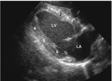
Fig 1 Right paraseternal B-mode view of the left ventricle. Six (1-6) segments for semi-quantitative evaluation of wall motion as descri-bed by Gehlenet al. (2005). LA = left atrium, LV = left ventricle
Rechte parasternale B-mode-Ansicht des linken Ventrikels. Gehlenet al. (2005) beschreiben sechs (16) Segmente für die semiquantitati ve Untersuchung der Wandbewegung. LA = linker Vorhof, LV = lin -ker Ventrikel
Fig 2 Echopac-processed images of the left ventricle in a right parasternal B-mode views at different heart rates. This program ena-bles to display cardiac cycles of different length at the same speed in order to further increase the possibility to detect wall motion abnor-malities.
Echopac-Bilder des linken Ventrikel in rechter parasternaler BmodeAnsicht bei unterschiedlichen Herzfrequenzen. Das Programm ermög -licht die Darstellung von Herzzyklen unterschiedlicher Längen bei der selben Geschwindigkeit, um die Erkennung abnormer Herzwandbe -wegungen zu verbessern.
Table 1 Wall motion scores applied in equine stress echocardiography according to Gehlenet al. (2005 c)
of healthy horses. The left ventricular M-mode parameters obtained after exercise were not significantly different from those measured after infusion of 50 µg/kg/min of dobutami-ne. However, all of the 14 horses tested showed various degrees of restlessness, three of them showed premature ven-tricular complexes and three of them showed venven-tricular tachycardia during the dobutamine challenge. Furthermore, two out of ten horses examined post-mortem showed histo-pathological lesions highly indicative of catecholamine-indu-ced myocardial toxicity.
In 2004, Gehlen et al. applied a low-dose dobutamine stress test (5µg/kg/min) to 16 horses with cardiac murmurs and to 27 horses without cardiac murmur. Cardiac auscultations as well as B-mode, M-mode, and Doppler echocardiographic images were recorded before and during the stimulation. When compared to baseline values, dobutamine at 5 µg/kg/min induced a significant increase of the left ventricu-lar fractional shortening, of the systolic interventricuventricu-lar septum thickness, and of the left ventricular free wall thickness. More-over, aortic and pulmonary peak flow velocities were signifi-cantly higher during than before dobutamine stimulation. These changes were similar in horses with and without ac murmurs. Two horses without murmur developed a cardi-ac murmur during dobutamine infusion, and in all horses with pre-existing murmur the intensity of the murmurs increased. In contrast, in horses with pre-existing valvular insufficiencies the Doppler echocardiographic examination revealed a decrea-sed prevalence and magnitude of regurgitant flows during sti-mulation. Even if this study demonstrated, that dobutamine at a dose of 5µg/kg/min appeared to be well tolerated and to induce significant increases in systolic wall thickening and in left ventricular fractional shortening it is difficult to compare it to exercise, since heart rate, a major determinant of cardiac work, was not significantly increased during the test. In human stress echocardiography, the target heart rate is 85% of the individual age-predicted maximal heart rate in order to avoid suboptimal cardiac workload which could lower the test’s sensitivity (Armstrong and Zoghbi 2005). The same is probably valid for the horse.
Since a high dobutamine infusion rate of 50 µg/kg/min appears to be potentially cardiomyotoxic in horses and since a low dobutamine infusion rate of 5µg/kg/min appears to induce an insufficient increase in heart rate an alternative protocol has been tested in healthy Shetland ponies (Sander -sen et al. 2005a). In this study, a group of seven ponies recei-ving a dobutamine infusion at a rate of 2 µg/kg/min during five minutes, followed by increasing steps of 5 µg/kg/min from 5 to 40 µg/kg/min was compared to a group of six ponies receiving twice 25 µg/kg of atropine followed by a dobutamine infusion at incremental rates of 1 µg/kg/min from 2 to 5 µg/kg/min, of 5 minutes duration each. In this latter study, cardiac output was measured as an indicator of global left ventricular function. As in the study of Frey et al. (2003), ponies that received the high dose of dobutamine showed excessive restlessness and ventricular arrhythmias during the test. In contrast, ponies receiving the low dose of dobutamine after atropine premedication showed less signs of restlessness, less inter-individual variability, and a higher increase in cardiac output, although they received a dobuta-mine infusion rate that was 8 times lower. This phenomenon might be explained by the atropine-induced inhibition of the
baroreceptor reflex that is induced by high-dose dobutamine. The protocol used in this study appeared to induce a cardiac stimulation of a similar magnitude than maximal exercise, to be well tolerated, and to give the possibility to record high quality images during an adequate cardiac stress test in hor-ses.
The protocol was then slightly modified and used with a lower dose of atropine as tested in a study by Sandersen et al. (2006), where seven horses were premedicated with 35µg/kg of atropine followed by a continuous infusion of increasing dobutamine rates up to a maximal rate of 6 µg/kg/min. Left ventricular M-mode parameters were recorded before and during each step of the pharmacological challenge. Systolic and diastolic interventricular septum thickness, systolic and diastolic left ventricular free wall thickness and left ventricular fractional shortening increased significantly, whilst systolic and diastolic left ventricular internal diameter significantly decreased during pharmacological stimulation.
The advantageous effect of atropine on low-dose dobutamine stress test has recently been used in another study by Geh -len et al. (2006), in which 10 healthy horses first received a dobutamine infusion of 7.5 µg/kg/min and then a bolus injection of 5 µg/kg of atropine during the dobutamine infu-sion. Two out of ten horses needed a second dose of 2.5 µg/kg of atropine in order to reach a target heart rate of more than 100 bpm. Left ventricular echocardiographic parameters were recorded before and during the pharmaco-logical challenge and were compared to echocardiographic values obtained after treadmill exercise. Both stressors indu-ced a significant decrease in left ventricular dimensions and in stroke volume, and a significant increase in left ventricular wall thicknesses. Those changes were more pronounced during the pharmacological stimulation than after exercise. The effects of various cardiac stress protocols on heart rate and echocardiographic parameters in horses are summarized in table 2.
Other pharmacological stressors have been tested in horses, but did not give satisfying results. G e h l e n et al. (2005c) tested adrenaline as a pharmacological stressor. M- and B- m o d e images were recorded in 10 healthy horses before and during infusion with 1mg/kg/min adrenaline during 6 minutes. M-mode derived systolic and diastolic left ventricular internal dia-meter significantly decreased and the diastolic interventricular septum thickness significantly increased during the stimulation with adrenaline when compared to baseline values. The volu-me of the left ventricle, based on B-mode volu-measurevolu-ments, was significantly smaller during stimulation with adrenaline. The tested horses showed multiple side effects: from the 10 tested horses, all showed sweating, 7 trembling, 3 headshaking, and 4 rapid movements of tail or limbs. The mean heart rate during the adrenaline infusion was 55 bpm. Even though adrenaline induced a significant increase in myocardial con-t r a c con-t i l i con-t y, icon-t did nocon-t appear con-to be an ideal pharmacological stressor for cardiac stress testing in horses, because adrenali-n e-iadrenali-nduced chaadrenali-nges are less importaadrenali-nt thaadrenali-n those observed after exercise and because horses showed multiple side effects during adrenaline infusion at a rate of 1 µg/kg/min. Noradrenaline has been tested on healthy Warmblood horses at an infusion rate of 1 µg/kg/min, but side effects were
simi-lar to those described after adrenaline administration. More-over, noradrenaline induced cardiac arrhythmias including severe multiple successive second degree atrio-ventricular blocs and sinus pauses of more than 3 seconds (Sandersen et al. 2005b).
Conclusion
In conclusion, stress echocardiography can be considered as a potential tool in the diagnosis and prognosis of cardiac disease in horses and can be performed either in the imme-diate post-exercise period or during a pharmacological sti-mulation. Pharmacological stress is best induced by a combi-nation of atropine and low doses of dobutamine and has several advantages over post-exercise imaging, noteworthy, the prolonged time-interval for image acquisition and the bet-ter image quality. The studies performed until now demon-strate that changes in left ventricular M-and B-mode para-meters are similar or even more pronounced during pharma-cological stimulation than those obtained after exercise. Futu-re studies should demonstrate the usefulness of this technique in horses suffering from cardiac disease.
References
Armstrong W. F. and Zoghbi W. A. (2005): Stress echocardiography. J. Am. Coll. Cardiol. 45, 1739-1747
Autenrieth G., Angermann C., Goss F. and Bolte H. D.(1977): Stress echocardiography in patients with coronary heart disease.Verh. Dtsch. Ges Inn. Med. 83, 231-236
Banister E. W. and Purvis A. D. (1968): Exercise electrocardiography in the horse by radiotelemetry. J. Am. Vet. Med. Assoc. 152, 1004-1008
Bassan L.and Ott W. (1968) Radiotelemetric studies of the heart rate in race horses at rest and in all paces (walk, trot, gallop)]. Arch. Exp. Veterinärmed. 22, 57-75
Blissit K. J. and Bonagura J. D. (1995a): Color flow Doppler echo-cardiography in normal horses. Equine Vet. J. Suppl. 19, 47-55
Blissit K. J.and Bonagura J. D. (1995b): Color flow Doppler echo-cardiography horses with cardiac murmurs. Equine Vet. J. Suppl. 19, 82-85
Carstensen S., Ali S. M., Stensgaard-Hansen F. V., Toft J., Haunso S., Kelbaek H.and Saunamaki K.(1995): Dobutamine-atropine stress echocardiography in asymptomatic healthy individuals. Circula-tion. 92, 3453-3463
Cerqueira M. D., Weissmann M. J., Dilzisian V., Jacobs A. K., Kaul S. , Laskey W. K., Pennel D. J., Rumberger J. A., Ryan T.and Verani M. S.(2002): Standardized Myocardial segmentation and nomenclatu-re for tomographic imaging of the heart. Circulation. 105, 539-542 Table 2 Left ventricular echocardiographic M-mode parameters reported in various studies on stress echocardiography in horses.
Cohen J. L., Greene T. O., Ottenweller J., Binenbaum S. Z., Wilchfort S. D.and Kim C. S.(1991): Dobutamine digital echocardiography for detecting coronary artery disease. Am. J. Cardiol. 67, 1311-1318
Dagenais G. R., Rouleau J. R., Christen A.and Fabia J.(1982): Sur-vival of patients with a strongly positive exercise electrocardio-gram. Circulation 65, 452-456
Durando M. M., Reef V. B.and Birks E. K. (2002): Right ventricular pressure dynamics during exercise: relationship to echocardiogra-phy. Equine Vet. J. Suppl. 34, 472-477
Frye M. A., Bright J. M., Dargatz D. A., Fettmann M. J., Frisbie D. D.,
Baker D. C. and Traub-Cargatz J. L. (2003): A comparison of dobutamine infusion to exercise as a cardiac stress test in healthy horses. J. Vet. Int. Med. 17, 58-64
Gehlen H., Becker J., Deegen E. and Stadler P. (2004): Veränderung echokardiographischer Funktionsparameter unter Dobutaminwir-kung bei Warmblutpferden mit und ohne Herzgeräusch. Wien. Tie-raerztl. Mschr. 91, 103-111
Gehlen H., Marnette S. and Stadler P. (2005a): Stressechokardio-graphie beim Warmblutpferd: aktive Stressinduktion durch Lauf-band- und Longenbelastung. Pferdeheilkunde 21, 303-310
Gehlen H., Marnette S. Rohn K. and Stadler P.(2005b): Echocardio-graphic Analysis of Segmental left ventricular wall motion at rest and after exercise in horses with and without heart disease. J. Equi-ne Vet. Science. 25, 468-479
Gehlen H., Marnette S. and Stadler P. (2005c): The influence of adrenaline on echocardiographic parameters of left ventricular function in the horses. Equine Comp. Exercise. Physiol. 2, 89-96
Gehlen H., Marnette S., Rohn K.and Stadler P. (2006): Stress echo-cardiography in warmblood horses: Comparison, of Dobutamin/ Atropine with Treadmill exercise as cardiac stressors. J. Vet. Int. Med. 20, 562-568
Geleijnse M. L, Elhendy A., Kasprzak J. D., Rambaldi R., van Dom -burg R. T., Cornel J. H., Klootwijk A. P., Fioretti P. M., Roelandt J. R.
and Simoons M. L. (2000): Safety and prognostic value of early dobutamine-atropine stress echocardiography in patients with spontaneous chest pain and a non-diagnostic electrocardiogram. Eur Heart J. 5, 397-406
Glendenning S. A. (1972): Significance of clinical abnormalities of the heart in soundness. Equine Vet. J. 4, 21-30
Henry T. D., Atkins J. M., Cunningham M. S., Francis G. S., Groh W. J., Hong R. A., Kern K. B., Larson D. M., Ohman E. M., Ornato J. P., Peberdy M. A., Rosenberg M. J.and Weaver W. D. (2006) ST-segment elevation myocardial infarction: recommendations on tri-age of patients to heart attack centers: is it time for a national poli-cy for the treatment of ST-segment elevation myocardial infarc-tion? J. Am. Coll. Cardiol. 47, 1339-1345
Kriz N. G., Hodgson D. R. and Rose R. J. (2000) Prevalence and cli-nical importance of heart murmurs in racehorses. J. Am. Vet. Med. Assoc. 9, 1441-1445
Leavitt J. I., Coats M. H. and Falk R. H. (1991): Effect of exercise on transmitral gradient and pulmonary artery pressure in patients with mitral stenosis or a prothetic mitral valve. J. Am. Coll. Cardiol.17, 1520-1526
Marr C. M., Bright J. M., Marlin D. J., Harris P. A. and Roberts C. A. (1999): Pre- and post exercise echocardiography in horses perfor-ming treadmill exercise in cool and hot/humid conditions. Equine Vet. J. Suppl. 30, 131-136
Martin B. B., Reef V. B., Parente E. J. and Sage A. D. (2002): Causes of poor performance in horses during racing, training or showing: 348 cases (1992-1996). J Am Vet Med Assoc. 216, 554-558
Marwick T. H. (2003): Stress echocardiography. Heart. 89, 113-118
Mitten L. A.(1996): Cardiovascular causes of exercise intolerance. Vet. Clin. North Am.: Equine Practice. 12, 473-494
Patesson M. W. and Cribbs P. J. (1993): A survey of cardiac auscul-tatory findings in horses. Equine Vet. J. 25, 409-415
Patteson M. W.(1995): Cardiac arrhythmias. In: PAtteson M.W. (ed) Equine Cardiology. Oxford: Blackwell Science. pp. 231-345
Picano E. (2004): Stress echocardiography. Expert. Rev. Cardiovasc. Ther. 2, 77-88
Picano E., Lattanzi F., Orlandi A., Marini C. and L’Abbate (1991): Stress echocardiography and the human factor: the importance of being an expert. J. Am. Coll. Cardiol.17, 666-669
Pierard L. A., De Landsheere C. M., Berthe C., Rigo P. and Kulbertus H E.(1990): Identification of viable myocardium by echocardio-graphy during dobutamine infusion in patients with myocardial infarction after thrombolytic therapy: comparison with positron emission tomography.J Am Coll Cardiol. 15, 1021-1031
Pipers F. Sand Hamlin R. L. (1977): Echocardiography in the horse. J Am Vet Med Assoc. 170, 815-819.
Poldermans D., Fioretti P. M., Forster T., Thomson I. R., Boersma E., el-Said E. M., du Bois N. A., Roelandt J. R. and van Urk H.(1993): Dobutamine stress echocardiography for assessment of periope-rative cardiac risk in patients undergoing major vascular surgery. Circulation. 87, 1506-1512.
Poldermans D., Arnese M., Fioretti P. M., Salustri A., Boersma E. , Thomson I. R., Roelandt J. R. and van Urk H.(1995): Improved car-diac risk stratification in major vascular surgery with dobutamine-atropine stress echocardiography. J Am Coll Cardiol. 26, 648-653
Previtali M., Poli A., Lanzarini L., Fetiveau R., Mussini A. and Ferrario M. (1993) Dobutamine stress echocardiography for assessment of myocardial viability and ischemia in acute myocardial infarction treated with thrombolysis. Am. J. Cardiol. 72, 124G-130G.
Reef V .B. (1992): Heart murmurs in horses. Determining the signifi-cance with echo- cardiography. In proceedings of the 10th Annu-al Americam College of Veterinary InternAnnu-al Medicine Forum, 1992. pp. 442-444
Reef V. B., Maxson A. D. and Lewis M. (1994): Echocardiographic and ECG changes in horses following exercise. Am. Coll. Vet. Int. Med. Annual Forum 12, 256-258
Reef V .B. (1997): Electrocardiography and Echocardiography in the Exercising Horse. in Robinson N.E. (ed.) Current Therapy in equi-ne mediciequi-ne. 4th ed. Saunders, London, pp. 234-239
Reef V. B.(2001): Stress echocardiography and its role in performance assessment. Vet. Clin. North Am.: Equine Practice. 17, 179-189
Sampson S. N., Tucker R. L. and Bayly W. M. (1999): Relationship between VO2max, heart score, and echocardiographic measure-ments obtained at rest and immediately following maximal exerci-se in Thoroughbred horexerci-ses. Equine Vet. J. Suppl. 30, 190-194
Sandersen C., Detilleux J., Delguste C., Pierard L., Ven Loon G. and
Amory H. (2005a): Atropine reduces dobutamine-induced side effects in ponies undergoing a pharmacological stress protocol. Equine Vet. J. 37, 128-137.
Sandersen C., Peters F., Pequito M., Vittoz S., Serteyn D. and Amory H. (2005b) Norepinephrine induces multiple 2nd degree atrio-ventricular blocks in healthy conscious horses. Pflugers Arch.-Europ. J. Physiol. 450, R10
Sandersen C., Detilleux J., De Moffarts B., Van Loon G.and Amory H.(2006) Effect of Atropine-Dobutamine Stress Test on left ventri-cular eechocardiographic parameters in untrained warmblood horses. J. Vet. Int. Med. 20, 575-580
Schiller N. B., Shah P. M., Crawford M., DeMaria A., Deveraux R. and
Feigenbaum H. (1989): Recommendations for quantification of the left ventricle by two-dimensional echocardiography. Eur. Heart J. 10, 910-916
Smart S. C., Sawada S., Ryan T., Segar D., Atherton L., Berkovitz K. , Bourdillon P. D.and Feigenbaum H. (1993): Lo w-dose dobutamine echocardiography detects reversible dysfunction after thrombolytic therapy of acute myocardial infarction. Circulation. 88, 405-415 Verdegaal L. J., Voorhut G., Van Loon G. and Sloet van
Oldruitenborgh-Osterbaan M. (2002): Herzgeräusch als Zufallsbefunde bei tierärzt-lichen Kauf- oder Verfassungsprüfungen –Befundung und Verlauf bei 77 klinisch gesunden Pferden. Pferdeheilkunde 18, 263-272
Watada H., Ito H., Oh H., Masuyama T., Aburaya M., Hori M., Iwa -kura M., Higashino Y., Fujii K. and Minamino T. (1994): Dobuta-mine stress echocardiography predicts reversible dysfunction and quantitates the extent of irreversibly damaged myocardium after reperfusion of anterior myocardial infarction. J. Am. Coll. Cardiol. 24, 624-630
Wu W. C., Aziz G. F.and Sadaniantz A. (2004): The Use of Stress Echocardiography in the Assessment of Mitral Valve Disease. Echocardiography. 21, 451-458
Charlotte Sandersen
University of Liege, Faculty of Veterinary Medicine Dept. of Clinical Sciences
Blvd. de Colonster 20, B41, 4000 Liege, Belgium [email protected]