The small Rho GTPase Rac1 controls normal human dermal fibroblasts
proliferation with phosphorylation of the oncoprotein c-myc
Ekaterina Nikolova a'b, Vanio Mitev b, Nikolai Zhelev c, Christophe F. Deroanne d, Yves Poumay a a Cell and Tissue Laboratory, URPHYM, University of Namur (FUNDP), B-5000 Namur, Belgium
b Department of Chemistry and Biochemistry, Medical University of Sofia, 1431 Sofia, Bulgaria c School of Contemporary Sciences, University of Abertay, Dundee, UK
Laboratory of Connective Tissues Biology, CBIGIGIGA Research Center, University of Liege, B-4000 Sart Tilman, Belgium
Abstract
Proliferation of dermal fibroblasts is crucial for the maintenance of skin. The small Rho GTPase, Rac1, has been identified as a key transducer of proliferative signals in various cell types, but in normal human dermal
fibroblasts its significance to cell growth control has not been studied. In this study, we applied the method of RNA interference to suppress endogenous Rac1 expression and examined the consequences on human skin fibroblasts. Rac1 knock-down resulted in inhibition of DNA synthesis. This effect was not mediated by inhibition of the central transducer of proliferative stimuli, ERK1/2 or by activation of the pro-apoptotic p38. Rather, as a consequence of the suppressed Rac1 expression we observed a significant decrease in
phosphorylation of c-myc, revealing for the first time that in human fibroblasts Rac1 exerts control on proliferation through c-myc phosphorylation. Thus Rac1 activates proliferation of normal fibroblasts through stimulation of c-myc phosphorylation without affecting ERK1/2 activity.
Keywords: Normal human skin fibroblasts; Rac1; Phospho-ERKl/2; Phospho-p38; Phospho-c-myc;
Proliferation
Fibroblasts are the main cells in the dermis and fulfill vital functions linked to connective tissue synthesis and maintenance. These functions require that fibroblasts perform chemotaxis, attachment on the matrix, cell proliferation and even tissue remodeling. Considerable efforts have been devoted to examine the physiological regulation of skin fibroblasts since altered fibroblast proliferation is observed in several normal [1,2] and pathological [3-5] conditions. Some aspects of signaling that regulates proliferation of dermal fibroblasts have not been studied. For instance, the precise role of Rac1 in the mechanisms underlying growth of human skin fibroblasts is still elusive. Rac1 is classified as a member of the Rho family of small GTPases, which function as regulated switches, cycling between inactive GDP-bound state and active GTP-bound state. Primarily, Rac1 has been found to control the organization of actin cytoskeleton components [6], but subsequently Rac1 has been identified as playing in the control of numerous other cellular processes, including cell growth [7,8]. In mouse fibroblasts, Rac1 signals to induce proliferation by an alternative pathway parallel to the potent mediator of cell cycle progression, the ERK cascade [9,10]. In mouse epidermal stem cells, the mechanism by which Rac1 promotes proliferation includes negative regulation of the transcription factor c-myc, a factor inducing epidermal differentiation [11].
In the present study, we aimed to assess the involvement of Rac1 in the regulation of cell growth in normal human dermal fibroblasts (NHDF). Previously, transfection of human fibroblast cell line WI-26 with active mutant of Rac1 has been found to induce activation of the ERK effector, Elk1 [12]. Thus indirect evidence has been provided that, in contrast to the established mechanism in mouse fibroblasts [9,10], Rac1 is able to control the ERK-signaling pathway in human fibroblasts [12]. Here, we investigated this question by mean of RNA interference (RNAi), a procedure intending to achieve selective inhibition of Rac1 expression without alteration of other Rho GTPases [8]. We analyzed the consequences of Rac1 inhibition on the phosphorylation of ERK1/2 and concomitant effects on the rate of DNA synthesis in human skin fibroblasts. The transcription factor c-myc plays as a central promoter of the cell cycle progression in fibroblasts [13], a role similar to that in autonomously proliferating human epidermal keratinocytes [14,15] but totally opposing to the pro-differentiation functions of c-myc established in epidermal stem cells [11]. This raises the second question which we examined in our study concerning the functional relationship between Rac1 and c-myc in human skin fibroblasts. As main results, we report three important new data: (1) Rac1 silencing inhibits NHDF proliferation; (2) Rac1 silencing does not disturb basal ERK1/2- and p38-phosphorylation in NHDF; (3) Rac1 silencing in NHDF decreases Ser62/Thr58 phosphorylation of c-myc without affecting c-myc protein expression. Thus we reveal for the first time that Rac1 activates proliferation in human skin fibroblasts with c-myc phosphorylation but without activation of ERK1/2.
Materials and methods
Cell culture. Primary cultures of NHDF were established from skin explants obtained after informed consent during abdominal plastic surgery from healthy donors (Dr. B. Bienfait, Clinique Saint-Luc, Bouge). Fibroblasts were cultured in DMEM medium (BioWhittaker, Cambrex) supplemented with 10% fetal bovine serum (FBS) and antibiotics (100 IU/ ml penicillin and 100 µg/ml streptomycin) at 37 °C in a humidified atmosphere containing 5% C02. Cells were seeded at density 1.25 × 10⁴ cell/cm2 and the medium was changed every 2-3
days. Fibroblasts were then passed at confluence by trypsinization with a solution containing 1 mg/ml of trypsin and 0.4 mg/ml of EDTA. Fibroblasts from fourth passage were used in this study. All cells were maintained in serum-free DMEM medium supplemented with BSA (0.1%) for 16 h before all experiments.
siRNA transfection. 21-nucleotide long small interfering RNA (siRNA) desalted, deprotected, and purified by polyacrylamide-gel electrophoresis (PAGE) was purchased from Eurogentec. The following oligoribonu-cleotides were used to inhibit Rac1: GAUAAAGACACGAUCGAGA-3' and
5'-UCUCGAUCGUGUCUUUAUC-3'. The sense and antisense oligoribonucleotides were annealed at
concentration 100 µM in 50 mM NaCl, 1 mM EDTA, and 10 mM Tris-HCl at pH 7.5. As a control we used the one recommended by the manufacturer: siRNA negative control duplex (Eurogentec, cat # OR-0030-NEG05). Human fibroblasts were transiently transfected with siRNAs by using oligofectamine transfection reagent according to the manufacturer's protocol (Invitrogen). One day before transfection, fibroblasts were split into 24-well plates at a density which ensures cells to reach 50% of confluence on the next day. Then, transient
transfection with Rac1-specific siRNA (siRac1) or RNA control duplex (siRNAcntrl) at 100 nM final concentration was carried out in medium without serum and antibiotics. The specific silencing of Rac1 was assayed 3 days after the transfection, as were the other assays.
DNA synthesis assay by [3H] thymidine incorporation. Fibroblasts were split in 24-well plate 1 day before transfection. On the next day, at about 50% confluence, fibroblasts were transiently transfected with siRac1 or siRNAcntrl as described above during a 2.5 h incubation. Then the cells were washed and re-incubated in normal culture medium. Forty-eight hours after transfection, the cells were left in serum-free medium for 16 h, including incubation during the last 4 h with 1 µCi/ml [methyl-3H]thymi-dine (MP Biomedicals Inc.) added in order to measure the DNA synthesis. Indeed, after the labeling period, the cells were rinsed twice with ice-cold PBS and incubated with 10% trichloroacetic acid (TCA) at 4 °C for 30 min. The precipitates were then washed three times with 5% TCA at 4°C and subsequently solubilized in 500 µl 1N NaOH/0.1% SDS. The radioactivity in 100 µl sample aliquots was counted in Beckman Liquid Scintillation Counter (Beckman Coulter Inc., Fullerton, Ca, USA), then the protein content in each sample was determined using the Bradford method and the results calculated as cpm/µg of protein.
Western blot analysis. After transfection as above, NHDF were cultured in normal medium for 48 h, then in serum-free medium for 16 h. Cells were then washed with cold (4 °C) PBS and lysed in lysis buffer (20mM Tris-HCl, pH 6.8, 2% SDS, 10% sucrose, and 50mM dithiothreitol). The protein concentration within the samples was measured using the Bradford assay (Bio-Rad). Proteins (50-120 µg) were separated by electrophoresis through 10% SDS-polycrylamide gel, then transferred onto PVDF membranes (Millipore). The blots were probed with specific primary antibodies: mouse anti-Rac1 (23A8, Upstate Biotechnology); mouse anti-Cdc42 (BD Biosciences); mouse anti-RhoA (sc-418, Santa Cruz); mouse anti-phospho-ERKl/2 (Thr202-Tyr204, cell signaling); rabbit anti-phospho-p38 (Thrl80-Tyrl82, cell signaling); mouse anti-c-myc (Invitrogen Life
Technologies, USA); rabbit anti-phospho-c-myc (Thr58/Ser62, cell signaling); and mouse anti-β-actin (AC-15, Sigma). Blots were visualized after incubation with secondary peroxidase-conjugated antibodies (goat anti-mouse-HRP and goat anti-rabbit-HRP both from Dako Cytomation) and by using chemiluminescence.
Results
Specific Rac1 siRNA reduces Rac1 protein level efficiently
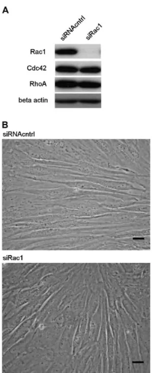
We applied siRNA technique to achieve posttranscriptional suppression of Rac1 protein expression in NHDF by transient transfection with Rac1-specific siRNA duplex (siRac1) in parallel with nonspecific control duplex (siRNAcntrl). Rac1 silencing was documented by immunoblotting (Fig. 1A) and the results reveal a coherent reduction in Rac1 protein level in the siRac1-treated fibroblasts. The targeted inhibition exhibits a highly specific effect, without alteration of the expression of other members of the Rho subfamily (RhoA and Cdc42) (Fig. 1A). In order to screen for any particular phenotype induced by siRac1, we looked at the cell morphology by phase-contrast microscopy. Silencing of Rac1 does not show dramatic effect on the fibroblasts morphology (Fig. 1B).
Rac1 protein suppression inhibits NHDF proliferation
A number of experimental observations have suggested a role of Rac1 in controlling cell proliferation in various cell types [10,16,17]. Since the function of Rac1 in controlling the proliferation of NHDF has not been assessed yet, we examined this question. Transient transfection of NHDF with siRac1 or siRNA control duplexes were performed, then 3 days later the consequences of Rac1 silencing on the rate of DNA synthesis were determined by radioactive [3H]thymidine incorporation. When compared to the control, the suppression of Rac1 expression
caused an essential 35% inhibition of the rate of cell proliferation (Fig. 2).
Fig. 1. siRac1 efficiently inhibit Rac1 expression without affecting the morphology of fibroblasts. NHDF were transiently transfected with siRac1 or siRNAcntrl. (A) siRac1 show highly efficient and specific action at reducing the expression of Rac1 in dermal fibroblasts. This experiment is representative of three independent repetitions. (B) Phase-contrast microscopy reveals no prominent difference in the morphology of siRac1-treated NHDF compared to control NHDF. Scale bars: 20 µm.
Rac1 silencing influences NHDF proliferation without changing the basal ERK1/2 phosphorylation and without inducing the phosphorylation of the p38 stress-activated protein kinase
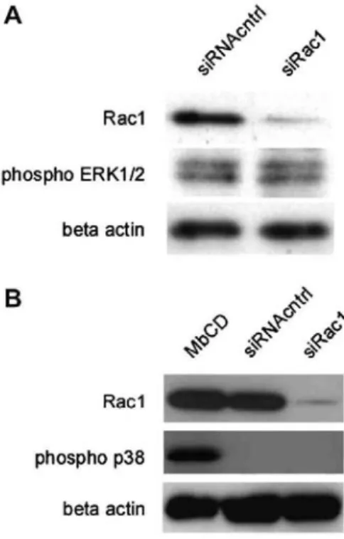
ERK1/2 has a central role in the transduction of proliferative stimuli in most cell types including NHDF [18,19]. Since we detected inhibition of NHDF proliferation as a result of Rac1 silencing we wondered whether this phenomenon has possibly negative effects on ERK1/2 phosphorylation. For this aim, transient transfection of NHDF with siRac1 or siRNAcntrl was done and 3 days after the transfection treated cells were harvested and their total protein content was extracted for Western blot analyses. Immuno-blotting with specific primary antibody against the active phosphorylated form of ERK1/2 revealed no effects on basal ERK1/2
phosphorylation after inhibition of Rac1 expression, when compared to the control (Fig. 3A).
In NHDF, the p38 MAPK (stress-activated protein kinase, SAPK) has pro-apoptotic effects opposing to ERK1/2. The p38 is phosphorylated in response to stress and subsequently induces cellular apoptosis [20]. Cholesterol depletion through treatment with methyl-β-cyclodextrin has been identified as a stress able to induce phosphorylation and was used here as a positive control. To investigate the effects of Rac1 inhibition on p38-phosphorylation, the total protein content extracted from NHDF 3 days after their transfection has been analyzed by Western blotting using a primary antibody specific for the phosphorylated active form of p38 (Fig. 3B). The inhibition of endogenous Rac1 protein does not induce phosphorylation of p38, whereas p38 can be
phosphorylated by the treatment with methyl-β-cyclodextrin.
Fig. 2. siRac1 inhibit DNA synthesis in fibroblasts. NHDF were transiently transfected with siRac1 or
siRNAcntrl. Forty-eight hours after the transfection, the cells were cultured in serum-free medium for 16 h and incubated during the last 4 h with 1 µCi/ml of radioactively labeled [3H]thymidine. The DNA synthesis was determined as the amount of radioactivity due to [3H]thymidine incorporated into cellular macro-molecules and was expressed in relation to the amount of cell proteins. siRac1-treatment inhibits the proliferation rate of NHDF by 35% in comparison with the control cells. Data are means ± SEM of three independent experiments.
Rac1 silencing in NHDF negatively influences the phosphorylation of c-myc
In the context of the cell-specific signaling events that control proliferation in mouse epidermal stem cells, Rac1 sustains the cellular proliferation through negative regulation of c-myc expression as c-myc activity induces epidermal differentiation [11]. However contrarily to its role in epidermal stem cells, c-myc fulfills different roles in fibroblasts as its activity promotes cell cycle progression [13].
Thus, since both Rac1 and c-myc function in order to favor proliferation in fibroblasts, we wondered whether they can functionally interact. To answer this question, the protein content of NHDF transfected with Rac1-specific siRNA has been harvested and analyzed by Western blot using antibodies Rac1-specific for total Rac1, total c-myc or Ser62/ Thr58-phosphorylated-c-c-myc proteins. After Rac1 silencing in NHDF, a strong decrease of basal c-myc phosphorylation is detected without affecting c-myc protein expression (Fig. 4).
Fig. 3. Rac1 silencing does not affect the phosphorylation of ERK1/2 and p38 MAPK. (A) NHDF were
transiently transfected with siRac1 or siRNAcntrl. Forty-eight hours after the transfection, the treated cells were cultured in serum-free medium for 16 h. Then, the transfected fibroblasts were harvested and their total protein content was extracted for analysis using Western blot and specific primary antibodies against Rac1- and phospho-ERKl/2. As a loading control the protein level of β-actin was determined. Data are confirmed by two independent experiments. (B) NHDF were transiently transfected and analyzed as above, except for the use of antibodies against Rac1 and phospho-p38. Extraction of the plasma membrane cholesterol by methyl-β-cyclodextrin (MbCD, 7.5 mM, 1 h) treatment is a positive control inducing cell stress and activation of p38. Data are confirmed by two independent experiments.
Fig. 4. Rac1 silencing inhibits the Ser62/Thr58 phosphorylation of c-myc. Transient transfection with siRac1 or siRNAcntrl of NHDF was performed. Forty-eight hours after the transfection the cells were cultured for 16 h in serum-free medium, then harvested. Their whole protein content was extracted and subjected to Western blot analyses using primary antibodies against Rac1 expression, c-myc expression and c-myc-Ser62/Thr58
Discussion
The role of Rac1 in progression of the cell cycle has been extensively studied in NIH-3T3 mouse fibroblast cell line [10,21]. Those studies revealed that a constitutively active Rac1-mutant increased cell growth and
conversely a dominant-negative Rac1-mutant slowed down growth. Additional experiments with different domain mutants of Rac1 clarified that this Rac1 function on cell growth in fibroblasts are independent of the effects of Rac1 on the cytoskeleton [10].
Here, we determined whether Rac1 also controls cell growth in low passage NHDF, and we investigated intermediates on which Rac1 exerts control [8]. For this purpose, we have chosen the method of RNA interference in order to selectively and efficiently inhibit the expression of Rac1 [8]. While assessing consequences of Rac1 inhibition on DNA synthesis, our results revealed a decreased cell proliferation which suggests that Rac1 regulates the cell cycle in NHDF.
Detailed studies with mouse NIH-3T3 have found out that Rac1 signaling promotes progression in the cell cycle, functioning as an alternative parallel transduction mechanism to the main proliferative Raf/MEK1,2/ERK1,2 pathway [9]. Indeed, in mouse fibroblasts Rac1 exerts control on the cell cycle without inducing ERK activity [22]. Only one sing1e study has previously given indirect evidence that Rac1 could control ERK1/2 pathway in human fibroblast cell line WI-26 [12]. This conclusion was made based on the observation that active Rac1-mutant induced activation of the well known ERK 1/2 target, Elk1. The functional significance of this activation in relation with the proliferation of NHDF has not been examined. In addition since Elk1 can be activated through phosphorylation not only by ERKs, but also by JNK and p38 MAP kinases [23,24], we looked in our study for more direct evidence of functional relationship between Rac1 and ERK1/2. Surprising1y, when we inhibited Rac1 expression, we did not detect any alteration in the phosphorylation of ERK1/2, suggesting that Rac1 is acting on other targets in order to inhibit proliferation of NHDF.
Another possible explanation for the effect of Rac1 suppression on the proliferation of NHDF could involve activation of the p38 MAPK pathway, as this activation has pro-apoptotic functions and suppresses cell proliferation [20], and thus could explain the higher spontaneous apoptotic population reported in Rac1-null mouse embryonic fibroblasts [9]. However, when we checked the phosphorylation of p38 after siRNA-mediated Rac1 inhibition in NHDF, we could not confirm this hypothesis. Moreover, our observations using phase-contrast microscopy confirm the absence of prominent morphological changes and signs of cell death in the siRac1-treated fibroblasts.
Unlike observations in fibroblasts, Rac1 promotes the proliferation of mouse epidermal stem cells, but in this cell type its effect results from negative regulation of the transcription factor c-myc through the mediator PAK2, and Rac1 deletion results in increased c-myc expression [11]. Although c-myc is a proto-oncogene, in epidermal stem cells it fulfills specific function, promoting differentiation. There are more evidences supporting the hypothesis that c-myc exhibits different features in stem cells differentiation [25]. However, in contrast to epidermal stem cells, c-myc emerges in fibroblasts as an important cell cycle promoter [13]. Since our data according1y suggested proliferative functions of Rac1 in a cell type different than stem cells, we examined in NHDF the possible existence of a functional interaction between Rac1 and c-myc in which both work to promote cell growth. Indeed after Rac1 silencing, we found inhibition of the phosphorylation of c-myc at the sites of growth factor-regulated phosphorylation, Ser62 and Thr58 [26]. In contrast to epidermal stem cells, Rac1 inhibition in NHDF does not alter the normal myc expression. The significance of this phosphorylation of c-myc in fibroblasts is well determined: Ser62/Thr58 phosphorylation serves as a mechanism for proper control of the transient accumulation of c-myc in early G1 following the stimulation of cell growth [26,27]. In a first step, the phosphorylation at Ser62 stabilizes c-myc as a premise for its accumulation. The following phosphorylation of Thr58 depends on the prior phosphorylation of Ser62 since mutation of Ser62 prevents c-myc phosphorylation at Thr58. Thr58 phosphorylation signals for myc degradation, the way to prevent cellular transformation by c-myc [26,28]. During early G1 phase, the elevated c-c-myc protein plays a critical role in promoting G1/S
progression [13,29-31]: c-myc induces cell cycle progression in fibroblasts acting as an upstream regulator of G1 cyclin-dependent kinases (Cdk) and antagonizing the action of the Cdk inhibitor, p27. Thus, the inhibition of c-myc phosphorylation observed in our results is directly or indirectly linked to the suppressed expression of Rac1, and is likely the reason for inhibition of proliferation.
In mouse fibroblasts ERK and Rac1 work in parallel to promote G1 progression by induction of cyclin D1 expression [9]. Our data suggest that c-myc may play a role in mediating these effects of Rac1 and ERK. Indeed, it was found in mouse fibroblasts that the ERK activator Ras and also myc impact the cell cycle machinery through the cyclins D [30].
We report here for the first time that in NHDF Rac1 inhibition affects c-myc phosphorylation at its growth factor-regulated phosphorylation sites (Ser62/Thr58). It has been proven previously that the inhibition of ERK with the selective inhibitor PD098059 also prevents c-myc phosphorylation at Ser62 [32], but since our experiments demonstrate that suppression of Rac1 inhibits c-myc phosphorylation without affecting ERK phosphorylation, we propose that Rac1 and ERK represent two mechanisms working in parallel which cooperate to regulate c-myc phosphorylation and cell growth in NHDF.
In conclusion, the effects of Rac1 silencing observed on NHDF proliferation and c-myc phosphorylation suggest that the signaling interactions between Rac1 and cell regulatory proteins are eventually mediated by c-myc, but differ between different cell types like skin fibroblasts and epidermal stem cells.
Acknowledgments
EN was holding a Ph.D. fellowship for foreign students from the University of Namur. Financial support through FRFC Grant 2.4506.01 to YP and through a grant from the Medical University of Sofia to VM. CFD is Research Associate of the Belgian FNRS.
References
[1] J. Mollenhauer, K. Bayreuther, Donor age related changes in the morphology, growth potential, and collagen biosynthesis in rat fibroblasts subpopulation in vitro, Differentiation 32 (1986) 165-172.
[2] N.H. Antoniades, T. Galanopoulos, J. Neville-Golden, C.P. Kiritsy, S.E. Lynch, Injury induces in vivo expression of platelet-derived growth factor (PDGF) and PDGF receptor mRNA in skin epithelial cells and PDGF mRNA in connective tissue fibroblasts, Proc. Nat. Acad. Sci. USA 88 (1991) 565-569.
[3] E.A. Bauer, N. Silverman, D.F. Busier, A. Kronberger, T.F. Deuel, Diminished response of Werner's syndrome fibroblasts to growth factors PDGF and FGF, Science 234 (1986) 1240-1243.
[4] A. Colige, B. Nusgens, CM. Lapière, Altered response of progeroid fibroblasts to epidermal growth factor, J. Cell Sci. 100 (1991) 649-655.
[5] N. Basset, A. Vie, J.M. Blanchard, J. J Guilhou, Expression of c-Myc, c-Myb, c-Erb-B and c-H-Ras oncogene mRNAs in fibroblasts cultured from psoriatic patients, Dermatologica 175 (1987) 296-299.
[6] A. Hall, Rho GTPases and the actin cytoskeleton, Science 279 (1998) 509-514.
[7] B.A. Jaffe, A. Hall, Rho GTPases: biochemistry and biology, Annu. Rev. Cell Dev. Biol. 21 (2005) 247-269.
[8] L. Wang, Y. Zheng, Cell type-specific functions of Rho GTPases revealed by gene targeting in mice, Trends Cell Biol. 17 (2007) 58-64.
[9] R.K. Assoian, MA. Schwartz, Coordinate signaling by integrins and receptor tyrosine kinases in the regulation of G1 phase cell-cycle progression, Curr. Opin. Genet. Dev. 11 (2001) 48-53.
[10] J.K. Westwick, Q.T. Lambert, G.J. Clark, M. Symons, L. van Aelst, R.G Pestell, C.J. Der, Rac regulation of transformation, gene expression, and actin organization by multiple PAK-independent pathways, Mol. Cell. Biol. 17 (1997) 1324-1335.
[11] S.A. Benitah, M. Frye, M. G1ogauer, F.M. Watt, Stem cell depletion through epidermal deletion of Rac1, Science 309 (2005) 993-995.
[12] J. Laboureau, L. Dubertret, C. Lebreton-De Coster, B. Coulomb, ERK activation by mechanical strain is regulated by the small G proteins rac-1 and rhoA, Exp. Dermatol. 13 (2004) 70-77.
[13] K. Berns, E.M. Hijmans, R. Bernards, Repression of c-Myc responsive genes in cycling cells caused G1 arrest trough reduction of cyclin E/CDK2 kinase activity, Oncogene 15 (1997) 1347-1356.
[14] M. Praskova, S. Kalenderova, L. Miteva, Y. Poumay, V. Mitev, The ornithine decarboxylase inhibitor, difluoromethylornithine, inhibits casein kinase II activity, c-Myc expression and normal human keratinocyte proliferation, Arch. Dermatol. Res. 293 (2002) 590-593.
[15] M. Praskova, S. Kalenderova, L. Miteva, Y. Poumay, V. Mitev, Ca(2+)/calmodulin-dependent protein kinase (CaM-kinase) inhibitor KN-62 suppresses the activity of mitogen-activated protein kinase (MAPK), c-myc activation and human keratinocyte proliferation, Arch. Dermatol. Res. 294 (2002) 198-202.
[17] S. Aznar, J.C. Lacal, Rho signals to cell growth and apoptosis, Cancer Lett. 165 (2001) 1-10.
[18] J.C. Chambard, R. Lefloch, J. Pouyssegur, P. Lenormand, ERK implication in cell cycle regulation, Biochim. Biophys. Acta (2006) (Epub ahead of print).
[19] R. Le Panse, V. Mitev, L.M. Houdebine, B. Coulomb, Protein kinase C-independent activation of mitogen-activated protein kinase by epidermal growth factor in skin fibroblasts, Eur. J. Pharmacol. 307 (1996) 339-345.
[20] S.P. Li, M.R. Junttila, J. Han, V.M. Kahari, J. Westermarck, p38 mitogen-activated protein kinase pathway suppresses cell survival by inducing dephosphorylation of mitogen-activated protein/extracellular signal-regulated kinase kinasel,2, Cancer Res. 63 (2003) 3473-3477.
[21] R. Khosravi-Far, P.A. Solski, G.J. Clark, M.S. Kinch, C.J. Der, Activation of Rac1, RhoA, and mitogen-activated protein kinases is required for Ras transformation, Mol. Cell. Biol. 15 (1995) 6443-6453.
[22] A. Minden, A. Lin, F.X. Claret, A. Abo, M. Karin, Selective activation of the JNK signalling cascade and c-Jun transcriptional activity by the small GTPases Rac and Cdc42Hs, Cell 81 (1995) 1147-1157.
[23] A.J. Whitmarsh, P. Shore, A.D. Sharrocks, R.J. Davis, Integration of MAP kinase signal transduction pathways at the serum response element, Science 269 (1995) 403-407.
[24] R. Janknecht, T. Hunter, Convergence of MAP kinase pathways on the ternary complex factor Sap-la, EMBO J. 16 (1997) 1620-1627.
[25] L. Frostesjo, O. Heby, Polyamine depletion up-regulates c-Myc expression, yet induces G(l) arrest and terminal differentiation of F9 teratocarcinoma stem cells, J. Cell. Biochem. 76 (1999) 143-152.
[26] J. Vervoorts, J. Lüscher-Firzlaff, B. Lüscher, The ins and outs of myc regulation by posttranslational mechanisms, J. Biol. Chem. 281 (2006) 34725-34729.
[27] R. Sears, F. Nuckolls, E. Haura, Y. Taya, K. Tamai, J.R. Nevins, Multiple Ras-dependent phosphorylation pathways regulate Myc protein stability, Genes Dev. 14 (2000) 2501-2514.
[28] S.E. Salghetti, S.Y. Kim, W.P. Tansey, Destruction of Myc by ubiquitin-mediated proteolysis: cancer-associated and transforming mutations stabilize Myc, EMBO J. 18 (1999) 717-726.
[29] E. Santoni-Rugiu, J. Falck, N. Mailand, J. Bartek, J. Lukas, Involvement of Myc activity in a G1/S-promoting mechanism parallel to the pRb/E2F pathway, Mol. Cell. Biol. 20 (2000) 3497-3509.
[30] Q. Yu, M.A. Ciemerych, P. Sicinski, Ras and Myc can drive oncogenic cell proliferation through individual D-cyclins, Oncogene 24 (2005) 7114-7119.
[31] P. Steiner, B. Rudolph, D. Muller, M. Eilers, The functions of Myc in cell cycle progression and apoptosis, Prog. Cell Cycle Res. 2 (1996) 73-82.