CBP and histone deacetylase inhibition enhance the transactivation
potential of the HOXB7 homeodomain-containing protein
Alain Chariot
1,3, Carine van Lint
2, Muriel Chapelier
1, Jacques Gielen
1, Marie-Paule Merville
1and Vincent Bours*
,11Laboratory of Medical Chemistry and Medical Oncology, CHU B35, University of Liege Sart-Tilman, 4000 Liege, Belgium; 2Laboratory of Biological Chemistry, Department of Molecular Biology, Free University of Brussels, Brussels, Belgium
Homeodomain-containing proteins are transcription fac-tors regulating the coordinated expression of multiple target genes involved in development, dierentiation and cellular transformation. In this study, we demonstrated that HOXB7, one member of this family, behaved as a transactivator in breast cancer cells. Deletion of either the HOXB7 N-terminal domain or the C-terminal acidic tail abolished this transcriptional eect, suggesting a combination of distinct functional transactivating do-mains. HOXB7 physically interacted both in vitro and in vivo with the coactivator CREB-binding protein (CBP). This interaction led to an enhanced transactivating potential and required the N-terminal of HOXB7 as well as two domains located at the C-terminal part of CBP. Moreover, trichostatin A, a deacetylase inhibitor, strongly enhanced the transcriptional properties of HOXB7. Our data therefore indicate that HOX proteins can directly interact with CBP and that acetylation/ deacetylation may regulate their transcriptional proper-ties.
Keywords: homeobox gene; CBP; coactivator; tran-scription; histone acetylation
Introduction
Genetic programs governing development, differentia-tion and cell growth imply the regulated transcripdifferentia-tion of a variety of genes through the action of multiple proteins de®ned as `general transcription factors' (GTFs), `activators', `repressors' and `mediators' or `cofactors', depending on their exact function (Sauer and Tjian, 1997).
The CBP (CREB-binding protein)/p300 proteins are coactivators which interact with the phosphorylated form of CREB (cAMP response element binding) (Chrivia et al., 1993) and with a number of other transcription factors including c-jun, c-fos, nuclear receptors, c-Myb. MyoD, YY1, Sap-1a, sterol regula-tory element binding protein (SREBP), E2F1/DP1 (see Shikama et al. (1996) for a review), NF-kB p65 (Rel A) (Perkins et al., 1997), p53 (Avantaggiati et al., 1997; Gu and Roeder, 1997a; Lill et al., 1997) and Smad
proteins (Feng et al., 1998; Janknecht et al., 1998). All these interactions suggest that CBP acts as a bridge or an `adaptator' between the activators or repressors and the initiation complex containing the GTFs and the RNA polymerase II (Eckner et al., 1994; Arany et al., 1995). CBP also interacts with other coactivators such as RAC3 (Li and Don Chen, 1998) and steroid receptor coactivator-1 (SRC-1) (Hanstein et al., 1996; Smith et al., 1996) and has been copuri®ed with the holoenzyme complex along with BRCA1 (Neish et al., 1998). CBP is now considered as an `integrator' (Kamei et al., 1996) or `co-integrator' (Chakravarti et al., 1996) of a variety of signaling pathways. Although both CBP and p300 proteins share similar functional properties in transient transfection experiments, they have distinct functions during retinoic-acid-induced dierentiation (Kawasaki et al., 1998).
An histone acetyltransferase activity has been attributed to the CBP/p300 proteins (Bannister and Kouzarides, 1996; Ogryzko et al., 1996). In other words, CBP can enhance removal of positive charges by acetylation of lysine residues at the N-terminal tail of histones, which presumably destabilizes the nucleo-some and facilitates the access of transcription factors to DNA. This histone acetyltransferase activity is required for in vivo transcription regulation (Marti-nez-Balbas et al., 1998). CBP also recruits P/CAF, a protein harboring a histone acetyltransferase activity as well (Yang et al., 1996). Moreover, CBP/p300 can also acetylate TFIIE, TFIIH (Imhof et al., 1997), the erythroid Kruppel-like factor (Zhang and Bieker, 1998), p53 and activate p53 biochemical function (Gu and Roeder, 1997b).
Homeodomain-containing proteins have been identi-®ed as transcriptional regulators controlling the expression of genes involved in development, differ-entiation and tumoral transformation (Levine and Hoey, 1988; Favier and Dolle 1997; Mark et al., 1997; Shimamoto et al., 1998). They share a highly conserved 60 amino acid DNA-binding domain (the `homeodomain') (Gehring et al., 1994). Although each member of this family exhibits in vivo speci®city as demonstrated by targeted gene knock-out experiments, they share very similar DNA-binding anities in vitro, suggesting that protein ± protein interactions mediate their speci®city. For instance, the homeodomain-containing extradenticle (exd)/Pbx gene products have been identi®ed as cofactors of HOX gene products (Mann and Chan, 1996). These interactions require the pentapeptide, a conserved domain located upstream from the DNA-binding domain of most HOX proteins (Chang et al., 1995) as well as the Hox cooperativity motif (`HCM'), a sequence carboxy terminal to the Pbx
*Correspondence: V Bours
3Current address: Laboratory of Immunoregulation, NIAID, NIH, Building 10, Room 11B17, 9000 Rockville Pike, Bethesda, Maryland, MD 20892, USA
Received 9 November 1998; revised 22 January 1999; accepted 22 January 1999
homeodomain (Chang et al., 1997). Since AbdB-like HOX proteins do not harbor the pentapeptide, most of these products cannot interact with Pbx (Shen et al., 1997), thus raising the possibility that other partners might be required. Indeed, a recent report demon-strated the formation of heterodimeric complexes between Hox and Meisl proteins (Shen et al., 1997). It is likely that other proteins yet to be identi®ed are involved in such processes.
HOXB7 was initially isolated from an SV40-transformed human ®broblast cDNA library (Si-meone et al., 1987) and proposed to be involved in a variety of developmental processes. The HOXB7 protein has also been implicated in hematopoietic dierentiation as well as in lymphoid development (Shen et al., 1989; Deguchi et al., 1991; Lill et al., 1995; Magli, 1998). The HOXB7 protein can bind DNA (Corsetti et al., 1992) and activate transcription from distinct promoters in a variety of cell lines (Care et al., 1996; Chariot et al., 1998; Sanlioglu et al., 1998). However, the functional domains that mediate HOXB7 transcriptional properties as well as its interacting partners remain to be identi®ed.
In this report, we demonstrated that both the N-terminal domain and the C-N-terminal acidic tail of HOXB7 are required for transactivation. Moreover, we illustrated the physical interaction between HOXB7 and the coactivator CBP in vitro and in vivo. We also showed that deacetylase inhibition by trichostatin A (TSA), potentiates the HOXB7 transactivating eect. Results
A combinatorial code mediates the HOXB7 transactivation eect
We previously demonstrated that the HOXB7 protein can activate transcription in MDA-MB231 cells (Chariot et al., 1998). Indeed, cotransfection of the pTCBS plasmid and the HOXB7 expression vector increased the luciferase activity 3.5- to 3.7-fold over basal activity in a dose-dependent manner (Figure 1b, left panel). A decrease in the transactivating eect was observed when 2 mg of HOXB7-expression vector was transfected, probably re¯ecting a squelching eect (Figure 1b, left panel). The commitment of the HOXB7 binding to the CBS sequence was indirectly con®rmed by cotransfection of the pT109 reporter plasmid.
To further map the domain(s) that mediate(s) HOXB7 transcriptional activity, expression vectors generating HOXB7 proteins deleted either in the N-or in the C-terminal domain were constructed (Figure 1a). `B7-DN18', `DN54' and `DN86' generated HOXB7 gene products lacking 18, 54 and 86 amino acids in the N-terminal region, respectively. These proteins har-bored the pentapeptide domain, whereas the `B7-DN129' construct encoded a HOXB7 gene product lacking the pentapeptide. The product `B7-DC12' lacked 12 amino acid in the C-terminal region and did not contain the acidic tail, whereas the constructs `B7-DC34'. `B7-DC80' and `B7-DC97' were deleted of 34, 80 and 97 amino acids, respectively. Neither `B7-DC80' nor `B7-DC97' products contained the home-odomain. In transient expression experiments, the
`B7-DN18' product transactivated (a 2.3- to 2.8-fold induction of luciferase activity) (Figure 1b, middle panel) almost as eciently as the wild-type HOXB7 protein. A further deletion of 54, 86 or 129 amino acids progressively decreased the transactivating ability of the HOXB7 protein, suggesting that the N-terminal domain of this gene product mediates its transactivat-ing eect. Interesttransactivat-ingly, the `B7-DC12' product did not exhibit any transactivation activity and a dose-dependent repressing eect was even observed (Figure 1b, right panel), indicating that the C-terminal acidic
Figure 1 (a) Schematic representation of the HOXB7 expression vectors. These vectors generate products deleted either in their N-terminal domain (`B7-DN18', `DN54', `DN86' and `B7-DN129') or in their C-terminal domain (`B7-DC12', B7-DC34', `B7-DC80' and `B7-DC97'). The homeodomain is illuatrated by a hatched rectangle while both the pentapeptide and the acidic C-terminal tail are also shown. The expected molecular mass of the resulting proteins is given on the right. (b) Analysis of the transcriptional properties of HOXB7 wild-type (left panel) and HOXB7 mutant gene products deleted either in their N-terminal domain (middle panel) or in their C-terminal domain (right panel). The pT109 does not contain any HOX-binding sequence and was used as a negative control. Cells were transfected with increasing amounts of HOXB7 wild-type or mutant expression vectors (0.5, 1 or 2 mg) together with 1 mg of reporter plasmid. The ®gure shows the relative luciferase activity over the activity observed with 1 mg of the pTCBS or pT109 reporter plasmids alone. Each value represents the mean (+s.d.) of at least three independent experiments after normalization to the protein concentration of the extracts
tail is also involved in the HOXB7-dependent transactivation eect. Therefore, we conclude that both the N-terminal domain and the acidic C-terminal tail are required for the HOXB7 transcriptional properties.
The HOXB7 protein physically interacts with the coactivator CBP in vitro
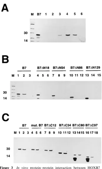
We investigated whether HOXB7 could interact with the coactivator CBP, which is a large and complex protein harboring distinct functional domains (Figure 2a). The GST-CBP fusion proteins (Figure 2b) were expressed in E.coli, puri®ed and incubated with in vitro-translated HOXB7 protein. HOXB7 was not precipitated by the GST-CBP `1' (Figure 3a, lane 1), a fusion protein harboring both the receptor-interact-ing domain (`RID') and the CREB bindreceptor-interact-ing domain (`KIX') of CBP nor by GST-CBP `2' (Figure 3a, lane 2) which harbors the CREB binding domain (`KIX'), suggesting that there was no physical interaction between HOXB7 and these parts of the CBP protein. However, when HOXB7 was incubated with the GST-CBP `3' fusion protein, a weak signal was detected (Figure 3a, lane 3), suggesting that the histone acetyltransferase domain contributes to the interaction with HOXB7 in vitro. Moreover, HOXB7 was clearly precipitated by the GST-CBP `4' fusion protein (Figure 3a. lane 4), suggesting that the cystein-histidine-rich `C/ H3' domain of CBP physically interacts with HOXB7. A signal was also observed when the GST-CBP `5' fusion protein which harbors the C-terminal domain of
CBP was incubated with the homeodomain-containing protein (Figure 3a, lane 5). These results suggest that HOXB7 interacts in vitro with two domains of CBP known to mediate physical interaction with other transcription factors such as c-fos, TFIIB and MyoD for the C/H3 sequence and p53 for the C-terminal domain (Figure 2a).
HOXB7 N-terminal domain is required for interaction with CBP
To further map the HOXB7 domain(s) involved in the physical interaction with CBP. various HOXB7 vectors were used as templates for in vitro translation, and translated products were incubated with the GST-CBP `4' fusion protein. As illustrated in Figure 3b, the deletion of the ®rst 18 amino acids clearly abolished HOXB7-CBP interaction (lane 5). Moreover, we could not detect any interaction between CBP and `B7-DN54', `B7-DN86' and `B7-DN129' (Figure 3b, lanes 8, 11 and 14). The N-terminal domain of HOXB7 is thus required for the interaction with CBP in vitro.
To determine whether other HOXB7 domains were involved in the interaction with CBP, several HOXB7 vectors deleted in the C-terminal domain were in vitro translated. As illustrated in Figure 3c, the mutant B7, which codes for a naturally occurring truncated HOXB7 protein lacking two amino acids within the acidic tail (Chariot et al., 1998) and the `B7-DC12' protein interacted with CBP, just as eciently as the wild-type HOXB7 gene product (lanes 5 and 8). The `B7-DC34', `B7-DC80' and `B7-DC97' proteins which
Figure 2 (a) Schematic representation of the CBP coactivator. All the proteins known to physically interact with CBP are mentioned next to their respective interacting CBP domains. `AR': androgen receptor; `cdk-2': cyclin-dependent kinase-2; `C/H3': cystein-histidine-rich domain 3; `ER': estrogen receptor; `GR': glucocorticoid receptor; `HAT': histone acetyltransferase; `PR': progesterone receptor; `RID': receptor interacting domain; `RXR': retinoid X receptor; `TBP': TATA-binding protein; `TR': thyroid receptor. (b) Schematic representation of the various GST-CBP fusion proteins used in this study
lack the acidic tail and a part or the entirety of the homeodomain could still interact with CBP in vitro (Figure 3c, lane 11, 14 and 17). The in vitro interaction between HOXB7 and CBP thus requires the N-terminal of this HOX protein.
CBP enhances the transactivation potential of HOXB7 in MDA-MB231 cells
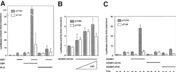
To determine whether HOXB7 and CBP interacted in vivo, we performed transient expression experiments in MDA-MB231 cells using either pTCBS or pT109 reporter plasmids, the CMX-CBP expression vector, and HOXB7 expression vectors (Figure 1a). CBP enhanced the HOXB7-mediated transactivation, since a 160-fold induction of luciferase activity was measured when both HOXB7-expression vector and a CMX-CBP expression vector were cotransfected with the pTCBS plasmid (Figure 4a). The luciferase activity observed with the pT109 reporter plasmid was much weaker
(Figure 4a), demonstrating that the eect is mainly mediated by the binding of HOXB7 to the CBS. We also constructed a CBP mutant named `CBPDC' that lacks the HAT, C/H3 and C-terminal domains (Figure 4a). The induction of luciferase activity was much less intense when we cotransfected this CBP mutant with the HOXB7-expression construct and the pTCBS plasmid, thus con®rming that these CBP domains are required for the interaction with HOXB7 in vivo as well as in vitro. Cotransfection of both the `B7-DN129' and CBP expression vectors with the pTCBS reporter plasmid did not signi®cantly enhance the eect of this deleted HOXB7 product (Figure 4b), con®rming that the N-terminal domain of HOXB7 is necessary for interaction with the coactivator as shown in vitro.
Modulation of HOXB7 transcriptional properties by protein acetylation/deacetylation was investigated by treating MDA-MB231 cells for 20 h with TSA, a deacetylase inhibitor. TSA strongly induced luciferase activity after transfection of both pTCBS and HOXB7 expression vectors (Figure 4c). Interestingly, TSA did not signi®cantly induce the transactivation eect of either the `B7-DN129' product or the `B7-DN18' protein, which still exhibited transactivation abilities (Figure 4c). Taken together, our results suggest that HOXB7 transcriptional properties might be regulated by acetylation/deacetylation through the N-terminal domain of this transcription factor.
We further investigated in vivo interaction between CBP and HOXB7 proteins by performing Mamma-lian two-hybrid system experiments in MDA-MB231 cells using both GAL4-CBP and HOXB7-VP16 expression vectors and the pSG5 reporter plasmid that harbors a GAL4 binding sequence upstream from a minimal E1B promoter and a CAT gene. No signi®cant induction of the CAT activity was measured when the HOXB7-VP16 expression vector was cotransfected with the GAL4 expressing con-struct and the reporter plasmid, whereas a 2.8-fold induction was observed when both GAL4-CBP and VP16 were expressed, probably because of the intrinsic transactivation ability of the wild-type CBP protein (Figure 5). A sixfold induction of CAT activity was measured when both GAL4-CBP and HOXB7-VP16 expressing constructs were expressed (Figure 5). Moreover, when the HOXB7-VP16 was cotransfected with the `GAL4-mCBP' construct that generates a GAL4-CBP fusion protein deleted of both the C/H3 and Q-rich domains of CBP, the induction of CAT activity was signi®cantly reduced. These results con®rmed that HOXB7 and CBP proteins interact in vivo through the C/H3 and Q-rich domain of the coactivator.
Discussion
In this paper, we have demonstrated that the transactivating eect of HOXB7 in MDA-MB231 cells requires both the N-terminal domain and the C-terminal acidic sequence, thus suggesting a `combina-torial code' that mediates HOXB7 transcriptional properties. This `combinatorial code' hypothesis is supported by other studies demonstrating that the transcriptional properties of other HOX proteins are also mediated by a unique combination of several
Figure 3 In vitro protein±protein interaction between HOXB7 and CBP. (a) The in vitro translated wild-type HOXB7 gene product (`B7') was incubated with the CBP`1' (lane 1), CBP`2' (lane 2), CBP`3' (lane 3), CBP`4' (lane 4), GST-CBP`5' (lane 5) or with the GST protein used as a negative control (lane 6). Lane `B7' represents the in vitro translated HOXB7 protein. (b) and (c): Incubation of the GST-CBP`4' (lanes 2, 5, 8, 11, 14, 17) or the GST protein (lanes 3, 6, 9, 12, 15, 18) with various in vitro translated HOXB7 proteins deleted either in their N-terminal (b) or in the C-terminal domain (c). In each case, 10% of in vitro translated material was run as control (lanes 1, 4, 7, 10, 13, 16)
domains (Schnabel and Abate-Shen, 1996; Zhang et al., 1996; Vigano et al., 1998).
Despite of the in vivo biological speci®city of each HOX gene product, all these transcription factors share very similar in vitro DNA-binding anities. Protein ± protein interactions thus likely play a major role in the regulation of their function. Partners for HOX proteins are, for instance, Pbx and Meis proteins (Mann and Chan, 1996; Shen et al., 1997). Since most of these
proteins have yet to be identi®ed, we investigated whether the coactivator CBP could modulate the transcriptional properties of HOXB7. Mammalian two-hybrid system experiments, transient transfections and in vitro studies indicated that HOXB7 and CBP interact in vitro and in vivo. Interaction between HOXB7 and CBP requires the HOXB7 N-terminal domain, which is also involved in interactions with other transcription factors such as Meis 1 (Shen et al.,
Figure 5 Interaction between the HOXB7 and CBP proteins by Mammalian two-hybrid system. The pG5 reporter plasmid is schematically represented. All the expression vectors that generate the fusion proteins are illustrated. The MDA-MB231 cells were transfected with 1 mg of the pG5 and 2 mg of expression vectors. The ®gure shows the relative CAT activity over the activity measured with 1 mg of the pG5 reporter plasmid alone. Each value represents the mean (+s.d.) of at least three independent experiments after normalization
Figure 4 CBP and a histone deacetylase inhibitor increase HOXB7 transcriptional activity. MDA-MB231 cells were transfected with HOXB7 and/or CBP expression vectors together with the pTCBS (hatched columns) or pT109 (white columns) reporter plasmids. Cellular extracts were prepared and luciferase activities determined. (a) Cotransfection with the HOXB7 expression vector (1 mg) together with expression vectors coding for wild-type (CBP) or mutant (CBPDC) protein (2 mg). (b) Cotransfection of the `B7-DN129' expression vector (1 mg) together with increasing amounts of the CBP expression vector (0.5, 1 or 2 mg). (c) After transfection with the HOXB7 expression vector (1 mg), the `B7-DN129; or the `B7-DN18' expression vectors (1 mg), MDA-MB231 cells were left untreated (±) or treated for 20 h with TSA (400 nM) as indicated in the ®gure
1997). Since this N-terminal domain is less conserved between the various homeodomain-containing proteins, it is tempting to postulate that interaction with CBP contributes to the in vivo speci®city of HOXB7. However, it is likely that CBP mediates the transcrip-tional eect of other homeodomain-containing pro-teins. In this context, a recent study has demonstrated that the pituitary-speci®c factor Pit-1 requires a co-activator complex that includes both CBP and p/CAF (Xu et al., 1998).
TSA enhanced the transactivating eect of the wild-type HOXB7 protein in MDA-MB 231 cells. This deacetylase inhibitor has been previously described as an activator of the gamma globin gene (McCarey et al., 1997) and the WAF1/Cip1 gene promoter through Sp1 sites (Sowa et al., 1997). Moreover, TSA can potentiate retinoid receptor action by altering the chromatin structure within the RARb2 promoter (Minucci et al., 1997). In vivo modi®cation of the chromatin conforma-tion at the HOX-binding sites may then facilitate the access of the homeodomain-containing proteins to regulating sequences. Moreover, our results raise the possibility that HOXB7 function may be regulated by its acetylation, a phenomenon already described for p53 (Gu and Roeder, 1997b), TFIIE, TFIIH (Imhof et al., 1997) and the erythroid Kruppel-like factor (Zhang and Bieker, 1998). The acetylation would occur in the N-terminal domain of HOXB7, since we demonstrated that TSA does not signi®cantly enhance the transactivation eect of two HOXB7 gene products which lack this region. Moreover, deletion of the CBP HAT domain almost completely abolished CBP functional interaction with HOXB7. Alternatively, the acetylation of a cofactor could be required for HOXB7 optimal transcriptional activity. Taken together, these observations suggest that the very strong transcriptional eect observed when HOXB7 and CBP were expressed simultaneously could be due to direct interaction between CBP and HOXB7 that would bridge HOXB7 with the transcription machinery and/or to a reaction of acetylation targeting either HOXB7 itself or a cofactor interacting with HOXB7 N-terminal domain.
The Rubinstein-Taybi syndrome (`RTS') is an autosomal dominant syndrome associated with point mutations in the CBP gene (Petrij et al., 1995) and is characterized by craniofacial malformations, broad thumbs, broad big toes and mental retardation. The mutations within the CBP gene lead to the expression of truncated proteins unable to interact with CREB. This hypothesis is supported by a recent study demonstrating a partial similarity to the `RTS' syndrome in embryos lacking a single Cbp allele (Tanaka et al., 1997). On the other hand, targeted gene knock-out experiments aecting one or two HOX genes clearly cause developmental malformations (Mark et al., 1997). Our data suggest that part of the RTS phenotype might be related to a loss of HOX protein activities during development. Further studies are certainly required to investigate whether a CBP-HOX protein complex is functional during embryogen-esis. Moreover, alterations of the CBP/p300 human gene sequences also lead to hematological malignancies (see Giles et al., 1998 for a review) whereas a variety of oncogenic translocations involves HOX genes (see Shimamoto et al., 1998 for a recent review). Taken together, these observations suggest that both CBP and
HOX proteins levels of expression are critical for cellular dierentiation and that CBP alteration may contribute to multiple diseases.
Materials and methods Cell line and treatment
The MDA-MB231 cell line was obtained from the American Type Tissue Culture Collection (Rockville, MD, USA) and maintained in RPMI medium. MDA-MB231 cells were treated with TSA 400 nM (Sigma, Bornem, Belgium) for 20 h before lysis.
Expression plasmids
Coding sequences of the HOXB7 gene and of a naturally occuring mutated allele lacking two amino acids in its C-terminal sequence (Chariot et al., 1998) were subcloned by polymerase chain reaction (PCR) into the expression vector pcDNA3 (Invitrogen. San Diego, CA, USA). Expression vectors generating truncated HOXB7 gene products were constructed by PCR (Figure 1a). The constructs `B7-DN18', `DN54', `DN86' and `DN129' generate HOXB7 gene products lacking the 18, 54, 86 and 129 N-terminus amino acids, respectively. The constructs `B7-DC12', `B7-DC34', `B7-DC80' and `B7-DC97' encode HOXB7 proteins lacking the 12, 34, 80 and 97 C-terminal amino acids, respectively. The pT109 and pTCBS reporter plasmids were provided by Dr Zappavigna (Laboratory of Gene Expression, Department of Biology and Technology, Istituto Scienti®co H.S. Raaele, Milan, Italy). The pTCBS plasmid contains an eightfold multimerized form of a homeodomain consensus binding sequence (CBS) cloned upstream from a HSV-TK promoter and of a luciferase (LUC) reporter gene whereas the pT109 construct does not contain the CBS sequence and is used as a negative control (Zappavigna, 1994).
The CMX-CBP expression vector was kindly provided by Dr Evans (The Gene Expression Laboratory, The Salk Institute for Biological Studies, La Jolla, CA, USA). A functional mouse full-length CBP protein is generated from this vector (Chrivia et al., 1993). A CBP mutant named `CBPDC' that lacks the HAT, the C/H3 and the C-terminal domains was ampli®ed by PCR and subcloned in the pcDNA3 vector.
In vitro translation
In vitro translations were performed using the `Wheat germ TNT' kit (Promega, Madison, WI, USA) with 1 mg of DNA template, 1 ml of T7 polymerase and [35S] methionine. Two ml
of the reaction products were separated by electrophoresis on a 12% polyacrylamide gel followed by autoradiography. In vitro protein ± protein interactions
Plasmids containing fusion genes of GST-CBP `1' (aa 1 ± 1099 of CBP), GST-CBP `3' (aa 1099 ± 1620) and GST-CBP `4' (aa 1620 ± 1877) constructs (Bannister and Kouzarides, 1996) were kindly provided by Dr Kouzarides (Wellcome/CRC Institute, Cambridge, UK) whereas the GST-CBP `2' (aa 390 ± 790) and GST-CBP `5' (aa 1990 ± 2441) plasmids (Gu and Roeder, 1997a) were kindly provided by Dr Roeder (Laboratory of Biochemistry and Molecular Biology, The Rockfeller University, New York, NY, USA).
Expression and puri®cation of GST fusion proteins were performed as described (Kaelin et al., 1991) with modifica-tions in the composition of the NENT buer (NaCl 250 mM,
EDTA 1 mM, Tris 20 mM pH 8, NP-40 1.5%). Protein ±
protein interactions were studied by incubating an aliquot of GST-CBP fusion protein bound to glutathione-Sepharose beads with 10 ml of in vitro translated protein in 200 ml of TWB buer (HEPES 20 mM pH 7.9, NaCl 60 mM, dithio-4012
threitol 1 mM, MgCl2 6 mM, 8.2% glycerin, EDTA 0.1 mM)
for 1 h at 48C. After six washes of the beads in NENTM buer, the precipitates were run on an SDS-polyacrylamide gel before autoradiography.
Transient transfections and luciferase assays
Transfections in MDA-MB 231 cells were performed using 1 mg of reporter plasmid and up to 2 mg of distinct expression vectors per 35-mm dish as described (Chariot et al., 1998). Cells were harvested 48 h after transfection and luciferase assays were performed as described (Chariot et al., 1998). Transfection eciency was assessed by transfection of a Luciferase reporter gene driven by the cytomegalovirus (CMV) promoter and by measuring the induction of the LUC activity after cotransfection of the pTCBS reporter plasmid with the HOXB7 expression vector. Luciferase activities were normalized to the protein concentration of the extracts.
Mammalian two-hybrid system
The coding sequence of the HOXB7 gene was subcloned in frame with the activation domain of VP16, whereas a GAL4-CBP expressing construct generating a fusion product containing the coding sequence of CBP in frame with the DNA-binding domain of GAL4 was kindly provided by Dr Evans. A GAL4-CBP construct harboring a stop mutation at amino acid 1630 of the CBP coding sequence and named
`GAL4-mCBP' was kindly provided by Dr Montminy (Laboratory of Advanced Genetic Techniques, Harvard Medical School, Cambridge, MA, USA). Transfection of DNA in MDA-MB 231 cells was performed as described (Chariot et al., 1998) and included up to 2 mg of expressing vectors with 1 mg of a pG5 reporter plasmid harboring ®ve GAL4 binding sequences upstream from a minimal E1B promoter and a CAT gene (Clontech, Palo Alto, CA, USA). Total amounts of DNA were kept constant by adding appropriate amounts of pcDNA3. Cells were harvested 48 h after transfection and CAT assays were performed as described (Neumann et al., 1987).
Acknowledgements
The authors are grateful to Dr Zappavigna for the gift of the pTCBS and pT109 plasmids, to Drs Kouzarides and Roeder for the GST-CBP plasmids and to Drs Evans and Montminy for the gift of both the CMX-CBP and the GAL4-CBP plasmids. V Bours and M-P Merville are Research Associates of the National Fund for Scienti®c Research (`FNRS', Belgium). A Chariot is a Research Assistant at the University of Liege and is supported by postdoctoral grants provided by both the NATO and the Fulbright Commission. This work was also supported by grants from the National Fund for Scienti®c Research. TeÂleÂvie (Belgium) and the Centre Anti CanceÂreux (Uni-versity of LieÁge, Belgium).
References
Arany Z, Newsome D, Oldread E, Livingston DM and Eckner R. (1995). Nature, 374, 81 ± 84.
Avantaggiati ML, Ogryzko V, Gardner K, Giordano A, Levine AS and Kelly K. (1997). Cell, 89, 1175 ± 1184. Bannister AJ and Kouzarides T. (1996). Nature, 384, 641 ±
643.
Care A, Silvani A, Meccia E, Mattia G, Stoppacciaro A, Parmiani G, Peschle C and Colombo MP. (1996). Mol. Cell. Biol., 16, 4842 ± 4851.
Chakravarti D, LaMorte VJ, Nelson MC, Nakajima T, Schulman IG, Juguilon H, Montminy M and Evans RM. (1996). Nature, 383, 99 ± 103.
Chang CP, Shen WF, Rozenfeld S, Lawrence HJ, Largman C and Cleary M. (1995). Genes Dev., 9, 663 ± 674.
Chang CP, De Vito I and Cleary ML. (1997). Mol. Cell. Biol., 17, 81 ± 88.
Chariot A, Senterre-Lesenfants S, Sobel ME and Castronovo V. (1998). J. Cell. Biochem., 71, 46 ± 54.
Chrivia JC, Kwok RP, Lamb N, Hagiwara M, Montminy MR and Goodman R. (1993). Nature, 365, 855 ± 859. Corsetti MT, Briata P, Sanseverino L, Daga A, Airoldi I,
Simeone A, Palmisano G, Angelini C, Boncinelli E and Corte G. (1992). Nucleic Acid Res., 20, 4465 ± 4472. Deguchi Y, Moroney JF and Kehrl J. (1991). Blood, 78,
445 ± 450.
Eckner R, Ewen ME, Newsome D, Gerdes M, DeCaprio JA, Lawrence JB and Livingston DM. (1994). Genes Dev., 8, 869 ± 884.
Favier B and Dolle P. (1997). Mol. Hum. Reprod., 3, 115 ± 131.
Feng XH, Zhang Y, Wu RY and Derynck R. (1998). Genes Dev., 12, 2153 ± 2163.
Gehring WJ, Aolter M and BuÈrglin T. (1994). Ann. Rev. Biochem., 63, 487 ± 526.
Giles RH, Peters DJ and Breuning MH. (1998). Trends Genet., 14, 178 ± 183.
Gu W and Roeder RG. (1997a). Nature, 387, 819 ± 823. Gu W and Roeder RG. (1997b). Cell, 90, 595 ± 606. Hanstein B, Eckner R, Direnzo J, Halachmi S, Liu H, Searcy
B, Kurokawa R and Brown M. (1996). Proc. Natl. Acad. Sci. USA, 93, 11540 ± 11545.
Imhof A, Yang X-J, Ogryzko VV, Nakatani Y, Wole AP and Ge H. (1997). Curr. Biol., 7, 689 ± 692.
Janknecht R, Wells NJ and Hunter T. (1998). Genes Dev., 12, 2114 ± 2119.
Kaelin Jr WG, Pallas DC, DeCaprio JA, Kaye FJ and Livingston DM. (1991). Cell, 64, 521 ± 532.
Kamei Y, Xu L, Heinzel T, Torchia J, Kurowaka R, Gloss B, Lin SC, Heyman RA, Rose DW, Glass CK and Rosenfeld MG. (1996). Cell, 85, 403 ± 414.
Kawasaki H, Eckner R, Yao TP, Taira K, Chiu R, Livingston DM and Yokoyama KK. (1998). Nature, 393, 284 ± 289.
Levine M and Hoey T. (1988). Cell, 55, 537 ± 540.
Li H and Don Chen J. (1998). J. Biol. Chem., 273, 5948 ± 5954.
Lill MC, Fuller JF, Herzig R, Crooks GM and Gasson JC. (1995). Blood, 85, 692 ± 697.
Lill NL, Grossman SR, Ginsberg D, DeCaprio J and Livingston DM. (1997). Nature, 387, 823 ± 826.
Magli MC. (1998). Biotherapy, 10, 279 ± 294.
Mann RS and Chan SK. (1996). Trends Genet., 12, 258 ± 262. Mark M, Rijli FM and Chambon P. (1997). Pediatr. Res., 42,
421 ± 429.
Martinez-Balbas MA, Bannister AJ, Martin K, Haus-Seuert P, Meisterernst M and Kouzarides T. (1998). EMBO J., 17, 2886 ± 2893.
McCarey PG, Newsome DA, Fibach E, Yoshida M and Su MS. (1997). Blood, 90, 2075 ± 2083.
Minucci S, Horn V, Bhattacharyya N, Russanova V, Ogryzko VV, Gbriele L, Howard BH and Ozato K. (1997). Proc. Natl. Acad. Sci. USA, 94, 11295 ± 11300. Neish AS, Anderson SF, Schlegel BP, Wei W and Parvin JD.
(1998). Nucleic Acids Res., 26, 847 ± 853.
Neumann JF, Morency C and Russian K. (1987). Biotechniques, 5, 444 ± 447.
Ogryzko VV, Schiltz RL, Russanova V, Howard BH and Nakatani Y. (1996). Cell, 87, 953 ± 959.
Perkins ND, Felzien LK, Betts JC, Leung K, Beach DH and Nabel GJ. (1997). Science, 275, 523 ± 527.
Petrij F, Giles RH, Dauwerse HG, Saris JJ, Hennekam RCM, Masuno M, Tommerup N, van Ommen GJB, Goodman RH, Peters DJM and Breuning MH. (1995). Nature, 376, 348 ± 351.
Sanlioglu S, Zhang X, Baader SL and Oberdick J. (1998). J. Neurobiol., 36, 559 ± 571.
Sauer F and Tjian R. (1997). Curr. Opin. Genet. & Dev., 7, 176 ± 181.
Schnabel CA and Abate-Shen C. (1996). Mol. Cell. Biol., 16, 2678 ± 2688.
Shen WF, Largman C, Lowney P, Corral JC, Detmer K, Hauser CA, Simonitch TA, Hack FM and Lawrence HJ. (1989). Proc. Natl. Acad. Sci. USA, 86, 8536 ± 8540. Shen WF, Montgomery JC, Rozenfeld S, Moskow JJ, Jerey
Lawrence H, Buchberg AM and Largman C. (1997). Mol. Cell. Biol., 17, 6448 ± 6458.
Shikama N, Lyon J and La Thangue NB. (1996). Trends Cell Biol., 7, 230 ± 236.
Shimamoto T, Ohyashiki K, Toyama K and Takeshita K. (1998). Int. J. Hematol., 67, 339 ± 350.
Simeone A, Mavilio F, Acampora D, Giampaolo A, Paiella A, Zappavigna V, D'Esposito M, Pannese M, Russo G, Boncinelli E and Peschle C. (1987). Proc. Natl. Acad. Sci. USA, 84, 4914 ± 4918.
Smith CL, Onate SA, Tsai MJ and O'Mailley BW. (1996). Proc. Natl. Acad. Sci. USA, 93, 8884 ± 8888.
Sowa Y, Orita T, Minamikawa S, Nakano K, Mizuno T, Nomura H and Sakai T. (1997). Biochem. Biophys. Res. Commun., 241, 142 ± 150.
Tanaka Y, Naruse I, Maekawa T, Masuya H, Shiroishi T and Ishii S. (1997). Proc. Natl. Acad. Sci. USA, 94, 10215 ± 10220.
Yang XJ, Ogryzko VV, Nishikawa JI, Howard BH and Nakatani Y. (1996). Nature, 382, 319 ± 324.
Vigano MA, Di Rocco G, Zappavigna V and Mavilio F. (1998). Mol. Cell. Biol., 18, 6201 ± 6212.
Xu L, Lavinsky RM, Dasen JS, Flynn SE, McInerney EM, Mullen TM, Heinzel T, Szeto D, Korzus E, Kurokawa R, Aggarwal AK, Rose DW, Glass CK and Rosenfeld MG. (1998). Nature, 395, 301 ± 306.
Zappavigna V, Sartori D and Mavilio F. (1994). Genes Dev., 8, 732 ± 744.
Zhang N, Shen WF, D Ho A and Lu M. (1996). Oncogene, 13, 1781 ± 1787.
Zhang W and Bieker JJ. (1998). Proc. Natl. Acad. Sci. USA, 95, 9855 ± 9860.