Oral Sessions
Saturday, 16 June 2018
Neurological manifestations of
systemic diseases
O101
EXPLORE: a prospective, multinational
natural history study of patients with
acute hepatic porphyria with recurrent
attacks
L. Gouya1, J. Bloomer2, M. Balwani3, D. Rees4,
D. Bissell5, H. Bonkovsky6, P. Ventura7, E. Sardh8,
P. Harper8, R. Desnick3, S. Rock9, Q. Dinh9, A. Chan9,
W. Querbes9, C. Penz9, A. Simon9, K. Anderson11,
J.-C. Deybach1
1Centre Francais des Porphyries, Paris, France, 2University
of Alabama, Birmingham, USA, 3Mt. Sinai Icahn School of
Medicine, New York, USA, 4King’s College Hospital, London,
United Kingdom, 5University of California , San Francisco,
USA, 6Wake Forest University, Winston-Salem, USA, 7Università degli Studi di Modena e Reggio Emilia,
Emilia-Romagna, Italy, 8Karolinska University Hospital, Solna,
Sweden, 10Alnylam, Cambridge, USA, 11University of Texas,
Medical Branch, Galveston, USA
Background and aims: Acute Hepatic Porphyrias (AHPs) are rare, genetic diseases caused by mutations in the heme pathway. Central to AHPs is the upregulation of aminolevulinic acid synthase1 (ALAS1), the first, rate-limiting enzyme which causes accumulation of neurotoxic heme intermediates, aminolevulinic acid (ALA) and porphobilinogen (PBG). This results in life-threatening attacks and chronic, debilitating manifestations due to injury to the nervous systems, including neurovisceral pain, fatigue, and motor weakness.
Methods: EXPLORE is the first observational study characterizing clinical management of patients with AHPs with ≥3 attacks/year, including patients receiving prophylactic treatment to prevent attacks. We will be presenting updated ≥12 month data.
Results: 112 patients enrolled from 13 countries. Patients reported a mean of 9.3 attacks in 12 months prior to the study, with pain (99%), mood/sleep, and digestive (each 96%) symptoms being the most common. Annualized attack rate on study was 4.9 attacks/person, of which 77% required treatment. For those on hemin prophylactically, mean attack rate/person-year=4.0. Chronic symptoms were reported by 65% of patients, with pain (63%), mood/sleep (44%), and digestive (36%) manifestations most frequent. The EQ5D quality of life score was 66 (1-100); 35% had some difficulty walking and 57% had difficulty with usual activities or anxiety/depression. Mean
ALA and PBG levels at screening (during non-attack) were increased to 8Xs and 20Xs ULN, respectively.
Conclusion: EXPLORE demonstrates that patients suffer from chronic symptoms in addition to frequent attacks that decrease quality of life. Given morbidity and mortality, there remains an unmet need for novel therapies to prevent attacks and treat chronic symptoms.
Disclosure: This is supported by Alnylam.
O102
Clinical characterisation of Wilson’s
Disease patients: a retrospective study at
a tertiary-care centre in Lisbon
J. Rosa, A. Sousa, P. Brás, M. Machado, M. Dias, M. Manita
Centro Hospitalar de Lisboa Central, Neurology, Lisbon, Portugal
Background and aims: Wilson’s Disease (WD) is an autosomal recessive metabolic disorder caused by ATP7B gene mutations, producing toxic copper accumulation, mainly in the liver and the brain. We aim to characterise the population of patients with WD followed at our centre and to identify possible factors that may correlate with neurological involvement in WD.
Methods: We identified all patients with the diagnosis of WD listed in our centre’s database between 2009 and 2017. We reviewed case records and collected clinical, laboratorial, genetic and imaging data.
Results: We identified 24 patients, 17 (81%) of them were females. Median age at diagnosis was 17 years (SD±14). ATP7B gene sequencing result reported c.2123T>C as the most frequent mutation. Mixed hepatic and neurological presentation was the most common form (45.8%, 11 cases). Pure hepatic and neurological presentations were found in 10 (41.7%) and 3 (12.5%) patients, respectively. Patients with neurological involvement were older at diagnosis than patients with only hepatic involvement (29 vs. 17 years). Rigidity, bradykinesia and tremor were the most reported neurological signs, with bradykinesia being more frequent in the younger patients. Normal liver transaminase levels at diagnosis correlated with presence of neurological disease (p=0.000034). Six patients with neurological symptoms presented brain MRI changes compatible with WD. Follow-up reported improvement with treatment in 8/11 (73%) patients with neurological symptoms.
Conclusion: Initial assessment of liver transaminase levels may help to identify WD patients who are more likely to develop in time neurological symptoms, alerting to the need of regular neurological evaluations.
Disclosure: Nothing to disclose
EUROPEAN
J
OURNAL
OF
O103
Impact of Patisiran, an investigational
RNAi Therapeutic, on nutritional status in
patients with hereditary
Transthyretin-Mediated Amyloidosis
L. Obici1, T. Coelho2, D. Adams3, A. Gonzalez-Duarte4,
W. O’Riordan5, C.-C. Yang6, T. Yamashita7, A. Kristen8,
I. Tournev9, H. Schmidt10, J. Berk11, K.-P. Lin12,
P. Gandhi13, M. Sweetser13, J. Chen13, S. Goyal13,
J. Gollob13, O. Suhr14
1Amyloidosis Research and Treatment Center, Fondazione
IRCCS Policlinico San Matteo, Pavia, Italy, 2Hospital Santo
António, Centre for the Study of Amyloidoses, Porto, Portugal, 3CHU Bicêtre, Neurologie Adulte - NNERF
(French Reference Center for FAP and other Rare Peripheral Neuropathies), Le Kremlin-Bicêtre, France, 4National
Institute of Medical Sciences and Nutrition - Salvador Zubiran(INCMNSZ), Mexico D.F., Mexico, 5eStudy Site,
Lamesa, USA, 6National Taiwan University Hospital, Taipei,
Taiwan, Chinese Taipei, 7Kumamoto University Hospital ,
Kumamoto, Japan, 8Heidelberg University Hospital,
Heidelberg, Germany, 9Sofia Medical University, Department
of Neurology, University Hospital Alexandrovska, Department of Cognitive Science and Psychology, New Bulgarian University, Sofia, Bulgaria, 10Universitätsklinikum
Münster, Münster, Germany, 11Boston University, Boston,
USA, 12Taipei Veterans General Hospital, Taipei, Taiwan,
Chinese Taipei, 13Alnylam Pharmaceuticals, Cambridge,
USA, 14Umeå, Sweden
Background and aims: Patients with hereditary transthyretin mediated amyloidosis (hATTR), a multi-systemic, rapidly-progressive, life-threatening disease, often have poor nutritional status and overall weight loss due in part to severe gastrointestinal manifestations as well as cardiac disease. In the phase 3 APOLLO study, Patisiran demonstrated significant improvements in neuropathy (mNIS+7) and quality of life (QOL) compared to placebo in hATTR amyloidosis patients with polyneuropathy, and was generally well-tolerated. We present the impact of Patisiran on nutritional status, as measured by modified body mass index (mBMI) the APOLLO study.
Methods: APOLLO was a multi-center, international, randomized (2:1), double-blind study of Patisiran 0.3mg/kg or placebo IV q3W in hATTR amyloidosis patients with polyneuropathy (NCT01960348). Primary endpoint was change from baseline at 18-months in mNIS+7. One of the secondary endpoints was change in mBMI, defined as the product of BMI and albumin levels.
Results: APOLLO enrolled 225 patients: mean age 60.5 years (24-83), 74% males and 43% V30M. At baseline, mBMI was similar in the Patisiran and placebo groups. In the placebo group, mBMI declined by a LS mean of 119.4 kg/m2 x g/L over 18-months relative to baseline, whereas LS mean decline was only 3.7 with Patisiran. This improvement compared to placebo was seen as early as 3-months of treatment with Patisiran.
Conclusion: Patients treated with Patisiran maintained their nutritional status over 18 months. The favorable effect of
Patisiran on mBMI relative to placebo indicates stabilization of mBMI decline in hATTR amyloidosis patients, therefore improving overall nutritional status.
Disclosure: This research was supported by Alnylam Pharmaceuticals.
O104
Impact of prior TTR stabilizer use in
patients with hereditary
Transthyretin-Mediated Amyloidosis in the APOLLO
phase-3 study of Patisiran
T. Coelho1, D. Adams2, A. Gonzalez-Duarte3,
W. O’Riordan4, C.-C. Yang5, T. Yamashita6, A. Kristen7,
I. Tournev8, H. Schmidt9, J. Berk10, K.-P. Lin11,
P. Gandhi12, M. Sweetser13, J. Chen12, S. Goyal12,
J. Gollob12, O. Suhr14
1Porto, Portugal, 2CHU Bicêtre, Neurologie Adulte - NNERF
(French Reference Center for FAP and other Rare Peripheral Neuropathies), Le Kremlin-Bicêtre, France, 3National
Institute of Medical Sciences and Nutrition - Salvador Zubiran(INCMNSZ) , Mexico D.F., Mexico, 4eStudy Site,
Lamesa, USA, 5National Taiwan University Hospital , Taipei,
Taiwan, Chinese Taipei, 6Kumamoto University Hospital ,
Kumamoto, Japan, 7Heidelberg University Hospital ,
heidelberg, Germany, 8Sofia Medical University, Department
of Neurology, University Hospital Alexandrovska, Department of Cognitive Science and Psychology, New Bulgarian University, Sofia, Bulgaria, 9Universitätsklinikum
Münster, Münster, Germany, 10Boston University, Boston,
USA, 11Taipei Veterans General Hospital , Taipei, Taiwan,
Chinese Taipei, 12Alnylam Pharmaceuticals, Cambridge,
USA, 13Biogen Idec, Cambridge, USA, 14Umeå, Sweden
Background and aims: Hereditary transthyretin-mediated (hATTR) amyloidosis is a multi-systemic, life-threatening disease caused by transthyretin (TTR) mutations resulting in TTR protein destabilization forming multi-organ amyloid fibril deposits. TTR tetramer stabilizers have been used in hATTR amyloidosis patients; however, some studies have shown disease progression is still observed. In the APOLLO study, Patisiran, an investigational RNAi therapeutic, resulted in significant improvement in neuropathy (mNIS+7) and Norfolk Quality of Life Diabetic Neuropathy (Norfolk QOL-DN) compared to placebo in hATTR amyloidosis patients and was generally well-tolerated. We evaluated the impact of prior treatment with TTR tetramer stabilizers on Patisiran efficacy from the APOLLO study. Methods: APOLLO was a Phase 3, randomized (2:1), double-blind, study of patisiran 0.3mg/kg or placebo IV q3W (NCT01960348) in hATTR amyloidosis patients with polyneuropathy. Primary endpoint was change from baseline at 18-months in mNIS+7. TTR tetramer stabilizer discontinuation was required 14 or 3 days prior to study entry for tafamidis or diflunisal, respectively.
Results: APOLLO enrolled 225 patients: mean age 60.5 years (24-83); 74% males; 43% V30M. Prior to study entry, 53% of patients had previously received a TTR stabilizer (Tafamidis: n=74(33%); Diflunisal; n=45(20%). An
improvement in mNIS+7 and Norfolk QOL-DN was seen in patients with or without prior stabilizer use (Table 1) at 18-months. Additional efficacy and safety data to be presented.
Table 1. mNIS+7 and Norfolk QOL-DN Results
Conclusion: Patisiran demonstrated significant benefit relative to placebo in mNIS+7 and Norfolk QOL-DN in patients with or without prior TTR tetramer stabilizer use, thus providing evidence that hATTR amyloidosis patients with prior stabilizer use may benefit from Patisiran. Disclosure: This research was supported by Alnylam Pharmaceuticals.
O105
Phase 1/2, randomized, placebo controlled
and open-label extension studies of
Givosiran an investigational RNA
interference (RNAi) therapeutic, in patients
with Acute Intermittent Porphyria
E. Sardh1, J. Phillips2, P. Harper3, M. Balwani4, P. Stein5,
D. Rees6, J. Bloomer7, D. Bissell8, R.J. Desnick4,
C. Parker2, H. Bonkovsky9, N. Al-Tawil10, S. Rock11,
Q. Dinh11, C. Penz11, A. Chan11, W. Querbes11,
A. Simon11, K. Anderson12
1Karolinska University Hospital, Solna, Sweden, 2University of
Utah, Salt Lake City, USA, 3Karolinska University Hospital,
Solona, Sweden, 4Mt. Sinai Icahn School of Medicine, New
York, USA, 5King’s College Hospital, London, United
Kingdom, 6King’s College Hospital, London, United Kingdom, 7University of Alabama, Birmingham, USA, 8University of
California, San Francisco, USA, 9Wake Forest University,
Winston-Salem, USA, 10Karolinska Trial Alliance Phase Unit,
Solna, Sweden, 11Alnylam, Cambridge, USA, 12University of
Texas, Medical Branch, Galveston, USA
Background and aims: Acute hepatic porphyrias (AHPs) are rare genetic diseases resulting from loss-of-function mutations that cause upregulation of Aminolevulinic Acid Synthase 1 (ALAS1), the first and rate-limiting enzyme in the heme pathway. The resulting accumulation of neurotoxic intermediates Aminolevulinic Acid (ALA) and porphobilinogen (PBG) leads to neurovisceral attacks and chronic symptoms. Common neurological symptoms include pain, fatigue, muscle weakness, peripheral neuropathy, and neuropsychological manifestations. Givosiran acts via RNA interference (RNAi) to inhibit liver ALAS1 synthesis. Methods: A Phase 1/2 (ClinicalTrials.gov Identifier: NCT02452372), multinational study evaluated the safety, tolerability, pharmacokinetics, and pharmacodynamics of subcutaneously administered Givosiran (2.5mg/kg monthly). Impact on clinical activity, including chronic symptoms, annualized attack rates, and hemin use were explored. Patients completing the study were eligible for the open label extension (OLE) study (NCT0294983).
Results: Givosiran was generally well tolerated with no clinically significant laboratory abnormalities related to study drug. One unexpected serious adverse event (SAE; hypersensitivity) related to Givosiran occurred. Urinary ALA and PBG were reduced by 77% and 76% versus baseline, respectively. Additionally, Givosiran decreased mean annualized attack rate versus placebo by 73% and decreased annualized hemin doses versus run-in period by 73%. The OLE (n=8) data showed maintenance of clinical activity as observed in Phase 1.
Conclusion: Givosiran was generally well-tolerated and resulted in rapid and durable lowering of neurotoxic intermediates. ALA and PBG lowering were associated with marked reductions in both the annualised attack rate and hemin use. Complete Phase 1/2 and interim OLE data will be presented.
Movement disorders 1
O107
Glucose dysregulation in advanced
Parkinson’s Disease: too much glucose or
not enough insulin?
A. Marques1, F. Dutheil2, E. Durand1, I. Rieu1,
A. Mulliez3, M.L. Fantini1, Y. Boirie4, F. Durif1 1Université Clermont-Auvergne, CHRU, NPSy-Sydo,
Neurology department, Clermont-Ferrand, France,
2Université Clermont-Auvergne, CHRU, CNRS, LaPSCo,
Stress physiologique et psychosocial, Santé Travail ENvironnement Witty-fit, Clermont-Ferrand, France,
3Université Clermont-Auvergne, CHRU, Direction de la
Recherche Clinique, Clermont-Ferrand, France, 4Université
Clermont-Auvergne, CHRU, INRA, UNH, Unité de Nutrition Humaine, service de Nutrition Clinique, Clermont-Ferrand, France
Background and aims: Glucose metabolism has recently been reported to be altered in Parkinson’s Disease (PD) as a non-motor consequence of the disease. Since insulin pancreatic production and secretion is modulated by the autonomic nervous system, the severity of dysautonomia in PD could be linked with blood glucose dysregulation. We aimed to detect changes in glucose regulation in PD patients compared to healthy controls in response to oral glucose intake.
Methods: Blood glucose and insulin kinetics during a 75-g Oral Glucose Tolerance Test were compared between 50 PD patients and 50 healthy controls (CT) matched for Body Mass Index (BMI), age and sex. Potential relationships between changes in glucose kinetics and clinical parameters were analyzed including PD severity and autonomic function using SCOPA-AUT (Scales for Outcomes in Parkinson’s disease, Autonomic dysfunction).
Results: Blood glucose was significantly higher at T90 (p=0.04) and T150 (p=0.01) in PD patients compared to CT. Moreover, the total area under time curve for blood glucose was significantly higher in PD patients compared to healthy controls (1187±229 vs 1101±201 mmol.min.l-1; p=0.05). Simultaneously, no significant increase of insulin levels was observed in PD patients compared to controls. Higher blood glucose levels were associated with higher BMI (p<0.001), female gender (p<0.033), longer duration of PD (p=0.001), lower dose of dopaminergic treatment (p=0.023), and higher score of dysautonomia (p=0.017).
Conclusion: Glucose control is impaired in advanced non-diabetic PD patients, due to impaired adaptive insulin response which may be a novel non-motor consequence of PD associated dysautonomia.
Disclosure: Nothing to disclose
O108
Skin nerve phosphorylated
α-synuclein
deposits in Parkinson’s disease with
orthostatic hypotension
V. Donadio1, A. Incensi2, F. Del Sorbo3, G. Rizzo1,
R. Infante1, C.L. Scaglione4, N. Modugno5,
E. Fileccia1, A. Elia6, F. Cencini7, R. Liguori8 1Bologna, Italy, 2IRCCS Istituto Scienze Neurologiche
Bologna, UOC Clinica Neurologica, Bologna, Italy,
3Fondazione IRCCS Istituto Neurologico Carlo Besta,
Milano, Italia, Milan, Italy, 4BOLOGNA, Italy, 5IRCCS
Neuromed Pozzilli, Isernia (Italy), Isernia, Italy, 6Milan,
Italy, 7IRCCS Istituto Scienze Neurologiche, Bologna (Italy),
Bologna, Italy, 8Department of Biomedical and Neuromotor
Sciences, Alma Mater Studiorum, University of Bologna, Bologna, Italy, Bologna, Italy
Background and aims: We aimed to investigate phosphorylated α-synuclein (p-syn) deposits in skin nerves and clinical characteristics in patients with Parkinson’s Disease (PD) and Orthostatic Hypotension (OH) vs PD patients without dysautonomia (PD-OH) to clarify the peripheral nerves involvement in these two conditions. Methods: We enrolled 28 idiopathic PD patients with abnormal nigro-striatal DatScan and cardiac MIBG: 1) 14 PD+OH; and 2) 14 disease duration matched PD-OH; 7 of them were re-evaluated over a long follow-up (4±2 years). Corrected Mini-Mental State Examination (MMSEc) was normal in all recruited patients. All patients underwent skin biopsy in proximal (i.e. C7 paravertebral spine region) and distal (i.e. thigh and leg) sites.
Results: PD+OH patients showed a higher incidence of REM sleep behavior disorder (RBD) than PD-OH. PD+OH showed a higher p-syn deposition than PD-OH with a widespread autonomic cholinergic and adrenergic skin nerves involvement. Over the follow-up PD-OH patients showed a marked increase in motor dysfunctions scores without autonomic symptoms and a slight increase of skin p-syn deposition but still lower than PD+OH.
Conclusion: 1) PD+OH showed a wide involvement of p-syn deposits in autonomic cholinergic and adrenergic skin nerves and higher incidence of RBD compared to PD-OH; 2) skin p-syn in PD-OH was mainly restricted to adrenergic fibers of skin vessels. A slight increase of skin p-syn deposition was found over a follow-up but still lower than PD+OH. These data supported a different pathogenesis between PD+OH and PD-OH and may help to identify a specific diagnostic trait for PD+OH patients.
O109
Clinical predictors of screen-defined
dementia in early Parkinson’s disease
F. Baig1, M. Lawton2, M. Rolinski3, C. Ruffmann1,
T. Barber1, J.C. Klein1, C. Lo1, Y. Ben-Shlomo4, M. Hu1 2University of Bristol, Department of Social Medicine,
Bristol, United Kingdom, 3University of Bristol, Neurology,
Bristol, United Kingdom, 4University of Bristol, Social
Medicine, Bristol, United Kingdom, 1University of Oxford,
Nuffield Department of Clinical Neurosciences, Oxford, United Kingdom
Background and aims: Predicting early dementia in Parkinson’s disease (PD) has important implications for individual prognosis, designing clinical trials and targeting novel treatments, but there remains a lack of evidence in this area. This study examined which clinical factors predicted early dementia in a large cohort of early PD subjects.
Methods: Parkinson’s patients assessed within 3.5 years of diagnosis were recruited between 2010-2015 (the Discovery cohort, UK) and then re-assessed after 18 months. The Montreal cognitive assessment was used to assess cognition, using a score of <23 for screen-defined dementia. A broad spectrum of other motor and non-motor symptoms were also assessed. A logistic regression model with a backward stepwise selection was used to determine which baseline clinical assessments were independent predictors of dementia at 18 months.
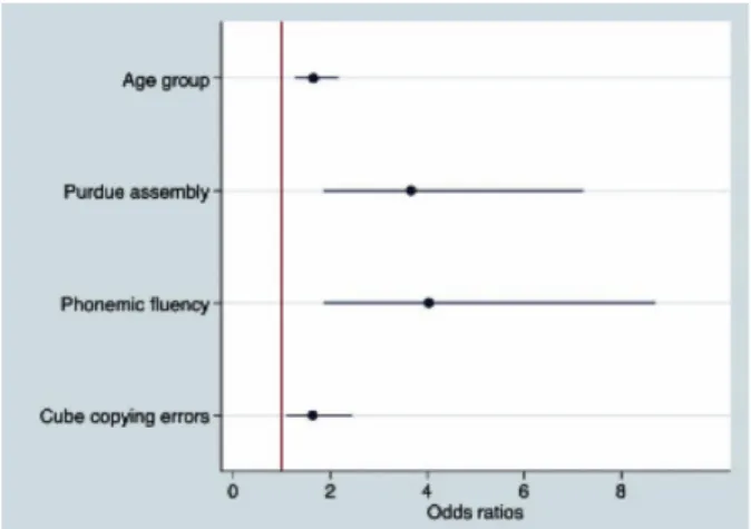
Results: 61 of the 488 included PD patients developed new dementia at 18 month follow-up. Older age at diagnosis with poor performance on phonemic fluency, cube copying, and the Purdue assembly task were all included in the final model as independent predictors of dementia (figure 1). The area under the ROC curve for this model is estimated at 0.81 (95% CI 0.74-0.89) (figure 2).
Figure 1 - Odds ratios of model for factors predicting dementia, with their 95% confidence intervals. Age at diagnosis was analysed as a 5 level ordinal variable, cube copying errors was analysed as a 3 level ordinal variable, and both phonemic fluency and purdue assembly were analysed as a dichotomous variables (poor performers <20th centile).
Figure 2 - ROC curve depicting model of factors predicting dementia including age at diagnosis, Purdue assembly task, phonemic fluency and cube copying connection errors.
Conclusion: Poor performance on three simple clinical tests performed early in PD (the Purdue assembly task, phonemic fluency and cube copying) can be used to predict early dementia. This has implications for both clinical practice and clinical trials.
Disclosure: This study was funded by the Monument Trust Discovery Award from Parkinson’s UK and supported by the National Institute for Health Research (NIHR) Oxford Biomedical Research Centre based at Oxford University Hospitals NHS Trust and University of Oxford, and the NIHR Clinical Research Network: Thames Valley and South Midlands.
O110
Temporal evolution of biomarkers in
isolated REM sleep behavior disorder and
early Parkinson’s disease
M.-L. Muntean1, B. Mollenhauer2, J. Zimmermann3,
N. Focke4, T. Wicke5, J. Ebentheuer1, M. Schaumburg5,
E. Lang5, W. Oertel6, C. Trenkwalder1, F. Sixel-Döring1 1Kassel, Germany, 2Göttingen, Germany, 3Psychologische
Hochschule Berlin, Berlin, Germany, 4University Medical
Center Göttingen, Clinical Neurophysiology, Göttingen, Germany, 5Paracelsus-Elena-Klinik, Kassel, Germany, 6Marburg, Germany
Background and aims: We aimed to study the temporal evolution of biomarkers of various modalities in healthy ageing controls (HC), the prodromal condition of idiopathic REM sleep behavior disorder (iRBD) and in Parkinson’s disease (PD).
Methods: We investigated previously identified biomarkers for early PD in 34 iRBD subjects and compared these to 88 HC and 91 PD patients. We stratified the PD group into 31 patients with RBD (PD+RBD) and 60 patients without (PD-RBD). Baseline and follow-up investigations after 24 months covered questionnaires On Non-Motor signs (NMS), cognitive testing, video-polysomnography (PSG), ECG, olfactory testing, magnetic resonance imaging with Voxel Based Morphometry (VBM) and Cerebrospinal Fluid (CSF) measures.
Results: Most biomarkers in the iRBD group lay between HC and the PD groups. ECG frequency, that was elevated in PD, was normal in iRBD and HC (p<0.01). Other biomarkers already showed abnormalities similar to PD: a high NMS burden (p<0.01) and a non-significant decrease of β-amyloid 1-42 and total tau protein in CSF. There was also a trend towards more abnormalities in iRBD patients compared to the PD group, but did not reach statistical significance: more severe hippocampal atrophy by VBM, more pronounced cognitive decline. The CSF levels of α-synuclein were lower in the iRBD compared to the PD+RBD group (p=0.03)
Conclusion: In prodromal PD abnormalities in NMS, imaging and fluidic markers are already obviously pointing towards the development of overt disease. Based on these results iRBD represents a prodromal state of various α-synuclein aggregation disorders and may develop into a specific, motor phenotype.
Disclosure: The study was supported by unrestricted research grants from the Paracelsus-Elena-Klinik, Kassel, TEVA Pharma/Lundbeck, GE Healthcare and the Parkinson Fonds Deutschland.
O111
Brain Lewy body density is associated
with a lower prevalence of
artherosclerotic cardiovascular disease
risk factors in patients with Parkinson’s
disease
E. Driver-Dunckley1, N. Zhang1, C. Adler1, G. Serrano2,
L. Sue2, H. Shill3, S. Mehta1, C. Belden2, E. Zamrini2,
K. Davis4, T. Beach2
1Mayo Clinic, Scottsdale, USA, 2Sun Health, Sun City, USA, 3Barrow’s, Phoenix, USA, 4Sun Health, Sun city, USA
Background and aims: Epidemiological studies suggest that Atherosclerotic Cardiovascular Disease (ASCVD) risk factors increase the risk of developing Parkinson’s disease (PD). However, conflicting data suggest lower rates of ASCVD in PD. The objective of this study is to determine, with data from a longitudinal clinicopathological study, whether ASCVD risk factors are associated with a PD diagnosis and/or brain alpha-synuclein pathology load. Methods: All subjects were enrolled in the Arizona Study of Aging and Neurodegenerative Disorders (AZSAND). Multivariable logistic regression models, including age, gender, and smoking history, were used to investigate the association of a PD diagnosis or brain alpha-synuclein pathology load with ASCVD risk factors
Results: 150 subjects were included (PD n=60, controls n=90). The regression models showed significant inverse associations. The multivariable Odds Ratio (OR) of brain alpha-synuclein pathology load for carotid artery disease was 0.93 (95% CI: .86 to .98; p=0.02), for anticoagulant use .95 (95% CI: .90 to .99; p=0.04) and for lower heart weight .96 (95% CI: .92 to .99; p=0.01).
Conclusion: This study shows a significant association of higher brain alpha-synuclein pathology load with a lower prevalence of both clinical and pathologic indices of ASCVD in PD subjects versus age-similar controls. We hypothesize this is due to alpha-synuclein pathology-induced sympathetic denervation in PD.
O112
Multimodal MRI markers modifications in
Multiple System Atrophy: a longitudinal
study
P. Péran1, M. Galitzy2, W. Meissner3, O. Rascol4,
A. Pavy-Le Traon4
1Toulouse, France, 2Centre d’Investigation Clinique (CIC),
CHU de Toulouse, Toulouse, France, 3Bordeaux, France, 4University Hospital of Toulouse, Toulouse, France
Background and aims: Multimodal MRI (mMRI) approach is based on combination of MRI parameters sensitive to different tissue characteristics (e.g. volume atrophy, iron deposition, and microstructural damage). The combination of different MR biomarkers could help to discriminate different pathologies with parkinsonian syndrome (Péran et al. Mov Disord.; 2018). Using mMRI, the aim of the study was to evaluate brain changes due to disease progression in Multiple System Atrophy (MSA) patients.
Methods: 19 MSA patients underwent 3-T MRI exam twice at time of inclusion and after one year of follow-up. This MRI comprised: T2*-weighted, T1-weighted and diffusion tensor imaging scans. We used the same method as in the previous work (Péran et al., Brain, 2010) to extract MRI markers (grey density, R2* value, mean diffusity (MD) and fractional anisotropy). The GD, R2*, MD, and FA maps were compared using non-parametric paired t-tests. Statistical significance threshold was set to p<.05 corrected for family wise error.
Results: Figure 1 shows changes due to disease progression from voxel-based analysis in R2* (red), MD (green) and FA (blue) maps. The main results showed significant increase of MD in brainstem and in cerebellum. MSA patients showed also lower FA mainly in left inferior longitudinal fasciculus. Additionally, MSA patients showed a decrease of R2* mainly in cerebellum and in fusiform gyrus. We did not find significant modifications for GD maps.
Figure 1
Conclusion: This study demonstrates that mMRI is able to detect longitudinal modifications after one year of MSA progression. Further analyses are on-going to determine the relationships between clinical and MRI markers.
MS and related disorders 1
O113
Spinal cord area is a stronger predictor of
physical disability than brain volume in
secondary progressive Multiple Sclerosis
F. De Angelis1, J. Stutters1, A. Eshaghi1, A. Garcia1,
F. Prados2, D. Plantone1, A. Doshi1, N.A. John1, A. Calvi1,
D. MacManus1, S. Ourselin2, S. Pavitt3, G. Giovannoni4,
R. Parker5, C.J. Weir5, N. Stallard6, C.P. Hawkins7,
B. Sharrack8, P. Connick9, S. Chandran9,
C.A. Gandini Wheeler-Kingshott1, F. Barkhof1,
J. Chataway1
1Queen Square MS Centre, UCL Institute of Neurology,
Neuroinflammation, London, United Kingdom, 2Translational
Imaging Group, Centre for Medical Image Computing (CMIC), University College London, Department of Medical Physics and Biomedical Engineering, London, United Kingdom, 3Dental Translation and Clinical Research Unit
(part of the NIHR Leeds CRF), University of Leeds, Leeds, United Kingdom, 4Blizard Institute, Queen Mary University
of London, London, United Kingdom, 5Edinburgh Clinical
Trials Unit, Usher Institute of Population Health Sciences and Informatics, University of Edinburgh, Edinburgh, United Kingdom, 6Statistics and Epidemiology, Division of Health
Sciences, Warwick Medical School, University of Warwick, Coventry, United Kingdom, 7Keele Medical School and
Institute for Science and Technology in Medicine, Keele University, Keele, United Kingdom, 8Department of
Neuroscience, Royal Hallamshire Hospital, Sheffield, United Kingdom, 9Centre for Clinical Brain Sciences, University of
Edinburgh, Edinburgh, United Kingdom
Background and aims: Spinal cord atrophy may be a more sensitive measure of disability worsening than brain atrophy in Multiple Sclerosis (MS).
We aimed at investigate the contribution of spinal cord area and brain volume to disability in people with secondary progressive MS (SPMS).
Methods: A group of SPMS patients randomised in a phase 2 clinical trial (MS-SMART) were included in this study. Patients underwent neurological assessments, brain and cervical cord MRI. We measured the following MRI and clinical parameters: Mean Upper Cervical-Cord Cross-Sectional Area (Mucca), Normalised Brain Volume (Nbv), Expanded Disability Status Scale (Edss), Ms Functional composite (MSFC), and Symbol Digit Modalities Test (SDMT). We analysed associations of MRI variables with clinical scores using multivariable linear regression models adjusting for age and gender. Fig.1A-B shows the MRI analysis pipelines.
Fig. 1A-B MRI analysis pipeline A: cross-sectional area of the cervical cord at C2-C3 level. B: normalised brain volume analysis.
Results: Sixty subjects were analysed. The baseline characteristics are shown in Table 1. Multivariable linear regression analyses (Fig.2) showed that MUCCA (standardised-beta= -0.35, standard-error [SE]=0.12,
p=0.005) and NBV (standardised-beta= -0.32, SE=0.14; p=0.02) were independently associated with EDSS. MUCCA, but not NBV, was significantly associated with MSFC (standardised-beta=0.28 SE=0.13; p=0.031). Both MUCCA (standardised-beta=0.3 SE=0.12; p=0.013) and NBV (standardised-beta=0.42, SE=0.14; p=0.006) were independently associated with SDMT.
Table 1. Baseline characteristics of the patients
Fig.2 Multivariable linear regression plots of the statistically significant associations. Plots are based on raw data (i.e. non-standardised data) for a better understanding of the relationships.
Conclusion: MUCCA was the strongest predictor of EDSS, NBV the strongest predictor of SDMT, and MUCCA the only predictor of MSFC. Our findings demonstrate that spinal cord area shows increasing promise as a marker of disability in progressive disease.
Disclosure: The MS-SMART (NCT01910259) trial is a project funded by the Efficacy and Mechanism Evaluation (EME) Programme, an MRC and NIHR partnership. It is also supported by the UK and National Multiple Sclerosis Society; the National Institute for Health Research University College London Hospitals Biomedical Research Centre and University College London; NIHR Leeds CRF (DenTCRU). CJW and RP were supported in this work by NHS Lothian via the ECTU. The remaining authors declare no conflict of interests with respect to this work.
O114
Long-term prognosis of disease evolution
and evidence for sustained Fingolimod
treatment effect by plasma neurofilament
light in RRMS patients
J. Kuhle1, J.A. Cohen2, H. Kropshofer3, R. Meinert4,
C. Barro1, M. Merschhemke3, D. Häring3, D. Leppert3,
D. Tomic3, L. Kappos1
1Neurologic Clinic and Policlinic, Departments of Medicine,
Clinical Research, Biomedicine and Biomedical Engineering, University Hospital and University of Basel, Basel,
Switzerland, 2Neurological Institute, Cleveland Clinic,
Cleveland, USA, 3Novartis Pharma AG, Basel, Switzerland, 4DATAMAP GmbH, Freiburg, Germany
Background and aims: Neurofilament light chain (NfL), an intracellular protein exclusively expressed by neurons, is elevated in the cerebrospinal fluid and blood of patients with multiple sclerosis (MS). We studied the mid- and long-term prognostic potential of plasma NfL for disease evolution and progression and long-term fingolimod effect on plasma NfL levels in patients with relapsing–remitting MS (RRMS).
Methods: Plasma NfL was measured at baseline (N=542), Month (M) 6 (N=467), M12 (N=471), M24 (N=225), and M120 (N=79) using Single Molecule Array (SIMOA) technology in participants from two Phase 3 studies (pooled FREEDOMS, TRANSFORMS) who continued fingolimod treatment in an extension study until M120. The relationship between NfL levels in the initial 12 months (NfL-area under the curve [AUC] classified as low, <30pg/mL; medium, 30−60pg/mL; and high, >60pg/mL) and MS outcomes was assessed using regression models adjusted for age, log[baseline NfL] and baseline characteristics.
Results: At M48, assignment to the high NfL-AUC category compared with low NfL-AUC predicted time to (TT) first relapse, mean cumulative number of new T2 lesions, annual rate of brain atrophy, TT EDSS>=4, TT SPMS and 6M-confirmed disease worsening. NfL levels in patients taking fingolimod were reduced and remained low relative to baseline (from 29.9 to 21.6pg/mL at M24 and from 30.6 to 18.4pg/mL at M120; p<0.0001, both). Conclusion: Our data support the value of plasma NfL as a mid- to long-term prognostic biomarker of disease evolution and progression in RRMS. The reduction of NfL levels achieved by fingolimod treatment was sustained over 10 years.
Disclosure: This study was funded by Novartis Pharma AG, Basel, Switzerland. Detailed disclosure of each author will be included in the poster/oral presentation.
O115
Alemtuzumab provides durable clinical
efficacy in patients with active rrms in the
absence of continuous treatment: 7-Year
follow-up of CARE-MS I (TOPAZ Study)
B. Van Wijmeersch1, P. Vermersch2, A. Boyko3,
J. de Seze4, H.-P. Hartung5, E. Kubala Havrdova6,
J. Said Inshasi7, P. McCombe8, X. Montalban9,
C. Pozzili10, M. Melanson11, N. Daizadeh11,
C. Rodriguez11, K. Selmaj12, O.B.O.T.C.-M.I.C.
and Topaz Investigators11
1Rehabilitation & MS-Centre Overpelt, BIOMED, Hasselt
University, Hasselt, Belgium, 2University of Lille, Lille,
France, 3Pirogov Russian National Research University &
Demyelinating Diseases Center, Usupov Hospital, Moscow, Russian Federation, 4University of Strasbourg, Strasbourg,
France, 5Heinrich-Heine University, Düsseldorf, Germany, 6First Medical Faculty, Charles University in Prague,
Prague, Czech Republic, 7Rashid Hospital and Dubai
Medical College, Dubai, United Arab Emirates, 8University
of Queensland, Brisbane, Queensland, Australia, 9St.
Michael’s Hospital, University of Toronto, Ontario and Centre d’Esclerosi Multiple de Catalunya (Cemcat), Hospital Universitario Vall d’Hebron, Barcelona, Spain, 10Sapienza
University of Rome, Rome, Italy, 11Sanofi, Cambridge, USA, 12Medical University of Lodz, Lodz, Poland
Background and aims: In CARE-MS I (NCT00530348), alemtuzumab 12 mg/day (baseline: 5 days; 12 months later: 3 days) significantly improved clinical/MRI outcomes versus SC IFNB-1a over 2 years in treatment-naive RRMS patients. Durable efficacy was observed in a 4-years extension (NCT00930553; 95% of CARE-MS I patients enrolled, 92% completed Y6), in which patients could receive alemtuzumab retreatment as-needed for relapse/ MRI activity or receive other DMTs per investigator’s discretion. Further evaluation is ongoing (TOPAZ extension; NCT02255656). We present efficacy/safety outcomes over 7 years (2 years core study plus 4 years extension and TOPAZ Y1) in alemtuzumab-treated patients from CARE-MS I.
Methods: Assessments: Annualised relapse rate (ARR); EDSS scores; 6-month confirmed disability worsening (CDW); 6-month confirmed disability improvement (CDI); no evidence of disease activity (NEDA); and AEs.
Results: 299 patients (93%) completed TOPAZ Y1. 59% received neither alemtuzumab retreatment nor other DMT after the initial 2 courses. ARR remained low (Y7: 0.13); 60% were relapse-free in Y3 7. The percentage with stable/ improved EDSS scores versus baseline remained high at Y7 (78% [improved, 21%; stable, 57%]). The mean change in EDSS score from baseline to Y7 was 0.09. At Y7, 74% were 6-month CDW-free; 37% achieved 6-month CDI. The majority of patients achieved NEDA each year (Y7: 61%). Overall AE incidence decreased over time.
Conclusion: Alemtuzumab efficacy was maintained for 7 years in treatment-naive patients, despite 59% receiving no additional treatment since the initial 2 courses. Alemtuzumab
safety profile remained consistent. Alemtuzumab provides a unique treatment approach for RRMS patients, offering durable efficacy without continuous treatment.
Disclosure: Study supported by Sanofi and Bayer HealthCare Pharmaceuticals.
O116
Effects of Fingolimod on MRI outcomes in
patients with paediatric-onset Multiple
Sclerosis: results from the Phase-3
PARADIGMS study
D.L. Arnold1, B. Banwell2, A. Bar-Or3, A. Ghezzi4,
B. Greenberg5, E. Waubant6, G. Giovannoni7,
J. Wolinsky8, J. Gärtner9, K. Rostasy10, L. Krupp11,
M. Tardieu12, W. Brück13, T. Stites14, G.L. Pearce15,
M. Merschhemke16, T. Chitnis17
1NeuroRx Research, Montreal, Qubec, Canada, 2The
Children’s Hospital of Philadelphia, Perelman School of Medicine, University of Pennsylvania, Philadelphia, PA, USA, 3Perelman School of Medicine, University of
Pennsylvania, Philadelphia, PA, USA, 4Ospedale di
Gallarate, Gallarte, Italy, 5Department of Neurology and
Neurotherapeutics, Department of Pediatrics, University of Texas Southwestern Medical Center, Dallas, TX, USA,
6Department of Neurology, University of California, San
Francisco, CA, USA, 7Blizard Institute, Barts and The
London School of Medicine and Dentistry, Queen Mary University, London, United Kingdom, 8McGovern Medical
School, UTHealth, Houston, TX, USA, 9Department of
Paediatrics and Adolescent Medicine, German Centre for Multiple Sclerosis in Childhood and Adolescence University Medical Centre, Göttingen, Germany, 10Division of
Paediatric Neurology, Children’s Hospital Datteln, University Witten/Herdecke, Datteln, Recklinghausen, Germany, 11Pediatric MS Center, NYU Langone, New York,
USA, 12Hôpital Bicêtre, Assistance Publique-Hôpitaux de
Paris, Paris, France, 13Department of Neuropathology,
University Medical Centre, Göttingen, Germany, 14Novartis
Pharmaceuticals Corporation, East Hanover, USA, 15GCE
Solutions, Bloomington, Illinois, USA, 16Novartis Pharma
AG, Basel, Switzerland, 17Partners Pediatric Multiple
Sclerosis Center, Massachusetts General Hospital, Boston, MA, USA
Background and aims: Approximately 3–5% of Multiple Sclerosis (MS) cases manifest in childhood and adolescence, characteristically with highly active inflammatory disease course. Paediatric-onset MS (POMS) has an impact on brain integrity and may increase Brain Volume Loss (BVL) above age-expected rates. This study assessed the effect of oral Fingolimod up to 0.5mg daily versus intramuscular interferon (IFN) beta-1a 30μg once weekly on MRI outcomes in POMS patients.
Methods: In this double-blind, double-dummy, active-controlled, multicentre study, patients with POMS (aged 10–<18 years) received either Fingolimod (dose adjusted for body weight; N=107) or IFN beta-1a (N=107) for up to 2 years. MRI was performed at baseline and every 6 months until the End Of The Study (EOS) core phase. Key MRI
outcomes were the number of new/newly enlarging T2 (n/ neT2) lesions and Gd-enhancing T1 (Gd+T1) lesions, Annual Rate Of Brain Volume Change (ARBVC), annualised rate of number of new T1 hypointense lesions, change in total T2 Hyperintense Lesion Volume (T2LV) and the number of Combined Unique Active Lesions (CUAL). Results: At the EOS, compared with IFN beta-1a, fingolimod significantly reduced the annualised rate of n/ neT2 lesions (52.6%; p<0.001), number of Gd+T1 lesions per scan (66.0%; p<0.001), ARBVC (−0.48% vs. −0.80%, p=0.014), annualised rate of number of new T1 hypointense lesions (62.8%; p<0.001), T2LV (percent change from baseline: 18.4% vs. 32.4%, p<0.001) and CUAL per scan (60.7%; p<0.001).
Conclusion: Fingolimod significantly reduced MRI activity and slowed BVL for up to 2 years vs. IFN beta-1a in paediatric-onset MS.
Disclosure: This study was funded by Novartis Pharma AG, Basel, Switzerland. Detailed disclosure of each author will be included in the poster.
O117
Characterizing the Slowly Evolving
Lesions (SELs) in a cohort of secondary
progressive Multiple Sclerosis patients
A. Calvi1, F. Prados2, C. Tur1, F. De Angelis1, N. John1,
A. Doshi1, J. Stutters1, D. MacManus1, S. Ourselin2,
O. Ciccarelli1, J. Chataway1, F. Barkhof2
1UCL Institute of Neurology, QS MS Centre. Department of
Neuroinflammation , London, United Kingdom,
2Translational Imaging Group, Centre for Medical Imaging
Computng (CMIC), University College London, Department of Medical Physics and Biomedical Engineering, London, United Kingdom
Background and aims: There is a need to develop markers of progression in Multiple Sclerosis (MS). Slowly Evolving Lesions (SELs) on MRI have been recently identified in longitudinal trials of Primary Progressive MS (PP-MS) using non-linear registration-based analysis techniques [1]. Magnetization Transfer Ratio (MTR) highly correlates with demyelination and axonal loss within MS lesions [2].
Methods: We included 79 secondary progressive (SP-MS) patients from the MS-SMART trial (NCT01912059) who underwent brain PD/T2, FLAIR and MTR scans at baseline, 24 and 96 weeks. Manually delineated lesions showing Jacobian expansion were selected as “candidates”. Final SELs were chosen through a sum score of their concentricity and constancy (figure 1). We calculated baseline MTR values within the different lesion types (SEL, non-SEL and non-candidates) and compared MTR changes from baseline to 96-week.
Results: From 4756 lesions screened, 1140 candidates were identified and ultimately 140 SELs (2.9%) were detected. Baseline MTR within SEL was lower compared to the non-SELs and non-candidates (24.51, 26.26 and 28.89, respectively; p-values<0.001). MTR decrease between baseline and week 96 within SELs was significantly greater compared to non-SELs (p=0.02) and to non-candidates (p=0.01) (Figure 2). In contrast, there were no significant differences in MTR change between SEL and non-candidates (p=0.50).
Conclusion: We confirm that, as in PP-MS, there are lesions in SP-MS that can be classified as SELs. Given their more destructive signature on MTR, SELs are promising biomarkers of chronic plaque evolution in progressive MS. Future studies will investigate whether there is a relationship between the occurrence of SELs and clinical disability. Disclosure: AC, FDA, FP, NJ, AD, JS, DM declare no conflicts of interests. CT acknowledges 2015 ECTRIMS fellowship. OC received research funding from: UK and National MS Society, Rosetrees trust, NIHR UCLH BRC, Biogen, Novartis, Roche, Genzyme, Teva. JC has received support from NIHR, UK MS Society and National MS Society, Receptos, Novartis, Biogen Idec, Roche, Merck, MedDay, Apitope. FB serves as as consultant for Bayer Shering Pharma, Sanofi-Aventis, Biogen-Idec, TEVA, Genzyme, Merck-Serono, Novartis, Roche, Synthon, Jansen Research, Lundbecok, BRC.
O118
Characterizing dynamic functional
network connectivity in the main clinical
phenotypes of Multiple Sclerosis
M. Hidalgo de la Cruz1, M.A. Rocca1, P. Valsasina1,
B. Colombo2, F. Esposito2, G. Comi2, M. Filippi1 1San Raffaele Scientific Institute, Vita-Salute San Raffaele
University, Neuroimaging Research Unit, Institute of Experimental Neurology, Division of Neuroscience, Milan, Italy, 2San Raffaele Scientific Institute, Vita-Salute San
Raffaele University, Department of Neurology, Milan, Italy
Background and aims: Resting-state (RS) dynamic Functional Network Connectivity (dFNC) in Multiple Sclerosis (MS) has rarely been studied. Here, we investigated dFNC changes occurring in MS patients according to their clinical phenotype.
Methods: RS fMRI data were acquired from 126 MS patients and 40 healthy controls (HC). There were 52 relapsing remitting (RR) MS, 16 benign (B) MS, 34 secondary progressive (SP) MS and 24 primary progressive (PP) MS patients. Between-group dFNC differences in 42 relevant networks were assessed: 1) in MS patients vs HC, and 2) among different clinical MS phenotypes.
Results: Clustering analysis revealed 3 dFNC states in HC and MS patients: State 1 (frequency=57%, low dFNC strength), State 2 (frequency=19%, middle-high dFNC strength), and State 3 (frequency=24%, low FNC strength except for high dFNC strength in the sensorimotor and visual networks). Compared to HC, MS patients showed an overall reduction of dFNC in the main sensorimotor, cognitive and subcortical networks, while increased dFNC was found for the frontal-attention network. The same pattern of dFNC changes was detected when comparing RRMS and PPMS patients vs HC. Compared to RRMS, SPMS showed strong dFNC reductions in most functional networks, mainly in States 2 and 3, and a markedly increased dFNC for the frontal-attention network in States 1 and 2. Conversely, frontal-attention dFNC was significantly decreased in BMS vs RRMS patients.
Conclusion: Significant dFNC changes contribute to explain MS phenotypic heterogeneity. While the prevalent reduction of dFNC might reflect the progressive accumulation of structural damage, compensatory/maladaptative mechanisms may take place in frontal/attentional circuits.
Disclosure: Partially supported by Fondazione Italiana Sclerosi Multiple (FISM2013/S/1).
Peripheral nerve disorders 1
O119
Axonal function predicts response to
subcutaneous immunoglobulin in chronic
inflammatory demyelinating
polyneuropathy: the PATH study
V. Bril1, N. van Geloven2, H.-P. Hartung3, G. Sobue4,
J.-P. Lawo5, O. Mielke5, B.L. Durn6, I.S. Merkies7 1University Health Network, University of Toronto,
Department of Medicine (Neurology), Toronto, Canada,
2Leiden University Medical Center, Department of
Biostatistics and Bioinformatics, Leiden, Netherlands,
3Heinrich-Heine Universität, Neurology, Düsseldorf,
Germany, 4Nagoya University Graduate School of Medicine,
Department of Neurology, Nagoya, Japan, 5CSL Behring,
Marburg, Germany, 6CSL Behring, King of Prussia, PA,
USA, 7Maastricht University Medical Center, Department of
Neurology, Maastricht, Netherlands
Background and aims: Chronic Inflammatory Demyelinating Polyneuropathy (CIDP) is an immune mediated disease starting with functional impairment and demyelination; in later stages, axonal degeneration may occur.
Methods: PATH was a randomised, double-blind study investigating 0.2 (low) and 0.4 g/kg (high) weekly doses of maintenance SCIG IgPro20 (Hizentra®, CSL Behring)
versus placebo (N=172). After Ig dependency testing, and IVIG restabilisation, patients were randomised to SCIG or placebo for 25 weeks or until early termination. Nerve conduction studies (NCS) were performed before study drug administration. Relapse rate (defined as a 1 point increase by adjusted Inflammatory Neuropathy Cause and Treatment score) comparisons were undertaken on patients with assumed non-axonal damage versus assumed axonal damage based on cut-off amplitudes at the distal stimulation site: 1 mV for the foot and 2 mV for the wrist.
Results: Patients with assumed non-axonal damage who received placebo had a 73% relapse rate versus 39% on low-dose and 19% on high-dose SCIG. Patients with assumed axonal damage had relapse rates of 25%, 30% and 19% for placebo, low-dose and high-dose SCIG, respectively.
Conclusion: CIDP patients with assumed non-axonal damage had a high relapse rate when switched from IVIG to placebo that was significantly reduced in patients switched to SCIG therapy. Relapse rates were lower in assumed axonal damage patients and were not influenced by SCIG. These findings could help in redesigning future trials including maintenance regimens based on NCS categorisation of patients.
Disclosure: This study was sponsored by CSL Behring.
O120
Corneal confocal microscopy and skin
biopsy in the evaluation of diabetic small
fiber neuropathy
J. Bednarik1, A. Rajdova2, I. Kovalova1, M. Horakova2,
S. Divisova2, J. Raputová3, E. Vlckova3
1University Hospital Brno, Department of Neurology, Brno,
Czech Republic, 2University Hospital Brno, Brno, Czech
Republic, 3Brno, Czech Republic
Background and aims: Corneal Confocal Microscopy (CCM) is a comparatively new diagnostic method that enables morphological evaluation of small sensory nerve fibers in the cornea. Currently, Intraepidermal Nerve Fiber Density (IENFD) is considered a gold standard for the diagnosis of Small Fiber Neuropathy (SFN). The aim was to compare sensitivity of CCM and IENFD in the detection of SFN in patients with Diabetic distal symmetrical Polyneuropathy (DPN).
Methods: A group of 81 patients with the diagnosis of definite DPN (mean age 58.2; 50 men, 31 women; 27 patients had painful DPN - pDPN) based on clinical signs and symptoms and nerve conduction studies, and a group of 32 healthy controls (HC) of similar age and gender were assessed using skin biopsy and CCM with evaluation of Corneal Nerve Fiber Density (CNFD), Length (CNFL), Branch Density (CFBD) and Tortuosity (CNFT).
Results: All CCM parameters showed significantly higher proportion of abnormal values not only in DPN group compared to HC (p<0.001), but the proportion of abnormalities of all CCM parameters (except CNFT) was significantly higher in pDPN subgroup compared to non-painful cases (p<0.05). CCM sensitivity in detection of SFN in DPN group was similar (72%) to that of IENFD (74%). Individual values of CCM parameters, however, showed insignificant correlation with IENFD values (p<0.05). Conclusion: CCM is able to prove significant involvement of small sensory nerve fibers in patients with symptomatic DPN with comparable sensitivity as IENFD obtained via semi-invasive skin biopsy procedure. Higher proportion of CCM abnormalities in pDPN possibly reflects higher severity of neuropathy in painful cases.
O121
Charcot-Marie-Tooth disease type 4B with
myelin outfoldings (CMT4B): a multicentre
retrospective study
D. Pareyson1, T. Stojkovic2, S. Leonard-Louis2,
MM. Reilly3, M. Laurà3, R. Horvath4, G. Ricci4,
Y. Parman5, E. Battaloglu5, M. Tazir6, M. Bellatache6,
N. Bonello-Palot7, S. Sacconi8, R. Guimarães-Costa2,
S. Attarian7, P. Latour9, A. Megarbane2, A. Schenone10,
G. Ursino10, M. Sabatelli11, M. Luigetti11 M.L. Santoro12,
F. Manganelli12, A. Quattrone13, P. Valentino19,
T. Murakami14, SS. Scherer15, L. Dankwa15, ME. Shy16,
CJ. Bacon16, DN. Herrmann17, C. Pisciotta1, S. Previtali1,
A. Bolino1
1Milan, 10Genoa, 11Rome, 12Naples, 13Catanzaro, Italy; 2Paris, 7Marseille, 8Nice, 9Lyon, France; 3London, 4Newcastle, UK; 5Istanbul, Turkey; 6Algiers, Algeria; 14Kurashiki, Japan; 15Philadelphia, 16Iowa City, 17Rochester,
USA.
Background and aims: Charcot-Marie-Tooth neuropathy B1 and B2 (CMT4B1/B2) are characterized by recessive inheritance, early onset, severe course, slowed nerve conduction, myelin outfoldings, loss-of-function mutations in Myotubularin-related protein-2 and -13 (MTMR2, MTMR13/SBF2), respectively, involved in phosphoinositides metabolism. We conducted a multicentre retrospective study to better characterise CMT4B in view of possible clinical trials.
Methods: In 16 centres, we collected clinical, genetic, instrumental data from CMT4B subjects.
Results: There were 44 patients (27 CMT4B1, 17 CMT4B2). CMT4B1 patients were younger and with earlier onset than CMT4B2. Onset age: 2.8+/-2.8 years (range 0-13) in CMT4B1, 7.6+/-8.7 (1-36) in CMT4B2; delayed motor milestones in 14/26 CMT4B1 and 4/17 CMT4B2 subjects. Twelve CMT4B1 but only two CMT4B2 patients became chair-bound. Both types are characterised by vocal cord involvement (10/25 CMT4B1, 9/17 CMT4B2); respiratory involvement was seen almost exclusively in CMT4B1 patients (n=8, four NIV, one tracheostomy; one CMT4B2 patient on NIV). Glaucoma (n=6) and buphthalmos (n=3) occurred only in CMT4B2.
CMTNS and CMTES-motor scores were significantly higher in CMT4B1 patients in spite of their younger age, indicating more severe disease: CMT4B1=CMTES mean 17.9+/-5.9 (n=20; range 9-28/28), CMTNS mean 30.1+/-4.7 (n=10; 19-36/36), CMTES-motor mean 13.2+/-2.9 (n=21; 8-16/16); CMT4B2=CMTES mean 15.8+/-4.4 (n=17; 6-24/28), CMTNS mean 23+/-5 (n=16; 13-32/36); CMTES-motor mean 9.2+/-3.9 (n=17; 4-16/16).
Conclusion: CMT4B1 is more severe than CMT4B2. MTMR2, a catalytically active phosphatase, interacts with MTMR13, which is known to increase MTMR2 enzymatic activity but is catalytically inactive. CMT4B2 nerves may have a residual enzymatic activity of MTMR2 which results in less severe phenotype than CMT4B1.
Disclosure: Partly supported by LAM Therapeutics
O122
Cryoglobulinaemia-associated peripheral
neuropathies: clinical characteristics and
prognosis from 20 years experience of
neuromuscular clinic
M. Mazzoli, A. Ariatti, M.T. Mascia, F. Valzania, P. Nichelli, G. Galassi
University of Modena and Reggio Emilia, Modena, Italy
Background and aims: Cryoglobulinaemia is associated with peripheral neuropathies. We assessed prognostic role of clinical and neurophysiological variables among 28 patients over 20 years.
Methods: 28 patients with cryoglobulinaemia-associated neuropathy were enrolled at University of Modena during period 1998-2017. Evaluated independent clinical variables were gender, age at onset, cryoglobulin type, HCV co-infection, type of neuropathy (axonal or demyelinating), copathologies, therapies. Degree of neurological involvement (mild, moderate, severe) was assessed using disability scales (MRC, INCAT, tremor rating scale) and electrophysiological examinations. Probability of death or neurological worsening was estimated from binomial, multinomial, ordered logistic regression, Cox models. P-values <0.05 were considered significant.
Results: 20 patients were female (71%; M:F ratio 1:2.5). Median age was 66 years (range 31-83). Median follow-up time was 33 months. Eighteen patients had type II cryoglobulins (64%), 7 type III (25%), 3 type I (11%). 16 patients had HCV-RNA (57%). Sensorymotor demyelinating neuropathy was prevalent (65%). Neuropathy was mild in 46.3%, moderate in 32.1%, severe in 23% of patients. Cumulative incidence of worsening over time was 39%. None of independent variables had predictive role on neurological worsening or death, except type II cryoglobulin at multivariable ordered logistic regression (OR 12.5, 95% CI 1.24-126, p 0.03). HCV co-infection showed borderline significance (OR 3.93, 95% CI 0.86-17.8, p 0.07). Kaplan-Meyer estimate of worsening in respect of stratified age at onset showed more severe course in subjects above 70 years.
Conclusion: Type II cryoglobulins was associated with more severe peripheral neuropathy especially in aged subjects.
Disclosure: Nothing to disclose
O123
Long-term efficacy and safety of
Inotersen in patients with hereditary
transthyretin (hATTR) amyloidosis treated
in the open-label extension of the phase-3
study NEURO-TTR
V. Plante-Bordeneuve1, T. Brannagan2, A. Wang3,
T. Coelho4, M. Waddington Cruz5, M. Polydefkis6,
P. Dyck7, M. Scheinberg8, J. Berk9, F. Barroso10,
D. Adams11, C. Whelan12, G. Merlini13, B. Drachman14,
S. Heitner15, I. Conceicao16, H. Schmidt17, G. Vita18,
J. Campistol19, J. Gamez20, P. Gorevic21, B. Monia22,
M. Benson23, M. Gertz7
1CHU Henri Mondor, Creteil, France, 2Columbia University
Medical Center, Neurology, New York, USA, 3University of
California, Irvine, Orange, USA, 4Centro Hospitalar do
Porto, Porto, Portugal, 5Federal University of Rio de
Janeiro University Hospital, Rio de Janeiro, Brazil, 6Johns
Hopkins University, Baltimore, USA, 7Mayo Clinic,
Rochester, USA, 8Associação de Assistência a Criança
Deficiente, Sao Paolo, Brazil, 9Boston University, Boston,
USA, 10FLENI, Ciudad Autónoma de Buenos Aires, Buenos
Aires, Argentina, 11CHU Bicetre, Universite Paris-Sud, Le
Kremlin-Bicêtre, France, 12University College London—
National Amyloidosis Centre, London, United Kingdom,
13Amyloidosis Center, IRCCS Policlinico San Matteo,
University of Pavia, Pavia, Italy, 14University of
Pennsylvania, Philadelphia, USA, 15Oregon Health and
Science University, Portland, USA, 16Hospital de Santa
Maria-CHLN, and IMM Faculty of Medicine, UL, Lisbon, Portugal, 17Universitätsklinikum Münster, Münster, Germany, 18A.O.U. Policlinico G. Martino—University of Messina,
Messina, Italy, 19Hospital Clinic, Barcelona, Spain, 20Hospital Universitari Vall D’Hebron, Barcelona, Spain, 21Mount Sinai Medical Center, New York, USA, 22Ionis
Pharmaceuticals, Carlsbad, USA, 23Indiana University
School of Medicine, Indianapolis, USA
Background and aims: hATTR is a rare, progressive, fatal disease manifested by systemic build-up of TTR protein, resulting in organ failure. The disease causes significant morbidity and progressive decline in Quality of Life (QOL) and robs patients of their independence owing to limitations on activities of daily living. We report results of the Open-Label Extension (OLE) study of NEURO-TTR, highlighting the long-term efficacy and safety of inotersen, an antisense oligonucleotide inhibitor of TTR protein production, in patients with hATTR.
Methods: Patients with hATTR who completed the double-blind, placebo-controlled, phase 3 study NEURO-TTR (NCT01737398) were eligible to receive Inotersen (300-mg weekly subcutaneous doses) for up to 5 years in this OLE.
The OLE monitored adverse events and change from baseline in the Norfolk Quality of Life—Diabetic Neuropathy (Norfolk QOL-DN) total score (136 points total, higher scores indicate worse QOL) and modified neuropathy impairment score +7 (mNIS+7) (346 points total, higher scores indicate worse neuropathy).
Results: At the time of the interim analysis, 114 patients had enrolled in the OLE. Most patients were white (95%) and male (70%), and, at OLE baseline, mean age was 61.4 years and 69% of patients had cardiomyopathy. Mean disease duration from time of symptom onset to OLE baseline was 81.8 months. Mean OLE baseline mNIS+7 composite scores and Norfolk QOL-DN total scores were 92.0 and 55.2, respectively. One-year OLE follow-up results will be presented.
Conclusion: Results of the OLE showed continued benefit, as measured by Norfolk QOL-DN and mNIS+7. No new safety concerns were identified.
Disclosure: This study was sponsored by Ionis Pharmaceuticals (Carlsbad, CA, USA).