DOCTORAT DE L'UNIVERSITÉ DE TOULOUSE
Délivré par :
Institut National Polytechnique de Toulouse (INP Toulouse)
Discipline ou spécialité :
Génie des Procédés et de l'Environnement
Présentée et soutenue par :
Mme MARINA ATGIE
le mardi 13 mars 2018
Titre :
Unité de recherche :
Ecole doctorale :
Composition and structure of gum Arabic in solution and at oil-water
interfaces
Mécanique, Energétique, Génie civil, Procédés (MEGeP)
Laboratoire de Génie Chimique (L.G.C.)
Directeur(s) de Thèse :
M. OLIVIER MASBERNATM. KEVIN ROGER
Rapporteurs :
Mme VERONIQUE SCHMITT, CNRS AQUITAINE
M. PETER FISCHER, EIDGENOSSISCHE TECH HOCHSCHURE ZURICH
Membre(s) du jury :
M. DENIS RENARD, INRA ANGERS, Président M. KEVIN ROGER, CNRS TOULOUSE, Membre
Mme ODILE AUBRUN, L'OREAL, Membre
M. OLIVIER MASBERNAT, CNRS TOULOUSE, Membre
Overview……… ... 10
Chapter I-
Literature review ... 14
I-1
Introduction ... 16
I-2
Literature review on gum Arabic ... 16
I-2-1
Structural composition of gum Arabic ... 17
I-2-2
Gum arabic properties in solution ... 30
I-2-3
Interfacial properties of gum Arabic ... 32
I-2-4
Conclusion and thesis objectives ... 41
Chapter II-
Gum Arabic: composition and multi-scale structure in solution ... 48
Chapter III-
Gum Arabic stabilized emulsions: composition of the interfacial film ... 82
Chapter IV-
Interfacial structuration in gum Arabic stabilized emulsions ... 110
Conclusion and perspective ... 142
Appendix: analytical techniques ... 150
I-
Emulsification ... 152
II-
Scattering ... 152
III-
Electrophoretic mobility ... 155
IV-
Interfacial tension ... 156
V-
Chromatography ... 156
Résumé………. ... 160
Cette thèse a été réalisée au Laboratoire de Génie Chimique, je tiens à remercier Béatrice
Biscans (ancienne directrice du laboratoire) et Pierre Aimar (actuel directeur du laboratoire), de
m’avoir permis de réaliser mes travaux de recherche dans ce laboratoire.
I would like to thanks all the jury members who kindly agreed to review my pHd thesis and for
the interesting discussion we had during the pHd defense: Véronique Schmitt, Peter Fischer,
Denis Renard, Sebastiaan Eeltink and Odile Aubrun.
Je tiens à adresser un remerciement tout particulier à mes deux directeurs de thèse : Olivier
Masbernat et Kevin Roger. Olivier je te remercie pour ton encadrement bienveillant et pour
l’enthousiasme dont tu as su faire preuve à chaque étape de cette thèse. J’aurai grand plaisir à
échanger de nouveau avec toi sur ce sujet passionnant qu’est la gomme arabique ou sur de
futurs projets. Kevin je tiens à te remercier pour ces trois années au cours desquelles tu m’as
appris énormément. Tu as su m’encadrer avec toute l’attention dont j’ai pu avoir besoin.
J’espère que nos routes se recroiseront à l’avenir pour discuter et travailler de nouveau
ensemble.
Une partie des expérimentations de ce travail de thèse a eu lieu aux IMRCP à Toulouse sous
l’encadrement de Jean-Christophe Garrigues. Je tiens à te remercier Jean-Christophe pour ton
accueil, ta disponibilité et nos échanges au cours de mes deux dernières années de thèse.
Je souhaite remercier la société Caragum International, partenaire de cette thèse dans le cadre
du labcom Sophy financé par l’ANR. Nous avons pu avoir des échanges riches au cours de cette
thèse notamment avec Vincent Dutaut et Fréderic Tur.
Je souhaite également remercier l’ensemble des permanents du laboratoire et tout
particulièrement de l’équipe de Génie des Interfaces et Milieux Divisés pour toutes les
discussions enrichissantes que nous avons pu avoir : Sébastien Teychene, Benjamin Lalanne,
Emmanuel Cid, Martine Meireles, Patrice Bachin, Micheline Abbas, Pierre Roblin…
Nous avons eu la chance de pouvoir réaliser des expériences de diffusion de rayonnement sur
différents sites. Pour cela je souhaite remercier le laboratoire Léon Brillouin pour l’accès au
réacteur Orphée et plus particulièrement aux spectrophotomètres PACE et PAXY. Un grand
merci à Alexis Chennevière pour son accompagnement lors de nos expérimentations et nos
échanges sur les résultats obtenus. Je remercie également le synchotron ERSF de nous avoir
donné accès à ID02 pour nos expérimentations. Enfin je souhaite remercier la fédération Fermat
pour les expérimentations que nous avons pu réaliser sur le spectrophotomètre XEUSS.
J’aimerais également remercier Sabine Heinisch et Florent Rouviere pour leur accueil chaleureux
sein de l’Institut des Sciences Analytiques à Lyon dans le cadre des expérimentations de
chromatographie bidimensionnelle.
Merci à l’ensemble du personnel technique et administratif du laboratoire sans qui rien ne
serait possible.
Je voudrais également remercier tous les doctorants et post-doctorants dont j’ai eu la chance de
croiser la route durant ces trois années à Toulouse. Nous avons pu partager des repas, des
parties de UNO endiablées, des soirées de danse, de barbecue et un karaoké inoubliable.
J’aimerais tous vous citer mais la liste serait beaucoup trop longue. Je voudrais exprimer un
remerciement particulier à tous ceux qui ont partagé mon bureau au laboratoire, où nous avons
pu partager nos questions, nos doutes et surtout nos joies : Brice, Fatma, Elise, Leo et Antoine.
Un grand merci à mes amis qui m’ont soutenu pendant cette thèse, merci pour votre écoute et
pour nos moments de retrouvaille qui sont toujours un bonheur : Marine, Léa B, Marie, Alice,
Audrey, Pauline C, Aurélie R, Maud, Anne-Sophie, Lilia, Kamal, Lionel, Vincent, Justine, Mathieu,
Hervé, Aurélie K, Pauline D, Joffrey, Charlotte, Kemie, Adèle, Léa G…
Le soutien de ma famille a été très important pendant ce long travail de recherche. Je souhaite
remercier mes oncles et tantes, mes cousins, cousines et bien sûr mes grands-parents, d’avoir
toujours été fiers de mon parcours. J’ai une pensée émue pour mes grands-parents partis trop
tôt qui auraient été très heureux de me voir terminer cette étape. Je voudrais remercier mon
petit frère pour sa curiosité et l’intérêt qu’il a su me porter tout au long de mes études. Un
énorme merci à mes parents pour leur soutien à toutes épreuves, leur compréhension, leur
écoute et leurs encouragements.
Enfin je porte un remerciement particulier à Maxime pour la compréhension dont il a su faire
preuve dans les moments difficiles et pour ses encouragements au quotidien. J’ai hâte de
commencer la prochaine étape à tes cotés.
An emulsion consists of a dispersion of a liquid as small droplets (with size ranging from several µm to nm) in another immiscible liquid. Emulsions are present in numerous industrial applications such as food products (milk, butter, mayonnaise or flavored beverages), cosmetic or pharmaceutical products (moisturizing creams, encapsulating active ingredients), bitumen for road coatings or in the paint industry. Such systems are widely used because they combine benefits of both phases. For instance, in a moisturizing cream the aqueous phase brings a fresh sensation while the oil phase contributes to the deposition of a protective film at the skin surface.
Several types of emulsion exist: direct emulsions (oil-in-water), inverse emulsions (water-in-oil) or multiple emulsions (water-in-oil in water for instance). Emulsions are thermodynamically unstable systems. Without the presence of interfacial agents, emulsions break to yield two separate phases. Several types of interfacial agents exist and are used for the stabilization of dispersed systems such as emulsions. A formulation may contain surfactants, nanoparticles, proteins, micro-gels, block-copolymers. All these species are able to adsorb at oil/water interface and to protect droplets against coalescence. The demand for natural additives in formulation is steadily increasing, regarding environmental and health issues. Hydrocolloids constitute a class of natural products such as proteins or polysaccharides, which sometimes possess emulsifying and stabilizing properties. Among these, gum Arabic, an acacia tree exudate, has been used for millennia due to its outstanding interfacial properties. This natural product is a complex mixture of biopolymers. Many attempts have been carried out, with limited success, to mimic interfacial properties of this product. A deeper understanding of the structure and composition of gum Arabic but also of its behavior at oil/water interfaces and of the stabilization mechanisms involved is thus required.
In the first chapter of this thesis, a literature review on the structure, behavior in solution and at interfaces of gum Arabic is presented. In the second chapter, we briefly introduce the analytical techniques used in the frame of this study to improve the understanding of gum Arabic adsorption and stabilization behaviors (emulsification, interfacial tension, chromatographic separation, light scattering and small angle X-ray and neutron scattering).
Chapter two deals with the structure of gum Arabic gum in solution as a component and multi-scale system, which has been investigated using a two dimensional chromatographic separation (size exclusion followed by hydrophobic interaction) and small angle scattering measurements (neutron and X-ray).
Chapter three focuses on the composition of gum Arabic layers adsorbed at oil droplets interfaces within an oil-in-water emulsion, as a function of the physico-chemical parameters (pH and salinity) involved in the formulation, using chromatographic analysis.
In the fourth chapter, the structuration of the gum Arabic adsorbed layer is addressed. We unravel a structuration of adsorbed gum amphiphilic moieties providing an elastic film capable to resist to extreme conditions (such as ultra-centrifugation).
Main results and findings of this work are summarized in the concluding section, and research prospects about the use of gum Arabic in formulation are proposed.
This PhD project was initiated through a collaboration between the Laboratoire de Génie Chimique (LGC) and CARAGUM International®, through the LabCom SOpHy, funded by ANR, the French National Research Agency. CARAGUM Int.® (Marseille, France) is a company specialized in the development and sale of food additives, including Gum Arabic.
16
I-1 Introduction
Hydrocolloids are water-soluble polymers which behave in solution more as colloidal particles than as polymers. They usually comprise a wide variety of bio-polymers, typically proteins and polysaccharides that are employed in a broad range of industrial sectors in order to provide specific functions [1]. These functions can include: thickening and gelling aqueous solutions, stabilizing foams, emulsions and dispersions or for the flavour controlled release. Hydrocolloids may originate from different sources: botanical, algal, microbial or animal. These materials are widely used in food industry to influence the texture or the organoleptic properties of the products. Many other industrial products take benefit of their properties such as cosmetics, inks, pharmaceutical or agricultural product [2]–[4].
Starch is the most commonly used product as thickening agent although xanthan gum is more and more employed particularly for its particular rheological properties. Gelatin is widely used as gelling agent. However, since the market demand for non-animal originating product increases, alternatives can be found with carrageenan.
Gum Arabic is a hydrocolloid involved in many industrial processes and especially in the food industry for the emulsification of flavor oil. Because of large fluctuation in the market price of gum Arabic starch-based substitutes to gum Arabic have recently emerged as well as protein-polysaccharides conjugates. The latter are formed through the Maillard reaction or by electrostatic interaction in order to reproduce the properties of gum Arabic. Yet, nowadays, no gum substitute has been found to possess the same exceptional properties as gum Arabic itself, in particular as an emulsion stabilizer. A better understanding of its structure and interfacial properties is still needed in order to mimic and substitute this natural product [4], [5].
The present study focuses on the structural and emulsifying properties of gum Arabic. We present in the following section a literature review on this hydrocolloid, its history, its chemical and structural composition, and finally its properties in solution and as an interfacial agent.
I-2 Literature review on gum Arabic
Gum Arabic is a sticky exudate from the stems and branches of Acacia trees when they are subjected to stress [3], [6]. It is collected as a white to pale amber coloured granular solid as illustrated in Figure I-1. Gum Arabic is named after its geographic location. Acacia trees are indeed present in a wide belt of semi-arid land stretching across sub-Saharan Africa, Sudan being the largest producer [7].
17
Figure I-1: Pictures of gum Arabic exudates from acacia trees (from Daoub et al., 2016) [8]
Gum Arabic is one of the oldest industrial gums. This natural gum was employed millenniums ago in South and North East Africa for its adhesive properties in tool manufacturing [9]. The ancient Egyptians also used gum Arabic as an adhesive when wrapping mummies and as a pigment binder in mineral paints when making hieroglyphs. Later, gum Arabic has been used as a binder in the formulation of metallo-gallic ink, used by painters between the 12th and the 19th century. Arabic gum has played an important role in the colonization of Africa by European countries, particularly France. Nowadays gum Arabic is still employed for various industrial purposes. Its is used for instance as a flocculating agent in ceramics, as an adhesive in the cosmetic industry, as an additive for micro-encapsulating processes, or for its emulsifying properties in lithography or food products. Recently gum Arabic has also used in the stabilization of individual carbon nanotubes [10]–[13].
I-2-1 Structural composition of gum Arabic
I-2-1-1 Polysaccharide/protein composition
Acacia gum consists of a mixture of polyelectrolytes associated with calcium, magnesium and potassium salts. This hybrid polyelectrolyte contains both proteins and polysaccharides subunits. It is composed of six carbohydrate moieties (galactopyranose, arabinopyranose, arabinofuranose, rhamnopyranose, glucuropyranosyl uronic acid and 4-O methyl glucuropyranosyl uronic acid) and also contains a small proportion of proteins (Table I-1). The main chain is composed of 1,3-linked β-D-galactopyranosyl units. These 1,3-linked β-β-D-galactopyranosyl units are composed of side chains linked to the main chain by 1,6-linkages. Both the main and side chains contain units of the carbohydrates moieties presented before, the uronic acid moieties being mostly end-units [6], [7], [14], [15].
18
Galatose Arabinose Rhamnose Glucuronic Acid
Table I-1: Main sugar units of gum Arabic
Gum Arabic is approximately composed of 39 to 42% of galactose units, 24 to 27% of arabinose units, 12 to 16% of rhamnose units, 15 and 16 of glucuronic acid units, 1.5 to 2.4% of protein moieties and finally 12 to 16% of moisture. These percentages vary from gums regarding acacia trees ages or locations [6].
The polysaccharide composition will influence gum Arabic solubility (percentage of polysaccharide units compared to that of polypeptide units) and its electrophoretic mobility (mostly influenced by the percentage of glucuronic acid moieties). Moreover, the percentage of protein moieties is a key parameter regarding the adsorption of gum Arabic species at oil/water interfaces.
I-2-1-2 Polydispersity and heterogeneity of gum Arabic species
The chemical composition of gum Arabic does not disclose the distribution of polysaccharide in size or mass and breakdown of proteins in the mixture. Gum Arabic structural composition has been intensively studied since the 1960’s. The heterogenous composition of the gum component has been evidenced using electrophoresis [16] or chromatographic techniques [17]. Since then, improved fractionation techniques such as size exclusion chromatography has been used in order to separate gum arabic moieties as a function of their molecular weight [18], [19].
The gum Arabic size exclusion chromatogram (UV detection at 214 nm) obtained by Vandevelde and Fenyo (1985) is presented in Figure 2 Authors showed that the relative mass proportion for each fraction was not related to the area under the curve, indicating a different adsorption behaviour for each gum fraction. Therefore, UV detection was only useful as a qualitative tool.
19
Figure I-2: Size-exclusion chromatography of A.senegal on sephacryl S-500gel in molar sodium chloride. UV detection at 214nm (from Vandevelde and Fenyo, 1985) [19]
The elution profile was shown to be dependent on the origin of the gum sample. However, as a common trend for all gum samples tested, size distributions were always ranging over a broad scale. Three fractions exhibiting differences in viscosity and nitrogen content were identified. The two first contained most of the nitrogen content of the gum and a high viscosity (fract 1 and 2 in Figure 2). The third fraction (fract 3) was mainly composed of low viscosity polysaccharides molecules.
Randall et al. (1988) applied an enzymatic degradation technique on a gum Arabic sample to eliminate the proteinaceous moieties from the gum. A diminution of the intensity of high molecular mass eluted fraction was observed, suggesting that most of the proteinaceous material of the gum was associated to these macromolecules [18].
The relative proportion of each species depends on the geography, soil, variety and age of the trees however the gross chemical and physicochemical characteristics of different gum is quite constant [20], [6], [21]–[23]. Depending on its origin and age, the averaged molar mass of the whole gum can vary from 3.0x10⁵ to 1.0x10⁶ g/mol.
Hydrophobic interaction chromatography was used by the same authors to fractionate gum Arabic [24]. An elution gradient of NaCl concentration going from 4.2 mol.l-1 NaCl to zero was applied. Four
fractions could be isolated the composition of which is reported in Table I-2. Most of the gum was found to have a low protein content and was referred to as arabinogalactan polysaccharides (AG) (80-90% of the total gum in weight) with an average molecular weight of 3x10⁵g.mol-1. The minor fractions constituting about 12% of the total mass were rich in proteins and were referred to as: arabinogalactan protein conjugates (AGP) (10% of the total gum in weight) with an average molecular weight of 1.45x10⁶g.mol-1 and, glycoproteins (GP) (1-2 w/w% of the total gum). The
20
authors revealed the presence of two possible types of glycoprotein with one of them having an average molecular weight of 2.5x10⁵g.mol-1.
Table I-2: Sugar and protein compositions of A.Senegal and isolated fractions (from Randall et al., 1989) [24]
Relatively small hydrodynamic radii were measured with respect to the high molecular weights, suggesting a highly branched structure. This observation was confirmed by NMR spectroscopy and methylation analysis [24]. Note however that in these experiments, gum Arabic sample was dissolved in a concentrated solution of salt (NaCl at 4.2 mol.l-1) and eluted using the same solution. The
question of the impact of such a salt concentration on the structure of the native gum species was not addressed.
Table I-3: Molecular mass and hydrodynamic radius of gum fraction separated by hydrophobic interaction chromatograhy (from Randall et al., 1989) [24]
Figure I-3 shows a HIC chromatogram of a gum Arabic sample carried out by Renard et al. (2006) using the same protocole as in Randall et al. (1989) .
21
Figure I-3: Elution curve of acacia gum following fractionation by hydophobic interaction chromatography on phenyl-sepharose CL-4B. Molecular fractions were eluted using 4.2M NaCl, 2M NaCl and distilled water. Left traces: neutral sugars (empty square) and uronic acids (black square) concentration C(mg/mL). Right trace:
absorbance at 280nm (black circle) (from Renard et al., 2006) [25]
The proteinaceous component of the bulk and the conjugate fractions were found to include similar amino acid distributions, with hydroxyproline and serine being the most abundant (Table 4). However the amino acid composition of the third fraction was different, with aspartic acid being the major amino acid [24], [26]. However studies based on 1H, 13C NMR spectroscopy and methylation analysis showed that there were no main differences between the sugar compositions of each fraction [15].
22
The addition of Yariv reagent in gum Arabic solutions precipitates arabinogalactan-protein conjugates. Osman et al. observed that all fractions precipitated clearly indicating the presence of conjugates in each fraction [27].
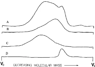
Several studies have dealt with the two-dimensional separation of gum Arabic macromolecular species. Fractions recovered from hydrophobic interaction chromatography are further separated by size exclusion chromatography [24], [25], [27]. Results from Randall et al. (1989) are displayed in Figure I-4. The less hydrophobic fraction (fraction 1 in Figure I-4 legend) was mainly composed of intermediate molecular mass species. Intermediate hydrophobicity fraction (fraction 2) was eluted first in SEC, and was thus composed of the higher molecular mass. Finally the most hydrophobic fraction (fraction 3A) was eluted last, indicating a composition of smaller molecular mass. However, all recovered fractions were distributed in molecular mass.
Figure I-4: Size exclusion chromatograms of gum arabic and its fractions (hydrophobic interaction chromatography): (A) whole gum arabic; (B) fraction 1; (C) fraction 2; (D) fraction 3A (from Randall et al., 1989)
[24]
Results from Osman et al. (1993b) also indicated that each fraction (of different hydrophobicity) was quite polydisperse in molecular mass. Indeed each fraction once again contained a broad range of molecular mass at different ratios. This result illustrated the hetero-poly-molecular nature of the gum.
Conclusions of Renard et al. (2006) were slightly different from those of Osman et al. (193b) and closer to those of R.C. Randall et al. (1989). In their study, the first fraction recovered from hydrophobic interaction separation was found to be rather monodisperse, contrasting with the other
23
fractions and especially the third fraction (higher hydrophobicity) spreading over the entire range of molecular mass as shown in Figure I-5.
Figure I-5: HPSEC chromatograms showing the elution profiles monitored by refractive index (RI) (C, g/mL) and light scattering (LS) (Mw, g/mol) for acacia gum and its molecular fractions collected after hydrophobic
interaction chromatography: RI (tick line), LS (thin line) (from Randall et al., 1989) [25]
The composition of gum issued from old acacia trees contains higher fractions of conjugates compared to that of the gum collected from younger trees. Such an evolution is also observed with aging gum after harvests. A process of gum Arabic maturation has been developed in order to increase the gum emulsifying activity and viscosity [28], [29]. The treatment is performed under strictly controlled temperature and humidity conditions and no chemical reagent is added. This maturation process was described by the authors as similar to the natural development of the gum.
Figure I-6: SEC chromatogram of control (FR-2876) and matured gum arabic. Light scattering detection at 90° (from Aoki et al., 2007a) [29]
24
Different matured gums were analyzed by size exclusion chromatography and it was shown that the maturation process was increasing the percentage of higher molecular weight molecules within the gum, from 2.5 x10⁶ g/mol for the native gum to 11.6 x10⁶ g/mol for the mature gum (Figure I-6). Such a process thus accelerates the biological process by associating glycoproteins with free arabinogalactan polysaccharides. Viscosity measurements indicated a highly branched structure. The authors observed no major changes in the gum species chemical structure after maturation [30].
All these studies on gum Arabic fractionation firstly revealed its heterogeneous composition in molecular mass. According to hydrophobic interaction chromatographic separation, the gum has been identified as a mixture of three fractions (arabinogalactan polysaccharides, arabinogalactan-protein conjugates and glycoarabinogalactan-proteins), which differ in their molecular mass and arabinogalactan-protein rate. Addition of Yariv reagent has shown that all species from the gum spectrum were present in the form of protein-polysaccharide conjugates. The hydrodynamic radius measured suggested that these macromolecules possess a hyper-branched structure. Finally, studies using two-dimensional separation of gum moieties have shown that the hydrophobicity of each fraction was more or less associated to a molecular mass, with the glycoprotein fraction exhibiting a broad range of molar masses.
I-2-1-3 Structural model of gum Arabic conjugates
Enzyme degradation experiments on gum Arabic samples were conducted by Connally et al. using a proteolytic enzyme, the pronase [31]. The pronase hydrolyses the protein, leading to the release of homogeneous polysaccharides chains of molecular mass similar to that of the arabinogalactan polysaccharides, making up the bulk of the gum. AGP complexes could be identified by a “wattle blossom” model, where carbohydrate blocks are covalently linked to a central polypeptide chain [24], [31]. It was proposed that each AGP molecule consists in average of five carbohydrate blocks of molecular weight around 2.105 g.mol-1, linked together by amino acid residues to form a very compact structure [24]. A schematic illustration is given in Figure I-7 and shows the conjugate resembles a block copolymer, which would explain its ability to stabilize emulsions by strong steric repulsion.
25
Figure I-7: Wattle blossom proposed model for the structure of arabinogalactan-protein conjugates (from Randall et al., 1988) [18]
An alternative to the “wattle blossom” model was proposed by Qi et al. [32]. Thanks to hydrogen fluoride deglycosylation of the high gum molecular weight fraction (recovered via size exclusion chromatography) there were able to recover a hydroxyproline-rich protein backbone (~400 amino acid residues). Further hydrolysis of the conjugates displayed that most of the carbohydrate groups were attached to the protein through hydroxyproline-galactose linkages. Further analysis of the conjugates structure (chromatographic analysis and TEM) led the authors to name it “twisted hairy rope” structure, schematized in Figure I-8. It consists of a rod-like molecule with small carbohydrate units attached at regular intervals to a polypeptide backbone.
Figure I-8: Hypothetical statistical model of the GAGP. A hypothetical repetitive block size of 7kD contains 10 amino acid residues: 1kD; 30 sugar residues: 4.44KD; 3 hyp-triarabinosides: 1.32kD. The
glucuronoarabinogalactan is probably a galactan backbone with glucuronic acid, rhamnose, and arabinose side branches (from Qi et al., 1991) [32]
This model was further amended by Goodrum et al. (2000) with a more symmetric structure as shown in Figure I-9, possibly the source of some properties of the gum. It was suggested that symmetry in peptide moieties may enhance molecular packing and self-association [33].
26
Figure I-9: Model of the GAGP consensus glycopetide according to L.J. Goodrum et al. This model depicts a symmetrical distribution of arabinosides and polysaccharide substituents which is direct by the palindrome-like arrangement of the Hyp residues in the peptide backbone; Ser-O is the palindromic center (from Goodrum et al.,
2000) [33]
An improved structure was proposed by Mahendran et al., illustrated in Figure I-10 [34]. Carbohydrate blocks are linked to the protein chain mainly by its serine amino acids and some linkages occurs with the hydroxyproline moieties. This model is consistent with the previous proposed models of wattle blossom (Connolly et al. 1987) and twisted hairy rope (Qi et al. 1991), with a modification in the molecular weight of the carbohydrate moieties attached along the polypeptide backbone. The authors obtained rather different polypeptide length through deglycosylation, with two chains of 45 and 250 amino acids. They also performed both enzymatic and reducing attacks of gum Arabic and claimed that polysaccharides blocks were much smaller than what was reported in enzymatic degradation studies. However, their conclusion does not actually seem to match their experimental data. Rather it seems that a soft reducing attack on arabinogalactan-protein, which mainly cuts O-serine bonds, gives both the polysaccharide blocks and the polypeptide, whereas enzymatic attacks cut the polypeptide. This scenario is consistent with the extensive enzymatic degradation study of Renard et al. [35] on their HIC fraction II, arabino-galactan protein conjugates. They show that this degradation yields similar sizes and structures are the population of arabinogalactan-peptides. The polypeptide chain is attacked with various degrees depending on the enzyme specificity, which can either enhance or degrade secondary structures observed using circular dichroism.
27
Figure I-10: Schematic illustration of the structure of the gum arabic arabinogalactan protein conjugate. The molecule has a molecular mass of (1-2).106 Da and consists of a polypeptide chain possibly containing ~250
amino acids with short arabinose side chains and much larger blocks of carbohydrate of molecular mass ~4.104Da and attached. The carbohydrate is highly branched and may adopt the thin oblate ellipsoid structure
proposed for the AG component. The amino acid sequence for the polypeptide chain has been determined by Goodrum et al. The molecule adopts a very compact conformation with Rg of ~36nm (from Mahendran et al.,
2008) [34]
I-2-1-4 Structural model of gum Arabic in solution
The structure of gum Arabic or gum Arabic fractions has been analyzed by two groups using Small Angle Neutron of X-ray Scattering techniques (resp. SANS and SAXS). Dror et al. have investigated the conformation of Arabic gum in solution at different concentration [36]. Above a concentration of 0.5 wt% a correlation peak was observed in the low-q range (large distance). This peak is characteristic of assemblies of scattering moieties with a given inter-aggregates correlation length. At the higher concentration studied (10-30 w/w%) the peak becomes broader as observed in Figure I-11. This result was interpreted as an overlapping of polymer aggregates, in agreement with rheology measurements in aqueous solutions of the gum. The same measurements were conducted with increasing concentrations of salt. It was shown that above a certain concentration (0.5 mol.L-1 NaCl) the correlation peak disappeared, indicating that electrostatic interactions determine the distance between the gum aggregates.
28
Figure I-11: SAXS spectra of gum arabic solutions at different concentrations (from Dror et al., 2006) [36]
Moreover, these scattering experiments revealed that the gum Arabic is possibly composed of many spheroidal structures intertwined with a small amount of large coils.
The conformation of the arabinogalactan fraction of gum Arabic (AG) in solution has been investigated by Sanchez et al. using SANS, transmission electron and atomic force microscopies [37]. The AG fraction was recovered in a preparative hydrophobic interaction chromatography column, with a sequence of two NaCl solutions at 4mol.l-1, 2 mol.l-1, then distilled water (as described above). The form factor of the AG fraction in 50 mM NaCl D2O solutions is reported in Figure I-12. At this salt
concentration, the polyelectrolyte charges are screened and the correlation peak is suppressed.
Figure I-12: Fraction 1 (AG) scattering form factor (1w/w%) at 25°C in D2O containing 50mM NaCl (from Sanchez et al., 2008) [37]
29
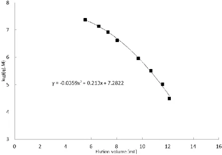
From the Guinier region (plateau in the low-q range) a radius of gyration of 6.4 nm was calculated. The pair distribution function of the gum fraction well compared with that of thin circular disks with a thickness of about 1.5 nm and a diameter around 20 nm. High-resolution microscopic measurements confirmed this structure. In the intermediate-q range, the scattering function decayed as q-2 interpreted by the authors as the signature of a disk-like particles population. Finally, in the high-q range the scattering intensity decayed as q-2.61, which was attributed to a dense and ramified internal structure.
SANS was also used to study the structure of the arabinogalactan-protein conjugate fraction (AGP) of the gum by Renard et al. [38]. The fraction was also recovered using hydrophobic interaction chromatography (HIC). Samples for scattering experiments were prepared in the same way as in the study of Sanchez et al. (2008). HPSEC-MALLS coupled on-line with viscometer showed the presence of two types of conformations with globular and elongated shape depending on the size of the carbohydrate branches.
Small angle scattering experiments revealed the presence of an elongated conformation corresponding to a triaxial ellipsoid shape (Figure I-13).
Figure I-13: Fraction 2 (AGP) scattering form factor (1w/w%) at 25°C in D2O containing 50mM NaCl (from Renard et al., 2012) [38]
In the low-q range the scattered function displayed a -1.1 power law, characteristic of a linear conformation. The -2.13 exponent in the intermediate-q range was attributed to a two-dimensional structure (disk or fractal). In the high-q the -3.85 slope which was identified as characteristic of a fractal structure of dimension 2.15.
30
The pair distance distribution function of this fraction revealed the presence of two maxima corresponding to half-distances of 10nm and 32nm, suggesting a two-dimensional conformation or the presence of a bi-dispersed population of particles.
Although TEM measurements of AGP moieties are quite sensitive due to their surface properties, Renard et al. have managed to perform AGP imaging using this technique. They noted different conformations according to their size, smaller species being spherical and larger ones more elongated [39].
The last HIC eluted fraction, the glycoproteins, was investigated using SAXS analysis by the same research team [40]. Samples for scattering experiments were prepared in an acetate buffer at pH=5 and 100mM of NaCl. SAXS measurements together with circular dichroism and TEM analyses led the authors to describe the shape of the glycoproteins fraction as an assembly of ring-like modules
Small angle scattering spectra of solutions of gum Arabic and its HIC recovered fractions displayed a correlation peak that was dependent on the gum concentration and disappeared when adding salt. This observation shows that electrostatic repulsions drive the structuration of gum moieties in solution. Moreover, studies focused on the characterization of gum fraction displayed structural differences between each fraction. The high molecular mass conjugate fraction (described as being responsible for the emulsifying and stabilizing properties of the gum) exhibits spheroidal and elongated shapes.
I-2-2 Gum arabic properties in solution
One practical advantage of gum Arabic is its high solubility in water, usually up to 50 w/w%. [31].
I-2-2-1 Rheology of gum Arabic solutions
Although gum Arabic is composed of high molecular weight molecules, the thickening property of gum Arabic remains limited compared to other common hydrocolloids. Indeed a 50 w/w% gum Arabic solution exhibits an apparent viscosity comparable to that of a 1.5 w/w% Xanthane gum solution [7]. Gum arabic solutions behaves as a shear-thinning fluid with a Newtonian plateau at large shear rates (>100s-1). In this regime, the apparent viscosity is an exponential growing function of the gum Arabic concentration, with a growth rate which is strongly dependent on the nature of the gum [13], [41]. Moreover an important increase in temperature will lead to an irreversible decrease of a gum solution viscosity due to denaturations of the protein-rich moieties [42].
More than shear-thinning gum arabic solutions are thixotropic, their response to deformation is time dependent. Sanchez et al. have observed an increase of the apparent viscosity as a function of
31
shearing time, especially significant as the shear rates is low [43]. The resting time of the solution prior to stress-strain measurements was also shown to increase apparent viscosity. This behavior was proved by the authors to result from a contribution of the solution sample surface rheology to the bulk stress response. This contribution arises from the adsorption process of proteinaceous species of the gum, building up with time a viscoelastic network at the sample-air interface. Replacing the water-air surface by an oil-water interface or working with a larger surface-to-volume ratio increases the time dependence of the rheological behavior, hence validating the proposed mechanism. In other rheological measurements performed by Li et al. (2009 and 2011), a PDMS film was deposited on the sample surface, to avoid evaporation [44], [45]. Although not mentioned by the authors, it is likely that this PDMS film also affected the adsorption rate of the proteinaceous species. In the first paper, the authors postulate the existence of an association-dissociation equilibrium of AG molecules which depends on the imposed shear rate of the resting time of the solution. The longer the resting time, the more compact are the AG aggregates, the faster is the deformation rate, the looser are the aggregates, tending towards a Newtonian behavior at large shear rates. This structuration mechanism is schematized in Figure I-14. Therefore, the history of the gum solution has a direct impact on its mesoscopic structure and its rheological behavior.
Figure I-14: State of gum arabic molecules with and without shear (from Li et al., 2009) [44]
In the second paper, the thixotropic property of the gum solution is investigated, submitting a 6 w/w% gum solution sample to a series of shear jumps inducing a sequence of stress jump responses [45]. The existence of an elastic contribution in the limit of low shear rate (<10s-1) is demonstrated due to the self-association mechanism of hydrocolloids, as well as a viscous contribution which takes over at larger shear rates, once the aggregates are dissociated. The stress relaxation curve after a sudden decrease of the shear rate clearly suggests a rapid development of an elastic network
32
resulting from molecular association within the gum. In fact, same experiments have been carried out removing the AGP moieties from the gum with single-walled nanotubes of carbon [46]. Without AGPs, the gum solution became fully Newtonian leading to the conclusion that this molecular association mechanism is driven by AGP and not to AG moieties.
I-2-2-2 Electrophoretic mobility
For the gum arabic in solution a value of approximately -1.5 µmcm/VS for the electrophoretic mobility was found above pH 4.5 and the value decreased close to zero as the pH was decreased to 2 (Figure I-15). This electrophoretic behavior was assigned to the carboxylic groups present on the polysaccharides glucuronic acid moities [47]. Moreover the pKa of glucuronic acid moieties is of 3.2 which is accordance with the measured electrophoretic mobility of gum Arabic as a function of pH (approximately 50% of the maximum electrophoretic mobility at pH 3) [48].
Figure I-15: Electrophoretic mobility of gum arabic and gum arabic strabilized emulsions as a function of pH (from Padala et al., 2009) [47]
I-2-3 Interfacial properties of gum Arabic
I-2-3-1 Emulsification and emulsion stability
It is now well admitted that the proteinaceous materials of the gum are responsible for its emulsifying properties. The treatment of the gum with a protease indeed inhibits its emulsifying activity [18].
Comparison of size exclusion chromatogram for gum Arabic solutions before and after emulsification has shown that a significant proportion of the high molecular mass species adsorbed at the interface as illustrated in Figure I-16 [18], [49]. It was also suggested that the proteinaceous moieties adsorbed at the interface and that carbohydrate groups extended into water to provide steric repulsion [24].
33
Figure I-16: Size exclusion chromatographic profiles of gum arabic in solution before (B) and after (A) emulsification as monitored by UV absorbance at 218nm (from Randall et al., 1988) [18]
Chikamai et al. demonstrated that heating a solution of gum Arabic at 100°C for 6h importantly destroyed the emulsifying properties of the gum whereas a temperature of 65°C did not really impact this property. [42]
Preparative size exclusion or hydrophobic interaction chromatography were used by Ray and coworkers to recover fractions of gum Arabic with different molecular masses or hydrophobicities [26]. It was found that the higher molecular mass fraction possessed the higher protein rate. The SEC recovered fractions were then used to stabilize oil-in-water emulsions at constant nitrogen concentration. Emulsions stabilized with fractions possessing the higher molecular weight and higher protein rate appeared to be the more stable. Also, it was observed that emulsion stability was increased when using a mixture of low and high molecular weight fractions. Finally, authors observed no differences in the zeta potential of the different emulsions and concluded that differences in stability were not originating from electrostatic effects.
Emulsifying properties of acacia gum were investigated by Buffo et al.. Using viscosity measurements on the oil-in-water emulsions they developed a model to estimate the thickness of the adsorbed species layer at the interface [50]. Layer thicknesses ranging from 190nm to 430nm were estimated according to the different gums studied (Senegal and Seyal). Gum processing such as pasteurization and demineralization were shown to enhance emulsion stability, due to an increase of molecular mobility and unfolding of the protein moieties at the interface.
Another emulsification stability study was carried out by H. Aoki and coworkers in 2007 [51]. Stabilities of oil in water emulsions stabilized by acacia senegal gum and mature gum Arabic were compared. According to accelerated stress conditions it was observed that emulsions stabilized with less than 20% of gum Arabic were not stable whereas when using a matured gum the stability was improved even with a concentration of 5%. Moreover when using a same concentration of native
34
gum or matured gum to stabilized an oil in water emulsion the authors observed that a matured gum provided emulsions with a finer droplet size. The same trend was observed in 2010 by O. Castellani et al. [52].
It was shown by Galazka and co-workers that protein unfolding is promoted under pressure [53]. Gum Arabic conjugates possess a small hydrodynamic radius with respect to their molecular mass suggesting a random coil structure unlike pectin for instance that is a semi-flexible hydrocolloid. Thus a high pressure could help the conjugates moieties to unfold and cover a greater surface at oil droplet interfaces (Figure I-17) [51].
Figure I-17: Proposed model by Aoki et al. of gum Arabic adsorption onto the oil droplet (from Aoki et al., 2007b) [51]
Depletion flocculation in gum Arabic stabilized oil-in-water emulsion may occur. Indeed, the non adsorbed species present in the continuous phase of the emulsion can act as depletants. Flocculation is harmful for emulsions stabilities, it causes an increase in the creaming rate due to the increase in particles sizes and it may enhance coalescence events because droplets are brought into contact. Above a critical concentration non adsorbed polymeric molecules can increase the attraction between emulsion droplets because of an exclusion effect of those polymers from the region surrounding the droplets due to an osmotic effect [54]. In 2001 R. Chanamoi et al. studied the depletion flocculation of diluted emulsions by gum Arabic [55]. They varied the concentration of gum Arabic added to a tween 20 stabilized emulsion in order to identify the critical flocculation that induce droplets flocculation for a given emulsion droplets size distribution. The authors identified
35
that reducing the droplets size distribution increased the critical flocculation concentration. However no study was performed on the critical flocculation concentration for gum Arabic stabilized emulsion.
I-2-3-2 Adsorption isotherms and recovery of amphiphilic species
Randall et al. have determined the amount of gum adsorbed at oil-water interfaces by recovering the aqueous phase of a centrifuged orange oil emulsion, and taking the difference between the weight of gum in solution before and after emulsification [18]. A surface coverage of about 4 mg/m2 was found for an emulsion stabilized with a 15 w/w% gum Arabic solution.
Yadav et al. have proposed a method for the purification of gum Arabic fractions adsorbing at oil droplets surface [12]. They used a tissue grinder to form gum Arabic stabilized hexadecane-in-water emulsions, let them cream, then broke these emulsions with an isopropanol solution to form a precipitate. After rinsing steps in various solvents, the precipitate was dried, dissolved in water, dialyzed against water and freeze-dried. In this study the emulsifying activity of different gums or gum sub-fractions was assessed from turbidity measurements. A correlation between the emulsifying activity of a gum and its lipid content was shown, although the amount of protein within each gum sample was not accounted for by the authors. The composition of the recovered gum sub-fractions displayed an important increase of the nitrogen content compared to that of the native gum, supporting the key role of proteins in the adsorption process. However, the emulsifying activity of the gum fraction recovered from the interface was not as important as expected. This result could be a consequence of a denaturation of the gum proteinaceous macromolecules during the separation process. The hydrophobic interaction chromatography analysis of this fraction indicated an increase in hydrophobicity compared to that of the native gum.
Another method to recover adsorbed species from gum Arabic stabilized emulsions was developed by M. Nakauma et al., using SDS surfactant to displace the proteinaceous material from oil-water interface [56]. Emulsions of medium chain triglycerides in gum Arabic solution were first centrifuged and the remaining emulsion was further mixed with a solution containing SDS and NaCl. The aqueous phase containing the displaced material was recovered and analyzed.
The surface concentration (in mg/m2) was deduced from the mass of adsorbed species and from the measurement of interfacial area (using a Static Light Scattering granulometer). Adsorption was found to be around 6mg/m2 and nearly independent of GA concentration (Figure I-18A). In return,
36
I-18B), although some inconsistency can be pointed out in these results (the value at the origin in Figure I-18B does not fit the value at a GA concentration of 10w/w% in Figure I-18A). Emulsions formed in the presence of salt were found to be more stable than emulsions prepared without. The increase of thus seems to impact the metastability of gum Arabic stabilized emulsion.
However, it is important to recall that SDS denaturates proteinaceous materials by altering their tertiary structure [57]. Therefore, the method proposed by Nakauma et al. is limited to the determination of the amount of adsorbed species but is not suited to study their structural composition.
Another method to recover species that adsorbed onto the surface of oil droplets as a function of hydrocolloids concentration was used in 2008 by M. Nakauma and coworkers [56]. Emulsions of medium chain triglyceride in water were prepared. For gum Arabic stabilized emulsion the concentration range was from 1% to 10%. In order to recover the species adsorbed the authors mixed the emulsions with a solution containing sugar and NaCl. The mixture was then centrifuged for to recover the lower aqueous phase and this step was repeated three times. The remaining emulsion was further mixed with a solution containing SDS and NaCl. The SDS surfactant was used to displace the proteinaceous material from the interface. The lower aqueous phase containing the displace material was recovered. Recovered fractions containing the species that adsorbed at the interface were then analysed. Again the use of SDS precludes any relevant structural analysis due to denaturation [56].
Figure I-18: Evolution of surface concentration in gum Arabic stabilized emulsions (triangle symbols) as a function of (A) GA concentration in solution, (B) Salt concentration for a concentration for a GA concentration of 10 w/w %). The pH of the aqueous phase was fixed at 3 prior to the addition to the oil (from Nakauma et al.,
2008) [56]
37
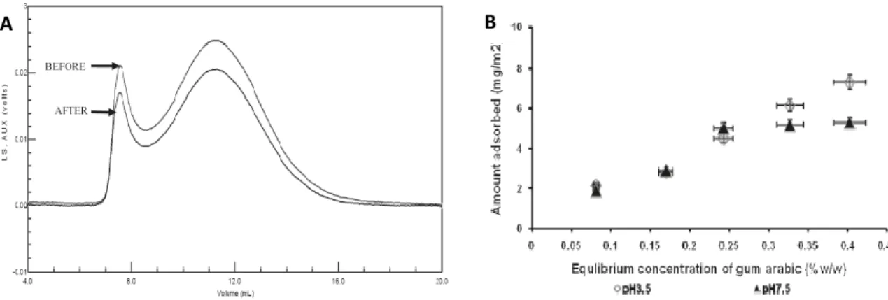
Padala et al. (2009) have determined the mass of gum adsorbed at oil/water interfaces using size exclusion chromatography with refractive index detection. Chromatogram areas were compared between gum Arabic solutions prior to and after emulsification (Figure I-19A) and adsorption isotherms could be determined at different gum Arabic concentrations and two pH (Figure I-19B).
Figure I-19:(A) SEC elution curves of gum arabic at pH 7.5 monitored by RI and the supernatant recovered after preparing an emulsion. (B) Isotherms for gum Arabic adsorbing onto limonene oil droplets from solution at pH
3.5 and 7.5 (from Padala et al., 2009) [47]
One important finding from their work was that adsorbed species at the interface were not limited to the high molecular mass species as it had been described before [18], [49] but instead covered a wide range of molecular weight. Results showed a dependence of the mass adsorbed at the interface upon aqueous phase pH before emulsification. This depedence was ascribed to variations of the degree of protonation of uronic acid carboxylic groups. Indeed as seen before the isoelectric point of the gum is around pH 2. Hence, decreasing the pH increases the protonation of carboxylic groups, resulting in a less charged polymer. This results in a diminution of electrostatic repulsion at the interface enabling more species to adsorb. Considering the amount of adsorbed species at the interface Padala et al. argued that this high value (compared to other systems) was resulting from a conformational change of the macromolecules at the interface or from the formation of multilayers. Such a mechanism is supported by the tendency of gum arabic species to self-aggregate in solution [43], [44].
I-2-3-3 Rheology of gum Arabic stabilized interfaces
First thermodynamic quantity impacted by adsorption process is interfacial tension. According to Gibbs’ relation, excess surface pressure is a growing function of surface concentration. Investigation of a possible correlation between interfacial tension and the nitrogen content (or protein content) of different acacia gums was carried out by Dickinson et al. [58]. Their results showed that two gums
38
with contrasted protein contents (1.86% and 7.5%) exhibited similar interfacial tension values after 15 hours of measurements. Moreover adsorption kinetics at short time did not indicate a correlation between interfacial tension and the protein content of each gum sample. Moreover no direct links were observed between the emulsifying capacity of a gum sample and its nitrogen content. Emulsion samples that displayed a lower size distribution and a better stability were the one with the higher and the lower nitrogen content. These observations are in contradictions with the study of Ray et al. Indeed here it is stated that there is not direct correleation between the protein rate and the emulsifying properties while in their study Ray et al. observed that they obtain the best emulsion metastability with the fractions having the higher protein rate [26].
Authors argued that regarding the effect of nitrogen rate on interfacial tension, two factors must be considered: the molecular weight of the molecules diffusing to the interface (the low molecular weight molecules diffusing more rapidly than the high molecular weight species) and the accessibility of the polypeptide moieties within the gum. Emulsion stabilized with a gum having the lower nitrogen content was quite fine and stable. The lowering of the interfacial tension with this sample was relatively quick due to the presence of low molecular weight molecules rapidly diffusing and adsorbing to the interface.
Interfacial tension seems to be a useful tool to probe the efficiency of a gum sample to diffuse towards an interface and to lower its energetic cost. Yet this measurement needs to be coupled with other analysis such as emulsifying activity (emulsion metastability) and chromatographic separation in order to fully probe the interfacial properties of a given gum Arabic.
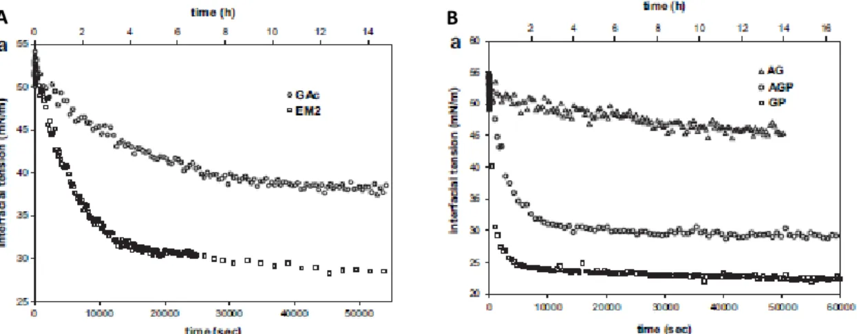
Interfacial rheology measurements on interfaces formed with gum Arabic and other hydrocolloids were performed using the pendant drop method [20]. It was shown that that matured gum Arabic (heated gum Arabic with a higher amount of high molecular weight conjugate) had a higher surface activity compared to native gum as illustrated Figure I-20A. The lowering of the interfacial tension was slow compared to other system, indicating that gum Arabic needs a certain time to diffuse towards the interface, adsorb and rearrange itself [59]. The elastic behavior of the adsorbed film was also increased in the case of matured gum.
39
Figure I-20: Linear plot of interfacial tension kinetics of (A)control acacia gum (GAc) and matured acacia gum (EM2) (B)matured gum components (GA, AGP et GP) at the n-hexadecane-water interface (pH 4.5 and 20°C,
0.5/L) (from Castellani et al., 2010a) [20]
A pH sensitivity of the interfacial tension between n-hexadecane and an aqueous solution of gum Arabic was also observed. Interfacial tensions decreased when reducing the gum solution pH, while the dilatational modulus was increased. It was argued that neutralization of carboxylic moieties from the gum at lower pH favors an increase of the amount of adsorbed gum and thus a larger elasticity.
In the same study the authors fractionated a matured gum using hydrophobic interaction chromatography. They recovered three fractions corresponding to the gum conjugates, glycoproteins and polysaccharides. Pendant drop tensiometry analyses were performed with these fractions (Figure I-20B). As expected regarding the protein content of each fraction, glycoproteins were the most efficient to lower the interfacial tension, polysaccharides providing almost no interfacial activity.
Interfacial rheology addresses shear and dilatational viscosity measurements, which respectively consist in a shearing deformation of surface element at constant area and in a surface expansion or compression at constant shape.
Adsorbed gum Arabic films exhibit high interfacial shear viscosity as shown by Dickinson et al. [60]. A method to exchange the sub-phase liquid during interfacial shear measurement was developed by Dickinson et al. It was used to see whether the non-adsorbed species from the gum present in the aqueous phase of an emulsion provided a positive or negative impact on its stability. It was observed that if the aqueous phase was diluted almost no change in surface viscosity occurred on a long time scale. These results indicated that some species from gum Arabic strongly adsorbed at an oil/water interface.
40
Interfacial rheological properties of adsorbed gum Arabic and modified starch at water/carvene interfaces were studied by Erni and co-workers [61]. It was demonstrated that acacia gum exhibits a significant shear elasticity of the interface.
Figure I-21: Time dependence of the interfacial shear moduli G' (storage) and G'' (loss) of acacia senegal gum and hydrophobically modified starch at the oil/water interface (T=20°C, deformation amplitude γ0 =1w%). The
storage modulus G' for modified starch was below the detection limit (from Erni et al., 2007) [61]
The authors observed that acacia gum layers were elastic over the whole range of time studied with the storage modulus above the loss modulus (Figure I-21). Moreover, measurements of the shear moduli frequency dependence revealed that gum Arabic layers remained elastic at all frequencies measured with a weak dependence on the oscillation frequency. Authors suggested an analogy with either “soft glassy” materials or gel networks.
41
Figure I-22: Deformation dependence of the interfacial shear moduli G' and G44 of acacia Senegal gum and hydrophobic ally modified starch at the oil/water interface (T=20°C, ω=1 rad.s-1, interface age t=20h) (from Erni
et al., 2007) [61]
As seen in Figure I-22 deformation dependence of interfacial moduli for gum Arabic layers showed a strong decrease of the elastic modulus above 1-2% deformations. This behavior suggests that the adsorbed film is a fragile network with a structural breakdown above a certain deformation rate. The authors thus concluded that the mechanical stability of the interfacial film must play a significant role in the stabilizing properties of the gum [61].
Spreading capacity (capacity to increase surface pressure when decreasing interfacial area) at air/water interface of different hydrocolloids using langmuir compression isotherms was studied by Castellani et al [62]. Matured gum had better spreading capacity and an acidification of the aqueous phase improved the spreading of native gum Arabic. More over the authors used AFM to measure the thickness of adsorbed film layers at air/water interface. According to their findings a decrease in the pH increased the thickness of the film layers (150nm at pH 3.1 over 30nm at pH 4.5). The AFM imagining of the adsorbed films at pH 3.1 revealed the presence of aggregates with localized zones of higher thickness. To complete their study, the authors performed ellipsometry measurements on adsorbed gum Arabic films at the air/water interface. Differences in ellipsometry angle for adsorbed film at pH 4.5 and pH 3.5 correlated with the thickness measured with AFM.
Interfacial tension and rheology studies that were carried out on gum Arabic samples revealed a non straightforward relationship between protein content and adsorption behavior, which contradicts other results showing the gum fraction having the higher protein rate providing the best emulsion metastabilities. The adsorption behavior of gum Arabic at oil/water interfaces appeared slow compared to other system and revealed a characteristic behavior of this complex system where molecules need time to adsorb and rearrange at the interface. Moreover, gum Arabic adsorbed films present an elastic behavior that might originate from the formation of a fragile network at the interface. Finally, these studies showed a dependence between gum adsorption and a decrease in pH of gum solutions, with increased interfacial properties (lower interfacial tension and higher elasticity).
I-2-4 Conclusion and thesis objectives
Arabic gum has been extensively studied over the past 40 years. This literature review shows the extremely heterogeneous structure and composition of the gum, as a complex mixture of polysaccharides and protein moieties. Two types of chromatographic separations (size exclusion and
42
hydrophobic interaction) coupled with different types of detection were used on different gum Arabic samples in order to improve the understanding of the molecular composition of the gum. A description of the gum containing three fractions was proposed, consisting of a bulk fraction composed of polysaccharides moieties, a fraction of protein-polysaccharides conjugates and a smaller fraction of glycoproteins [7], [15], [21], [24], [25], [27], [63]. For each fraction (separated through preparative hydrophobic interaction chromatography), their hydrodynamic radius, molecular masses, polydispersity, viscosity, emulsification behavior, interfacial tension, structure in solution were thoroughly investigated.
The role of the proteinaceous moieties of the gum on its interfacial properties was evidenced and it was shown that the high molecular masses species appeared to preferentially adsorb at the interface proving steric repulsion [18], [24], [64]. Although gum Arabic is composed of polyelectrolytes, ionic repulsions were not described as the main mechanism for emulsion droplets stabilization [26]. Gum Arabic amphiphilic species were shown to strongly adsorb at the interface and a dilution of a gum Arabic stabilized emulsion does not seem to affect its metastability [60]. One direct consequence is that the non-adsorbed species of gum Arabic do not influence the emulsion stabilization mechanisms. Interfacial rheology measurements with gum Arabic or its fractions displayed an elastic behavior of the adsorbed film when submitted to perturbations [60], [61]. This macroscopic observation reflects a given structuration of the interface which needs deeper insights. The effect of physico-chemical parameters such as pH or salinity of gum Arabic solutions on the metastability of the emulsion, or the adsorption behavior have been demonstrated (interfacial tension measurements, isotherms of adsorption) [47], [56], [62], [65].
Despite this impressive amount of work, many questions remain largely open as recently reviewed by Sanchez and coworkers [13]. This notably includes two points treated in this thesis:
- A unified description of structures displayed by the various species composing gum Arabic, both in solution and at interfaces
- A comprehensive understanding of the impact of formulation on these structures and thus on emulsifying properties
43
References
[1] D. Saha and S. Bhattacharya, “Hydrocolloids as thickening and gelling agents in food: a critical review,” J. Food Sci. Technol., vol. 47, no. 6, pp. 587–597, Dec. 2010.
[2] E. Dickinson, “Hydrocolloids at interfaces and the influence on the properties of dispersed systems,” Food Hydrocoll., vol. 17, no. 1, pp. 25–39, Jan. 2003.
[3] A. Nussinovitch, “Exudate gums,” in Hydrocolloid Applications, Springer, Boston, MA, 1997, pp. 125–139.
[4] G. O. Phillips and P. A. Williams, Handbook of Hydrocolloids. Elsevier, 2009.
[5] C. Schmitt and S. L. Turgeon, “Protein/polysaccharide complexes and coacervates in food systems,” Adv. Colloid Interface Sci., vol. 167, no. 1, pp. 63–70, Sep. 2011.
[6] O. H. M. Idris, P. A. Williams, and G. O. Phillips, “Characterisation of gum from Acacia senegal trees of different age and location using multidetection gel permeation chromatography,” Food Hydrocoll., vol. 12, no. 4, pp. 379–388, Oct. 1998.
[7] A. M. Islam, G. O. Phillips, A. Sljivo, M. J. Snowden, and P. A. Williams, “A review of recent developments on the regulatory, structural and functional aspects of gum arabic,” Food Hydrocoll., vol. 11, no. 4, pp. 493–505, Oct. 1997.
[8] R. M. A. Daoub, A. H. Elmubarak, M. Misran, E. A. Hassan, and M. E. Osman, “Characterization and functional properties of some natural Acacia gums,” J. Saudi Soc. Agric. Sci., May 2016.
[9] A. M. Zipkin, M. Wagner, K. McGrath, A. S. Brooks, and P. W. Lucas, “An Experimental Study of Hafting Adhesives and the Implications for Compound Tool Technology,” PLOS ONE, vol. 9, no. 11, p. e112560, Nov. 2014.
[10] A. Chevalier, “Sur la production de la Gomme arabique en Afrique occidentale française.,” J. Agric. Tradit. Bot. Appliquée, vol. 4, no. 32, pp. 256–263, 1924.
[11] R. Bandyopadhyaya, E. Nativ-Roth, O. Regev, and R. Yerushalmi-Rozen, “Stabilization of Individual Carbon Nanotubes in Aqueous Solutions,” Nano Lett., vol. 2, no. 1, pp. 25–28, Jan. 2002.
[12] M. P. Yadav, J. Manuel Igartuburu, Y. Yan, and E. A. Nothnagel, “Chemical investigation of the structural basis of the emulsifying activity of gum arabic,” Food Hydrocoll., vol. 21, no. 2, pp. 297–308, Mar. 2007.
[13] C. Sanchez et al., “Acacia gum: History of the future,” Food Hydrocoll., Apr. 2017. [14] S. C. Churms, E. H. Merrifield, and A. M. Stephen, “Some new aspects of the molecular
structure of Acacia senegal gum (gum arabic),” Carbohydr. Res., vol. 123, no. 2, pp. 267–279, Nov. 1983.
[15] P. A. Williams, G. O. Phillips, and A. M. Stephen, “Spectroscopic and molecular comparisons of three fractions from Acacia senegal gum,” Food Hydrocoll., vol. 4, no. 4, pp. 305–311, Dec. 1990.
[16] B. A. Lewis and F. Smith, “The heterogeneity of polysaccharides as revealed by electrophoresis on glass-fiber paper,” J. Am. Chem. Soc., vol. 79, pp. 3929–3931, 1957. [17] M. Jermny, “Chromatography of acidic polysaccharides on Deae-Cellulose,” Aust. J. Biol.
Sci., vol. 15, no. 4, pp. 787–792, 1962.
[18] R. C. Randall, G. O. Phillips, and P. A. Williams, “The role of the proteinaceous component on the emulsifying properties of gum arabic,” Food Hydrocoll., vol. 2, no. 2, pp. 131–140, Jun. 1988.
[19] M.-C. Vandevelde and J.-C. Fenyo, “Macromolecular distribution of Acacia senegal gum (gum arabic) by size-exclusion chromatography,” Carbohydr. Polym., vol. 5, no. 4, pp. 251–273, 1985.
![Table I-2: Sugar and protein compositions of A.Senegal and isolated fractions (from Randall et al., 1989) [24]](https://thumb-eu.123doks.com/thumbv2/123doknet/2991709.83086/21.918.137.760.161.296/table-sugar-protein-compositions-senegal-isolated-fractions-randall.webp)
![Figure I-12: Fraction 1 (AG) scattering form factor (1w/w%) at 25°C in D2O containing 50mM NaCl (from Sanchez et al., 2008) [37]](https://thumb-eu.123doks.com/thumbv2/123doknet/2991709.83086/29.918.260.651.729.1035/figure-fraction-scattering-form-factor-containing-nacl-sanchez.webp)
![Figure I-14: State of gum arabic molecules with and without shear (from Li et al., 2009) [44]](https://thumb-eu.123doks.com/thumbv2/123doknet/2991709.83086/32.918.279.642.551.850/figure-i-state-gum-arabic-molecules-shear-li.webp)
![Figure I-16: Size exclusion chromatographic profiles of gum arabic in solution before (B) and after (A) emulsification as monitored by UV absorbance at 218nm (from Randall et al., 1988) [18]](https://thumb-eu.123doks.com/thumbv2/123doknet/2991709.83086/34.918.200.755.114.279/exclusion-chromatographic-profiles-solution-emulsification-monitored-absorbance-randall.webp)