To link to this article: DOI:
10.3109/03639045.2015.1103746
http://dx.doi.org/10.3109/03639045.2015.1103746
This is an author-deposited version published in: http://oatao.univ-toulouse.fr/
Eprints ID: 16535
To cite this version:
Pereira Camelo, Sarah Regina and Franceschi-Messant, Sophie and Perez,
Emile and Girod Fullana, Sophie and Ré, Maria Inês Factors influencing
the erosion rate and the drug release kinetics from organogels designed as
matrices for oral controlled release of a hydrophobic drug. (2016) Drug
Development and Industrial Pharmacy, vol. 42 (n°6). pp. 985-997. ISSN
0363-9045
Open Archive Toulouse Archive Ouverte (OATAO)
OATAO is an open access repository that collects the work of Toulouse researchers and
makes it freely available over the web where possible.
Any correspondence concerning this service should be sent to the repository
administrator:
staff-oatao@listes-diff.inp-toulouse.fr
RESEARCH ARTICLE
Factors influencing the erosion rate and the drug release kinetics from
organogels designed as matrices for oral controlled release of a
hydrophobie drug
Sarah Regina Pereira Camelo1, Sophie Franceschi2, Emile Perez2, Sophie Girod Fullana3, and Maria lnês Ré1 1Mines Albi, CNRS, Centre RAPSODEE, Campus Jarlard, Université De Toulouse, Albi CT Cedex, France, 2Laboratoire Des l.M.R.C.P., Université Paul Sabatier, Toulouse, France, and 3Faculty of Sciences Pharmaceutiques, CIRIMAT INPT-CNRS-UPS, Université Toulouse, Toulouse, France
Abstract
This article proposes solid-like systems from sunflower oil structured with a fibrillar network built by the assembly of 12-hydroxystearic acid (12-HSA), a gelator molecule for an oil phase. The resulting organogels were studied as oral controlled release formulations for a lipophilic drug, Efavirenz (EFV), dissolved in the oil. The effects of the gelator concentration on the thermal properties of the organogels were studied by Differential Scanning Calorimetry (DSC) and showed that drug incorporation did not change the sol-gel-sol transitions. The erosion and drug release kinetics from organogels under conventional (filling gelatin capsules) or multiparticulate (beads obtained by prilling) dosage forms were measured in simulated gastric and intestinal fluids. EFV release profiles were analyzed using model-dependent (curve-fitting) and independent approaches (Dissolution Efficiency DE). Korsmeyer-Peppas was the best fitting release kinetic model based on the goodness of fit, revealing a release mechanism from organogels loaded with EFV different from the simple drug diffusion release mechanism obtained from oily formulations. From organogels, EFV probably diffuses through an outer gel layer that erodes releasing oil droplets containing dissolved EFV into the aqueous medium .
Keywords
Efavirenz, lipid formulation, multiparticulate dosage form, oral drug delivery, organogel, poorly soluble drugs, prilling process, release profile models
Introduction
Interest in lipid-based drug delivery has developed over the past 15 years, largely driven by the growing need for novel drug delivery systems to deal with the vast majority of the new chemical entities (NCE) that have poor solubility, or permeability or to improve the delivery of existing drugs 1•
The diversity of commercialized lipid excipients (e.g. fatty acids, glycerides, polyoxyethylene glycols derivatives, ethoxy-
lated glycerides and polyalcohol fatty acid esters) has created a large platform for lipid-based formulation design. Depending on the choice of lipid excipients and formulation techniques, it is possible to obtain a variety of systems in the forms of emulsions, microemulsion s, micelles, liquid crystalline particles, solid lipid particles and various self-emulsifying systems.
Successfully marketed oral lipid-based drug products, cur- rently conceming S4% of the global pharmaceutical market, are mostly formulated as bulk liquid solutions or liquid-filled gelatin capsules2• Clinical translation of novel lipid-based formulations is clearly related to the rate of discovery of pipeline drugs exhibiting poor aqueous solubility, which is predicted to increase from 30%
Address for correspondence: Maria Inês Ré, PhD, Mines Albi, CNRS, Centre RAPSODEE, Campus Jarlard, Université De Toulouse, 81013 Albi CT Cedex 09, France. Tel: +33 5 6349 3299. Fax: +33 5 6349 3025.
E-mail: maria-in es.re@mines-albi.fr
to 70%3. Major barriers for attaining full commercial potential for lipid-based formulations are associated with challenges in their physical stability, non-ideal drug encapsulation and unpredictable delivery performance.
The primary mechanism of action by which a lipid formulation leads to improved bioavailability is usually avoidance of the slow dissolution process that limits the bioavailability of hydrophobie drugs from solid dosage forms. A formulation keeping the drug in a dissolved state throughout its transit in the gastrointestinal tract (Gl tract) appears preferable. In this context, a gel vehicle and especially an organogel could be useful to improve the bioavail- ability of a hydrophobie drug. Ingeneral, a gel may be defined as a semi-solid formulation having an external solvent phase, apolar (organogel) or polar (hydrogel) immobilized by a 3D-network scaffold. Organogels may be regarded as bi-continuous systems consisting of a gelator and an apolar solvent. The gelators may undergo physical or chemical interactions so as to form self- assembled entangled fibrous structures leading to the formation of a three-dimensional scaffold. The 3D-networked structure, hence formed, entraps and immobilizes the apolar phase.
In the last 20 years, there has been an explosive growth in research on organogels, and on publications related on them4- 11 as another category of lipid system. Most of these publications report the discovery and/or the synthesis of new organogelators, the properties of organogels including the gel microstructures and the manner in which the gelator molecules could be arranged
in 3-D network structures. The large diversity in terms of the possible microscopie structures available for organogels origi- nated the interest for applications of these soft materials in the petrochemical industry for gelling flammable solvents for storage and transport 12, in the food industry for structuring edible oils13, in the cosmetic field14 and more recently as part of pharmaceut- ical drug delivery systems. As far as drug delivery is concerned, organogels appear as suitable carriers for lipophilic drugs. Consequently, in recent years, the possibility of using organogels as drug delivery vehicles has been explored for parenteral 9 and transdermal delivery8·10·". Nevertheless, so far, only few studies have been carried out on oral drug delivery 15-17.
Starting from these considerations, this study proposes the application of organogels to integrate attractive and nove! lipid- based delivery systems for controlled release of a hydrophobie drug, Efavirenz (EFV), via oral administration. EFV is a non- nucleoside reverse transcriptase inhibitor, used for the treatment of the human immunodeficiency virus (HIV). EFV is a molecule of class IIof the biopharmaceutical class system18 that presents low aqueous solubility (4 µ g/ml)19 and high permeability. Efavirenz (EFV) is a first-line anti-HIV drug largely used as a non-nucleoside reverse transcriptase inhibitor as part of antiretro- viral therapies. EFV as active pharmaceutical ingredient (APT) is commercially available as micronized powder. Sorne papers20-22 have been published focusing on EFV innovative solid forms, but there is no clear evidence of solubility or dissolution improve- ment. Other studies23-27 have led to an amorphous system that was inherently unstable, could recrystallize and had low physical stability. Organogels for oral administration of EFV could represent an innovative drug delivery system, particularly under multiparticul ate forms.
Among the different physical methods available to obtain multiparticulate forms, such as spray drying, fluid bed coating or extrusion, prilling is able to produce microparticles or beads (prills) with very narrow dimensional range and high encapsula- tion efficiency. These beads are produced by breaking apart a laminar jet of molten organogel into a row of monosized drops by means of a vibrating nozzle device. The aim of this study was also to show that prilling technique could be used as a simple method to prepare organogel beads with narrow particle size distribution loaded with Efavirenz, as multiparticulate dosage forms. Multiparticulate dosage forms are known to overcome the poor and variable GI tract absorption of drugs and have shown the ability to reduce or eliminate the influence of food on bioavailability 28.
ln view of the above, the control release of EFV from sunflower oil-based organogel structured with 12-hydroxystearic acid (12-HSA) as gelator has been studied. The 12-HSA was chosen because it is one of the most studied molecular gelators for cosmetics29 and more recently for food 17 and drug delivery applications 15 in part due to its inexpensive cost, versatility in gelling numerous solvents30 and safety29. The effect of different parameters (gelator concentration, temperature of the gel forma- tion, EFV loading, composition of the release media) on the thermal properties of the gels and drug release kinetics was investigated. The erosion rate was also investigated for the gel capsules.
Materials and methods Materials
A food grade sunflower oil (density of 0.92 g/mL) provided by a local supplier was chosen as the vegetable oil for organogel preparation. Sunflower oil is mainly composed of linoleic acid (60--703w/w), but it also contains oleic acid (14-353w/w), palmitic acid (3-103w/w) and stearic acid (1-103w/w)31.
12-Hydroxystearic acid (12-HSA 853, C18H3603, MW 300.48 g/mol) was purchased from Alfa Aesar (France) and used as gelling agent. Efavirenz (EFV) was obtained from Cristalia S.A. (!tapira, Brazil). Acetonitrile used in solubility, pepsin and pancreatin used in the erosion/drug release studies were supplied by Sigma-Aldrich (France). Sodium dodecyl sulfate (SDS) was supplied by VWR (Prolabo, France).
Methods
Drug solubility in gel and oil phases
EFV soluhility in molten organogels. The enthalpies of solution of EFV in molten organogels were carried out isothermally (90.0
±
0.5 °C) using a heat flux in a Calvet calorimeter model- C80 (Setaram, France) equipped with two mixture cells, one for reference. Each ceII has an upper and lower compartment separated by a displaceable lid that allows the connection of compartments during the analysis. The sample cell was filled with accurately weighed amount of drug (30, 60, 70, 75 and 80mgEFv/ g0rganoge1) in the lower compartment and 1g of organogel in the upper compartment. After thermal stabilization, the vesse! was rotated by 180° several times, which displaces the lid between the drug and solution leading to their mixing. The signal was automatically recorded on the strip chart recorder. Experiments were conducted in duplicate.EFV soluhility in sunjlower oil. The solubility of Efavirenz in sunflower oil was determined at 37
±
1 °C by adding excess drug (5 g) to the oil (50 g). The suspensions were equilibrated in a constant temperature shaking water bath during 24, 48 and 72 h. An aliquot of the filtered samples ( 1 mL) was transferred to a volumetric flask for appropriate dilution with acetonitrile (ACN); subsequently, 1mL of the resultant solution was diluted with the mobile phase acetonitrile: acetate buffer (60:40) and analyzed by High Performance Liquid Chromatography (HPLC) (LaChrom Elite, Hitachi) to determine the amount of dissolved EFV. The HPLC was equipped with reversed-phase column C18, 250 x 4.6 mm, 5 µm (Kinetex™ - Phenomenex, France) and the HPLC column operating conditions were optimized [mobile phase ACN: acetate buffer (60:40), 1.0mL/min flow rate, volume injection of 40 µL, UV-detector 252 nm]. Experiments were conducted in triplicate.Thermal analys is
12-HSA was added to sunflower oil over a range of compositions (2-303 w/w). Samples were prepared by dissolving the weighed solid organogelator in a weighed amount of liquid oil at 85 °C under stirring (300rpm). The heated solutions were subsequently cooled to room temperature (25 °C) and stored at this temperature for further analysis. For the formulations including EFV in the organogel (30 or 60 mg EFV/g0rganoge1), both EFV and 12-HSA were weighed and added to sunflower oil. The mixture was then heated at 85 °C with gentle stirring during 30 min to melt the organogel and dissolve EFV. The heated solutions were subse- quently cooled to room temperature and stored under the same conditions for further analysis. AU concentrations are expressed as a weight percentage (wt3).
Thermal properties of organogels, unloaded or loaded with EFV, were determined by Differential Scanning Calorimetry (DSC) using a FRS5 calorimeter (Mettler Toledo, France). Samples (5-10mg) were hermetically sealed in aluminum standard pans. Temperature was heated and cooled at a rate of 3 °C/min from 15 to 85 °C for two cycles. Before the second cycle, the system was kept al 15°C for 20 min. A nitrogen gas purge was used at 20 mL/min. Melting (Tm) and crystallization
(Tc) points were determined from DSC data (onset, peak and end temperatures as well as the associated specific enthalpies). Erosion and release kinetics from organogel in a conventional dosage form (cylindrical shape)
The erosion mode of the delivery system is one of the factors controlling drug release. In order to understand the release kinetics in relation to their weight changes, both erosion rate and release rate were determined and correlated.
Molten organogels loaded with EFV were solidified in hard shell gelatin capsules (size #00: i.e. 0.95 mL of capacity volume; length of 23.3mm and diameter of 8.2mm) at 25 or 5 °C. The dissolution of gelatin capsules after five minutes of contact with the dissolution media did not affect erosion nor release rate. Two gelator concentrations were used (5 and 20 wt% of 12-HSA). Erosion and drug release were measured from the organogels in a SOTAX AT7 (Switzerland) dissolution bath at 37
±
0.5 °C. The organogels capsules were weighed, placed in a USP dissolution basket and immersed in 500 mL dissolution media at 50rpm. The simulated gastric solution comprised pepsin (800 U/L of enzyme activity) dissolved in 0.1 N hydrochloric acid (pH 1.2) and the simulated intestinal solution included pancreatin (1750 U/L of protease activity and 140 U/L of lipase activity)32 dissolved in phosphate-buffered saline (pH 6.8). Both solutions contained 1.0wt% SDS according to dissolution monograph of Efavirenz. The organogels were removed from the test solution (1, 2, 3, 4, 6 and 8 h after starting) and dried under ambient conditions during 24 h. Determination s were performed in duplicate.Organogel erosion was determined by difference in weight, taking the amount of the drug release into account. Percentage of organogel erosion (E) at time t was calculated from Equation (1).
E(%) = ( mo - m, - mEFVreleased ) X lOO (l) mo
Where m0 is the initial weight of the organogel, m1 the weight of the organogel subjected to erosion for time t and mEFVreleased the weight of EFV released at time t.
When the organogel was removed from the test solution, 5 mL of the medium was also withdrawn from the incubation vesse!. The amounts of EFV dissolved were determined by HPLC (LaChrom Elite, Hitachi), utilizing a reversed-phase column Cl8, 250 x 4.6 mm, 5 µ m (Kinetex™ - Phenomenex, France); UV- detector 252 nm; 1.0mL/min flow rate; 40 µ L volume injection and mobile phase ACN: acetate buffer (60:40).
Methods used ta compare EFV release profiles from the
cylindrical organogels
Efavirenz (EFV) release profiles were analyzed using model- dependent (curve-fitting) and independent approaches (Dissolution Efficiency - DE). The free open source software KinetDS®33 was used to fit the release curves to the Korsmeyer- Peppas, Weibull and Hixson-Crowell mathematical models [Equations (2-4), respectively, Table 1). The accuracy and prediction ability of these models were compared by calculation of coefficient of determination (R2), AIC (Akaikes's information criterion) and RMSE (root mean square error).
The dissolution (or release) efficiency (DE), calculated by KinetDS® according to Equation (8) (Table 1), is a model- independent parameter that was also used to evaluate the EFV release performance under different formulations and test condi- tions. The differences for DE were statistically examined by one- way analysis of variance (ANOVA) followed by Tukey test, in order to find the source of difference (p
<0.05).
ln this method, DE (n =2) was the dependent variable, and Formulation was thefactor. The calculations were performed using the software Statistica version 12.0 (Data Analysis Software system, StatSoft, Inc., Tulsa, OK). Throughout the study, p ::;0.05 was used as the criterion to assess statistical significance.
Organogel beads for drug delivery purpose
Preparation method. Before beads production, the organogel formulation was preliminary selected. The concentration of 12- HSA was fixed at 20 wt% and the drug loading at 60 mg/g, an interesting composition for controlled drug release. Blank organogels (without drug) were also prepared under the same conditions for comparison. Organogel beads were generated using prilling equipment (Spherisator M, BRACE - GmbH, Germany). After melting 12-HSA in sunflower oil at 90°C, EFV was added to the melt under stirring up to its complete dissolution in the molten matrix. By applying air pressure (50 mbar), the mixture was extruded towards a thermostated nozzle (inner diameter: 0.4 mm). The extruded liquid jet was broken up into droplets of equal size by the application of a vibrational frequency (250 Hz). The droplets were allowed to form and fall into a thermostated vesse! containing water at 25 °C. The distance between the nozzle and the collector bath was adjusted to 90 mm. A surfactant (0.2 wt% Tween-20) was added into the collector bath to prevent the formation of bead aggregates. The beads were collected immediately, rinsed thoroughly with distilled water and then stored in dry atmosphere at ambient temperature until further experiments.
Characterization methods
Bead size and shape. Beads were observed by optical micros- copy (OM) and scanning electron microscopy (SEM). The OM was performed by numerical microscope (Keyence VHX-5000, France) equipped with objective-glass Z20 and ZlOO. Concerning SEM, a XL30 ESEM- FEG equipment (Philips, USA) was utilized . At least 1OO beads were analyzed from each batch. An average value for ail beads has been calculated as the mean particle size.
Raman microscopie mapping. To further characterize the beads, complementary Raman analyzes were undertaken focusing on the evaluation of the spatial distribution of the components within the gel beads. Raman microscopie mapping was performed using the Alpha 300AR Confocal Raman-AFM microscope (Witec, Germany) equipped with a double laser Nd:Y AG at a wavelength of 532 nm. Cross-sections of beads were scanned by a 50 x long working distance objective lens, which allowed 432 nm of lateral resolution and 1320 nm of axial resolution. Ail spectra were acquired using an exposure time of 10s and a step size of 0.2 µ m. The area mapped was approximately 15 µ m x 30 µ m. The result- ing images provide information about the distribution of EFV within the organogel beads.
Drug release studies from organogel beads
Dissolution profile of the organogel beads generated by prilling, composed with 20 wt% of 12-HSA and loaded with 60 mg/g of Efavirenz was performed using an USP4 apparatus (Sotax CH- 4008, Switzerland) equipped with three flow-through cells. Twenty beads were placed into each cell operating at a flow rate set of 16mL/min in a closed circuit. In order to simulate the GI tract, two dissolution media were successively used according to Pharmacopeia recommendations (2 h at pH 1.2 and then 8 h at pH 6.8). About 1wt% SDS was added in the media as previously described. According to the literature, the equilibrium concentra- tion of the drug in simulated gastrointestinal fluid containing l wt% is close to 2.5 mg/g,01ution at 37 °C34. The drug
Table 1. Dependent and independent kinetic models applied to analyze the drug release data. Approach Method Model-dependent Korsmeyer-Peppas Mt = KKP.t" (2) Equations Weibull Hixson-Crowell Higuchi Mt = 1-exp(-(•Ti)b) (3) (1 -Mt/Mo) 113= KHc.t (4) Mt = KHi.t05 NE(5) Best mode/ criteria RM.SE = L.,
'
·=-' (yiobs-yipred)2 ( 6)
n
AIC = 2k
+
n.
[zn
( (yiobs -yipred ) 2) ] (7)Model-lndependent Dissolution efficiency (DE) DE = ( fMi Mtdt) x 100 (8)
trnax .t
Mt: amount of drug released in time t.
Mo: initial amount of drug in the dosage form. KHi is the release constant in Higuchi mode!.
KHc is the release constant in Hixson-Crowell mode!.
KKP is the release constant incorporating structural and geometric characteristics of the drug-dosage for; n is the diffusional exponent indicating the drug-re ease mechanism.
Mtmax = maximum amount of drug released (=100%).
N = number of time points.
Ti = lag time before the onset of the release process (in most cases will be zero). a = time scale of the process.
b = shape parameter.
concentration used in the dissolution tests was 0.04 mg/gsotution·
Samples were withdrawn at predefined time intervals up to 10h
2.5 ---
and directly analyzed. The amount of drug released at each time
point was determined by the previously described HPLC-UV
method. Ali experiments were performed in triplicate.
The free open source software KinetDS®25 was used to fit the
release curves to the Korsmeyer-Peppas, Weibull, Hixson-Crowell
and Higuchi mathematical models [Equations (2-5), Table l].
The accuracy and prediction ability of these models were compared
2
...,
15=
'."
C'
.
"
..
i
"'
..
...···.±-
·
·
·
·
··/
·
..
+
-+
=:·
.
f.
---
+
·
·
·
·
··
·
...·
·
·
. ....··
f
·
by calculation of coefficient of determination (R2), AIC and RMSE.
0.5
0 ....·
..
..
..··.···Results and discussion
Drug solubility in molten organogels
Organogels were prepared for prilling at 90 °C. The determination of the drug solubility in the molten organogel (for the highest organogelator concentration tested) was necessary to ensure the complete dissolution of the drug in the molten organogel and prevent nozzle clogging during prilling.
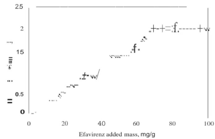
Solution calorimetry measured the heat change generated from the dissolution of the solid EFV by the molten organogel constituted of sunflower oil and 20 wt% of 12-HSA. The creation of a saturated solution at 90 °C involved dissolving the solute (EFV) up to the maximum and constant concentration that also corresponds to a constant value of heat of solution. As shown in Figure 1, incremental amounts of EFV added to the molten organogel resulted in a straight line increase in the magnitude of
the heat of solution until a plateau was reached when the
concentration of the solute equaled the saturation point. From
Figure 1, the EFV mass where the two straight lines cross
corresponds to the amount of solute that saturates the solvent contents in the vesse!. The solubility value corresponds to 75 mg/g for EFV in the molten organogel at 90 °C. From these results, we choose to work with a concentration below the saturation (30 and 60 mg of EFV per gram of organogel).
0 20 40 60 80 100
Efavirenz added mass, mg/g
Figure 1. Enthalpy of dissolution of EFV in molten organogel comprising 12-HSA (20 wt%) and sunflower oil at 90 °C. Means with error bars for
two experiments (n = 2) are shown.
It is also important at this stage to keep in rnind the organogel concept, in which oil is entrapped in a network self-assembly of the gelator molecules creating a gel. In this study, sunflower oil is entrapped in the self-assembled fibrillar networks of 12-HSA. The aim is to entrap EFV amounts smaller than its solubility lirnits at 37 °C in the oil phase in order to keep the drug always dissolved when exposed to the release medium. This condition is achieved for the EFV concentrations equal or lower than 60 mg per gram of organogel containing 20% 12-HSA (corresponding to 75 mg per gram of sunflower oil), since the equilibrium concentration determined experimentally after 72 h is equal to 79.8 ± 0.6 mg of EFV per gram of sunflower oil as shown in Table 2.
Thermal analysis
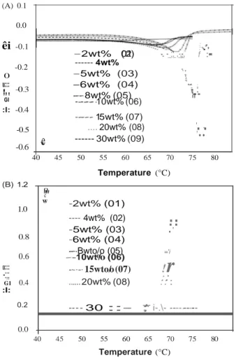
The effects of the gelator concentration and the drug incorp- oration on the thermal properties of the organogels were studied by DSC. DSC thermograms for the second
=
:
:
.
-
.
.
..
e
Table 2. EFV equilibrium concentration in sunflower oil at 37 ± 1°C as a fonction of time.
Equilibrium lime (h) 24 48 72 Median value*
EFV concentration (mg/g oil) 82.47 ± 2.18 79.14 ± 3.49 79.78± 0.65 80.02
*The median of ail measured values from 24 to 72 h because we cannot see differences among concentrations al different limes, the differences being smaller than standard deviations.
(A) 0.1 0.0
êi
-0.1 --2wt% (0021) ··.. -_;:_._-_._,-, . ..·.
, f :The thermal point of technical-grade 12-HSA is roughly 76 °C
with a melting enthalpy of 182J/g35. 12-HSA/sunflower organo-
gels display the following melting characteristics: as gelator
concentration is increased, so does the melting enthalpy and the
onset of melting, i.e. the gel melts at a higher temperature. For
example, a 2 wt% HSA sunflower oil gel has a Tm of
approxi-0 u:::
"
GI'
-0.2 -0.3 --- 4wt% --5wt% (03) --6wt% (04) ···-··- 8wt% (05) -··-··-··- ·10wt% (06) ·., " · . \ · : ··,.
.
:.
;"
mately 57 °C, against 75 °C for a 20 wt% HSA. It is perhaps
appropriate to remember that the main goal of this study is the
optimization of a structured organogel matrix in which EFV could
be solubilized to control its delivery in the GI tract after oral
:I: -0.4 -0.5 -0.6 w -·-·-·-·- 15wt% (07) .... 20wt% (08) --- 30wt% (09) ' ,
.
..
.
.administration . Thus, the best formulation for EFV-loaded
organogels should take into account the melting temperature of these systems. In fact, organogels being thermoreversible, they should guarantee both the thermal stability and solid-like behavior
40 45 50 55 60 65 70 75 80 at a temperature higher than or equal to the human body
(B) 1.2 1.0 0.8 0.6 u:::
-:;;
G1 0.4 :I: 0.2 0.0 Temperature (°C) 40 45 50 55 60 65 70 75 80 Temperature (°C)temperature, and even higher than 40 °C to combine environ-
mental and storage aspects. All fractions of 12-HSA used in this
study to produce sunflower oil gels are able to lead to a structured
network of 12-HSA since the onset for melting is approximately 48 °C for the lower gelator concentration (2 wt%).
Besides the gelator and its concentration, the effect of the drug on the gel structures should be an important parameter to be taken
into account when developing organogels for drug delivery
purposes. In previous studies reported in the literature, the drug
was mostly suspended in the gel medium and had a little or no
impact on the structures of the drug-loaded gels, which possessed
nearly identical physical properties with blank gels15-17• In other
cases, although no impact was observed on the gelation tempera-
ture, the presence of some dissolved drug in the gel apparently
shortened the time of phase inversion while raising drug
content36.
The effect of the drug incorporation in the thermal transition
temperatures of the organogels was also studied by DSC. EFV
was incorporated (30 mg/g0rganogel or 60 mg/g0rganogeJ in 12-HSA/
Figure 2. DSC second-heating for gelled sunflower oil over the range of 12-HSA compositions from 2% to 30wt%. Melting (A) and Crystallization (B).
heating-cooling cycle of the organogels without EFV (Samples
01-09) are shown in Figure 2(a) and (b), respectively. The
position of the melting and the crystallization peaks shifts to
higher temperatures as the concentration of 12-HSA is
increased and they become broader and shallower at low
concentrations of 12-HSA as shown in the bottom curves in
Figure 2(a) (melting) and in the top curves in Figure 2(b)
(crystallization). As expected, the organogels were thermo-
reversible as identical thermal parameters were obtained over
the two melting/crystallization cycles (data not shown).
Table 3 shows the thermal parameters that describe the
crystallization and melting behavior of organogels over the range
of compositions from 2% to 30% (w/w) HSA. Tm(onset)> Tm and
Tm(enctset) are the temperatures at the beginning, peak and end of
the melting endotherm, respectively. Tc(onset) • Tc and Tc(endset) are
the temperatures at the beginning, peak and end of the crystal-
lization exotherm, respectively. AHm and AHc are the melting and
crystallization enthalpies, respectively, determined by integrating
the area of each peak, melting and crystallization respectively.
sunflower oil gels containing different gelator concentrations (5%,
10%, 20% and 30wt%). The DSC thermograms for the second
heating-cooling cycle of EFV-loaded organogels formed with
20%wt HSA are shown in Figure 3(a) and (b). The same trend was
systematically seen for the whole gelator concentrations studied.
The temperatures and heats of melting (i.e. Tm and AHm) and
crystallization (Tc and AHc ) are reported in Table 3.
The organogel melting enthalpy (AHm ) combines at least two
distinct energetic contributions: (l) the melting enthalpy of the
fibers (which should be more or Jess constant and independent of
the solvent used if the fibers comprise pure gelator molecules
organized in a similar and regular structure) and (2) the enthalpy of dissolution of the gelator in the solvent phase, which is related
to the molecular affinity between the two species and should
depend on the solvent used37'38. A decrease of the melting
enthalpy could be taken as a sign of some re-arrangements in the
microstructure and/or packing of the 12-HSA molecules occur-
ring in the presence of the dissolved drug in the oil. However, the
data shown in Table 3 suggest that the introduction of EFV in the
gelling system did not result in evident changes on AHm or AHc of
the formulations, but apparently reduced the thermal transition
temperatures (Tm, Tc) by a few degrees while raising drug content.
This is quite visualized for low gelator concentrations, the
differences decreased when the HSA fraction increased, both Tm
0
"
c"
w -2wt% (01) ---- 4wt% (02).
..
.
-5wt% (03).
' -6wt% (04) ···-·Bwto/o (05) ='i · ··-···10wt0/o (06) -·-·-·15wto/o (07)!
r
... 20wt% (08) ::
::
: --- - 30 ::- ;* i-.\- ----·----Table 3. Thermal parameters obtained from the melting and crystallization thermograms of organogel with different concentrations of 12-HSA in sunflower oil, neat and loaded with EFV.
Organogel composition
and conditions of preparation Thermal properties measured by DSC
Melting (Tm)* Crystallization (Tc)*
*Standard errors in brackets.
(A) 0.1 0.0 -0.1
:§
-0.2 :i: 0 i;::: -0.3 niG> ...··
...._
((_-
---!
f
'
\
!
!
-- 20wt% (08) \\ .. 1!
: --... 20wt% (OEFV3) --- 20wt% (OEFV7)and Tc are almost constant and only fonction of the gelator amount. A similar effect in onset temperatures was reported for soy organogel with different gelators: myristic, palmitic, stearic and arachidic acids39.
The onset of Tc is the last point associated with the solution state and the onset of Tm is the last point of the gel state. Anyway, the peak melting Tm and onset of crystallization temperatures of the gels increased as 12-HSA concentrations increased, whatever the gel studied, in the presence and absence of drug. Both thermal parameters scaled in a logarithmic fashion in relation to the
12-:J: -0.4 -0.5 0 )( -0.6 w 40 45 50 55 60 65 70 75 80 Temperature (°C)
HSA concentration (Figure 4). The sol-gel transition is some- times interpreted as dissolution or melting of the "crystal " gelling molecules. In this sense, the solubility of "crystals gelling" corresponds to the concentration of gelling agent in an
ideal solution at a given temperature by the following Equation
(9), called Schroeder-van Laar equation37•40•41 . -ôHm (B) 1.2
.,,
c L1nC =--+ constant e R.Tm (9) w 1.0l
êi
0.8 :i: 0.6 0 i;::: --20wt% (08) ...20wt% (OEFV3)..
--- 20wt% (OEFV7)Where R is the universal gas constant and /'<;.Hm corresponds to the melting enthalpy apparent (sol-gel) of the pure gelling and C its
molar concentration in the gel. According to this equation, a plot of L111 C versus 1/ Tm should be a straight line. This linear dependency is confirmed in Figure 5. The melting and crystal- lization enthalpies likewise increased in a linear manner with an
increase of the 12-HSA concentration (Figure 6). Similar
ni G> :J: 0.4 0.2 ,' ' • / > _
)
'
\
behaviors were already reported in the literature using the 12- HSA dissolved in various organic phases42.
0.0
1-
...,.==
===-
-- o --- Organogel erosion and drug release kinetics (cylindricalshape)
40 45 50 55 60 65 70 75 80
Temperature (°C)
Figure 3. DSC second-heating for gelled 12-HSA (20wt%) - sunflower oil loaded with EFV (30 mg/gorganogel or 60 mg/gorganogeÙ· Melting (A) and Crystallization (B).
To clarify the mechanism of drug release from organogels, the weight loss and the amount of drug released were measured as a fonction of time. Experimental data plotted in Figure 7 highlights an initial slow release during the first hour, during which probably the drug diffuses slowly, followed by a second step characterized
by a faster release. This faster release could be attributed to the
onset of an erosion phenomenon, which makes the entrapped drug
Sample HSA
fraction Sunflower EFV Onset Peak Endset ôHm Onset Peak Endset ôHc
ID (w/w) oil (w/w) (mg/goranoe1) (oC) (OC) (OC) (J/g) (°C) (OC) (°C) (J/g)
01 0.02 0.98 48.3 (0.9) 56.6 (0.7) 61.4 (0.8) 0.75 (0.1) 54.0 (0.4) 51.0 (0.4) 43.6 (0.6) -0.54 (0.1) 02 0.04 0.96 53.9 (0.1) 61.5 (0.1) 67.7 (0.4) 0.90 (0.1) 57.7 (0.2) 55.0 (0.5) 49.6 (0.6) -0.80 (0.1) 03 0.05 0.95 55.5 (0.4) 66.9 (0.1) 70.7 (0.1) 4.5 (0.1) 63.7 (0.1) 62.2 (0.1) 59.2 (0.2) -5.2 (0.5) 04 0.06 0.94 55.01 (0.4) 67.3 (0.3) 72.2 (0.4) 7.5 (0.1) 65.4 (0.2) 63.6 (0.1) 57.5 (1.5) -8.l (0.3) 05 0.08 0.92 59.9 (0.9) 69.1 (0.4) 72.8 (0.2) 8.4 (1.1) 66.0 (0.1) 64.5 (0.1) 61.3 (0.5) -9.3 (0.1) 06 0.10 0.90 64.5 (0.1) 71.8 (0.5) 74.4 (0.4) 12.l (0.9) 68.6 (0.1) 65.4 (0.5) 62.7 (2.2) -13.5 (0.1) 07 0.15 0.85 66.2 (0.8) 72.2 (0.2) 75.2 (0.7) 16.3 (0.7) 68.5 (0.2) 67.6 (0.1) 66.l (0.1) -15.9 (0.1) 08 0.20 0.80 72.3 (2.5) 75.3 (0.2) 77.05 (0.8) 29.6 (1.7) 70.2 (0.1) 68.8 (0.1) 67.5 (0.1) -26.3 (0.1) 09 0.30 0.70 74.2 (2.1) 77.4 (1.1) 78.6 (0.8) 41.2 (2.1) 71.0 (0.16) 70.5 (0.1) 68.7 (0.1) -41.2 (0.3) OEFVI 0.05 0.95 30 55.2 (6.5) 62.5 (0.4) 70.8 (3.0) 5.1 (0.1) 61.0 (0.4) 58.8 (0.1) 55.5 (1.2) -4.8 (0.2) OEFV2 0.10 0.90 30 60.2 (0.4) 69.7 (0.6) 73.2 (0.3) 1 1.0 (0.2) 66.6 (0.1) 64.2 (0.4) 60.2 (0.7) -10.1 (0.9) OEFV3 0.20 0.80 30 68.4 (1.0) 74.3 (1.1) 76.2 (0.6) 28.5 (1.1) 68.9 (0.3) 67.2 (0.1) 64.9 (0.2) -27.7 (0.1) OEFV4 0.30 0.70 30 72.8 (2.5) 75.6 (0.3) 77.1 (0.2) 41.4 ( 1.5) 70.3 (0.1) 69.6 (0.1) 68.0 (0.1) -39.0 (0.5) OEFV5 0.05 0.95 60 50.9 (5.9) 58.0 (2.6) 61.8 (5.1) 5.7 (0.4) 56.0 (0.8) 52.0 (1.5) 47.4 (0.2) -5.0 (0.3) OEFY6 0.10 0.90 60 59.2 (0.2) 69.4 (1.1) 72.0 (0.3) 13.7 (1.0) 62.6 (0.1) 61.1 (0.6) 59.1 (0.6) -13.9 (0.1) OEFY7 0.20 0.80 60 70.0 (4.4) 73.6 (1 .0) 75.01 (0.3) 27.6 (1 .2) 67.1 (0.1) 65.2 (0.1) 64.6 (0.6) -28.3 (0.1) OEFV 8 0.30 0.70 60 72.0 (2.8) 74.8 (0.1) 76.6 (0.1) 40.4 (1.4) 69.7 (0.4) 68.2 (0.1) 66.7 (0.2) -38.8 (0.3)
.
Y
•
E.
•
...
2
85 80 75 -)..J 70é
65 -.;:"
...
60"
c..
---
-
---
1
--·- --
:-=
--%· ---·--o---r-·oTc (withoutFv) / l:.Tc (30 mg/g EFV)...
=
<I 50 40/
30 '// / 20 /. OOHo(-"•oo<EFV) E 55 F! 50 40 e DTc (60 mg/g EFV)0 •Tm (without EFV)
-"Tm (30 mg/g EFV)
li li.He (30 mg/g EFV)
D6.Hc (60 mg/g EFV)
10
i
/
•li.Hm (without EFV)6. Hm (30 mg/g EFV) 0
-
45 •Tm (60 mg/g EFV) •li.Hm (60 mg/g EFV) 0 5 10 15 20 25 30 35 0 10 15 20 25 30 35Concentration of 12-HSA, wto/o
Figure 4. Transition phase diagrams of organogels in different 12-HSA concentrations made without active pharmaceutical ingredient, with 30mg/g and 60mg/g of Efavirenz. Blank symbols correspond to crystallization and black symbols correspond to melting. Means with error bars for two experiments (n =2) are shown.
Concentration of 12-H SA, wto/o
Figure 6. Enthalpy of melting and crystallization of 12-HSA calculated from DSC peak area for 12-HSA series (organogels without active pharmaceutical ingredient and with 30 or 60mg/g of Efavirenz). Blank symbols correspond to crystallization and black symbols correspond to melting. Means with error bars for two experiments (n
=
2) are shown.-0.4 -0.6 -0.8 0 -1.0
ô
-1.2.
..
s
.
:
,
-1.4 -1.6 -1.8fT
l
•
without EFV 1 30 mgEFV 60 mgEFVfluid. The corresponding organogel erosion percents were 4 and 12%, respectively. lt was also observed that the same organogels when formed at 5 °C eroded more easily (7 and 22% mass losses in gastric and intestinal conditions, respectively), although minor differences were observed on the amounts of drug released (24 and 34%), compared to those at 25 °C.
In summary, the data presented here evidenced that both erosion and drug diffusion from the 12-HSA sunflower organogel are dependent on the matrix network (directly correlated to HSA concentration and temperature of gel formation) and slightly affected by the EFV presence, especially at low concentration of HSA. Therefore, the more structured the system the lower should be the matrix erosion, yielding to a lower drug release as a final result. More structured gels were obtained with the higher gelator
2.85 2.90 2.95 3.00 3.05
1000/Tm, K'1
Figure 5. Representation of Schroeder-van Laar equation for the analysis of the influence of gelling agent concentration on the sol-gel transition temperature. Means with error bars for two experiments (n =2) are shown.
more available. Combined drug diffusion and erosion may contribute to a faster release than erosion alone16.
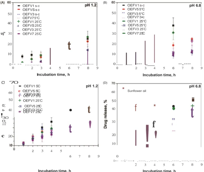
The data shown in Figure 7 include the influence of four different parameters: gelator concentration (5 or 20 wt% HSA), temperature of gel formation (5 °C or 25 °C), and composition of the release media (pH 1.2 containing pepsin or pH 6.8 containing pancreatin) to simulate the GI tract.
At the end of 8h of release, the mass lost (erosion) and the amount of drug released were more pronounced for organogels less concentrated in 12-HSA (5 wt%) formed at lower temperature (5 °C). In fact, organogels containing 5 wt% HSA (OEFVl) formed at 5 °C released 34% of EFV under simulated gastric conditions against 51% in simulated intestinal fluid. The corres- ponding organogel erosion percents were 25 and 41%, respect- ively. The temperature of gel formation influenced the amount of drug released and the organogel mass Joss. lncreasing the temperature of organogel formation to 25 °C led to an increased drug release (36% of EFV released in gastric and 45% in intestinal conditions; 27 and 39% of organogel mass Joss, respectively).
In tum, organogels formed at 25 °C, loaded with 20 wt% 12- HSA (OEFV7) provided the smaller amounts of drug released and mass losses. They released approximately 24% of EFV under simulated gastric conditions against 32% in simulated intestinal
concentration (20 wt% HSA) and the higher temperature of gel formation (25 °C). Both erosion and drug diffusion from more structured gels were less affected by the pH of the medium. These findings are also in agreement with the few works found in the literature related to drug delivery from organogels in simulated gastric and intestinal test solutions 15-17.
Release profiles from the cylindrical organogels Dependent models (curve fitting )
ln a general manner, the drug release from organogels may
involve processes of diffusion, erosion or drug dissolution 16•17.
Diffusion and erosion are expected to be the processes controlling drug release from the organogels since EFV at the concentrations
tested, is completely dissolved within the organogels, as expected from the measured solubility data and confirmed by DSC studies. The drug is then immediately available for diffusion.
A large number of models (both empirical and mechanistic mathematical models) have been proposed to describe drug release mainly for polymer matrices characterized by a 3D network. Even though organogels have different structures, the models already proposed for more investigated polymeric carriers can be useful to fit experimental values to compare the release profiles obtained from different formulation (gelator concentration, EFV loading) and conditions (gel formation temperature) studied here.
The in vitro drug release data from the different organogels contaimng EFV were evaluated kinetically using the Korshmeyer-Peppas, Weibull and Hixson-Crowell models [Equations (2-4), Table 1]. Korsmeyer-Peppas mode!is expected to be valid only up to 60% cumulative drug release, so the data for analysis were restricted to this range. The coefficient of
• • 50 "" 40 ....
+
<Ill OEFV1 s·c OEFVS s c OEFV3 s·c OEFV7 5°C OEFV1 25'C OEFVS 25'C uj" 30 ... OEFV3 25'C • OEFV7 25'C 20 10o--
--
...,
..
t
---..---50 • • "" .... OEFV 1 s·c OEFV5 5°C OEFV3 5°C OEFV7 5•c pH 6.8 40 30 • OEFV1 25°C <Ill OEFV5 25°C ... OEFV3 25'C • OEFV7 25'C 20 10o ... ...
---- ,,
--..----Cl•
'
'
(B) 0(
C
)
7
0
2 3 4 5 6 7 8 9 Incubation time, h 0 (D) 2 3 4 5 6 7 8 9 Incubation time, h • OEFV1 S'C pH 1.2 60 e .t. OEFV5 5OEFV3 5'C'C T OEFV7 5'C 60*
Sunflower oil pH 6.8 50+
OEFV1 25'C 50i
o;
<Ill OEFV5 25'C 40 ... OEFV3 25'C G> • OEFV7 25'C 40f
•
t
*
•
•
+
t
!
30.
::.
::.
1t
30!
f
""
...
c
20 10 2 3 4 5 20 10 6 7 8 9 0 0+- --1'---+ .----...----.---..----..----.----..---, 2 3 4 5 6 7 8 9Incubation time, h Incubation time, h
Figure 7. Weight Joss and release studies of organogels containing EFV at 37 °C. (a, c) Simulated gastric solution at pH 1.2 inclucling pepsin 800 U/L enzyme activity; (b, d) Simulated intestinal solution at pH 6.8 including pancreatin 1750 U/L protease activity. Means with error bars for two experiments (n
=
2) are shown.determination (R2), root-mean-square error (RMSE) and Akaike's
information criterion (AIC) values of these models were
determined for evaluation of accuracy and prediction ability of these models using KinetDS 3.0 rev. 2010 software (Krakow, Poland). The result of the curve fitting into varions mathematical models is given in Table 4. The best fitting of models was based on goodness of fit, i.e. the highest coefficient of determination
(R2) (doser to unity) and lowest RMSE and AIC values
(definition in Table 1).
From the obtained results, it is observed that for all formula- tions the drug release mechanism is best described by Korsmeyer- Peppas model. This model is a semi-empirical mode!, relating
exponentially the drug release to the elapsed time. Tt is used to
analyze the release of pharmaceutical polymeric dosage forms,
when the release mechanism is not well known or when more than
one type of release phenomena could be involved43.
The release exponent (n) is used in order to characterize different release mechanism. Ifthe n value is 0.5 or Jess, the release mechanism follows Fickian diffusion. Higher values of n (0.5
<
n<
1) correspond to a non-Fickian model denominated anomalous transport The drug release follows Zero-order drug release or Case-II transport if then value is 1. For the values of n higher than 1, the mechanism of drug
release is regarded as super Case-II transport, describing the influence
of polymeric hydration and swelling on drug release for polymeric and swellable systems, and which can be related to matrix erosion for non-
swellable systems.
The values of diffusional exponent (n) determined from Korsmeyer-Peppas model ranged from 0.81 to 1.39 (Table 4) for all organogels loaded with EFV (Fl-Fl6, refer Table 5 for formulation code description), indicating that the drug release from these systems followed the super case-II transport mechan- ism. However, the n value determined from this mode! for Reference (when EFV-60 mg/g0;1 is dissolved in non-gelled oil) is
0.30 (Fl7, Table 4), denoting a Fickian diffusion mechanism.
In conclusion, the release mechanism from organogels loaded with EFV is different from the diffusion mechanism obtained with simple oil formulations. From organogels, EFV probably diffuses through an outer gel layer that erodes, releasing oil droplets containing dissolved EFV into the aqueous medium. This
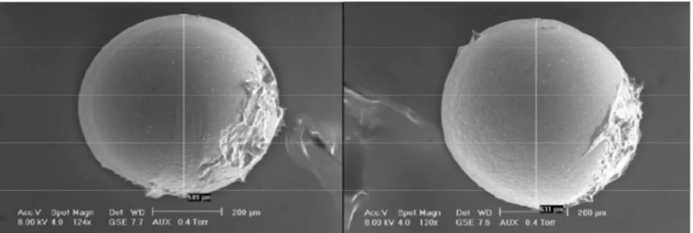
conclusion is supported by the visual observation of the cylin- drical organogels before and after release in the test solutions. The photographs shown in Figure 8 were taken at the end of the erosion/release studies. It is clearly visible that the organogels undergo changes in thickness (decrease in diameter) mainly for organogels containing 5 wt% HSA, while the capsule length remained essentially unchanged. These findings evidence erosion occurring at the organogel surface (Figure 8) depending on the
gelator concentration.
The results from the evaluation of drug release mechanism using varions kinetic models confirmed the experimental obser- vations that both erosion and drug diffusion mechanisms take place, depending on the matrix network .
Table 4. Results of curve fitting of the in vitro release data from different cylindrical organogels containing EFV. Formulation code (Table 5)
Model-dependent method Kinetic parnmeters FI F2 F3 F4 F9 FlO Fi l Fl2
Test solution: gastric solution (H 1.2) al 37 °C comprising pepsin
R 0.9992 0.9999 0.9993 0.9999 0.9993 0.9997 0.9534 0.9998 RMSE 1.2990 0.8705 l.8703 0.2703 l.2988 l.2349 2.6259 1.3810 Korshmeyer-Peppas AIC 21.28 15.68 26.38 -0.69 21.28 20.57 31.14 22.14 K 4.33 3.67 4.25 2.90 4.33 5.84 4.82 4.64 n 1.02 0.98 0.93 1.05 1.02 0.82 0.96 0.81 DR (%)4h 17.81 14.28 15.51 12.45 17.81 18.18 18.28 14.21 R2 0.9990 0.9998 0.9992 0.9999 0.9990 0.9997 0.9520 0.9998 Weibull RMSE 2.5639 1.0580 2.0647 0.6355 2.5639 1.5196 11.019 1.3133 AIC 30.80 18.41 27.71 11.27 30.69 23.48 51.22 21.43 R2 0.8024 0.7844 0.7692 0.7854 0.8024 0.7306 0.7625 0.7186 Hixson-Crowell RMSE 7.5353 5.8859 6.8725 4.5060 7.5354 7.8429 10.248 6.101 AIC 45.89 42.44 4.46 38.70 45.89 46.46 50.20 42.94
Model-independent method DE 17.68 14.36 15.57 12.65 17.68 17.54 17.83 13.92
Number of time points 7 7 7 7 7 7 7 7
Test solution: intestinal solution (pH 6.8) at 37 °C comprising pancreatin
Formulation code (Table 5)
Model-dependent method Pararneters F5 F6 F7 F8 F13 F14 F I S Fl6 F17
Table 5. Comparison of EFV-loaded organogel release profiles using the
independent mode! values of dissolution efficiency DE (done using
one-way [ANOVA] followed by Tukey's test).
*Values represent the mean of two independent determinations. Same letters
at the same colurnn indicate that there is no statistical difference (p <0.05).
lndependent model
Dissolution efficiency (DE) results are shown in Table 5. The
analysis of variance (ANOVA) revealed a statistical difference
(p
<0.05)
between the formulations. Differences in the DEpH 6.8 including pancreatin 1750 U/L protease activity. (A) 20 wt% HSA and (B) 5 wt% HSA. Both organogels were loaded with EFV (60mg/g). A
calibrated vernier cal iper was used for diameter and thickness evaluation
of the gel capsules.
data may be related to the release profiles since factors such as
12-HSA concentration, drug loading, gel formation temperature
and pH of the release medium may influence the release of the
drug molecule from the organogels.
R2 0.9989 0.9999 0.9993 0.9992 0.9997 0.9998 0.9998 0.9997 0.9975 Korshmeyer-Peppas RMSE 1.0503 l.2482 1.5821 0.7897 1.2178 1.5781 1.6493 1.5383 3.4737 AIC 18.3 2.07 24.04 14.32 20.38 24.08 24.63 23.65 24.45 K 5.48 4.52 7.82 3.37 9.01 4.10 4.21 1.83 29.20 n 1.02 0.89 0.65 1.07 0.85 0.99 1.05 1.39 0.30 DR (%)4h 22.55 15.46 19.22 14.94 29.24 16.16 18.03 12.63 44.00 R2 0.9999 0.9998 0.9994 0.9987 0.9999 0.9998 0.9997 0.9995 0.9980 Weibull RMSE 1.1189 1.2339 1.5218 l.3811 0.9910 1.6369 2.057 3.4710 3.251 AIC 19.19 20.56 2.35 22.14 17.49 24.52 27.72 35.04 23.84 Rz 0.7726 0.7573 0.6457 0.7931 0.7228 0.7665 0.7890 0.8352 0.4353 Hixson-Crowell RMSE 8.199 6.6980 8.0967 5.4566 11.380 6.1924 6.9713 3.8616 21.05 AIC 47.08 44.25 46.90 41.38 53.67 43.15 44.81 36.54 42.51
Model-independent method DE 22.86 15.33 18.04 15.20 28.64 16.18 18.44 13.95 40.74
Number of time points 7 7 7 7 7 7 7 7 5
Dissolution Formulation Formation EFV efficiency, DE
code temperature mg HSA% pH mean (n =2)*
Fl7 (oil formulation) 25 60 0 6.8 40.38" Fl4 5 60 5 6.8 30_97•.b F13 5 30 5 6.8 29_07•.b FS 25 30 5 6.8 22.86a,b F9 5 30 5 1.2 21.46•-b FIS 5 30 20 6.8 19.29•.b F7 25 30 20 6.8 18.04b FI 25 30 5 1.2 17.68b Fll 5 30 20 1.2 17.43b FIO 5 60 5 1.2 17.43b F3 25 30 20 1.2 15.57b F8 25 60 20 6.8 15.53b F6 25 60 5 6.8 15.33b F16 5 60 20 6.8 14.46b F2 25 60 5 1.2 14.36b
Fl2 5 60 20 1.2 14.17b Figure 8. 12-HSA sunflower oil gel formed in gelatin capsules (size #OO)
Figure 9. SEM image of organogel beads produced by prilling process.
600 1200 1800 2400 3000
Wavenumbers, cm·'
Figure 1O. Raman spectra and mapping for EFV-loaded organogel beads.
To evaluate the difference among the 17 formulations, a post
hoc Tukey test was performed on the results of ANOVA. With the
application of the Tukey test it was possible to observe that, with a
level of significance of 5%, DE of formulations Fl, F2, F3, F4, F6, F7, F8, FlO, Fll, F12 and F16 were equal and significantly
smaller than the DE found for the formulation F17. Moreover, Fl7
is not significantly different from F5, F9, Fl3, F14 and F15
(p
>
0.05), which are not significantly different from one other.From these results, it could be concluded that the drug release
was delayed by the gelled oil as already discussed, i.e. an oil-
structured system is expected to present lower drug release rate.
However, the different parameters studied such as the gelator
concentration, the drug loading, the temperature of organogel
formation and even the pH of the release media showed no
significant difference on DE.
Organogel beads for drug delivery purposes
Beads characterization
Size and shape. Figure 9 reveals that the organogel beads consist of large micron-scale beads, spherical in shape and very uniform
in diameter {610
±
5 µm). / / / / / I / 1660 I 2400 Wavenumber, cm·• 3000 28 2900 1068 12991..0 2250 2250 J.."
C>
Table 6. Results of curve fitting of the in vitro release data from organogel beads containing EFV.
Test conditions: media at 37 °C containing 1wt% SDS with changing pH values (2 h at pH 1.2 and then 8 h at pH 6.8), without enzyme
Model-dependent method
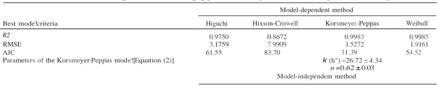
Best mode!criteria Higuchi Hixson-Crowell Korsmeyer-Peppas Weibull
R2 0.9750 0.8672 0.9983 0.9985
RMSE 3.1759 7.9909 1.5272 1.9161
AJC
Parameters of the Korsmeyer-Peppas mode![Equation (2)]
61.55 83.70 31 .39
k (h") =26.72 ± 4.34
n =0.62 ± 0.03
54.52 Dissolution efficiency (DE, %)
Model-independent method
56.38 (number of time points = 13)
100 90 80 70 C> 60 ë)
so
0:: ::R 40 30 20 10 0 0 2 3 4 • experimental data--Korshmeyer-Peppas fit
----Higuchifit
--Weibull fit
5 6 7 8 9 10 11
40% during the first 2 h under simulated gastric condition s (pH
1.2). Despite an increase in the EFY solubility at pH 6.845, drug
dissolution exhibited a slow release reaching 80% after 4 h of immersion into the simulated intestinal fluid.
The data obtained during the release test were examined
according to Korsmeyer-Peppas, Weibull, Hixson-Crowell and
Higuchi equations [Equations (2-5), respectively, Table l]. The coefficient of determination (R2), root-mean-square error (RMSE) and Akaike's information criterion (AlC) values of these models
were determined for evaluation of accuracy and prediction ability
of these models using KinetDS 3.0 rev. 2010 software. The result of the curve fitting into various mathemati cal models is given in Table 6. Figure 10 also confirms the correlation of the EFV release profile with the theoretical profile predicted by these
model s (see fitting curves). The best fitting was based on goodness of fit, i.e. the highest coefficient of determination (R2;
Incubation time, h
Figure 11. Release amount of EFV from the organogel beads containing
20%wt 12-HSA as gelling agent during the release test at 37 °C (2 h in pH 1.2, 8 h in pH 6.8). Means with error bars for three experiments (n = 3)
are shown.
Raman mapping. The Raman spectra of the individual compo-
nents (HSA, sunflower oil and organogel beads) are compared in
Figure 10. The Raman spectrum of 12-HSA displays a strong peak
at around 2850-2900cm- 1 (C-H), peaks at 1440cm-1 (CH2) and
two peaks around 1068-1135 cm-1 characteristics of C-0
stretching bands. In turn, the characteristic peaks are identified
in the Raman spectrum of the sunflower oil: at 1660cm-1 (C =C
stretching bands) and the peaks at 1306 and 1272cm-1 ( =C-H
deformation), already reported in the literature44. For the Raman
spectrum of pure EFV, the CF3 stretching modes have been
assigned at 1146cm-I, with a typical C =0 stretching bands at
1791cm-1, one distinguishing intensive band at 2250cm- 1
assigned to C C stretching bands, which was not evident in the
other gel components.
The pure component Raman spectrum of 12-HSA could not be
separated from that of sunflower oil using cluster analysis due to
the homogeneous (molecular) dispersion of EFV within the
organogel. Comparison of the Raman spectra of the samples
revealed that, except for the EFV vibrational bands at 2250 cm-I,
there were no distinguishable vibrations bands. EFV was found
uniformly distributed within the organogel. Release studies from organogel beads
Figure 11 shows the cumulative amount of EFV release profiles
from organogel beads comprising 20wt% of 12-HSA and loaded
with 60mg/g of EFV. The organogel beads exhibited a slow
dissolution profile with 80% of the drug being released after 8 h,
closer to unity) and lowest RMSE and AlC values (Table 1). From the obtained results, it is observed that the drug release
mechanism is also best described by Korsmeyer-Peppas model.
The release exponent n calculated from Korsmeyer-Peppas mode!
for predicting drug release mechanism was 0.62, corresponding to
a non-Fickian mode! denominated anomalous transport, revealing
that the EFV release is strongly influenced by the size of the
organogel system. For the organogel beads an anomalous
transport is the main release mechanism, whereas for cylindrical
organogels (larger size) erosion and diffusion are the mechanisms
controlling the release. The 3D-fibrillar network with 12-HSA as
gelator is well documented in the literature and already observed
in some of our previous works46-49. The driving force of
controlled release is the entanglement of the network which represents a physical barrier to the diffusion of the entrapped oil containing the dissolved drug. Sorne interaction between EFV and
12-HSA could delay the free EFY diffusion through the 12-HSA
network. However, it was not detected in this study by the Raman mapping of the organogel beads.
Conclusions
This study underlines the interest of gelling sunflower oil with 12-
HSA for the controlled release of a hydrophobie drug like EFV.
With organogels of different shapes and sizes, exposed to different release conditions, it could be demonstrated that the drug release
was controlled mainly by diffusion with a minor participation of erosion in absence of enzymes, or controlled by more than one process, a coupling of diffusion and erosion mechanisms, the later
accelerated by enzymatic activities.
Based on the drug release kinetics and mechanism models,
EFV release from organogels could be described by the
Korsmeyer-Peppas mode!with the highest coefficient of deter-
mination and lowest RMSE and AIC values. The release exponent
with sunflower oil-based delivery systems: drug diffusion- controlled (sunflower oil, n
=
0.30), anomalous transport (orga-nogel beads, n =0.62) and super Case-II transport (cylindrical organogels 0.81<n <1.39). Even though further data will be necessary to investigate
EFV
release k:inetics in the12-HSA
sunflower oil gel more deeply, the obtained results have &bown that is possible to modify the drug release from the oil phase by
gelling the system.
Furthermore, prilling is a useful and simple technique to
prepare organogel beads with spherical shape and narrow size distribution. The beads could be applied fur the manufacture of multiparticulate dosage furms where a suitable controlled release rate could be designed to takc advantage of beads properties. The
multiparticulate
nature
of beadsas a
dosage form (filled into capsules) can offer important pharmacological and technological advantages over conventional single-unit solid dosage forms. Further optimization of formulation wi11 be required before moving onto any in vivo studies.Acknowledgements
Authors thank Gala® technological platform (France) for technical
support, Cristali.a (Brazil) fur providing the drug (EFV) and EA-
CIDAM (Conception Ingénierie et Développement de /'Aliment et du
médicament) for dissolution studies.
Declaration
of
interestThe authors report no conflicts of interest. The authors alone are responsible for the content and writing of the paper. This study was partially financially supported by the CNPq (Brazil). References
1. Jannin V, MUBakhanian J, Marchaud D. Approaches fur the
development of solid and semi-solid lipid-based formulations. Adv Drug Deliv Rev 2008;60:734-46.
2. Porter CJH, Trevaskis NL, Charman WN. Lipids and lipid-based formulations: optimizing the oral delivery of lipophilic drugs. Nat Rev Drug Discov 2007;6:231-48.
3. Hauss Dl Oral lipid-based formulations. Adv Drug Deliv Rev 2007; 59:667-76.
4. Goto S, Kawata M, Suzuki T, et al. Preparation and evaluation of Eudragit gels. 1: Eudragit organogels containing drugs as rectal
sustamed-release preparations. J Pharm Sei 1991;80:9581. 5. Kawata M, Suzuki T, Kim NS, et al. Preparation and evaluation of
Eudragit gels. Il:invitro release of salicylic acid, sodium salicylate, and ketoprofen from Eudragit Land Sorganogels. J Pbarm Sei 1991; 80:1072-4.
6. Zia H, SU7.Ctte RF, Mohammed Q, et al. Ketorolac tromethamine and
Ketoprofen suppositorics: relcase profiles and bioavail.ability of a cocoa butter base formula in rabbits. Int J Pharm Compd 1998;2: 390--3.
7. Pisal S, Shellœ V, Mahadik
K.
Kadam S. Effect of organogel components on in vitro Illlllal delivery of propranolol hydrocbloride.AAPS PhannSciTech 2004;5:92-100.
8. Kumar R, Katare OP. Lccithin organogels as a potential phospho- lipid-structured system fur topical drug delivery: a review. AAPS PharmSciTech 2005;6:E298-310.
9. Motulsky A, Lafleur M, Couffi.n-Hoarau A.C, et al. Characterization and biocompatibility of organogels baSC<l on 1-alaninc for parenteral drug delivery implants. Biomaterials 2005;26:6242-53.
10. Lim PFC, Liu XY, Kang L, et al. Pbysicochemical effects of
terpenes on organogel for transdermal drug delivery. lnt J Pharm 2008;358: 102-7.
11. Bhatia A, Singh B, Raza K, et al. Tamoxifen-loaded lecithin organogel (LO) fur topical application: development, optimization and cbaracterization. lnt J Pharm 2013;444:47-59.
12. Abdallah DJ, Weiss RG. Organogels and low molecular mass
organic gelators. Adv Mater 2000;12:1237-47.
13. Pernetti M. van Malssen KF, Flôter E, Bot A. Structuring of edible oils by alternatives to crystalline fat. Curr Opin Colloid Interface Sei 2007;12:221-31.
14. Anand B, Pisal SS, Paradkar AR, Mahadik KR. Applications of organogels in pharmaceuticals. J Sei Ind Res 2001;60:311-18. 15. lwanaga
K.
Sumizawa T, Miyaz.aki. M, Kakemi M. Cbaractcrizationof organogel as a novel oral controlled release formulation for
lipophilic compounds. Int J Pharm 2010;388:123--8.
16. Iwanaga K, Kawai M. Miyazaki M, Kakemi M. Application of organogels as oral controlled release formulations of bydrophilic drugs. Int J Pharm 2012;436:869-72.
17. Lupi FR, Gabriele D, Baldin.o N, et al. Olive oil/policosanol organogels for nuttaceutical and drug delivery purposes. Food Funct 2013;4:1512-20.
18. Amidon GL, Lennemiill H, Shah VP, Crison JR. A theoreti.cal basis for a biopharmaceutic drug classification: the correlation of in vitro drug product dissolution and in vivo bioavailability. Pharm Res
1995;12:413--20.
19. Chiappetta DA, Hocht C, Taira C, Sosnik A. Efavirenz-loaded polymeric micelles for pediatric anti-IIlV pbarmacotherapy witb significantly higher oral bioavailability [corrected]. Nanomedicine (Lond) 2010;5:11-23.
20. Chiappetta DA, Alvarez-Lorenzo C, Rey-Rico A, et al. N-Alkylation of poloxamines modulates micellar assembly and encapsulation and release of the antiretroviral Efavirenz. Eur J Pharm Biopbarm 2010; 76:24-37.
21. Chiappetta DA, Hocht C, Taira
C.
Sosnik A. Oral pharmacokinetics of the anti-IIlV efavirenz encapsulated within polymeric micelles. Biomaterials 2011;32:2379--87.22. Chiappetta DA, Facorro G, Rubin de Celis E, Sosnik A. Synergistic encapsulation of the anti-IIlV agent Efavirenz within mixed
poloxamine/poloxamer polymeric micelles. Nanomedicine Nanotechnol Bio!Med 2011;7:624-37.
23. Chowdary KPR, Naresh A. Formulation development of Efavirenz
tablets employing j3 cyclodextrin- PVP K30- SLS: a factorial stu.dy.
J Appl Pbarm 2011;1:130-4.
24. da Costa MA, Seiceira RC, Rodrigues CR, et al Efavirenz
dissolution enhancement I: co-micronization. Pharmaceutics 2013; 5:1-22.
25. Chowdary KPR, Enturi V. Preclinical pharmacokinetic evaluation of Efavirenz solid dispersions in two new modified starches. J Appl Pbarm Sei 2013;3:S89-92.
26. Patel GV, Patel VB, Patbak A, Rajput SJ. Nanosuspension of efavirenz for improved oral bioavailability: formulation optimiza- tion, in vitro, in situ and in vivo evaluation. Drug Dev Ind Pbarm 2014;40:80-91.
27. Alves IDS, de La Roca Soares MF, de Albuquerque CT, et al. Solid dispersion of efavirenz in PVP K-30 by conventional solvent and kneading methods. Carbohydr Polym 2014;104:166--74.
28. Digenis G. The in vivo behavior of multiparticulate versus single unit dosage formulations. In: Ghebre-Sellassie 1,ed. Multiparticulate
oral drug delivery. New York: Marcel Dekker; 1994:333-55. 29. CRI CIR. Am.ended final report on tbe safety assessm.ent of
hydroxystearic acid. Int J Toxicol 1999;18(Suppl I):l-10. 30. Gao J. Solvent induced modifications to fiber nanostructure and
morphology fur 12HSA molecular gels. New Brunswick (NJ): The State University of New Jersey; 2014.
31. Anvisa AN de VS. Resoluçio ROCn° 482, de 23 de setembro de 1999. Aprova o Regulamento Técnico para Fixaçâo de Identidade e Qualidade de Ôleos e Gorduras Vegetais; 1999.
32. USP35. United States Phannacopeia 35 - National Formulary 30; 2012.
33. Mendyk A, Jachowicz R, Fijorek K, et al. KinetDS: an open source software for dissolution test data analysis. Dissolution Tcchnol 2012; 19:6-11.
34. Panikumar AD, Venkat RY, Sunitha G, et al. Development of biorelevant and cliscriminating method for dissolution of Efavirenz and its formulations. Asian J Pharm Clin Res 2012;5:220--3. 35. Eloundou JP, Girard-Reydet E, Gérard J-F, Pascault J-P.
Calorimetric and rheological studies of 12-hydroxystearic acid/ digycidyl etber of biBphenol A blends. Polym Bull 2005;53:367-75.
36. Jiao T, Wang Y, Zhang Q, et al. Self-assembly and headgroup effect
in nanostructured organogels via cationic amphiphile-graphene
oxide composites. PLoS One 2014;9:e101620.
37. Murata K, Aoki M, Suzuki T, et al. Thermal and light control of the sol-gel phase tranBition in cbolesterol-base<l organic gels. Novel Helical Aggregation Modes As Detected by Circular DichroWn and
Electron Microscopie Observation. J Am Chem Soc 1994;116:
![Table 5. Comparison of EFV-loaded organogel release profiles using the independent mode! values of dissolution efficiency DE (done using one- way [ANOVA] followed by Tukey's test)](https://thumb-eu.123doks.com/thumbv2/123doknet/3130141.89060/10.892.61.794.192.523/comparison-organogel-release-profiles-independent-dissolution-efficiency-followed.webp)