Aguilar-Uscanga et al. 2013 : 2 (6) 2462-2468
Journal of Microbiology, Biotechnology and Food Sciences
...
International peer-reviewed scientific online journal
...
EFFECT OF CULTURE MEDIUM ON BACTERIOCIN PRODUCTION BY LACTOBACILLUS RHAMNOSUS HN001
AND LACTOBACILLUS REUTERI ATCC 53608
Aguilar-Uscanga B. R.*
1, Solís-Pacheco J. R.
1, Plascencia L.
1, Aguilar-Uscanga M. G.
3, García H. S.
3and Lacroix M.
2Address(es): Blanca Rosa Aguilar Uscanga, PhD.
1Centro Universitario de Ciencias Exactas e Ingeniería. Universidad de Guadalajara, Jalisco, México. Boulevard Marcelino García Barragán #1421, Col. Olímpica.
Guadalajara, Jalisco C.P. 44430 México.
2
INRS-Institut Armand Frappier, Research Laboratories in Sciences, Applied to Food, Canadian Irradiation Centre. 531 Boulevard des Prairies, Laval, Québec, Canada, H7V 1B7.
3
Instituto Tecnológico de Veracruz, UNIDA. Laboratorio de Bioingeniería. Miguel Angel de Quevedo 2779. Col. Formando Hogar. C.P. 91860, Veracruz, Ver. *Corresponding author: agublanca@gmail.com
ABSTRACT
Keywords: Bacteriocin, Lactobacillus rhamnosus, Lactobacillus reuteri, probiotics, food safety
INTRODUCTION
Bacteriocins have attracted attention as potential substitutes for antibiotics to cure and/or prevent bacterial infections and are widely employed in food preservation. This can be particularly useful in preservation or food safety applications, but also has implications for the development of desirable flora in fermented food (Riley, 2000). Nowadays, new research to produce probiotics with antimicrobial activity in inexpensive culture media, different from MRS broth, such as molasses or fructo-oligosaccharides in substitute for glucose is very important. Some studies have shown the impact of Agave fructans on the microbial ecology of the human gut and it has been reported as a potential prebiotic which increased populations of bifidobacteria and lactobacilli compared to glucose and others carbon sources used as a control (Gómez et al.,
2010).
Mexico has a high diversity of species of Agave plants and the most important is Agave tequilana Weber var. azul, as it is the raw material for the tequila elaboration process. During Agave development, which lasts several years, these plants accumulate reserves of sugars consisting of fructans. These carbohydrates are fructose bound polymers frequently with a single glucose moiety. Agave tequilana Weber var. azul is an economically important species not only because it is the sole plant allowed for tequila production but because it is a potential source of prebiotics (López et al., 2003). On the other hand, cane molasses is an inexpensive waste that has been used in various fermentation processes, mainly for improving kinetics growth and metabolite production in bacteria and other microorganisms of industrial interest (Yu-Peng et al., 2003;
Demirci et al., 2008).
Lactobacillus reuteri is an obligatory heterofermentative resident in the gastrointestinal tracts of humans and most other animals examined to date
(Kandler et al., 1980). These bacteria produced an antimicrobial agent
(bacteriocin) active both in a bacteriocidal and bacteriolytic manner against sensitive cells. This bacteriocin has been designated reutericin 6, with apparent molecular size of 3800 Da as determined by SDS-PAGE studies (Kawai et al.,
2001). Reutericin 6 was shown to be a class II cyclic bacteriocin which retained
activity after heating at 100°C for 20 min (Toba et al., 1991). It is a compound with antimicrobial activity towards a broad range of pathogens and food spoilage
organisms, including Gram- positive and Gram- negative bacteria, yeasts, fungi and protozoa, which makes it very attractive for use in food or as conservative auxiliary therapeutic agent (Doleyres et al., 2005).
Lactobacillus rhamnosus HN001 is a Gram-positive species, a facultative anaerobe, non-motile and non-spore forming this strain was identified as a potential probiotic, by the New Zealand Dairy Research Institute and analyzed by molecular methods such as DNA/DNA homology, SDS-PAGE and PCR (Gill et al., 2000). L. rhamnosus GG was selected for studies in animals and humans for its ability to survive atlow pH levels such as 2.5 and in order to survive in this harsh environment, must prevail over host defense mechanisms, such as gastric activity and bile (Shu and Gill 2002).
Several studies have been documented to L. rhamnosus HN001 satisfies the most stringent requirements for probiotic properties (Gill et al., 2000). In order to assess its safety it has been tested in the animal studies where it was found to be non-pathogenic, non-toxic and has no adverse effect on animal health. Hence this strain is safe for human consumption (Gill et al., 2000; Shu and Gill 2002;
Zhou et al., 2001). Few studies have been published about bacteriocin produced
by L. rhamnosus. Some strains such as ST461BZ and ST462BZ produced bacteriocins with a broad spectrum of inhibitory activity toward various pathogenic bacteria (Todorov and Dicks 2005). Some studies have also reported that L. rhamnosus GG (LGG), ATC 53103 and HN001 are probiotics that have been isolated from the human gastrointestinal tract and widely used against diarrhea in children and animals (Guandalini et al., 2000; Weese and Anderson
2002).
Some investigators have isolated and partially purified bacteriocin from different species of lactobacilli predominantly from food and some in human feces
(Millette et al., 2007; Theppangna et al., 2007). Alongside with the discovery
of new bacteriocins, several interesting aspects of these peptides have been elucidated so bacteriocins have attracted attention as potential substitutes for antibiotics to cure and/or prevent bacterial infections (Toure et al., 2003; Line et al., 2008). These microorganisms are used in food process fermentations and in probiotic therapy and their beneficial properties are widely recognized. (Abee et al., 1995; O’Sullivan et al., 2002; Dortu and Thonart 2009).
The aim of this work was to evaluate the effect of different carbon sources (fructan from Agave, sugar cane molasses and glucose) in different culture media The aim of this study was to evaluate the effect of media on bacteriocin production by Lactobacillus rhamnosus HN001 and
Lactobacillus reuteri ATCC 53608 using three different media: YPM, YPF and MRS supplemented with glucose and K2HPO4. The
optimum temperature was 37°C and initial pH 6.5. Bacteriocin-like substances produced by tested bacteria in MRS medium supplemented with glucose and K2HPO4 exhibited a broad antimicrobial spectrum determined by well diffusion assay against indicator
bacteria Listeria monocytogenes, Lactobacillus sakei, Enterococcus faecium, Lactobacillus delbrueckii, Lactobacillus acidophilus, but no antimicrobial spectrum against E. coli O157:H7, Salmonella typhimurium, Staphylococcus aureus, Bacillus cereus was detected. Bacteriocin was sensitive to protease IV, trypsin, pepsin and α-amylases, but resistant to lipase. It was also resistant to detergents such as Tween 80, Triton-X and SDS. This bacteriocin was thermo-stable (resistant at 60°C, 90°C and 100°C for 30 min). Tested bacteria showed the best antimicrobial (bacteriocin-like) activity after growth in MRS medium. Bacteriocin substances produced by tested bacteria showed promising thermo-stable technological properties.
ARTICLE INFO Received 4. 3. 2013 Revised 22. 4. 2013 Accepted 25. 4. 2013 Published 1. 6. 2013 Regular article
(MRS, YPM and YPF) for bacteriocin production through fermentation of Lactobacillus rhamnosus HN001 and Lactobacillus reuteri ATCC 53608, besides testing the spectrum of antimicrobial activity and bacteriocin production by these strains.
MATERIAL AND METHODS
Microorganisms
Lactobacillus rhamnosus HN001 and Lactobacillus reuteri ATCC 53608 were used. Lyophilized cultures of both strains were grown in MRS broth at 37°C and stored at -80°C in appropriate media containing 20% glycerol (w/v). Before experimental use, the cultures were propagated twice in Man Rogosa Sharpe (MRS) broth. The bacterial indicator strains used in this study for antimicrobial activities were maintained at -80°C in appropriate media containing 20% glycerol (w/v), and were propagated in their respective culture broth at 37°C for 24 h, as indicated in Table 4. Agar diffusion method was used to examine the antimicrobial capacity (MRS, TSA, 1.5% (w/v) agar).
Culture Media
The media used for bacteriocin production were: A) MRS broth (Difco), supplemented with 5 gL-1 glucose (Bioxon) and 10 gL-1 K2HPO4 (Fermont), total
sugar, 24 gL-1 glucose; B) YPM broth (10 gL-1 yeast extract (Bioxon), 10 gL-1
bactopeptone (Bioxon), 2.0 gL-1 ammonium sulfate (JT Baker), 10 gL-1 K2HPO4
(Fermont), 1% (v/v) Tween 80 (Golden Bell) and sugar cane molasses (total sugars, 21 gL-1 equivalent to 15 gL-1 sucrose, 3 gL-1 glucose and 3 gL-1 fructose) and C) YPF broth (10 gL-1 yeast extract (Bioxon), 10 gL-1 bactopeptone (Bioxon),
10 gL-1 K2HPO4 (Fermont), 1% (v/v) Tween 80 (Golden Bell) and 20 gL-1 Agave Tequilana fructan. The culture media pH values were adjusted to 6.5 by the addition of 5 M NaOH and finally it was autoclaved for 15 min at 121°C.
Inoculum preparation and growth conditions
The inoculums were prepared adding 1 mL cellular suspensions of L. rhamnosus HN001 and L. reuteri ATCC 53608, in 50 mL MRS broth (Difco). Incubates for 12 and 18 h at 37°C. Subsequently, cell concentration was determined by a previously prepared CFU/mL vs. O.D calibration curve at 660 nm. Bacteria cultures were carried out with inoculums of approximately 0.25 to 0.35 O.D, corresponding to a cell concentration ≈ 1.6x10⁶ to 2.1x10⁶ CFU/mL, a reading of O.D660 ≈ 0.25 to 0.35 according to the calibration curve.
Kinetic production of bacteriocin
Bacteriocin production and growth kinetics of L. rhamnosus HN001 and L. reuteri ATCC 53608 was carried out Erlenmeyer flask in a 500 mL with MRS broth, inoculated with 2% inoculum. Growth kinetics was monitored with a Genesis 200 spectrophotometer (Termo spectronic) at O.D. 660. Three hundred ml MRS, YPM and YPF medium were inoculated in triplicate with L. rhamnosus HN001 or L. reuteri ATCC 53608 in the different media, followed by incubation at 37°C for 40 h fermentation. Initial pH of the culture broth was 6.5. Ten mL samples were removed from the cultures at 8, 12, and 20 h for bacterial growth and bacteriocin activity analyses. Bacterial growth was monitored by measuring culture absorbance at 660 nm, followed by a bacteria count (CFU/mL) on MRS agar incubated for 40 h at 37°C.
Biomass production
Bacteria growth was measured in triplicate batch cultures for all strains in the different media, monitored by measuring the absorbance at 660 nm with a Genesis 200 spectrophotometer (Termo spectronic). Appropriate dilution of cell suspension was made so linearity remained between dry mass and OD at 660 nm (i.e. from 0 to 0.4 units at 660 nm). Biomass was determined gravimetrically by filtering 10 mL culture samples on pre-weighed nitrocellulose filters (pore size 0.45 μm; Sartorius AG, Goettingen, Germany). The filters were washed with demineralized water and dried in an oven set at 80°C. Dry mass was established by repeatedly weighing the filters until a constant value was reached (after 18-24 h). Biomass was determined using the standard OD 660 curve against dry cell weight.
Calculation of the Kinetic parameters
Biomass yield (YX/S in g/g): X = (X-X0/S-S0)
Substrate consumption (S in gL-1) = S-S0/f (t)
Where X0, and X are initial and final biomass concentrations (gL-1) and So and S
are initial and final substrate concentrations (gL-1).
Specific growth rate (µ in h-1) = 1/X (dX/dt)
Sugar consumption rate (rS in g. gL-1. h-1): rS =dS/dt
Duplication time (td=h) = μmax/ Ln 2
Substrate consumption and metabolite production
The analysis of substrate consumption and metabolite production was made on 2 mL sample from different culture media. The samples were centrifuged at 4500 rpm for 10 min and the supernatant filtered with a 0.45µm Millipore filter. Analysis was by high performance liquid chromatography (HPLC), using an index refraction (IR) detector (Varian 356-LC), a column packed with Metacarb Plus Ca for sugar analysis and a column packed with Metacarb H Plus for alcohol and organic acid analysis.
The operating conditions of the chromatograph (Varian Pro Star) for the analysis of substrate consumption were: water HPLC grade mobile phase, 90°C temperature, 0.6 mL/min flow. For metabolite analysis (alcohols and organic acids), a mobile phase of 0.01N H2SO4 (Golden Bell), 54°C temperature, 0.6
mL/min flow were used. Injected sample volume was 20 µL. A calibration curve was performed using as standards: glucose (Sigma), sucrose (Caledon), fructose (Sigma), lactic acid (Sigma-Aldrich), acetic acid (Sigma-Aldrich), butyric acid (Sigma-Aldrich), propionic acid (Sigma-Aldrich) and ethanol (Golden Bell).
Preparation of Culture Supernatants
Sterile cell-free cultures were obtained by centrifugation (10000g for 15 min at 4°C) and filtration through a 0.45-μm pore-size filter (Millipore). They were adjusted to pH 7.0 with NaOH 2M to eliminate any effect of acidity.
Determination of antimicrobial spectrum and bacteriocin activity assay
Growth and bacteriocin activity of L. rhamnosus HN001 and L. reuteri ATCC 5360 strains cultured in MRS broth, YPM and YPF media were measured during growth phase (4, 8, 12 h) and at the beginning of stationary phase (20 h). Bacteriocin antibacterial spectra of supernatant strains were determined using paper dish and well diffusion assay as described by Toba et al (1991). Thirty ml MRS agar was inoculated with 106 CFU/mL of each indicator strain
(ATTC-American Type Culture Collection, Rockville, MD, USA; LSPQ-Laboratoire de Santé Publique, Ste-Anne de Bellevue, QC, Canada; FRDC-Food Research and Development Center, St-Hyacinthe, QC, Canada; HPB-Health Product Branch, Santé Canada; CHUM-Centre Hospitalier Universitaire de Montreal; UG-University of Guadalajara), then poured into Petri dishes and allowed to gel for 30 min. Six mm diameter wells were cut and 80 µL crude extract bacteriocin (CEB) producing strains were placed into each well. After 24 h incubation at a temperature optimal for the indicator strain, clear zones of inhibition appeared when the strain was sensitive. Inhibition was scored positive if the width of the clear zone around the well was ≥ 0.5 mm. Plates were examined for the apparition of growth inhibition zones in the clear zones around the strains tested. The diameter of the inhibition zone were recorded as follows: ≥ 20 mm (+++), 10-5 mm (++), 5-1 mm (+) and 1-0 mm (-) no inhibition zone.
Antimicrobial activity was expressed in arbitrary units (AU/mL), one AU being the reciprocal of the highest level of dilution which exerted total inhibition of the indicator lawn, expressed in activity units (AU) per milliliter (Dortu and
Thonart 2009). Residual activity was calculated in comparison to L. rhamnosus
HN001 and L. reuteri ATCC 53608 CEBs corresponding to 100% antimicrobial activity.
Equation 1:
1UA/mL = (16/1) x (1mL/0.080 mL) = 200
Equation 2:
1UA/mL = (8/1) x (1mL/0.080 mL) = 100
Effect of enzymes, detergents and temperature
Active substance sensitivity to enzymes was tested on the supernatants of L. rhamnosus HN001 and L. reuteri ATCC 53608 CEBs grown on MRS broth which was treated with 1% (w/v) of the following enzymes: proteinase K, α-amylase, pepsin, lipase and trypsin, all incubated for 2 h at 37°C. The surfactants tested were sodium dodecyl sulphate (SDS), Tween 80, Triton X-100, urea all of them at 1% (w/v) concentration and incubated for 2 h at 37°C. Aliquots of the CEB were exposed to heat treatments of 30°C, 60°C, 80°C, and 100°C for 30 min and 121°C for 20 min. The activity of each sample was compared to CEB activity at pH 6.5 for L. rhamnosus HN001 and L. reuteri ATCC 53608 supernatants.
Molecular weight determination in SDS-PAGE
Molecular weight of L. rhamnosus HN001 and L. reuteri ATCC 53608 bacteriocins grown on MRS broth were determined in a volume of 20 µL crude CEB analyzed with Mini- PROTEAN Tetra cell (BIO-RAD) with a NuPAGE 12% Bis-Tris gel kit (InVitrogen, Burlington, ON, Canada) used as described by the manufacturer at 200 V constant for 40 min. Molecular weight marker Novex Sharp Protein standard with a size range from 3.5 to 260 kDa kit (Invitrogen) was used. At the end of electrophoresis (60 mA for 1.5 h), the first gel was stained
with Coomassie blue R-250 (Invitrogen). A duplicate gel was used for plate overlay assay. The gel was placed onto a plate containing growing cells of L. sakei ATCC 15521, and 10 mL tempered soft agar (0.8% agar) was added containing 106 CFU/mL indicator strain, then cooled and incubated at 37°C for 24 h. After incubation, the formation of an inhibition zone indicated the position and size of active bacteriocin in the gel. The plate overlay assay was conducted to estimate the molecular weight of the antimicrobial compounds as described by
Bhunia et al., (1987).
Statistical Analysis
Values were expressed as means (from 3 samples) ± standard deviation (SD). Differences between groups were determined by one-way analysis of variance (ANOVA). A value of p < 0.05 was considered to indicate statistical significance.
RESULTS AND DISCUSSION
Growth kinetics and production metabolites
Figure 1 and 2 show L. rhamnosus HN001 and Lactobacillus reuteri ATCC 53608 growth in the three culture media (MRS, YPM and YPF), MRS broth being the best culture media for both strains. L. rhamnosus HN001 grows up to 2.2 E+09 CFU/mL in MRS at 12 hours of cultivation, 8.8 E+08 CFU/mL in YPM at 8 hours of cultivation and 9.8E+08 CFU/mL in YPF at 12 hours of cultivation (Figure 1B). Biomass decreased 80% and 75% in YPF and YPM media respectively (Figure 1A) and the same behavior is observed in cell count (Figure 1B). More living cells are observed in the MRS cultures than in the other two, as is a decrease in stationary phase cells.
(A)
(B)
Figure 1 Growth kinetics of biomasses (gL-1) (A) and CFU/mL (B) of Lactobacillus rhamnosus HN001 in the three culture media (MRS ,YPM and YPF).
Moreover, L. rhamnosus HN001 shows a similar specific growth rate (µ) in MRS broth and YPM medium; despite an observed increase in production and biomass yield in MRS cultures (Figure 1A). This result is due to the growth rate being calculated in growth phase which takes place very fast in YPF medium in a short time, compared to MRS which has a similar slope, bacteria growth resulting
in an increase in biomass production and yield but in a longer period which influences the result of growth rate. L. rhamnosus HN001 biomass (gL1)
decreases 71% and 98% when grown in YPM and YPF media respectively. The same effect was observed in L. reuteri ATCC 53608, these strains grow in all three media (Figure 2, A and B). L. reuteri ATCC 53608 grows up to 2.8E+07 CFU/mL in MRS, 1.0E+07CFU/mL in YPM and 1.5E+07 CFU/mL in YPF, all with a maximum of 12 hours of cultivation (Figure 2B). Biomass production and yields are poor compared to the kinetics of L. rhamnosus HN001 mainly in MRS medium (Table 1), L. reuteri ATCC 53608 biomass being 59% lower in MRS. L. reuteri ATCC 53608 growth rate in MRS media and YPF is similar (0.148 and 0.143 h-1) because these bacteria consume glucose in MRS and
fructan in YPF practically at the same speed, but the production of biomass was different (Table 1).
(A)
(B)
Figure 2 Growth kinetics of biomasses (gL-1) (A) and CFU/mL (B) Lactobacillus reuteri ATCC 53608 in the three culture media (MRS ,YPM and YPF).
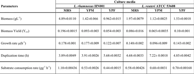
Results showed a better development for L. rhamnosus HN001with significant differences (α = 0.05) in biomass (gL-1) performances in MRS, YPM
and YPF media (4.89, 1.42 and 0.962 g-1 respectively). Other kinetic parameters
such as growth rate µ(h-1) and duplication time (h) for MRS and YPM did not show significant differences (α = 0.05); however, they were observed in YPF medium. For L. reuteri ATCC 53608, YPM medium did show meaningful differences (α = 0.05).
This study demonstrated that growth, biomass production, substrate consumption and the kinetic parameters of L. rhamnosus HN001 and Lactobacillus reuteri ATCC 53608 in different culture medium (MRS, YPM and YPF), change notably depending on the medium used. There is much information on the benefits of probiotic bacteria involving the use of such strains, with a particular focus on emerging probiotic therapies for humans, livestock, and aquaculture; however little has been published with respect to the kinetic behavior of these bacteria grown in different culture media. Parente and Ricciardi
(Parente and Ricciardi, 1999), explain that bacteriocin production by lactic acid
bacteria is associated to growth, and the yield of bacteriocin production per biomass unit is affected by several factors, including the producing strain, media
(carbohydrate and nitrogen sources, cations, etc.) and fermentation conditions (pH, temperature, agitation, aeration and dilution rate in continuous
fermentations).
Table 1 Effect of mediums: MRS, YPM and YPE on Lactobacillus rhamnosus HN001 and L. reuteri ATCC 53608 on the growth parameters
Parameters
Culture media
L. rhamnosus HN001 L. reuteri ATCC 53608
MRS YPM YPF MRS YPM YPF
Biomass (gL-1) 4.89±0.0110 1.42±0.066 0.962±0.015 1.97±0.0079 1.12±0.0025 1.53±0.0018
Biomass Yield (Yx/s) 0.196±0.0015 0.093±0.003 0.054±0.003 0.086±0.016 0.065±0.0035 0.10±0.001
Growth rate µ(h-1) 0.178±0.001 0.177±0.009 0.122±0.007 0.148±0.002 0.096±0.009 0.143±0.002
Duplication time (h) 3.89±0.0049 3.91±0.0026 5.68±0.0032 4.68±0.0033 7.22± 0.0018 4.85±0.0042
Substrate consumption rate (gg-1 h-1) 1.10±0.00436 0.53±0.0026 0.44±0.0015 0.58±0.00426 0.68±0.0031 0.70±0.00168
Furthermore, we observed that glucose was totally consumed by Lactobacillus rhamnosus HN001 in MRS at 25 hours of fermentation (Figure 3A) with a substrate consumption rate of 0.98 (g/g*h). However, in YPM and YPF media, the carbon source was not consumed in its entirety, leaving 50% Agave fructan and approximately 40% sugar cane molasses, with a substrate consumption rate of 0.86 (g/g*h) in YPM medium and 5.6 (g/g*h) in YPF medium, respectively. Lactobacillus reuteri ATCC 53608 in MRS broth only consumes 62% glucose, with a substrate consumption rate of 3.2 and 1.2 g/g*h for YPM and YPF media respectively (Figure 3B).
(A)
(B)
Figure 3 Consumption of carbon source (gL-1) of Lactobacillus rhamnosus HN001 (A) and Lactobacillus reuteri ATCC 53608 (B) in the three culture media (MRS, YPM and YPF).
Table 2 shows organic acid (acetic and lactic acids) and ethanol production in the different culture media with the two bacteria. L. reuteri ATCC 53608 produces acetic acid, lactic acid and ethanol when grown in MRS and YPM media, but in YPF medium it does not produce these metabolites, probably because its metabolism is directed only to biomass production and to maintain its viability. These bacteria begin to die when entering stationary phase (after 10 hours). Results of the metabolites produced by L. rhamnosus HN001 showed that the bacterium was capable of producing lactic and acetic acid in MRS broth, but did not produce ethanol in any medium (Table 2). L. rhamnosus HN001 was observed not to produce acetic acid when grown in YPM and YPF media; however, it was observed that the bacteria survived with a population of 4.1E+08 and 2.4E+08 CFU/mL respectively during stationary phase, up to 36 hours of fermentation (Figure 1B). Subsequently, the cells remained in a steady state with no increase in the population; however, at 36 hours the number of cells dropped to 1.4E+09 CFU/mL. It should be noted that although a decrease in bacterial growth was observed; a number of viable colonies (CFU/mL) remained alive, resisting the change of a culture medium now containing high lactic acid concentrations.
Table 2 Production of metabolites (gL-1) during fermentation.
Metabolite (gl-1)
Culture media
L. rhamnosus HN001 L. reuteri ATCC 53608
MRS YPM YPF MRS YPM YPF
Acetic acid 4.05±0.021 0±0 0±0 2.96±0.005 1.3±0.001 0±0
Lactic acid 9.71±0.050 4.77±0.047 1.98±0.001 7.84±0.031 4.85±0.028 0±0
Ethanol 0±0 0±0 0±0 3.15±0.020 0.51±0.001 0±0
Also, our results demonstrated that L. rhamnosus HN001 and Lactobacillus reuteri ATCC 53608 can grow and produce metabolites in diverse culture media with different kinetic parameters and consequently changing metabolite production such as bacteriocins. This is normal because those potential probiotic bacteria species differ in terms of their bioavailability, metabolic activity, and mode of action (Gänzle et al., 1998). The metabolism and the physiological properties of Lactobacillus from sourdoughs are highly adapted to their natural substrate and several studies suggest that the production of antagonists may further account for their dominance in the dough environment (Gopal et al.,
2001).
Antimicrobial spectrum and bacteriocin activity.
MRS broth was used to confirm bacteriocin production. The effect of growth media on bacteriocin activity was measured during growth and early stationary phases (8, 12 and 20 h). In general, an increased bacteriocin activity between 8 to 20 hours fermentation (Table 3), specifically in exponential phase, was observed, at the same time as the greatest number of cells (CFU/mL) (Figures 1 and 2). However, approximately 90% reduction of antimicrobial activity was noted when bacteria entered stationary phase (data not shown).
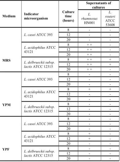
The highest antimicrobial activity was recorded for supernatants of L. reuteri ATCC 53608 and L. rhamnosus HN001 at 12 hours in MRS broth against L. acidophilus ATCC 43121 and L. delbrueckii subsp. lactis ATCC 12315 (Table 3), but not against L. casei ATCC 393. In YPM (sugar cane molasses) inhibition activity at 8 hours of culture was observed against the strains. YPF medium alone showed antibacterial activity in the culture with L. reuteri ATCC 53608 in
exponential phase between 4 and 8 hours against L. acidophilus ATCC 43121, where an inhibition zone of 2 mm was exhibited. L. rhamnosus HN001 antimicrobial activity was noted, but not for L. reuteri ATCC 53608 strain.
Table 3 Inhibitory spectrum of the cell free supernatants (CEB) of L. rhamnosus
HN001 and L. reuteri ATCC 53608 in MRS, YPM and YPF media at different times of culture, measured by well diffusion assay.
Medium Indicator microorganism Culture time (hours) Supernatants of cultures L. rhamnosus HN001 L. reuteri ATCC 53608 MRS L. casei ATCC 393 8 - - 12 - - 20 - - L. acidophilus ATCC 43121 8 + + - 12 + + - 20 + + - L. delbrueckii subsp. lactis ATCC 12315 8 + + + 12 + + + 20 + + - YPM L. casei ATCC 393 8 - - 12 - - 20 - - L. acidophilus ATCC 43121 8 + + 12 - - 20 - - L. delbrueckii subsp. lactis ATCC 12315 8 - - 12 - - 20 - - YPF L. casei ATCC 393 8 - - 12 - - 20 - - L. acidophilus ATCC 43121 8 + - 12 + - 20 - - L. delbrueckii subsp. lactis ATCC 12315 8 - - 12 - - 20 - -
ATTC-American Type Culture Collection, Rockville, MD, USA. Diameter of the inhibition zone ≥ 20 mm (+++), 10-5 mm (++) and 5–1 mm (+), 1-0 mm (-) no inhibition zone.
L. rhamnosus HN001 supernatant at 8, 12 and 20 hours of culture in supplemented MRS broth was inhibited slightly by L. acidophilus ATCC 43121 and L. delbrueckii subsp. lactis ATCC 12315 producing a 5 mm inhibition zone (Figure 4); and the supernatant of YPF media produced an inhibition of 2 mm against L. acidophilus ATCC 43121 (Figure 4). However, after 20 hours of fermentation, the antimicrobial activity decreased considerably or was not present, in the mediums YPM and YPF. The YPM medium supernatants of L. reuteri ATCC 53608 and L. rhamnosus HN001 produced a 2 mm inhibition zone against L. acidophilus ATCC 43121 but no inhibition was observed for L. delbrueckii subsp. lactis ATCC 12315, and L. casei ATCC 393. Additionally, L. rhamnosus HN001 supernatants on YPF medium did not exhibit inhibition for any indicator microorganism, but L. reuteri ATCC 53608 supernatants in this medium produced a small zone of inhibition (1mm) against L. acidophilus ATCC 43121.
(A) (B)
Figure 4 Inhibition zone of the cell free supernatant (CEB) of L. rhamnosus
HN001 in MRS broth, against L. delbrueckii subsp. lactis ATCC 12315 (A) and of the cell free supernatant in YPF medium, against L. acidophilus ATCC 43121 (B).
Results of antimicrobial bacteriocin produced in supplemented MRS broth (Table 4) showed a broad antimicrobial spectrum determined by well diffusion assay against all strains of Listeria monocytogenes and Lactobacillus sakei ATCC 53103, Enterococcus faecium CHUM, L. delbrueckii subsp. lactis ATCC 12315, L. acidophilus ATCC 43121, but no effect against E. coli O157:H7 EDL933, Salmonella typhimurium EDL933, Bacillus cereus LSPQ 2872, Staphylococcus aureus ATCC 29213 amongst others. Inhibition zones obtained in this trial measured 2 mm to 5 mm (Table 4).
Table 4 Inhibitory spectrum of the cell free supernatants (CEB) of the MRS
broth of L. rhamnosus HN001 and L. reuteri ATCC 53608 in exponential phase (12 hours of cultivation), by well diffusion assay.
Indicator
microorganism Source Medium
L. rhamnosus
HN001
L. reuteri
ATCC 53608
Bacillus cereus LSPQ 2872 TSA - -
Enterococcus faecalis ATCC 29212 TSA - + +
Enterococcus faecium ATCC 19434 TSA - + +
Escherichia coli ATCC 25922 TSA - -
Escherichia coli O157:H7 EDL933 TSA - -
Lactobacillus casei ATCC 393 MRS - -
Lactobacillus curvatus CRDA V32 MRS - + +
Lactobacillus helveticus ATCC 15807 MRS - -
Lactobacillus rhamnosus GG
ATCC
53103 MRS - -
Lactobacillus sakei ATCC 53103 MRS + + + +
Lactococcus lactis ATCC 11454 MRS - -
Listeria monocytogenes HPB 1043 TSA + + +
Listeria monocytogenes HPB 2569 TSA + + + +
Listeria monocytogenes HPB 2739 TSA + + + +
Listeria monocytogenes HPB 2812 TSA + + + +
Listeria innocua LSPQ 3285 TSA + -
Pseudomonas fluorescens FRDC V491 TSA - -
Pseudomonas putida FRDC V376 TSA - -
Pseudomonas fragi FRDC V378 TSA - -
Salmonella typhimurium SL1344 TSA - -
Salmonella enterica
subsp. enterica
ATCC
49217 TSA - -
Staphylococcus aureus ATCC 29213 TSA - -
Lactobacillus delbrueckii
subsp. lactis
ATCC
12315 MRS + ++
Lactobacillus acidophilus ATCC 43121 MRS + ++
ATTC- American Type Culture Collection, Rockville, MD, USA; LSPQ-Laboratoire de Santé Publique, Ste-Anne-de Bellevue, QC, Canada; FRDC-Food Research and Development Center, St-Hyacinthe, QC, Canada; HPB-Health Product Branch, Santé Canada; CHUM-Centre Hospitalier Universitaire de Montreal. Diameter of the inhibition zone ≥ 20 mm (+++), 10-5 mm (++) and 5-1 mm (+), 1-0 mm (-) no inhibition zone.
As for antimicrobial spectrum and bacteriocin activity, L. rhamnosus HN001 and L. reuteri ATCC 53608 were observed to produce an antimicrobial compound when grown in modified MRS broth, but in the supernatants of YPM and YPF media, inhibition activity lessens greatly. Studies have been published about bacteriocins produced by different strains of L. rhamnosus and L. reuteri
(Gänzle et al., 1998; Gänzle et al., 2000; Talarico and Dobrogosz, 1989), but
most authors deal with bacteria cultured in MRS broth or modified MRS broth with the addition of salts or other compounds but few authors have studied other culture media for bacteriocins production.
For example, Gänzle et al., (2000) studied the characterization of reutericyclin produced by Lactobacillus reuteri LTH2584 in MRS broth. They observed that fatty acid supply in MRS broth for the growth of these bacteria had a strong effect on reutericyclin production and its distribution between producer cells and the culture supernatant. On the other hand, Talarico et al., (1989) studied Lactobacillus reuteri 1063 cultured in modified MRS broth, and observed that it converted glycerol into a potent cell growth inhibitor. This substance, termed reuterin, inhibits the growth of Gram positive and Gram negative bacteria as well as yeasts, fungi, and protozoa.
It is known that bacteriocin production is deeply affected by type and level of the carbon, nitrogen and phosphate sources, cations, surfactants and inhibitors.
In our case, the obtaining of antibacterial compound (bacteriocin) was not totally satisfactory when L. rhamnosus HN001 and L. reuteri ATCC 53608 were grown in YPM and YPF culture media, probably due to the lack of some nutrient in the media or the carbon source used (cane molasses and Agave Tequilana fructan), which probably affected the growth and production of the antimicrobial substance.
It has been reported by other authors (De Vuyst and Vandamme 1992), that bacteriocins can be produced after producer strain growth in media containing different carbohydrate sources (glucose, sucrose and xylose). For example Nisin Z can be produced in media enriced with glucose, sucrose and xylose by L. lactis IO-1 (Chinachoti et al., 1997), but better results were obtained with glucose compared to xylose. Sucrose can be a better carbon source than glucose for bacteriocin production as forenterocin 1146 production by Enterococcus faecium DPC1146, but fructose or lactose result in comparable levels of biomass but lower levels of bacteriocin (Parente and Ricciardi, 1994; Parente et al., 1997).
Our results reveal that bacteriocins are produced during or at the end of exponential growth, the bacteriocins usually being extracellular products, but part of this activity may be retained within the cell (Parente et al., 1997). The most important factors influencing bacteriocin production and activity are physiological state of cells, culture medium, pH and temperature (Leal-Sánchez et al., 2002; Todorov and Dicks, 2005).
Effect of enzymes, detergents and temperature
Active substance sensitivity to enzymes was testedin supernatantof CEB from L. rhamnosus HN001 and L. reuteri ATCC 53608 using supplemented MRS broth. The results showed that antimicrobial bacteriocin was sensitive to protease IV, trypsin and pepsin, but was resistant to lipase and -amylases (Table 5). It was also resistant to detergents such as Tween 80, Triton-X, SDS and urea (Table 6).
Table 5 Effect of enzymes on bacteriocin-like substances from L. rhamnosus HN001 and L. reuteri ATCC 53608 cultured in MRS broth
Enzymes
Supernatant (CEB)
L. rhamnosus HN001 L. reuteri ATCC 53608 AU/mL Residual activity % AU/mL Residual activity % Pepsin 0 0 0 0 Lipase 50 50 25 12.5 Trypsin 0 0 0 0 Protease XIV 0 0 0 0 α-amilase 0 0 0 0 Control 100 100 200 100
Regarding the results obtained, an increase of 400 % of residual activity in supernatant extract (CEB) from L. rhamnosus HN001 and L. reuteri ATCC 53608 with Triton-X was obtained compared with others detergents used in this work. Others studies have reported higher values to 100 % of residual activity with Triton-X, observing that it was because there were probably too many bacteriocins containing hydrophobic links which tend to form large aggregates. Therefore, these macromolecules can be broken by using surface-active compounds (Tween, Triton, etc) and the disaggregation may cause a significant increase of antimicrobial activity (Ivanova et al., 1998; Seatovic et al., 2011).
Table 6 Effect of detergents on bacteriocin-like substancesfrom L. rhamnosus HN001 and L. reuteri ATCC 53608 cultured in MRS broth.
Detergent
Supernatant (CEB)
L. rhamnosus HN001 L. reuteri ATCC 53608 AU/mL Residual activity % AU/mL Residual activity % Tween 80 100 100 200 100 Triton-X 100 100 400 200 SDS 100 100 200 100 Urea 100 100 200 100 Control 100 100 200 100
Active antimicrobial bacteriocin sensitivity to enzymes was also tested and our results showed that antimicrobial bacteriocin was sensitive to all enzymes tested, but was resistant to the effect of different temperatures (Figure 5). Bacteriocin biochemical properties differ greatly with respect to their sensitivity or inactivation, mainly changes in pH (3 to 10) and temperature (60 °C to 100 °C) as well as the activity of some enzymes (Rogelj and Bogovic 1994). Many of the bacteriocins produced by lactic acid bacteria (LAB) are stable at neutral or acidic pH and inactivated at pH values above 8.0. This suggests that heat resistance of bacteriocin activity is based on their relatively small molecular structures. However, all bacteriocins are inactivated with at least one protease
(Rogelj and Bogovic 1994).
Different temperatures produced results that showed inhibition activity of supernatant of CEB, resistant to temperatures of 60°C, 90°C and 100°C for 20
minutes, but not at 121°C (Figure 5). Inhibitory activity at 100 °C was shown to be not affected in the supernatants of both bacteria, their inhibitory activity against indicator microorganism being maintained for 20 minutes heating.
It is noteworthy that a decrease in bacteriocin activity was observed during storage of the cell free L. rhamnosus HN001 and L. reuteri ATCC 53608 supernatants (CEB) in refrigeration; this may be attributed to degradation by proteases that were produced by the same bacteria during their growth. Some researchers refer to the decrease in bacteriocin inhibitory activity when pH decreases, so not surprisingly bacteriocin reduction is sometimes observed in fermentations without pH control (Yang and Ray, 1994).
Figure 5 Effect of temperature in the cell free supernatant (CEB) from L.
rhamnosus HN001 and L. reuteri ATCC 53608 of MRS broth.
Molecular weight determination
The molecular weight of the Lactobacillus rhamnosus HN001 and Lactobacillus reuteri ATCC 53608 bacteriocins in supplemented MRS broth was determined by electrophoresis on SDS-PAGE gel onto an MRS agar overlay as described in Material and Methods. The results obtained (data not shown) indicated that both bacteriocins have small molecular weight (less than 3.5 kDa), and that the inhibition zones were found below this molecular marker on the plate containing the indicator Lactobacillus sakei ATCC 15521 indicator.
The molecular weight of bacteriocin produced by Lactobacillus rhamnosus HN001 and Lactobacillus reuteri ATCC 53608 in the supplemented MRS broth is less than 3.5 kDa. Probably the antimicrobial compound produced by these bacteria, correspond to enterocin A, of type II class. The bacteriocins of this study had a weight similar to other studies, such as Plantaricin SA6 (Rekhif et al., 1995) and Sakacin B (Samelis et al., 1994), which produce bacteriocins with 2.5 kDa and 3.4 kDa molecular weight, respectively. Bacteriocins from lactic acid bacteria are ribosomally produced peptides (usually 30 to 60 amino acids). Ribosomal production of these small (2 at 6 kDa) antimicrobial peptides as a defense mechanism against other organisms is well documented (Nissen-Meyer,
1997). Class IIa bacteriocins can be considered as the major subgroup of
bacteriocins from lactic acid bacteria, not only because of their large number, but also because of their activities and potential applications (Saïd et al., 2000).
CONCLUSION
Both bacteria cultivated in all culture media produced bacteriocins with different antimicrobial capacity and these bacteria may be potential antimicrobial agents to be possibly used for safety in the food industry. The ability of L. rhamnosus HN001 and L. reuteri ATCC 53608 strains to produce inhibitory compounds capable of inhibiting Listeria monocytogenes species is a good reason to continue with selective studies for bacteriocin production, mainly to optimize culture conditions and culture medium. Finally, continuation of this work on bioreactor in batch or continuous culture, in order to control kinetic parameters and bacteriocin production would be of interest.
Acknowledgments: The authors acknowledge the economical support from the
Jalisco State Council of Science and Technology (COECYTJAL) and the critical reading of Patricia M. Hayward-Jones M Sc. and Dulce Ma. Barradas-Dermitz, M Sc.
REFERENCES
ABEE, T., KROCKE, L., HILL, C. 1995. Bacteriocins: modes of action and potentials in food preservation and control of food poisoning. Inter. J. Food. Microbiol, 28, 169-185.
BHUNIA, A.K., JOHNSON, M.C AND RAY, B. 1987. Direct detection of an antimicrobial peptide of Pediococcus acidilactici in sodium dodecyl sulfate-polyacrylamine gel electrophoresis. Journal Ind. Microbiol, 2, 319-322.
CHINACHOTI, N., MATSUSAKI, H., SONOMOTO, K., ISHIKAZI, A. 1997. Utilization of xylose as an alternative carbon source for nisin Z production by Lactococcus lactis IO-1. J. Fac. Agric. Kyushu. Univ, 42, 171-181.
DEMIRCI, A., POMETTO, A.L., BYUNGTAE, L AND HINZ, P.N. 1998. Media Evaluation of Lactic Acid Repeated-Batch Fermentation with Lactobacillus plantarum and Lactobacillus casei Subsp. rhamnosus. J. Agri. Food Chem, 46(11), 4771-4774.
DE VUYST, L AND VANDAMME, E.J. 1992. Influence of the carbon source on nisin production in Lactococcus lactis subsp. lactis batch fermentation. J. Gen. Microbiol, 138, 571-578.
DOLEYRES, Y., BECK, P., VOLLENWEIDER, S., LACROIX, C. 2005. Production of 3-hydroxypropionaldehyde using a two-step process with Lactobacillus reuteri. Appl. Microbiol. Biotechnol, 68, 467-474.
DORTU, C AND THONART, P. 2009. Les bactériocines des bactéries lactiques: caractéristiques et intérêts pour la bioconservation des produits alimentaires. Biotechnol Agron Soc Environ, 13(1), 143-154.
GÄNZLE, M., EHMANN, G.M AND HAMMES, W.P. 1998. Modeling of growth of Lactobacillus sanfranciscensis and Candida milleri in response to process parameters of the sourdough fermentation. Appl. Environ. Microbiol, 64, 2616-2623.
GÄNZLE, M.G., HÖLTZEL, A., WALTER, J., GÜNTHER, J AND HAMMES, W.P. 2000. Characterization of Reutericyclin Produced by Lactobacillus reuteri LTH2584. Appl. Environ. Microbiol, 66(10), 4325-4333.
GILL, H.S., RUTHERFURD, K.J., PRASAD, J., GOPAL, P.K. 2000. Enhancement of natural and acquired immunity by Lactobacillus rhamnosus (HN001), Lactobacillus acidophilus (HN017) and Bifidobacterium lactis (HN019). British. J. Nut, 83, 167-176.
GÓMEZ, E., TUOHY, K.M., GIBSON, G.R., KLINDER, A., COSTABILE, A. 2010. In vitro evaluation of the fermentation properties and potential prebiotic activity of Agave fructans. J. App. Microbiol, 108(6), 2114–2121.
GOPAL, P., PRASAD, J., SMART, J., GILL, H.S. 2001. In vitro adherence properties of Lactobacillus rhamnosus DR20 and Bifidobacterium lactis DR10 strains and their antagonistic activity against an enterotoxigenic Escherichia coli. Int. J. Food Microbiol, 67, 207-216.
GUANDALINI, S., PENSABENE, L., ZIKRI, M.A. 2000. Lactobacillus GG administered in oral rehydration solution to children with acute diarrhea: a multicenter European Trial. J. Pediatr. Gastroenter. Nutr, 30, 54-60.
IVANOVA, I., MITEVA, V., STEFANOVA, T.S., PANTEV, A., BUDAKOV, I., DANOVA, S., MONCHEVA, P., NIKOLOVA, I., DOUSSET, X., BOYAVAL, P. 1998. Characterization of a bacteriocin produced by Streptococcus thermophilus 81. International Journal of food microbiology, 42, 147-158.
KANDLER, O., STETTER, K.0. AND KOHL, R. (1980). Lactobacillus reuteri sp. nov., a new species of heterofermentative lactobacilli. Zentralbl. Bakteriol. Mikrobiol. Hyg. Abt. 1 Orig. C 1, 264-269.
KAWAI, Y., ISHII, Y., UEMURA, K., KITAZAWA, H., SAITO, T. AND ITOH, T. 2001. Lactobacillus reuteri LA6 and Lactobacillus gasseri LA39 isolated from faeces of the same human infant produce identical cyclic bacteriocin. Food Microbiol, 18, 407-415.
LEAL-SÁNCHEZ, M., JIMÉNEZ-DÍAZ, R., MALDONADO-BARRAGÁN, A., GARRIDO-FERNÁNDEZ AND RUIZ-BARBA, L. 2002. Optimization of Bacteriocin Production by Batch Fermentation of Lactobacillus plantarum LPCO10. Appl. Environ. Microbiol, 68(9), 4465-4471.
LINE, J.E., SVETOCH, E.A., ERUSLANOV, B.V. 2008. Isolation and purification of enterocin E-760 with broad antimicrobial activity against Gram-positive and Gram-negative bacteria. Antimicrobial Agents and Chemotherapy, 52 (3), 1094-1100.
LOPEZ, M.G., MANCILLA-MARGALLI, N.A AND MENDOZA-DIAZ, G. 2003. Molecular Structures of Fructans from Agave tequilana Weber var. azul. J. Agri. Food Chem, 51(27), 7835-7840.
MILLETTE, M., DUPONT, C., ARCHAMBAULT, D AND LACROIX, M. 2007. Partial characterization of bacteriocins produced by human Lactococcus lactis and Pediococccus acidilactici isolates. J. App. Microbiol, 102, 274-282. NISSEN-MEYER, J.I.F. 1997. Ribosomally synthesized antimicrobial peptides: their function, structure, biogenesis, and mechanism of action. Arch Microbiol, 167, 67-77.
O’SULLIVAN, L., ROSS, R.P., HILL, C. 2002. Potential of bacteriocin-producing lactic acid bacteria for improvements in food safety and quality. Biochimie, 84, 593-604.
PARENTE AND RICCIARDI, A. 1994. Effect of nitrogen and carbohydrate sources on lactic acid and bacteriocin production by Enterococcus faecium DPC1146. Agro Industry Hi-Tech, 5, 35-39.
PARENTE, E., BRIENZA, C., RICCIARDI, A., ADDARIO, G. 1997. Growth and bacteriocin production by Enterococcus faecium DPC1146 in batch and continuous culture. J. Ind. Microbiol. Biotechnol, 18, 62-67.
PARENTE, E AND RICCIARDI, A. 1999. Production, recovery and purification of bacteriocins from lactic acid bacteria. Appl. Microbiol. Biotechnol, 52, 628-638.
PRASAD, J., SMART J.B., GOPAL P.K., GILL H.S. 1998. Selection and characterization of Lactobacillus and Bifidobacterium strains for use as probiotics. Int. Dairy. J, 8, 993-1002.
REKHIF, N., ATRIH, A., LEFEBVER, G. 1995. Activity of plantaricin SA6, a bacteriocin produced by Lactobacillus plantarum SA6 isolated from fermented sausage. Journal of Applied Bacteriology, 78, 349-358.
RILEY, M.A AND WERTZ, J.E. 2002. Bacteriocins: evolution, ecology, and application. Ann Rev Microbiol, 56, 117-137.
ROGELJ, I AND BOGOVIC, M.B. (1994). Bacteriocins of Lactic Acid Bacteria Properties, Range of Inhibitory Activity and Methods of Detection. Prehrambeno-tehnol Biotehmol, 32 (4), 171-175.
SAÏD, E., TOSHIHIRO, S., KENJI, S., AYAAKI, I. 2000. Class IIa bacteriocins: biosynthesis, structure and activity. FEMS Microbiology Reviews, 24, 85-10. SAMELIS, J., ROLLER, S., METAXOPOULOS, J. 1994. Sakacin B, a bacteriocin produced by Lactobacillus sake isolated from Greel dry fermented sausages. Journal of applied bacteriology, 76, 475-486.
SEATOVIC, S,. JOVANOVI, J., ZAVISIC, G., RADULOVIC, Z., GAVROVIC-JANKULOVIC, M., JANKOV, R. 2011. The partial characterization of the antibacterial peptide bacteriocin G2 produced by the probiotic bacteria
Lactobacillus plantarum G2. Journal of the Serbian chemical society, 76 (5),
699-707.
SHU, Q AND GILL, H.S. 2002. Immune protection mediated by the probiotic Lactobacillus rhamnosus HN001 (DR20) against Escherichia coli O157:H7 infection in mice. FEMS Immun. Med Microbiol, 34, 59-64.
TALARICO, T.L AND DOBROGOSZ, W.J. 1989. Chemical Characterization of an Antimicrobial Substance Produced by Lactobacillus reuteri 1063. Antimicrob. Agents. Chemother, 33(5), 674-67.
TOBA, T., SAMANT, SK., YOSHIOKA, E., ITOH, T. 1991. Reutericin 6, a new bacteriocin produced by Lactobacillus reuteri LA 6. Lett Appl Microbiol, 13, 281-286.
TODOROV, S.D AND DICKS, L.M.T. 2005. Growth parameters influencing the production of Lactobacillus rhamnosus bacteriocins ST461BZ and ST462BZ. Ann Microbiol, 55(4), 283-289.
TOURE, R., KHEADR, E., LACROIX, C., MORONI, O AND FLISS, I, 2003. Production of antibacterial substances by bifidobacterial isolates from infant stool active against Listeria monocytogenes. J. Appl. Microbiol, 95, 1058-1069. THEPPANGNA, W., MURASE, T., TOKUMARU, N., CHIKUMI, H., SHIMIZU, E., AND OTSUKI K. 2007. Screening of the enterocin genes and antimicrobial activity against pathogenic bacteria in Enterococcus strains obtained from different origins. Journal of Veterinary Medical Science. 69 (12), 1235-1239.
WEESE, S.J AND ANDERSON, M.E.C. 2002. Preliminary evaluation of Lactobacillus rhamnosus strain GG, a potential probiotic in dogs. Can Vet J, 43, 771-774.
YANG, R AND RAY, B.1994. Factors influencing production of bacteriocins by lactic acid bacteria. Food Microbiol, 11, 281-291.
YU-PENG, L., ZHENG, P., ZHI-HAO, S., YE NI., JIN-JUN, D., LEI-LEI Z. 2008. Economical succinic acid production from cane molasses by Actinobacillus succinogenes. Bioresource Technology, 99 (6), 1736-1742.
ZHOU, J.S., GOPAL, PK., GILL, H.S. 2001. Potential probiotic lactic acid bacteria Lactobacillus rhamnosus (HN001), Lactobacillus acidophilus (HN017) and Bifidobacterium lactis (HN019) do not degrade gastric mucin in vitro. Int. J. Food Microbiol, 63, 81-90.