Université de Sherbrooke
Identification and isolation of multipotent stromal cells from human skeletal muscle
Par
Jennifer Downey Programme de Pharmacologie
Mémoire présenté à la Faculté de médecine et des sciences de la santé en vue de l’obtention du grade de Maître ès Sciences (M.Sc.)
en Pharmacologie
Sherbrooke, Québec, Canada Août, 2013
Membres du jury d ’évaluation
Pr. Guillaume Grenier, Ph.D., Département de Chirurgie Pr. Klaus Klarskov, Ph.D., Département de Pharmacologie
Dr. Frédéric Balg, M.D., Département de Chirurgie
Pr. Mannix Auger-Messier, Ph.D., Département de Pharmacologie Pr. Eileen M. Shore, Ph.D., Department o f Orthopaedic Surgery,
Perelman School o f Medicine, University o f Pennsylvania © Jennifer Downey, 2013
1+1
Published Héritage Branch Direction du Patrimoine de l'édition 395 Wellington Street Ottawa ON K 1A0N 4 Canada 395, rue Wellington Ottawa ON K1A 0N4 CanadaYour file Votre référence ISBN: 978-0-499-00385-0 Our file Notre référence ISBN: 978-0-499-00385-0
NOTICE:
The author has granted a non-
exclusive license allowing Library and Archives Canada to reproduce, publish, archive, preserve, conserve, communicate to the public by
télécomm unication or on the Internet, loan, distrbute and sell theses
worldwide, for commercial or non- commercial purposes, in microform, paper, electronic and/or any other formats.
AVIS:
L'auteur a accordé une licence non exclusive permettant à la Bibliothèque et Archives Canada de reproduire, publier, archiver, sauvegarder, conserver, transmettre au public par télécomm unication ou par l'Internet, prêter, distribuer et vendre des thèses partout dans le monde, à des fins com merciales ou autres, sur support microforme, papier, électronique et/ou autres formats.
The author retains copyright ownership and moral rights in this thesis. Neither the thesis nor substantial extracts from it may be printed or otherwise reproduced without the author's permission.
L'auteur conserve la propriété du droit d'auteur et des droits moraux qui protégé cette thèse. Ni la thèse ni des extraits substantiels de celle-ci ne doivent être imprimés ou autrement
reproduits sans son autorisation.
In compliance with the Canadian Privacy A ct some supporting forms may have been removed from this thesis.
W hile these forms may be included in the document page count, their removal does not represent any loss of content from the thesis.
Conform ém ent à la loi canadienne sur la protection de la vie privée, quelques
form ulaires secondaires ont été enlevés de cette thèse.
Bien que ces form ulaires aient inclus dans la pagination, il n'y aura aucun contenu manquant.
RÉSUMÉ
IDENTIFICATION ET ISOLEMENT DE CELLULES STROMALES MULTIPOTENTES DU MUSCLE SQUELETTIQUE HUMAIN
Par
Jennifer Downey Programme de Pharmacologie
Mémoire présenté à la Faculté de médecine et des sciences de la santé en vue de l’obtention du diplôme de maître ès sciences (M.Sc.) en Pharmacologie, Faculté de médecine et des
sciences de la santé, Université de Sherbrooke, Sherbrooke, Québec, Canada, J 1H 5N4 Le muscle squelettique humain est une source essentielle de cellules progénitrices ayant plusieurs applications thérapeutiques potentielles. Les cellules stromales mésenchymateuses du muscle squelettique humain (hmrMSCs) semblent être impliquées dans des pathologies telles l’ossification hétérotopique, la dégénérescence graisseuse et la fibrose. L’identification de la population cellulaire à l’origine de ces pathologies permettrait de mieux comprendre les mécanismes derrières celles-ci et aiderait à la création de traitements plus efficaces. Nous avons d ’abord mis au point une méthode d ’isolement et déterminer des conditions de culture pour la prolifération et le maintien en culture de la fraction cellulaire adhérente dérivée du muscle squelettique humain. Par le biais de la cytométrie en flux et des marqueurs connus des cellules stromales mésenchymateuses (MSC), nous avons pu enrichir les cellules stromales multipotentes. Le potentiel ostéogénique, adipogénique et chondrogénique des populations cellulaires enrichies a été évalué par des essais de différenciation. La sous-population de cellules CD73+CD105+CD90' a montré une multipotence robuste sur les trois lignées étudiées. Des essais de différenciation clonale ont confirmés que les trois lignées obtenues proviennent tous d ’un progéniteur multipotent commun. De plus, cette sous-population cellulaire avait la capacité de se différencier en cellule de gras brun, démontrée par une expression élevée d’UCPl au niveau génique et protéique suivant une stimulation continue avec le rosiglitazone (ROS). Ce résultat suggère que cette sous-population cellulaire pourrait également représenter un modèle pour l’adipogenèse vers le gras brun. La méthode d’enrichissement présentée représente une nouvelle technique afin d ’obtenir des hmrMSCs. Elle semble prometteuse pour de futures applications cliniques employant ces cellules, étant donné qu’elles sont amplifiées dans un milieu défini permettant une reproductibilité inter laboratoire. De plus, les marqueurs de phénotype choisis pour l’enrichissement par cytométrie en flux sont bien conservés entre individus, limitant la variabilité inter-donneur.
Mots clés : Cellules stromales mésenchymateuses, Cytométrie en flux, Gras brun, Maladies dégénératives, Multipotence, Muscle squelettique humain
IDENTIFICATION AND ISOLATION OF MULTIPOTENT STROMAL CELLS FROM HUMAN SKELETAL MUSCLE
By
Jennifer Downey Pharmacology Program
Thesis presented to the Faculty o f medicine and health sciences for the completion o f the Master’s degree diploma [Maitre ès Sciences (M.Sc.)] in Pharmacology, Faculty o f Medicine and Health Sciences, Université de Sherbrooke, Sherbrooke, Québec, Canada,
J1H 5N4
Human skeletal muscle is an essential source o f various cellular progenitors with potential therapeutic perspectives. Muscle-resident mesenchymal stromal cells (mrMSCs) are thought to be involved in the development o f several regenerative disorders such as fatty degeneration, heterotopic ossification and fibrosis. Identifying the cell population responsible for these pathologies will help better understand the underlying mechanisms and lead to more efficient treatment. We first developed an isolation method and culture conditions for the prolifération and maintenance o f the adhèrent fraction o f human skeletal muscle derived cells. To further enrich the cell population as multipotent progenitors, we used fluorescent-activated cell sorting (FACS) and known mesenchymal stromal cell (MSC) markers. The enriched cell populations obtained were tested for their multipotent capabilities towards the ostéogénie, adipogénie and chondrogenic lineages. The CD73+CD105+CD90' subset of human skeletal muscle adhèrent cells displayed robust multipotence to ail three lineages under the appropriate différentiation conditions. Clonal différentiation assays confirmed that ail three lineages stem from a single multipotent progenitor. Furthermore, this cell subset was able to differentiate into brown adipocyte-like cells, expressing UCP1 at the RNA and protein levels following prolonged stimulation with rosiglitazone (ROS). This resuit suggests that this cell subset could also represent a human cell model for brown adipogenesis. The cell isolation and enrichment method presented in this thesis represent a novel technique to obtain human mrMSCs. This method holds great promise for future clinical applications with the enriched cell populations since they are expanded in a defined medium, which supports inter-laboratory reproducibility. Furthermore, the phenotypic markers chosen for the FACS isolation are well conserved amongst donors in the proposed conditions, limiting donor-to-donor variability.
Keywords: Brown adipogenesis, Fluorescent-activated cell sorting, Human skeletal muscle, Mesenchymal stromal cells, Multipotency, Regenerative disorders
To my family, who has always encouraged me to be curious
R É SU M É ... Il S U M M A R Y ... III TABLE OF C O N T E N T S...V LIST OF FIGURES... IX LIST OF TABLES... X LIST OF A BBR EV IA TIO N S... XI FO R E W A R D ... 1 4 1 . IN TR O D U C TIO N ...2
1.1. Skeletal muscle tissu e ...2
1.1.1. Structure of skeletal muscle tissu e ... 2
1.1.2. Satellite cells... 4
1.2. Skeletal muscle régénération... 5
1.2.1. Degenerative p h a se ... 6
1.2.2. Regenerative phase... 6
1.3. Skeletal muscle regenerative disorders...8
1.3.1. Fibrosis and fatty degeneration... 10
1.3.2. Heterotopic ossification... 11
1.3.2.1. Fibrodysplasia ossificans progressiva... 11
1.3.2.2. Traumatic heterotopic ossification... 12
1.3.2.3. HO pathogenesis...12
1.4. Mesenchymal stromal c ells...13
1.4.1. Tissue sources of MSCs... 15
1.4.2. Identification of MSCs...16
vi 1.4.3.1. CD73... 17 1.4.3.2. CD105 (endoglin)... 17 1.4.3.3. CD90 (Thy-1)... 18 1.4.4. Multilineage potential of MSCs...18 1.4.4.1. Osteoblastic différentiation... 19 1.4.4.2. Adipogénie différentiation... 19 1.4.4.3. Chondrogenic différentiation... 19
1.5. Skeletal muscle résident mesenchymal stromal cell populations... 20
1.5.1. Pericytes...21
1.5.2. Muscle-derived stem cells (MDSCs)... 21
1.5.3. Myoendothelial cells (MECs)... 22
1.5.4. Skeletal muscle mesenchymal progenitors... 23
1.5.5. M uscle-resident strom al cells (mrSCs)... 24
1.5.6. Traumatized muscle mesenchymal progenitor cells...24
1.6. Other skeletal muscle progenitor populations...28
1.6.1. Brown adipocytes...28
1.6.1.l.O rigin of BAT... 28
1.6.1.2. Factors regulating human brown adipogenesis...29
1.6.1.3. Adult BAT...30
1.6.1.4. Brown adipocyte progenitor populations in skeletal m u scle...30
1.6.2. M yofibroblasts...31
1.6.2.1. TGFpi and myofibroblastic différentiation...31
1.6.2.2. Myofibroblast identification... 32
2 . HYPOTHESIS... 3 3 3 . OBJECTIVES...3 3 4 . MATERIAL A N D M E T H O D S ...3 4 4.1. Human adult skeletal muscle sam ples... 34
4.2. Tissue préparation...35
4.3. Cell culture... 35
4.6. Clonal growth and différentiation...39
4.7. RNA extraction... 40
4.8. Complementary DNA and reverse transcription...40
4.9. Quantitative PCR (qPCR)...40
4.10. Immunofluorescence...41
4.11. Western blot analyses...42
4.12. Myofïbroblastic différentiation... 43
4.13. Collagen gel contraction a ssa y ... 43
4.14. Statistical Analysis... 43
5 . RESULTS...4 4 5.1. Enrichment strategy for human muscle résident mesenchymal stromal cells... 44
5.1.1. Isolation and culture conditions... 44
5.1.2. A sub-population of human m uscle-resident cells expresses strom al progenitor m arkers ...45
5.2. Différentiation potential of human muscle résident mesenchymal stromal c e lls ...47
5.2.1. Only CD73+CD105+CD90 cells are m ultipotent in v itro...47
5.2.2. Clonal progenies of CD73+CD105+CD90_ hmrMSCs are m ultipotent... 49
5.2.3. The effect of BMP9 on ostéogénie différentiation of hum an muscle derived cells... 50
5.2.4. Decreased myogénie cells in the CD73+CD105+CD90 subset... 51
5.3. Other progenitor populations présent in human skeletal m u scle... 53
5.3.1. Prospective brown adipocyte progenitors in the CD73+CD105+CD90- s u b s e t... 53
5.3.2. Unsorted muscle résident cells contain myofïbroblastic cells... 55
6 . D IS C U S S IO N ... 5 7 6.1. Defined culture conditions are suitable for human muscle-derived MSC expansion...57 6.2. MSC phenotypic markers can be used to identify mrMSCs 59
6.3. Human skeletal muscle-derived CD73+CD105+CD90 MSCs are multipotent in v itro 60 6.4. Multipotent clonogenic assays with CD73+CD105+CD90- cells...62 6.5. BMP9 stimulation hinders ostéogénie différentiation in human muscle-derived c e lls 63 6.6. Prospective brown adipocyte progenitors in the CD73+CD105+CD90- human skeletal muscle su b se t...64 7.C O N C L U SIO N A N D PERSPECTIVES...6 6 Remerciements... 68 8 . LIST OF REFERENCES... 6 9 A N N E X I ...8 7 A N N E X I I ... 8 8 A N N E X III... 8 9
Figure 1.1: Skeletal muscle structure and organization... 3
Figure 1.2: Internai structure o f a muscle fib e r... 4
Figure 1.3: The myofîber and its satellite cells...5
Figure 1.4: Muscle tissue régénération... 6
Figure 1.5: Key factors involved in myogénie différentiation... 7
Figure 1.6: MSCs and skeletal muscle regenerative disorders... 9
Figure 1.7: Multipotent mesenchymal stromal cell characteristics...14
Figure 4.1: Semitendinosus tendon with associated muscle tissue (arrowhead)... 34
Figure 5.1: Prolifération and morphology o f adhèrent human muscle derived cells in sérum versus defined m ed ia...44
Figure 5.2: Flow cytometry sorting strategy o f human skeletal muscle stromal cell subpopulations...47
Figure 5.3: CD73+CD105+CD90‘ cells demonstrate superior ostéogénie, adipogénie and chondrogenic activity in vitro...48
Li s t o f t a b l e s
Table 1: Summary of the différent mesenchymal progenitor cell populations described from
skeletal m uscle...26
Table 2: Summary o f human muscle biopsies used for experim ents... 35
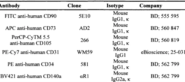
Table 3: Summary o f primary antibodies used for flow cytom etry... 37
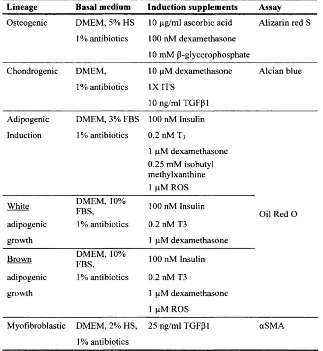
Table 4: Summary o f the multilineage différentiation media com position... 39
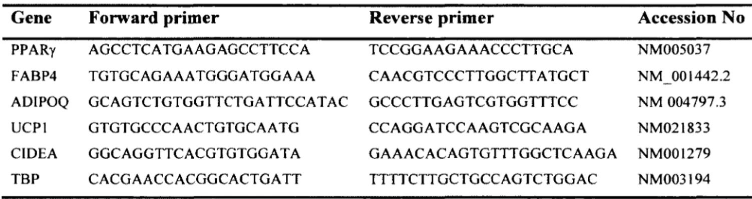
Table 5: Primer sequences used for gene expression analysis... 41
Abs Absorbance
ACL Anterior cruciate ligament
ADIPOQ Adiponectine
AlkP alkaline phosphatase
ASC Adipose stromal cell
BAT Brown adipose tissue
BMP Bone morphogenetic protein
CFU-F Colony-forming unit fibroblast
CHUS Centre hospitalier universitaire de Sherbrooke CIDEA Cell death-inducing DFFA-like effector A
cMEC clonal myoendothelial cells
Col2al Type II collagen
COX Cytochrome c oxidase
DMD Duchenne muscular dystrophy
DMEM Dulbecco’s modified eagle medium
ECM Extracellular matrix
FAB4 Fatty acid binding protein 4
FACS Fluorescence-activated cell sorting
FAP Fibro/adipogenic progenitors
FBS Fetal bovine sérum
FOP Fibrodysplasia ossificans progressiva
FSC Forward scatter
GAPDH Glyceraldehyde-3-phosphate dehydrogenase
GPI Glycosyl-phosphatidylinositol
GS Glycine-serine
hMAD human multipotent adipose derived stem cells
hmrSC Human muscle résident stromal cell
HRP HSC HSMM Ig MDSC MEC MHC MMP MPC MPC MRP mrSC MSC NHDF NSAID OC Osx PDGFRa P G C la PPARy PRDM16 PRE ROS Runx2 SP SSC TBP TGFpl THA Horseradish peroxidase Hematopoietic stem cell
Human skeletal muscle myoblast Immunoglobulin
Muscle-derived stem cells Myoendothelial cells Myosin heavy chain Matrix metalloproteinase Myogénie progenitor cell Mesenchymal progenitor cell Myogénie related factor muscle-resident stromal cell Mesenchymal stromal cell Normal human dermal fibroblast Nonsteroidal anti-inflammatory drugs Osteocalcin
Osterix
Platelet-derived growth factor receptor alpha
Peroxisome proliferator activated receptor gamma coactivator l a
Peroxisome proliferator activated receptor-y
PRD1-BF-1-RIZ1 homologous domain containing protein-16
PPARy-response element Rosiglitazone
Runt-related transcription factor 2 Side population
Side scatter
TATA-box binding protein Transforming growth factor-pi Total hip arthroplasty
WAT White adipose tissue
F
o r e w a r dDue to its marked regenerative capacity, skeletal muscle is one o f the most studied tissues in regenerative biology. Despite significant advances in understanding the process o f muscle tissue régénération after injury or disease, the identity o f the progenitor cells involved has been a matter of considérable debate, particularly with respect to regenerative disorders such as fibro/adipogenic degeneration and heterotopic ossification (HO).
Mesenchymal stromal cells (MSCs) are a population o f multipotent adult progenitor cells with many properties that make them attractive for use in the field o f regenerative medicine (Aldahmash et al. 2012). These cells are inherently plastic, enabling them to differentiate along différent lineages. They also appear to exhibit a number o f trophic properties that promote régénération in the surrounding tissue (Caplan 2009). Furthermore, MSCs can be harvested from a variety o f adult tissues (da Silva Meirelles et al. 2006), and of particular interest in this thesis from skeletal muscle (Huard 2008).
In spite of the potentially far-reaching promise o f MSCs in many aspects o f regenerative medicine, approaches using these cells are limited by the availability of a suitable MSC population in a clinical setting. Another obstacle arises from the identification o f MSC populations due to a lack o f standardization with respect to methods for their isolation and expansion.
This thesis focuses on the enrichment and characterization o f a muscle résident stromal progenitor cell population from adult human skeletal muscle.
1.1. Skeletal muscle tissue
Skeletal muscle is the largest tissue mass in the body, making up approximately 40% of the total body weight (Huard et al. 2002). As a form o f striated muscle tissue, its main function is to perform locomotor activity under the voluntary control o f the somatic nervous system. In addition, it is documented to have an important rôle in metabolism and its perturbation may contribute to metabolic diseases such as metabolic syndrome (Wells et
al. 2008).
1.1.1. Structure o f skeletal muscle tissue
As the name suggests, most skeletal muscles are attached to the bones by tendons. Although skeletal muscle is composed primarily o f contractile material, notably muscle fibers, it is a composite tissue o f blood vessels, connective tissue, and nerves. Several layers o f connective tissue encompass the muscle. The outer layer, the épimysium, groups several fascicles that surround the entire muscle and extend into the m uscles’ tendons. Each fascicle, made up o f parallel myofibers, also called muscle fibers, is enclosed within the périmysium. The endomysium, also referred to as the basai lamina, is the connective tissue that surrounds each individual myofiber (Fig. 1.1). The later separates myofibers from the surrounding connective tissue through which blood vessels run to provide nutrition, nerves to induce fiber contraction, and muscle résident stromal cells which maintain tissue integrity (Gillies and Lieber 2011).
8
Following the MPCs prolifération phase, myogenin is expressed during terminal différentiation and fusion. Myogenin expression enables MPCs to exit the cell cycle, through p21 expression. Unlike the other transcription factors discussed, the loss o f myogenin leads to severe skeletal muscle deficiencies, highlighting its essential rôle in the différentiation of MPCs during embryogenesis (Venuti et al. 1995). Myogenin is linked to muscle fiber fusion and maturation. The différentiation process is then completed by the activation o f muscle spécifie proteins such as myosin heavy chain (MHC), the motor protein of muscles thick filaments, leading to functional muscle fibers. Histologically, newly formed myofibers are characterized by central nuclei (Charge and Rudnicki 2004). Once MPC fusion is complété, newly formed myofibers increase in size due to functional protein synthesis, and myonuclei move to the periphery o f the muscle fiber (Fig. 1.4).
After injury, tissue régénération involves not only the replenishment or replacement o f parenchymal cells but also that o f supporting structures including blood vessels, nerves, connective tissue and stromal cells. Little is known about how this network o f associated cells, notably MSCs, coordinates myofiber growth, homeostasis and repair in skeletal muscle.
1.3. Skeletal muscle regenerative disorders
Although tissue régénération is ensured by tissue-specifïc stem cells, efficient tissue repair requires activation o f the surrounding stromal microenvironment, mainly a-smooth muscle actin (aSMA) positive myofibroblasts, to produce growth and survival factors, pro- inflammatory chemokines and components o f the extracellular matrix, such as collagens. However, inappropriate activation o f the stromal compartment can lead to persistence o f aSM A+ collagen-overproducing cells, extracellular matrix (ECM) accumulation and overgrowth o f fibrous and/or adipogénie tissue, thereby compromising tissue recovery and impairing function (Wynn 2008). Therefore, tissue repair culminâtes in either complété restoration of tissue integrity, defined as régénération, or in a process that leads to the génération o f stromal structures that replace functional tissue, which I will entitle ‘regenerative disorders’ in this thesis. The three most commonly occurring outcomes of
10
1.3.1. Fibrosis and fatty degeneration
After chrome injury, muscle is often replaced by a mix of fibrous tissue and white adipocytes in a process termed fatty degeneration. This fibro/adipogenic infiltration compromises muscle function and alters the tissue microenvironment, potentially limiting the success o f regenerative approaches.
Pathophysiologic fibrosis is a defining characteristic in most reparative disorders involving chronic inflammation (Porter et al. 2002). It is characterized by excessive accumulation of ECM in which collagen type I is the major component. It is the end resuit o f a cascade o f events proceeding from tissue injury via inflammation, and resulting in permanent scar formation. The accumulated ECM replaces normal parenchymal tissue and can affect tissues and organ Systems, resulting in anatomical anomalies as well as reduced functional capacities. In skeletal muscle, fibrosis is most often associated with the muscular dystrophies. As well as the muscular dystrophies, aging is associated with loss o f skeletal- muscle mass and function with concomitant fibrosis and ECM déposition (Mann et al.
2011).
The accumulation o f fat in damaged tissue leading to “fatty degeneration” is another common regenerativedisorder (Moyer and Wagner 2011). Usually, fat is found in newly formed adipocytes infiltrating the tissue, most often associated with concurrent fibrotic matrix déposition, and associated with injuries and defective repair processes (Wallace and McNally 2009). The appearance o f both fat accumulation and fibrosis has been shown in macrophage-suppressed mice (Warren et al. 2005; Segawa et al. 2008). Fat accumulation and fibrosis are also observed in mice lacking Myf5, a myogénie transcription factor (Gayraud-Morel et al. 2007). Furthermore, it is widely known that an increase in fatty and fibrous connective tissue is a hallmark o f advanced Duchenne muscular dystrophy (DMD), which is caused by a mutation o f the dystrophin gene (Carpenter and Karpati 2001 ).
The poorly defined fibroblast or myofibroblast, the effector cell component o f connective tissue, is generally thought to be responsible for producing excessive collagen and other ECM proteins (Hinz and Gabbiani 2010). However, the origin o f fibrosis in
skeletal muscle has not been elucidated (Mann et al. 2011). It has been suggested that both adipogénie and fibrogenic cells originate from myogénie cells through alternative lineage choices dictated by a pathological environment (Li et al. 2004; Shefer et al. 2004; Brack et
al. 2007). As an example, muscle side population (SP) cells isolated from dystrophic or
cardiotoxin-injured muscle, which normally contribute to myogenesis and muscle régénération, failed to undergo myogenesis and instead gave rise to fibroblasts and adipocytes (Penton et al. 2013), suggesting that muscle damage affects lineage choices o f muscle SP cells for a possible rôle in fibrosis and fat déposition. The fate o f these progenitors is therefore heavily dépendent on their microenvironment. This local microenvironment dictâtes whether these cells provide trophic support to satellite cells, the endogenous myogénie stem cells, to yield complété régénération o f injured muscle or whether they generate the components o f the fibro-fatty tissue infiltrâtes often found in degenerating muscle tissue. On the other hand, a population of skeletal muscle résident mesenchymal progenitors expressing platelet-derived growth factor receptor alpha (PDGFRof) has been shown to contribute to ectopic fat formation and fibrosis in mouse skeletal muscle under pathogenic conditions (Joe et al. 2010; Uezumi et al. 2011). Also, chronic activation o f PD G FRa+ cells leads to widespread organ fibrosis in mice (Oison and Soriano 2009).
1.3.2. Heterotopic ossification
HO is another regenerative complication involving the formation o f cartilage and bone outside o f the normal skeleton, within soft tissue such as muscle. Overall, it decreases range of motion and can cause partial or complété joint ankylosis for which surgical intervention is required. Usually, the development o f HO is either genetic (Shore and Kaplan 2010) or acquired following a traumatic injury. (Vanden Bossche and Vanderstraeten 2005; Porter et al. 2007).
1.3.2.1. Fibrodysplasia ossificans progressiva
The most severe form o f HO is manifested in the rare, autosomal dominant genetic disorder, fibrodysplasia ossificans progressiva (FOP), in which heterotopic bone forms
12
progressively throughout the life o f the individual, resulting in devastating effects on health, life expectancy, and quality o f life (Kaplan et al. 2004; Vanden Bossche and Vanderstraeten 2005). It was discovered that FOP results from the missense mutations in the highly conserved glycine-serine (GS) regulatory domain of the bone-morphogenetic protein (BMP) Type I receptor 1 A/activin-like kinase-2 (ACVR1/Alk2) (Shore et al. 2006; Fukuda et al. 2009), which renders the BMP signalling pathway hypersensitive or independent o f BMP ligands (Shore et al. 2006; Billings et al. 2008; Fukuda et al. 2009). Injury is also a trigger for HO in FOP; however, in this genetically susceptible background, even mild soft tissue trauma can cause pronounced heterotopic skeletogenesis (Kaplan et
al. 2008).
1.3.2.2. Traumatic heterotopic ossification
Traumatic HO is the most common type o f HO, observed following fractures, bums, surgical traumas, especially total hip arthroplasty (THA) (Nilsson and Persson 1999). For example, HO is diagnosed on average in 53% o f patients after THA. O f this number, 10% of patients suffer from severe HO with pain in the area o f the operated joint combined with a decrease in the range o f motion, leading to functional impairment (Brooker et al. 1973; Thomas 1992). Not to mention, HO is also prévalent in patients with severe extremity wounds following a polytraumatic injury, in fact in these cases the incidence o f HO increases by 57% (Potter et al. 2007). The incidence o f HO is also increased following central nervous system injuries (Newman et al. 1987; van Kuijk et al. 2002; Sakellariou et
al. 2012). The range o f these conditions suggests that factors released by distant tissues
such as the brain may have an osteoinductive effect on soft tissues either directly or by stimulating local cells to produce ostéogénie growth factors (Gautschi et al. 2009).
1.3.2.3. HO pathogenesis
Although the pathogenesis o f HO is not fully understood, it is generally agreed that an inciting event, such as a trauma induces local inflammation (McCarthy and Sundaram 2005). This is followed by the recruitment o f surrounding ostéogénie progenitor cells that differentiate into chondrocytes, undergo hypertrophy and are replaced by endochondrale
bone (Shore and Kaplan 2010). Therefore, HO is thought to resuit from the inappropriate différentiation of tissue résident ostéogénie progenitor cells that are induced by a pathological imbalance of local or systemic factors. However, the précisé cell origin and environmental eues leading to HO have not been fully elucidated. Since the most common cases of HO are in muscle and soft tissues (Nilsson and Persson 1999), it is important to understand the ostéogénie properties o f cells résident in skeletal muscle.
Among local tissue résident populations, satellite cells have received considérable attention as a possible cell-of-origin, primarily because of their muscle-restricted distribution and their capacity for BMP-dependent ostéogénie différentiation in culture (Katagiri et al. 1994; Wada et al. 2002). Cre/lox-based lineage analysis in the mouse, however, demonstrated that satellite cells in vivo do not appreciably contribute to HO lésions (Kan et al. 2009; Lounev et al. 2009).
1.4. Mesenchymal stromal cells
Friedenstein et al. was the first group to isolate multi-potential stromal precursor cells from the bone marrow (BM-MSCs). They described adhèrent, spindle-shaped, colony- forming unit fibroblasts (CFU-Fs) capable o f differentiating into adipocytes, chondrocytes and osteocytes both in vitro and after transfer in vivo (Friedenstein et al. 1974) (Fig. 1.7). It was subsequently demonstrated that these cells were also capable o f multi-lineage différentiation at the clonal level (Pittenger et al. 1999). A clonal différentiation assay is important since it supports the hypothesis that the différentiation potential belongs to a single multipotent progenitor rather than unipotent subpopulations.
also secrete trophic (pro-growth and pro-survival) factors that augment the endogenous régénération process (Tang et al. 2004; Lozito et al. 2009).
Therefore, multipotent mesenchymal stromal cells can be defined as a heterogeneous population o f cells that proliferate in vitro as plastic-adherent cells, have fibroblast-like morphology, form colonies in vitro and can differentiate into bone, cartilage and fat cells (Horwitz et al. 2005). Since their original description, MSCs categorized based on trilineage (osteoblast, adipocyte and chondrocyte) potential in vitro have been isolated from the adhèrent fraction o f many adult tissues in multiple species (da Silva Meirelles et al. 2006).
1.4.1. Tissue sources o f MSCs
In addition to bone marrow aspirate, MSCs have been isolated from a variety o f other adult tissues, including adipose tissue (Gimble and Guilak 2003; Meliga et al. 2007), skin (Toma et al. 2001), the marrow space o f long bones (Kuo and Tuan 2003; Tuli et al. 2003), trabecular bone (Tuli et al. 2003; Song et al. 2005), synovial membranes (De Bari et al. 2001), and periosteum (Nakahara et al. 1991; Choi et al. 2008) to name a few. However the limited availability of these tissues may limit their clinical potential. Similarly, MSCs have been harvested from tissues that are lost as a resuit o f development, such as the umbilical cord (Lee et al. 2004; Baksh et al. 2007) and umbilical cord blood/W harton’s Jelly (Cetrulo 2006; Troyer and Weiss 2008).
O f particular interest, there have also been several reports o f harvesting MSC-like cells from murine and human adult skeletal muscle. Human muscle tissue used to harvest cells was obtained from healthy muscle tissue biopsies (Zheng et al. 2007), surgical waste tissue from orthopaedic reconstructions, or surgically debrided muscle tissue following orthopaedic trauma (Nesti et al. 2008). Given that these cells can be obtained from surgical waste tissue or from minimally invasive biopsy procédures, there is growing evidence that skeletal muscle may be an important clinical source o f MSCs for use in therapeutic applications.
16
1.4.2. Identification o f M SCs
Identifying MSCs in vivo is difficult, due to their low abundance. Consequently, the majority o f work studying the properties o f MSCs has been performed using cultured MSCs which are selected by adhérence to tissue culture plastic, followed by différentiation assays to test their multilineage potential. This sélection method makes it difficult to compare MSC populations from différent species, isolation techniques, culture conditions, and donor sites.
Multipotence and adhérence to tissue culture plastic are important characteristics o f MSCs, yet there is no définitive, agreed-upon marker to positively identifÿ a population that is capable o f these functions (Kolf et al. 2007). Instead, human MSCs are identified based on the expression o f a number o f surface markers comprising CD105, CD90, CD73, CD71, CD44 and Stro-1 (Moroni and Fomasari 2013). Adhésion molécules like CD 166, CD 106, ICAM-1, and CD29 are also reported to be expressed on MSCs, while hematopoietic surface markers, such as CD45, CD34, CD 14 and CD11, are not. MSCs are also characterized by a négative expression of co-stimulatory markers like CD86, CD80, and CD40, as well as spécifie adhésion molécules related to platelet/endothelial cells (CD31 or PECAM-1), to neural adhésion (CD56), and to leukocyte function (CD18 or LFA-1) (Moroni and Fomasari 2013).
These markers were essentially developed for cells harvested from the bone marrow. Therefore, additional tissue-specific criteria may also apply to this cell population from other tissues. In fact, these surface markers are not homogeneously expressed throughout stromal cultures and they may vary with tissue source (Al-Nbaheen et a l 2013). This raises the question o f whether MSCs from différent anatomical locations, selected by classic adhérence and in vitro culture methods are biologically équivalent.
Interestingly, MSCs derived from various embryonic and postnatal tissues using identical culture conditions display significant différences in colony forming morphology, différentiation potential and gene expression (Lee et al. 2004; Panepucci et al. 2004; da Silva Meirelles et al. 2006; Kaltz et al. 2010). Furthermore, studies have characterized and
compared the immunophenotype o f cultured adipose-derived stromal cells (ADSCs), in early and later passages, and found that the expression profile changes during culture time. It has been repeatedly shown that freshly isolated ADSCs express différent surface markers than those in later passages (Mitchell et al. 2006; Varma et al. 2007). Many o f the markers, particularly surface proteins, may be differentially up- or down-regulated in vitro, possibly influenced by culture conditions such cell confluence and medium, which may be especially true when using serum-based culture medium.
1.4.3. Cell surface markers fo r MSCs
Although MSCs do not express unique phenotypic markers, the International Society for Cellular Therapy proposed minimal criteria (Dominici et al. 2006) for defîning MSCs based on their plastic adhérence, phenotype and trilineage multipotence in order to help standardize their identification amongst researchers. The phenotype définition requires the expression o f CD73, CD90 and CD 105, together with a lack o f expression o f haematopoietic progenitor and endothélial cell markers (CD34), a leukocyte marker (CD45) and several others markers spécifie for other cell types. However, these markers were identified in studies done using mostly human bone marrow MSCs and it is still to be determined whether they apply to human muscle-derived MSCs.
1.4.3.1. CD73
CD73 is an ecto-5’-nucleotidase, which is known to be involved in bone marrow stromal interactions (Barry et al. 2001), mesenchymal stem cell migration (Ode et al. 2011), and potentially, MSC modulation o f adaptive immunity (Eckle et al. 2007).
1.4.3.2. CD 105 (endoglin)
Several groups have reported the expression o f the cell surface receptor CD105 (endoglin) to be correlated with stem cell capacity within mesenchymal cells o f adipose tissue and bone marrow origin (Rada et al. 2011). Mechanistically, CD 105 has been shown to function as a TGFpl co-receptor (Castonguay et al. 2011; Zhang et al. 2011). CD105 has also been found to be involved in a variety o f other biological processes. Studies have
18
linked CD 105 to angiogenesis and neovascularization, the development o f pre-eclampsia, scleroderma, and psoriasis (Castonguay et al. 2011; Holmes et al. 2011; Nassiri et al. 2011; Pohl et al. 2011)
1.4.3 3. CD90 (Thy-1)
CD90 (Thy-1) is a glycosyl-phosphatidylinositol (GPI)-linked membrane protein which has been shown to be associated with osteoprogenitor cells (Chen et al. 1999; Chan
et al. 2009; Nakamura et al. 2010). CD90 is a member o f the immunoglobulin (Ig)
supergene family and is also expressed on the surface o f thymocytes, peripheral T cells, fibroblasts, épithélial cells, neurons, and hematopoietic stem cells (R eif and Allen 1964; Blankenhom and Douglas 1972; Williams 1982). Interestingly, CD90 expression varies with species (Dalchau and Fabre 1979; Kemshead et al. 1982) as well as the State o f différentiation (Chen et al. 1999).
1.4.4. Multilineage potential o f MSCs
Beyond their ability to generate osteoblasts, adipocytes and chondrocytes in vitro, MSCs give rise to bone and cartilage after ectopic implantation in vivo (Ashton et al. 1980; Haynesworth et al. 1992) and have been documented to contribute to bone régénération in animal models o f genetic bone disorders (Li et al. 2010).
Many studies have further reported MSC différentiation into multiple other cell types o f mesodermal and non-mesodermal origin, including endothélial cells (Oswald et al. 2004), cardiomyocytes (Makino et al. 1999), hépatocytes (Snykers et al. 2009) and neural cells (Phinney and Prockop 2007; Arthur et al. 2008). Although the multipotent capabilities o f MSCs are controversial, their différentiation into the ostéogénie, adipogénie and chondrogenic lineages are now widely accepted and well documented.
1.4.4.1. Osteoblastic différentiation
Osteoblasts develop through a sériés o f phases, initiated by cellular prolifération, followed by ECM déposition and matrix mineralization. This process o f cell maturation can be induced in vitro by the addition o f BMPs, such as BMP2 (Diefenderfer et al. 2003), or the addition o f a différentiation cocktail o f dexamethasone, ascorbate and (3- glycerophosphate (Jaiswal et al. 1997). A range o f transcription factors are known to be involved in the régulation o f ostéogénie différentiation (Marie 2008), with the two most popular being runt-related transcription factor 2 (Runx2/Cbfal) and Osterix (Osx). Runx2 is considered the major transcription factor controlling osteoblast commitment and différentiation. It is expressed early in skeletal development and throughout osteoblast différentiation. Osx, another important transcription factor involved in osteoblast commitment, appears to act downstream of RunX2. Osx is thought to act in the régulation o f numerous osteoblast genes including, osteocalcin (OC). The latter is a bone spécifie protein synthesized by osteoblasts and represents a good marker for ostéogénie maturation (Nakamura et al. 2009).
1.4.4.2. Adipogénie différentiation
Adipocytes mature though a sériés o f increasingly committed cell types, before expressing adipocyte spécifie markers such as fatty acid binding protein 4 (FAB4) (Samulin
et al. 2008) and forming lipid vesicles which can be detected by oil red O staining.
Peroxisome proliferator activated receptor-y (PPARy) is a nuclear hormone receptor, thought to be the master regulator of adipogenesis. There are two isoforms o f PPARy, PPARy 1 is ubiquitously expressed whilst PPARy2 is restricted to adipose tissues and appears to be a more potent stimulator o f adipogenesis (Mueller et al. 2002). PPARy is expressed early in the différentiation o f adipocytes, with forced overexpression of PPARy inducing adipogenesis in cultured fibroblast (Tontonoz et al. 1994).
1.4.4.3. Chondrogenic différentiation
Chondrogenic différentiation in vivo requires an initial condensation of the MSCs, which is mimicked in vitro by culturing MSCs as micromass pellets. Chondrogenic
20
différentiation can then be induced by the presence o f transforming growth factor-p (TGFP) resulting in the appearance o f a chondrocyte-like phenotype characterized by the up- regulation o f cartilage-specific molécules such as collagen type II and IX, aggrecan, versican, biglycan and decorin (Pelttari et al. 2008). Chondrocyte maturation evolves though a sequence of defined steps; initially differentiating cells are termed chondroblasts and are proliferative. This stage is then followed by a hypertrophie stage, marked by the expression of collagen type-X, which is vital in vivo for vascular invasion, osteoblast différentiation, and bone formation. Sox9 is a high-mobility group box-containing transcription factor and is considered the master regulator o f chondrogenesis. The Sox9 gene is expressed in ail chondrocyte progenitors and chondrocytes, but its expression is completely tumed off in hypertrophie chondrocytes (Zhao et al. 1997). This expression parallels that o f the gene for type II collagen (C ol2al), a spécifie marker o f chondrocyte différentiation.
1.5. Skeletal muscle résident mesenchymal stromal cell populations
It was originally suggested that satellite cells had the ability to differentiate into lineages other than the myogénie lineage, but recent studies have demonstrated that they are committed to the myogénie lineage and do not spontaneously adopt non-myogenic fates (Starkey et al. 2011). Also, several reports suggest that stem/progenitor cell populations other than satellite cells participate in skeletal muscle repair/regeneration. Several groups have identified muscle-resident mesenchymal progenitor cells with varying levels o f multipotency from mouse and human skeletal muscle. Interestingly, ail these described populations share a common characteristic: to be closely associated with small blood vessels within the interstitial tissue (Crisan et al. 2008; Crisan et al. 2012). Importantly, one must keep in mind that most o f the différent published sub-populations may be directly related to each other and/or share a common progenitor although they were not isolated and cultured the same way. Below are the main muscle résident mesenchymal progenitors described to date.
1.5.1. Pericytes
Pericytes are defmed by their location, closely encircling endothélial cells in small vessels and capillaries (Andreeva et al. 1998). Recent studies suggest that pericytes include progenitors o f différent cell types, capable o f differentiating along several lineages including ostéogénie and chondrogenic (Collett and Canfield 2005). However these populations represented pericyte-containing cultures and not purified pericytes. Dellavalle
et al. demonstrated that pericytes sorted from human skeletal muscle based on alkaline
phosphatase (AlkP) expression are myogénie precursors, distinct from satellite cells, which support skeletal muscle régénération (Dellavalle et al. 2007). Also, AlkP+ pericyte-derived cells can differentiate into osteoblasts and adipocytes in vitro.
Pericytes were sorted from multiple human organs, including fetal and adult skeletal muscle, by FACS. These perivascular cells were identified and sorted based on CD146+ expression, whereas contaminating myogénie (CD56+), hematopoietic (CD45+) and endothélial (CD34+) cells were eliminated. Sorted human perivascular cells (CD146+CD56' CD45'CD34') expressed recognized markers o f mesenchymal stem cells, including CD73, CD90 and CD105. Additionally, these cells can be expanded in vitro. Also, when cultured in appropriate inductive conditions, the sorted perivascular cells differentiated into chondrocytes, adipocytes and osteocytes at the clonal level in vitro and developed into bony nodules when transplanted into a skeletal muscle pocket in a murine model (Crisan et al. 2008).
1.5.2. Muscle-derived stem cells (MDSCs)
Murine skeletal muscle contains a population o f stem cells termed muscle-derived stem cells (MDSCs) (Qu-Petersen et al. 2002). MDSCs are isolated from the muscle homogenate using a pre-plating technique, which éliminâtes the contaminating more adhèrent cell types (Gharaibeh et al. 2008). The more slowly adhèrent MDSCs have demonstrated enhanced différentiation potential, and can be induced to become osteoblast, adipocytes and chondrocytes in vitro. Also, these MDSCs have the ability to differentiate into myoblasts in vitro and to promote muscle régénération in vivo. Interestingly, a
22
comparable population of MDSCs has not been harvested from human muscle tissue solely on the basis o f their adhésion characteristics.
1.5.3. Myoendothelial cells (MECs)
In addition to satellite cells (CD56+) and endothélial cells (CD34+CD144+), human skeletal muscle contains a rare (<0.5%) population o f cells that coexpress myogénie and endothélial cell markers (CD56+CD34+CD144+), which has been identified using FACS to isolate the cells that are positive for CD56+CD34+CD144+. Myoendothelial cells (MECs) proliferate long term, retain a normal karyotype, and survive better under oxidative stress than myogénie (CD56+) cells. Although most satellite cells are committed to the myogénie lineage, the small subset of satellite cells that co-expresses the endothélial markers is capable o f multilineage différentiation into myogénie, chondrogenic, and ostéogénie cells
in vitro. Also, these cells appear to have enhanced myogénie potential in vivo, participating
in myofiber régénération. MECs are associated with the vasculature in human skeletal muscle, specifically located in the intimai compartment of blood vessels within human skeletal muscle (Zheng et al. 2007).
A major limitation in characterizing the multipotent potential o f MECs was the likely heterogeneous nature in “stemness”, therefor it was investigated whether chondrogenic and ostéogénie potential was preserved at the clonal level. Clonal myoendothelial cells (cMECs: CD56+CD34+CD144+CD45') were purified (from ffesh human muscle biopsies by FACS. cMECs displayed robust multipotency in vitro and in vivo (Zheng et al. 2013), including chondrogenesis, osteogenesis, and adipogenesis, in addition to myogenesis. cMECs seem to represent a human counterpart of murine MDSCs, as one o f the multilineage mesodermal stem cell populations residing in the vascular niche within the adult skeletal muscle.
/. 5.4. Skeletal muscle mesenchymal progenitors
The PDGFRa was used to identify mesenchymal progenitors in adult skeletal muscle, responsible for ectopic fat cell formation (Uezumi et al. 2010). PDGFRa+ mesenchymal progenitors are distinct from satellite cells and are located in the muscle interstitium. The expression of PDGFRa appears to be a characteristic o f undifferentiated MSCs (Bail et al. 2007). According to Uezumi et al., o f the muscle-derived cell populations, only PD G FR a+ cells show efficient adipogénie différentiation both in vitro and in vivo (Uezumi et al. 2010). In a subséquent study, it was shown that PD G FR a+ mesenchymal progenitors could also differentiate into ECM-producing cells. Clonal analysis showed that these cells possess the ability to differentiate into both collagen type-I-producing cells and adipocytes (Uezumi
et al. 2011).
Moreover, murine fibro/adipogenic progenitors (FAPs) sorted by FACS (Lin’a7'Sca- 1+) efficiently generate both fibroblasts and adipocytes at the clonal level, but fail to generate other mesenchymal lineages. Interestingly, Lin'a7‘S ca-l+ cells were uniformly PDGFRa+ (Joe et al. 2010).
Recent lineage tracing studies have implicated the vascular endothélium in HO, as cells expressing the angiopoietin receptor, Tie2, robustly contribute to ail stages o f BMP- induced skeletogenic lésions (Lounev et al. 2009; Medici et al. 2010). A population o f Tie2+PDGFRa+S cal+ cells was identified as multipotent mesenchymal progenitors that résidé in the murine skeletal muscle interstitium and represent a significant cell o f origin for HO. These progenitors résidé in the interstitium o f skeletal muscle and other tissues, and are distinct from the endothélium. Intramuscular transplantation, together with clonal analysis in culture, revealed that these progenitors are multipotent, exhibiting the capacity for both BMP-dependent skeletogenic différentiation and spontaneous adipogénie différentiation (Wosczyna et al. 2012).
Recently, a group suggested that PD G FR a+ mesenchymal progenitors residing in human skeletal muscle are the major cell origin o f HO. In this study, both CD56+ myogénie cells and PDGFRoC mesenchymal progenitors showed comparable ostéogénie
24
différentiation potential in vitro. However, in an in vivo ectopic bone formation model, only PDGFRa+ cells formed bone-like tissue and showed successful engraftment (Oishi et al. 2013)
1.5.5. Muscle-resident stromal cells (mrSCs)
Our laboratory identified and isolated a population o f murine muscle-resident stromal cells (mrSCs) which are also closely associated with the vasculature (Scime 2005, Leblanc 2011). The common stromal cell marker Seal was used to identify the mrSC population, whereas hematopoietic (Lin+) and endothélial (C D 31) cell lineages were discarded. Depending on their culture conditions, this cell population took on an endothélial (Grenier
et al. 2007), an adipogénie (Scime et al. 2005), a fibrogenic (Trensz et al. 2010) or an
ostéogénie fate (Leblanc et al. 2011). This subpopulation is also augmented in chronic (Trensz et al. 2010) and acute muscle damage (Grenier et al. 2007; Leblanc et al. 2011), a condition which is known to lead to fibrosis and fatty degeneration. Not only are they known to express RunX2 and AlkP during ostéogénie différentiation in vitro, mrSCs have been shown to contribute to HO in vivo in a murine model for HO which consisted o f injecting BMP9 within a damaged muscle (Leblanc et al. 2011).
1.5.6. Traumatized muscle mesenchymal progenitor cells
A population of multilineage mesenchymal progenitor cells was identified in human traumatized muscle, capable o f undergoing ostéogénie différentiation in vitro (Nesti et al. 2008). These cells were isolated and enriched from digested muscle tissue based on their high adhésion characteristics to tissue culture plastic. Moreover, they have a cell surface epitope profile that is characteristic o f BM-MSCs (Jackson et al. 2009), although they appeared to demonstrate limited lineage commitment compared to their bone marrow counterparts. It was shown that damage triggers the activation and prolifération o f a higher number o f progenitors within muscle and that those cells were more responsive to ostéogénie eues (Jackson et al. 2011)
The ongoing research is focused on improving skeletal muscle régénération, and limiting regenerative disorders, by investigating the hypothesis that cells related to the vascular wall in muscle tissue can (under appropriate conditions) give rise to potent “stem” cells that possess strong multilineage potential. For the time being, there is a lack o f consensus in the field on the appropriate MSC isolation technique, caused largely by the absence o f a truly sélective and universally adopted MSC marker, especially in human skeletal muscle.
1.6. Other skeletal muscle progenitor populations
1.6.1. Brown adipocytes
Humans have two types o f adipose tissue: white adipose tissue (WAT), which not only stores energy in the form o f triglycérides but is also recognized as an important endocrine and immune organ, and brown adipose tissue (BAT), which is specialized in energy expenditure by buming lipids to generate heat (thermogenesis). W hile WAT structure is characterized by a single, large, lipid droplet in vivo and few mitochondria, BAT contains several small lipid droplets (multilocular), many mitochondria, and uniquely expresses high levels o f uncoupling protein 1 (UCP1) (Cannon and Nedergaard 2010; Nedergaard et al. 2010; Richard and Picard 2011). UCP1 is a proton channel o f the inner mitochondrial membrane that allows proton influx into the mitochondrial lumen. UCP1 acts to uncouple oxidative phosphorylation from ATP production, thereby releasing energy as heat (termed thermogenesis). BAT plays a pivotai rôle in adaptive thermogenesis, a physiological process during which energy is dissipated in response to environmental changes, such as cold température and diet (Lowell and Spiegelman 2000).
1.6.1.1. Origin o f BA T
It has been suggested that BAT and skeletal muscle may share a common developmental ancestry (Timmons et al. 2007) . Myf5-expressing progenitors can give rise to both skeletal muscle and interscapular brown fat (Seale et al. 2008). In accordance with these observations, BAT progenitors have also been identified in human skeletal muscle (Crisan et al. 2008). These cells provide the potential for increasing the oxidative capacity o f these tissues by targeting these endogenous precursor cells to differentiate in vivo into energy-dissipating brown adipocytes. Though, it is now clear that two différent types o f brown adipocytes exist, which have distinct developmental origins. Classical brown adipocytes residing in the interscapular and perirenal régions develop from myofibroblastic-like Myf5-positive precursors (Seale et al. 2008) that differentiate into brown adipocytes through the action o f transcriptional regulators PRDM16 and C/EBPb (Kajimura et al. 2009); whereas, pockets o f a second, distinct type o f UCP1-positive
adipocyte are found sporadically in the WAT of adult animais that have been exposed to chronic cold or p-adrenergic stimulation. These inducible brown-like adipocytes (beige or brite cells) possess many o f the biochemical and morphological characteristics o f classical brown adipocytes, including the presence o f multilocular lipid droplets (Frontini and Cinti 2010). However, they arise from a non-Myf5 cell lineage, hence, have a distinct origin. Also, it has been shown that epidydimal WAT-derived brite cells that are induced by rosiglitazone (ROS) do not express myocyte-enriched genes (Petrovic et al. 2010).
1.6.1.2. Factors regulating human brown adipogenesis
Among factors regulating human brown adipogenesis, most of them have been identified and characterized in mouse models in vitro as well as in vivo. Within these signaling pathways, Hedgehog and BMP7 were described as modulators o f brown adipogenesis (Tseng et al. 2008; Pospisilik et al. 2010; Schulz et al. 2011). When tested on human multipotent adipose derived stem cells (hMADs) as a model for brown adipogenesis, both Hedgehog and BMP7 did not have any significant effect on hMADs “brite” conversion, indicating discrepancies between mouse and human brown adipocyte régulation (Pisani et al. 2011). However, it has been shown that the activation o f PPARy by synthetic ligands induces a brown fat-like gene program in WAT (Fukui et al. 2000; Sell et
al. 2004; Rong et al. 2007; Petrovic et al. 2010). Among members o f the thiazolidinedione
family, ROS is known as a high-affinity ligand o f PPARy. Mechanistically, these drugs function by directly binding to and activating PPARy and PPAR-response elements (PREs) on the promoter and/or enhancer o f brown fat-selective genes (Sears et al. 1996; Viswakarma et al. 2007).
Recent research has also identified several dominant transcriptional regulators o f brown adipocyte development and function, including peroxisome proliferator activated receptor gamma coactivator l a (PGC1 alpha) and PRD1-BF-1-RJZ1 homologous domain containing protein-16 (PRDM16) (Kajimura et al. 2010). P G C -la is highly expressed in BAT compared with WAT and is responsible for regulating mitochondrial biogenesis and thermogenesis. Mice déficient in P G C -la are cold-sensitive with low expression o f UCP1 and a morphologically abnormal BAT (Lin et al. 2004). It is generally accepted that
PGC-30
l a is the critical regulator o f adaptive thermogenesis in responsive tissues. PRDM16 on the other hand is a 140-kDa zinc-finger PR domain-containing protein that induces a program o f gene expression as well as mitochondrial biogenesis/oxygen consumption, consistent with the brown phenotype, when ectopically expressed in white preadipocytes in culture or white dépôts in the animal (Seale et al. 2007). Also, PPARy functions to regulate the underlying adipogénie programs common to both white and brown adipocytes. It is possible that its activity can favor one phenotype over the other. Several recent studies have shown that exposure o f white adipocytes, either in culture or in animais, to potent PPARy ligands such as ROS induces a “browning” of the white cells, as characterized by an increase in mitochondrial mass and structure as well as a markedly enhanced oxygen consumption and lipid oxidation (Wilson-Fritch et al. 2003; Wilson-Fritch et al. 2004). This process is likely due to PPARy ligand-associated induction o f mitochondrial genes, including UCP1 and cytochrome c oxidase (Cox) (Wilson-Fritch et al. 2004).
1.6.1.3. Adult BAT
In humans, BAT is présent in abundant quantity in newboms (Lean et al. 1986), but it was traditionally believed that BAT was nonexistent or non-functional in adult humans. However, this dogma was reversed by evidence from nuclear medicine (Nedergaard et al. 2007; Cypess et al. 2009; Saito et al. 2009; van Marken Lichtenbelt et al. 2009; Virtanen et
al. 2009), which showed active BAT in adult humans. Indeed, fluorodeoxyglucose (18F-
FDG) positron émission tomography (PET) studies allowed visualization in humans o f highly dynamic adipose tissue dépôts. Their metabolism was stimulated by cold. These dépôts were proposed to represent BAT that had been undetected until now (Nedergaard et
al. 2007). Since then, there has been a flurry o f new data surrounding BAT function and
therapeutic potential (Enerback 2010; Ravussin and Galgani 2011; Muller and Bosy- Westphal 2013).
1.6.1.4. Brown adipocyte progenitor populations in skeletal muscle
In adult humans, it remains to be determined which population o f progenitors gives rise to brown fat. Although a group identified a subpopulation o f adipogénie progenitors
(S ca-l+/CD457M acl'; referred to as Sca-1+ cells) residing in murine brown fat, white fat, and skeletal muscle. These Sca-1+ cells from skeletal muscle and subcutaneous white fat are highly inducible to differentiate into brown-like adipocytes upon stimulation with BMP7 (Schulz et al. 2011).
1.6.2. Myofibroblasts
Following tissue injury, fibroblasts are recruited during the inflammatory phase o f tissue régénération. These cells are then activated and acquire a smooth muscle cell-like phenotype; they are consequently called myofibroblasts. These myofibroblastic cells synthesize and deposit ECM components. They also exhibit contractile properties, due to the expression o f a-smooth muscle actin (aSM A ) in microfilament bundles or stress fibers, playing a major rôle in the contraction and maturation o f the granulation tissue (Hinz 2010). The presence o f aSMA represents the most reliable marker o f the myofibroblastic phenotype (Tomasek et al. 2002). During the final phase o f healing, proteolytic enzymes, essentially matrix metalloproteinases (MMPs) and their inhibitors (tissue inhibitor o f metalloproteinases [TIMPs]) play an essential rôle. Following which the number o f vascular cells and myofibroblasts is dramatically reduced by the process o f apoptosis (Desmouliere et al. 1995). Persistent fibrosis is often due to an imbalance between ECM synthesis and dégradation by myofibroblasts due to chronic inflammation.
1.6.2.1. TGFfil and myofibroblastic différentiation
Several cytokines and growth factors have a rôle in wound healing. Among these, TGFpl is a potent inducer of myofibroblastic différentiation (Desmouliere et al. 1993). Beyond induction of aSMA expression, TGF(31 also promotes the déposition o f large amounts of ECM and reduces MMP activity by promoting TIMP expression (M icallef et al. 2012). Hence, TGFpi plays a key rôle in inducing the formation o f fibrotic tissue that limits muscle healing after severe injury.
32
1.6.2.2. Myofibroblast identification
One limitation that has hindered studies o f fibrosis is the lack o f good markers to label fibroblasts/myofibroblasts. It is well established that in non-muscle Systems, activated fibroblasts may be identified by their increased prolifération, migratory ability, enhanced contractility, increased expression o f vimentin and, in particular, aSM A, a contractile protein o f stress fibers. When aSM A stress fîbers contract, they exert mechanical tension on the ECM, which in tum provides a mechanically résistant support, hence the name “myofibroblast”. These cells are associated with tissue repair and fibrosis in many tissues and organs, including muscle, skin, liver, lung, bone and cartilage (Hinz et al. 2007). However, despite their relevance in these diseases, it remains unclear whether myofibroblasts are présent in fibrotic skeletal muscle, or whether they are instead mature fibroblasts actively producing ECM components (Lieber and Ward 2013). The identification o f the myofibroblastic progenitor in skeletal muscle would help détermine their rôle in fibrosis.
The identity and régulation o f the multipotent progenitors residing within the skeletal muscle are topics o f great interest and intense study. Developing a method to obtain an enriched muscle résident stromal cell population from human skeletal muscle would help understand their rôle during normal skeletal muscle régénération as well as the mechanisms behind their aberrant activation during regenerative disorders. Furthermore, the multipotent capabilities of these cells also make them excellent candidates for potential applications in cellular therapy and/or tissue engineering.
We believe that FACS can be used as an efficient enrichment method, to isolate the stromal subpopulation from the adhèrent fraction o f human muscle-derived cells by using multiple mesenchymal stromal cell markers identified in other tissue sources. We propose that these markers will enable the identification o f the human mrMSC subset as they do in other tissue sources and enable the isolation o f a cell subpopulation with superior multipotent capabilities. Furthermore, we propose that a defined culture system that enables the prolifération and maintenance o f these cells in culture will be more compatible with future applications and foster inter-laboratory reproducibility.
To test this hypothesis, we have established three objectives:
3 . O
b j e c t i v e s1) Develop a reproducible enrichment strategy for the isolation and the culture o f the adhèrent human muscle résident mesenchymal stromal cell fraction
2) Identify the multipotent stromal cell subpopulation
Table 2: Summary of human muscle biopsies used for experiments
Sample No Sex Age (years) Weight (g)
M37 M 21 3.84 M38 F 37 3.23 M39 F 28 4.49 M41 M 24 6.30 M52 F 41 7.35 M58 M 36 7.35 M60 F 52 5.88 M63 M 21 5.72 M68 M 34 4.58 Average 32.7±8.1 5.4±1.2 4.2. Tissue préparation
Skeletal muscle samples were carefully dissected leaving them free o f any tendon and/or fat tissues. They were then minced and digested with 1 mg/mL collagenase type I (Sigma) in DMEM containing 10% FBS, for 30 min at 37°C at a ratio o f 4g o f tissue for 30ml of lmg/ml collagenase type I in medium. The tissue slurry was diluted with medium and passed through a 70-pm and then a 40-pm cell strainer (Becton Dickenson) prior to centrifugation (325xg, 6 min, 4°C). Cell pellets were resuspended in 8ml o f fresh medium.
4.3. Cell culture
Primary human muscle derived adhèrent cells were grown in Mesencult-XF® medium (StemCell Technologies, Vancouver, Canada) supplemented with 1% antibiotics. This medium is referred to as ‘defined medium’. Collected cells were counted and allowed to attach in a 100-mm tissue culture plate coated with Mesencult-XF® attachment substrate (StemCell Technologies) for approximately 7 days without a medium change on day 4. The cells isolated from approximately 4g o f tissue were left to adhéré in a single 100mm pétri dish. Roughly 7 x l0 5 adhèrent cells were recovered per gram o f tissue. At 80% confluence,