International Journal of Gynecology & Obstetrics 57 (1997) 295-303
GYNECOLOGY
&OBSTETRICS
Article
Radiation therapy duration influences overall survival in
patients with cervical carcinoma
J.F. Delaloye”,” , P.A. Couckeb, S. Pampallona’,
G. Peltecu”, P. De Grandi”
“DLpartement de Gynkologie et d’Obst&que, Centre Hospitalier Uniuersitaire Vaudois, L,ausanne, Swi~zerlundbSeruice de Radio-Oncologic, Centre Hospitalier Uniuersitaire Vaudois, Lausnnne, Switzerland ‘forMed Statistics for Medicine, Geneva, Switzerland
Received 31 July 1996: revised 5 February 1997; accepted 21 February 1997
Abstract
Objective: This article analyses the influence of treatment duration on survival in patients with invasive carcinoma of the cervix treated by radical radiation therapy. Method; Three hundred and sixty patients with FIG0 stage IB-IIIB carcinoma of the cervix were treated in Lausanne (Switzerland) with external radiation and brachytherapy as first line therapy. Median therapy duration was 45 days. Patients were classified according to the duration of the therapies, taking 60 days (the 75th percentile) as an arbitrary cut-off. Results: The 5-year survival was 61% (SE. = 3%) for the therapy duration group of less than 60 days and 53% (SE. = 7%) for the group of more than 60 days. In terms of univariate hazard ratio (HR), the relative difference between the hvo groups corresponds to a 50% increase of deaths (HR = 1.53, 95% CI = 1.03-2.28) for the longer therapy duration group (P = 0.044). In a multivariate analysis, the magnitude of estimated relative hazards for the longer therapies are confirmed though significance was reduced (HR = 1.52, 95% CI = 0.94-2.45, P = 0.084). Conclusion: These findings suggest that short treatment duration is a factor associated with longer survival in carcinoma of the cervix. 0 1997 International Federation of Gynecology and Obstetrics
Keywords: Treatment time; Overall survival; Radiation therapy; Cervical carcinoma
1. Introduction [l-3]. External irradiation and brachytherapy are combined in order to increase local control and Carcinoma of the uterine cervix can be effec- hence survival. Both treatment modalities may be tively treated with definitive radiation therapy applied. One of the unanswered questions is how to combine these two radiotherapeutic modalities. Should they be given sequentially or concomi-
*Corresponding author. Tel.: +41 21 3143269; fax: + 41 21 tantly? Overall treatment time will depend upon
3143263. the choice of the treatment sequence.
0020-7292/97/$17.00 0 1997 Intcmational Fcdcration of C~ynecology and Obstetrics
296 J.F. Delaloye et al. /International Journal of Gynecology & Obstetrics 57 (1997) 295-303
Recently the importance of overall treatment
time has been highlighted in large series of
patients treated by radiation therapy for cervical
cancer [4-71. The importance of this particular
prognostic factor for the outcome is supported by
in vivo evidence of a short potential doubling
time CT,,,) in cervical cancer [8]. This Tpot value,
measured prior to treatment, is probably closely
related to the kinetics of proliferative clonogenic
cells during treatment, which is generally thought to be at the origin of failure to control the disease
locally. To check the intrinsic importance of the
sequencing of both treatment modalities and
hence treatment duration, we decided to retro-
spectively analyze a consecutive series of patients, treated by external radiation and brachytherapy. 2. Methods
2.1. Patients population
From January 1971 to December 1992, 612
consecutive patients with primary FIG0 stage
IB-IIIB carcinoma of the cervix were treated with
radiation therapy. Only 360 of them received both
external radiation and brachytherapy, and were
considered for the current study. Of the 252 patients excluded, 114 received either external
radiation or brachytherapy alone, 40 had been
diagnosed too recently or had incomplete base-
line data, and 98 had undergone total hysterec- tomy. Patients characteristics are summarized in Table 1. The median age was 60 years (range 20-90 years). Baseline evaluation included physi-
cal and pelvic examination, cervical biopsies, com-
plete blood count, chemistry profile, chest X-ray,
intravenous pyelogram or computerized tomogra-
phy, cystoscopy and rectoscopy. Treatment vari-
ables are described below and summarized in
Table 2.
2.2. Radiation therapy
External radiation therapy was delivered ini-
tially with a betatron (45 MeV) and later with a linear accelerator (6-18 MeV). The pelvic volume was approached using a four-field box technique.
The dose per fraction generally was 180-200
cGy/fraction, 5 days per week. Patients with stage
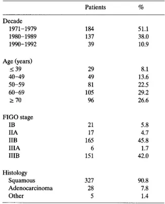
Table 1 Patients characteristics Decade 1971-1979 1980-1989 1990-1992 Age (years) I 39 40-49 50-59 60-69 2 70 FIG0 stage IB IIA IIB IIIA IIIB Histology Squamous Adenocarcinoma Other Patients % 184 51.1 137 38.0 39 10.9 29 8.1 49 13.6 81 22.5 105 29.2 96 26.6 21 5.8 17 4.7 165 45.8 6 1.7 151 42.0 327 90.8 28 7.8 5 1.4
IB-IIA received 45 Gy to the whole pelvis. Patients
with advanced stage IIB-IIIB received 45 Gy to
the pelvic volume and a lo-15 Gy boost to the
involved parameter(s). Intracavitary brachyther-
Table 2 Treatment characteristics Intracavitary curietherapy Radium Cesium Mode of therapy Concomitant (Con) Sequential (Seq) Treatment duration 5 60 days > 60 days Total effect I 1164 Gy2 > 1164 Gy2 Patients % 172 47.8 188 52.2 291 80.8 69 19.2 271 75.3 89 24.7 87 24.2 273 75.8
J.F. Delaloye et al. /International Journal of Gynecology & Obstetics 57 (1997) 295-.103 297
apy delivered a boost of 30 Gy to point A in l-3 fractions for LDR and in 3 or 4 fractions over 3
or 4 weeks for HDR. In the first decade
(1971-1979), low dose-rate brachytherapy (LDR:
90 cGy/h at point A) was systematically used,
whereas from the second decade on, all patients
were submitted to high dose-rate (HDR: 30
cGy/min at point A). The isotope used for LDR
was radium, and the isotope used for HDR was cesium. The rectal dose was measured during the insertion of the source and the total rectal dose for the whole period of brachytherapy was com-
puted. External radiation therapy and intracavi-
tary brachytherapy were performed concomitantly (Con) or sequentially (Seq). Sequential curiether- apy was given at weekly intervals, 5-20 days fol-
lowing completion of external radiation. From
1990 to 1993 radiation therapy was Seq in 92% of the patients.
2.3. Total effect
In 1980, when HDR curietherapy was intro-
duced, the original Nominal Standard Dose (NSD)
from Ellis and the modification proposed by Or-
ton were used to calculate the biological equiva- lence of HDR vs. LDR [9,10]. For the present analysis the treatment related factors (total dose, number of fractions) were introduced in the lin-
ear quadratic model modified according to the
proposal of Fowler in order to calculate the Total
Effect (TE) [ll]. For the curietherapy a correc-
tion for the TE was introduced with the continu- ous repair model from Thames [12]. No correc-
tions were made for overall treatment duration
because no individual data are available either on
pre-treatment potential doubling time, or on time
of kick-off repopulation as suggested by Withers
[13,14]. To facilitate the discussion, all doses will be expressed as TE in Gray squared (Gy2> for both external and brachytherapy treatments (see addendum). One should be aware that this TE is different from the real dose given at point A. It is
a measure of the radiobiological effectiveness of
the treatment for tumor tissue. For these calcula- tions some assumptions have been made concern-
ing repair half-time (T1,2) and related continuous
repair factor (g-factor). A T,,, of 1.5 h has been
considered and the corresponding g-factor has
been extrapolated according to the exposure time for both HDR and LDR. Moreover, the choice of
the a/P factor, required for calculation of TE,
does not really influence the results because the
same value has been chosen for HDR as for
LDR. This factor is merely a measure of the intrinsic sensitivity of the tumor tissue to a change in fraction size and is unrelated to the dose-rate. Total effect could not be calculated for 14 patients
due to incomplete information on radiotherapy
doses administered. Median total effect was 1283
Gy”. For the purpose of this analysis patients were classified according to whether they had
received treatment with rather low total effect?
taking 1164 Gy* (the 25th percentile) as an ar-
bitrary cut-off.
2.4. Therapy duration
This included the duration of external radiation therapy, the time interval between the former and
brachytherapy, and the duration of brachyther-
apy. Therapy duration was calculated in days.
Median therapy duration was 45 days. For the purpose of the analysis, patients were classified according to whether they had rather long thera-
pies, taking 60 days (the 75th percentile) as an
arbitrary cut-off.
2.5. Statistical methods
The primary endpoint of this study was overall survival (OS), which was defined as time from beginning of radiation therapy to death. For the purpose of this analysis, survival status for all patients was updated during the summer of 1993. Statistical analyses were carried out by means of the software package Stata [151. Survival per- centages over time have been calculated by the
Kaplan-Meier method [161 and their correspond-
ing standard errors (S.E.) with Greenwood’s for- mula [17]. For univariate analysis of OS the P-val- ues from the log-rank test are reported for each comparison considered [181. Estimated hazard ra- tios (HR) of death, with respect to the indicated reference group, their 95% confidence intervals (95% CD and P-values were calculated with pro-
298 J. F. Delaloye et al. /International Journal of Gynecology & Obstetrics 57 (I 997) 295-303
portional-hazard regression with appropriate bi-
nary variables to identity each group of interest
[19]. For the multivariate analysis, some cate-
gories of variables have been collapsed to allow for small numbers of deaths in the respective subgroups. Values of HR greater than unity indi- cate increased rates of death with respect to the chosen reference category. For analyses other
than on OS, chi-squared tests (for categorical
variables) or Kruskall-Wallis tests (for continu-
ous variables) have been used [20]. All probability values are for two-sided tests. For the purpose of the analyses of survival and unless otherwise indi- cated, observations have been censored at 5 years in order to limit the effect of competing causes of
mortality and also to avoid having only patients
registered during the earlier periods contribute to the right tail of the survival curves. Five-year survival is also considered a relevant endpoint for cervical cancer patients.
3. Results
3.1. Total effect and therapy duration
Among patients treated with the sequential
modality, median total effect was 1183 Gy2 as
opposed to 1283 Gy2 in the group treated with
the concomitant modality (P < 0.001). Similarly,
as concerns therapy duration, the sequential and
concomitant groups had a median duration of 72
and 44 days, respectively (P < 0.001).
The association among type of therapy on the one hand and total effect and duration on the other was also visible in terms of the percentage of patients in each therapy group with a total effect less than 1164 Gy2 (namely 38% for Seq
and 21% for Con, P = 0.004) or with a therapy
duration longer than 60 days (namely, 74% for
Seq and 13% for Con, P < 0.001). In addition, of
the therapies of more than 60 days, 32% had a total effect of less than 1164 Gy2, as opposed to
22% of the therapies of less than 60 days (P =
0.064).
FIG0 stage I was more frequently treated with
shorter therapies (P < 0.038) and with lower total
effect (P < 0.001). No other associations between
therapy descriptors and other clinical characteris- tics were identified.
3.2. Survival analysis
Median follow-up for the studied patients was
in excess of 12 years. Median survival for the
whole group was 7.5 years and the 5-year survival was 59% (S.E. = 3%). Fig. 1 displays the survival experience of the whole group. For the following analyses, survival has been censored at 5 years, as explained in the Statistical Considerations. When considering sub-groups, the 5-year survival for the Con and Seq groups was 60% (S.E. = 3%) and
58% (S.E. = 8%), respectively (P = 0.226). Simi-
larly, for the total effect groups less than 1164 Gy2 and more than 1164 Gy2, the 5-year survival was 47% (SE. = 6%) and 63% (SE. = 3%), re-
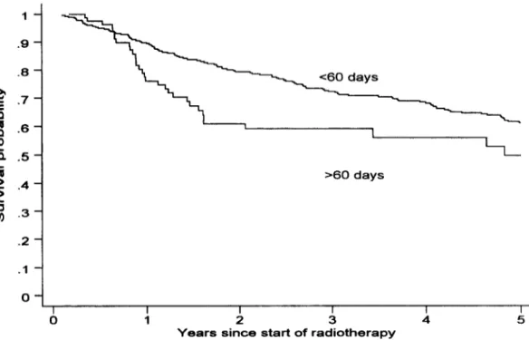
spectively (P = O.OlO), while for the therapy dura-
tion groups of less than 60 days and more than 60 days we observed a 5-year survival of 61% (S.E. =
3%) and 53% (S.E. = 7%), respectively (P =
0.044). In terms of univariate hazard ratio (HR), the relative difference between the two therapy
duration groups corresponds to an increase in
hazard of death of about 50% (HR = 1.53, 95% CI = 1.03-2.28) for the more than 60 days group with respect to the less than 60 days group. These
and additional univariate results are displayed in
Table 3. Figs. 2-4 display the survival experience
of the sample, according to type, duration and
total effect of therapy.
In light of the associations observed among the various descriptors considered, which may also
have an important effect on survival, a multivari-
ate analysis has been performed to establish the residual effect of each of the variables studied
after simultaneous adjustment of all other factors
considered. The results are displayed in Table 4
and confirm the association in magnitude of
longer therapies with increased risk of death. The estimated relative hazard for longer therapies is
as in the univariate analysis (HR = 1.52, 95%
CI = 0.94-2.45) but with only borderline signifi-
cance (P = 0.084). The indication of the univari-
J.F. Delaloye et al. / InternationalJournal of Gynecology & Obstettics 57 (1997) 295-303 299 Table 3
Five-year overall survival and univariate analysis
Age (years) 139 40-49 50-59 60-69 270 FIG0 stage IB II III Histology Squamous Other Decade 1970-1970 1980-1989 1990~1992 Type of therapy Sequential Concomitant Total effect I 1164 Gy* > 1164 Gy* Therapy duration 5 60 days > 60 days
Syear survival Log-rank Univariate Cox regression
Deaths 7% SE P-value HR 95% CI 11 60 21 52 28 63 33 67 43 47 3 85 56 66 77 47 122 59 51 69 63 61 53 6 65 21 58 115 60 42 47 94 63 104 61 32 53 9 7 5 5 6 7 4 4 3 7.6 4 4 13 8 3 6 3 3 I
effect of therapy is not confirmed. Stage is still a strong predictor of survival. An exploratory analy- sis has been performed considering the whole of the follow-up available on each patient (no cen-
soring at 5 years). A multivariate regression con-
firms the effect of longer therapies (HR = 1.52,
95% CI = 1.03-2.26, P = 0.037).
4. Discussion
The main issue of this study was to determine whether overall treatment time was an authentic prognostic factor, as it was already reported [4-61. Differing from other reports, besides clinical de-
0.227 Reference 1.14 0.95 0.82 1.37 Reference < 0.001 2.51 4.86 0.475 0.81 Reference 0.341 Reference 1.29 1.02 Reference 0.226 0.74 1.62 0.010 Reference Reference < 0.044 1.53 0.555238 0.47F1.92 0.41~1.62 0.71-2.67 0.78-X.03 153~15.42 0.91-1.82 0.43-2.38 0.46-1.18 1.12-2.33 1.03-2.x
scription we have studied the role of this factor together with that of total dose (expressed in total
effect) and type of therapy (concomitant and se-
quential) which may be acting as confounders of the association of interest. Strong associations
among treatment descriptors were observed.
The univariate analysis showed that overall
treatment time and total effect are of prognostic
significance. The multivariate analysis confirmed
the prognostic importance of overall treatment
time and FIG0 stage, but not the role of total
effect. The relative risk of death increased sig-
nificantly when overall treatment time exceeded
300 J.F. Delaloye et al. /International Journal of Gynecology & Obstetrics 57 (1997) 295-303 -2 .I 0 I 1 I I I I I I I 012345 10 15 20
Years since start of radiotherapy
l- .9 - .a - E = .7- 0 1 -6 h .5- T .- .4 - z ; .3- .2 .l 0 3
Fig. 1. Overall survival in all patients.
Seq
i
1
A
A
4
Years since start of radiotherapy
1- .9 - .8 - 2 = .7- 0 H -6 ti .5- f .- .4 - il ; .3- .2 - .I - 0- 1 .8 .I 0
J.F. Delaloye et al. /International Journal of Gynecology & Obstetrics 57 (1997) 295-303 301
MO days
I
0
I I I I I
1 2 3 4 5
Years since start of radiotherapy
Fig. 3. Overall survival according to duration of therapy: > 60 days or 2 60 days.
cl164 GY
I
0
I I I I I
1 2 3 4 5
Years since start of radiotherapy
302 J.F. Delaloye et al. / InternationalJournal of Gynecology & Obstetrics 57 (1997) 295-303
Table 4
Multivariate analysis of survivaI censored at 5 years
Age (years) I 49 50-59 60-69 2 70 FIG0 stage IB-II III Histology Squamous Other Decade 1970-1979 1980-1992 Type of therapy Sequential Concomitant Total effect I 1164 Gy* > 1164 Gy’ Therapy duration I 60 days > 60 days HR 95% CI P-value Reference 0.65 0.62 1.03 Reference 2.03 0.78 Reference Reference 0.58 Reference 0.88 1.23 Reference Reference 1.52 0.38-1.10 0.108 0.38-1.02 0.061 0.64-1.65 0.913 1.41-2.91 0.001 0.45-1.36 0.378 0.22-1.53 0.274 0.49-1.58 0.661 0.83-1.82 0.299 0.94-2.45 0.084
ment time on outcome after radiation therapy has been extensively studied in head and neck cancer
[ll]. Any prolongation of radiation treatment is
potentially deleterious for local control and sur-
vival. Fowler calculated a median rate of loss of local control of 14% (range 3-25%) per week of extra overall time [ll]. In cervical cancer, Girin- sky et al. [6] reported a loss of local control of
approximately 1% per day, when treatment time
exceeded 52-62 days. Lanciano et al. E51 showed that total treatment time duration was predictive for both local control and survival especially for
FIG0 stage III. Fyles [4] also showed a highly
significant association between pelvic control and
treatment duration. The hypothesis of a rapid
proliferation of clonogenic cells during the course
of radiation therapy was highlighted by Withers
[13,14]. We hypothesized that potential doubling
time (T,,,) was shorter in cervical cancer than in head and neck cancer, pointing to rapid prolifer-
ating tissue potentially escaping the treatment
when gaps or unnecessary delays are introduced.
The multivariate analysis in our series confirmed
therapy duration as a significant prognostic fac- tor.
Several sources of bias may have affected the results of this retrospective analysis. Chronologic time (more than 20 years have elapsed since the first patient was registered), patient selection, di- agnostic accuracy, tailoring of total dose accord-
ing to stage and response during treatment, or
data quality might have acted as confounders. There are no obvious reasons to suggest that
these factors would not have similarly affected all
subgroups. However, other considerations may
also contribute to the strength of the observed
results. In particular, our patients have been
treated by a single institution, where traditionally,
treatment choices have been dictated primarily by
the therapeutic philosophies of successive teams
of clinical staff. Despite this, we have considered
in the multivariate analysis, most of the prognos-
tic factors which may also have affected treat- ment choice. In addition, we have simultaneously been able to address the issues of treatment duration and total effect in patients treated with
either the concomitant or sequential modalities
and allowing for the effect of surgery, thus ac-
counting for most of the variation in survival
attributable to treatment. Our study does not
allow us to conclude that treatment time must be less than 60 days, since this threshold was ar- bitrarily chosen as a cut-off. Based on our results,
we decided to apply brachytherapy (30 Gy to
point A) concomitantly with the external irradia-
tion (45 Gy to the pelvic volume and a boost of lo-15 Gy to the involved parameters). In order to
optimize the geographical distribution of the dose
within the tumor, this brachytherapy is applied at
the end of the external treatment. This therapy
schedule therefore reduces overall duration to
J.F. Delaloye et al. /International Journal of Gynecology & Obstetrics 57 (1997) 295-303 303
5. Addendum
Calculation of total effect (TE) expressed in Gy * For HDR or LDR treatment
TE, =((~/P+dxg)nxd d = dose per application n = number of applications g = continuous repair factor a/P = 10 Gy
g-values, assuming a repair half-time of 1.5 h (obtained by extrapolation from tabulated values) [ 121
g uDR = 0.88 (application duration rt 20 min)
g,,, = 0.16 (application duration * 24 h)
For external radiation therapy
TE,=(cw/P+d)n xd
d = dose per fraction y1= number of fractions a/P = 10 Gy
Cumulative total effect (TE, > TE,. = TE, + TE,
Examples:
1. HDR (4 x 10 Gy)
TE, = (10 Gy + 10 Gy x 0.88)4 x 10 Gy = 752 Gy”
2. External radiation therapy (25 x 1.8 Gy
= 45 Gy)
TE, = (10 Gy + 1.8 Gy)25 X 1.8 Gy = 531 Gy2 TE, = TE, + TE, = 1283 Gy2
References
[II PI
[31
[41
Brady LW, Markoe AM, Micaily B, Damsker Jl, Karlsson UL. Amendola BE clinical treatment planning in gyne- cologic cancer. Radiother Oncol 1987; 21: 302-332. Souhami L, Melo JAC, Pareja VG. The treatment of stage III carcinoma of the uterine cervix with telecobalt irradiation. Gynecol Oncol 1987; 28: 262-267.
Perez CA, Kuske RR, Camel HM, Galakatos AE, Hed- erman MA, Kao MS, Walz BJ. Analysis of pelvic tumor control and impact on survival in carcinoma of the uterine cervix treated with radiation therapy alone. Int J Radiat Oncol Biol Phys 1988; 14: 613-621.
Fyles A, Keane TJ, Barton MB, Simm J. The effect of treatment duration in the local control of cervix cancer. Radiother Oncol 1992; 25: 273-279. 151 f61 [71 ml [91 [lOI [Ill 1121 1131 [141 [la [I61 1171 [181 [191 [201
Lanciano RM, Pajak TF, Martz K, Hanks GE. The influence of treatment time on outcome for squamous cell cancer of the uterine cervix treated with radiation: a patterns-of-care study. Int J Radiat Oncol Biol Phys 1993; 2s: 391-397.
Girinsky T, Rey A, Roche B, Haie C, Gerbaulet A. Randrianarivello H, Chassagne D. Overall treatment time in advanced cervical carcinomas: a critical parame- ter in treatment outcome. Int J Radiat Oncol Biol Phys 1993; 27: 1051-1056.
Keane TJ, Fyles A, O’Sullivan B, Barton MB, Maki E, Simm J. The effect of treatment duration on local control of squamous carcinoma of the tonsil and carci- noma of the cervix. Sem Rad Oncol 19Y2: 2: 26-28. Wilson GD, McNally NJ, Dishes S. Saunders Ml. Desrochers C, Lewis AA, Bennett MH. Measurement of cell kinetics in human tumors in vivo using bromod- eoxyuridine incorporation and flow cytometry. Br J Can- cer 1988; 58: 423-431.
Ellis F. Low to high dose rate by TDF system. Br J Radio1 1980: 17: 146656.
Orton CG. Time-dose factors (TDFs? in brachytherapy. Br J Radio1 1974; 47: 603-607.
Fowler J, Lindstrom MJ. Loss of local control with prolongation in radiotherapy. Int J Radiat Oncol Biol Phys 1992; 23: 4S7--467.
Thames HD. Withers HR, Peters W. Tissue repair capacity and repair kinetics deduced from multifraction- ated or continuous irradiation regimen with incomplete i-epair. Br J Cancer 1984; 49 (suppl VI): 263269. Withers HR. Taylor JMG, Maciejewski B. The hazards of accelerated tumor clonogens repopulation during ra- diotherapy. Acta Oncol 1988; 27: 131-46.
Withers HR. Treatment induced accelerated human tu- mor-growth. Semin Radiat Oncol 1993; 3: 135 - 143. Stata. Computing Resource Center Reference Manual. 1991.
Kaplan EL, Meier P. Nonparametric estimation from incomplete ohservations. J Am Stat Assoc 1958; 53: 457-m481.
Greenwood M. Reports on public health and medical subjects: the natural duration of cancer. HMSO 1926: 33: 1-16.
Peto R, Pike MC, Armitage P, Breslow NE, Cox DR, Howard SV, Mantel N, McPherson K. Peto J, Smith PG. Design and analysis of randomized clinical trials requir- ing prolonged observation of each patient. II. Analysis and examples. Br J Cancer 1977; 35: l-39.
Cox DR. Regression models and life tables. .I Roy Stat Sot B 1972: 34: 1877220.
Armitage P, Berr G. Statistical Methods in Medical Research. 2nd ed. Oxford: Blackwell. lY87: 205-211.