AVIS
Ce document a été numérisé par la Division de la gestion des documents et des archives de l’Université de Montréal.
L’auteur a autorisé l’Université de Montréal à reproduire et diffuser, en totalité ou en partie, par quelque moyen que ce soit et sur quelque support que ce soit, et exclusivement à des fins non lucratives d’enseignement et de recherche, des copies de ce mémoire ou de cette thèse.
L’auteur et les coauteurs le cas échéant conservent la propriété du droit d’auteur et des droits moraux qui protègent ce document. Ni la thèse ou le mémoire, ni des extraits substantiels de ce document, ne doivent être imprimés ou autrement reproduits sans l’autorisation de l’auteur.
Afin de se conformer à la Loi canadienne sur la protection des renseignements personnels, quelques formulaires secondaires, coordonnées ou signatures intégrées au texte ont pu être enlevés de ce document. Bien que cela ait pu affecter la pagination, il n’y a aucun contenu manquant.
NOTICE
This document was digitized by the Records Management & Archives Division of Université de Montréal.
The author of this thesis or dissertation has granted a nonexclusive license allowing Université de Montréal to reproduce and publish the document, in part or in whole, and in any format, solely for noncommercial educational and research purposes.
The author and co-authors if applicable retain copyright ownership and moral rights in this document. Neither the whole thesis or dissertation, nor substantial extracts from it, may be printed or otherwise reproduced without the author’s permission.
In compliance with the Canadian Privacy Act some supporting forms, contact information or signatures may have been removed from the document. While this may affect the document page count, it does not represent any loss of content from the document.
REGULATION OF INTESTINAL CHOLESTEROL TRANSPORT AND METABOLISM BY HIGH GLUCOSE LEVELS
RÉGULATION INTESTINALE DU TRANSPORT ET DU MÉTABOLISME DU CHOLESTÉROL PAR LE GLUCOSE
par
Rosa Zaava Ravid Leibovici
Département de pathologie et biologie cellulaire Faculté de Médecine
Mémoire présenté à la Faculté des études supérieures en vue de l'obtention du grade de Maître ès sciences (M.Sc.)
en biologie cellulaire
Août, 2008
Identification du jury
Université de Montréal Faculté des études supérieures
Ce mémoire intitulé:
REGULATION OF. INTESTINAL CHOLESTEROL TRANSPORT AND METABOLISM BY HIGH GLUCOSE LEVELS
présenté par:
Rosa Zaava Ravid Leibovici
a été évalué par un jury composé des personnes suivantes :
Dr. Lucian Ghitescu président-rapporteur Dr. Moïse Bendayan directeur de recherche Dr. Emile Levy codirecteur Dr. Edgard Delvin membre du jury
Abstract
Growing evidence suggests that the small intestine may contribute to excessive postprandial lipemia, which is highly prevalent in insulin-resistantltype 2 diabetic individuals increasing the risk of cardiovascular disease. The aim of the present study was to determine the role of high glucose levels on intestinal cholesterol absorption, cholesterol transporter expression, enzymes controlling cholesterol homeostasis and the status of transcription factors. To this end, we employed highly differentiated and polarized intestinal cells, Caco-2 cells, plated on permeable
polycarbonate filters. Four major technical approaches were used,
immunocytochemistry in electron microscopy, western blot, RT-PCR and the assessment of enzymatic activities. The levels of cellular cholesterol uptake were measured by radio-Iabeling.
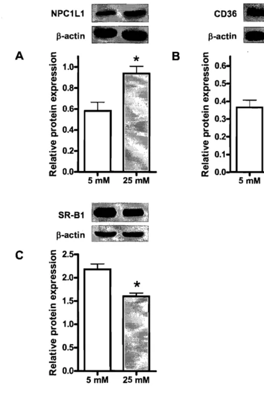
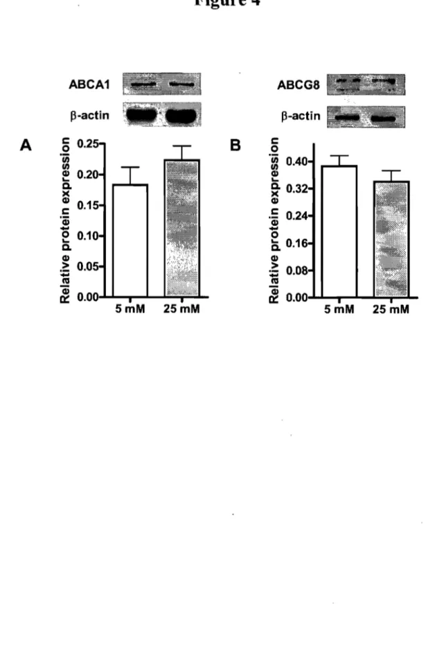
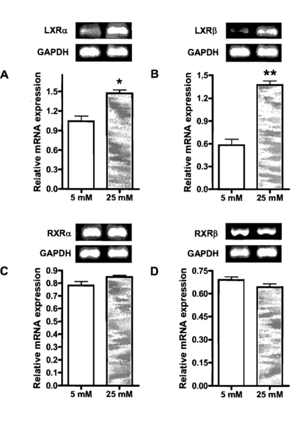
In the presence of radiolabeled cholesterol, glucose at 25 mM stimulated its uptake as compared to 5 mM glucose. The high concentration of glucose enhanced the protein expression of the critical cholesterol transporters NPCILI and CD36 and concomitantly decreased SR-BI protein expression. No significant changes were observed for ABCAI and ABCG8, which act as efflux pumps favoring cholesterol export out of absorptive cells. At the same time, HMG-CoA reductase activity was significantly decreased, whereas ACAT activity remained unchanged. Finally, increments were noted in the transcription factors LXRa, LXRP, pp ARp and PPARy along with a drop in the protein expression of SREBP-2.
Collectively, our data indicate that glucose at high concentrations may regulate intestinal cholesterol transport and metabolism, thus suggesting a potential influence on the cholesterol absorption process in type 2 diabetes.
Key
words: ABCAI, ABCG5/G8, SR-BI, CD36, NPCILl, PPAR, LXR, SREBP, ACAT and HMG-CoA reductaseRésumé
Plusieurs évidences suggèrent que l'intestin contribue à l'hyperlipidémie postprandiale fortement répandue chez les individus souffrant de résistance à insuline et de diabète de type-2, augmentant ainsi les risques de développer des maladies cardio-vasculaires. Le but de notre étude fut de déterminer, au niveau intestinal, le rôle du glucose dans l'absorption du cholestérol, l'expression des transporteurs du cholestérol et celle des enzymes qui régulent l'homéostasie du cholestérol et le statut des facteurs de transcription. À cet effet, nous avons ensemencé des cellules intestinales différenciées et polarisées, des cellules Caco-2, sur des filtres en polycarbonate perméables. Quatre grandes approches techniques ont été utilisées, l'immuno-cytochimie en microscopie électronique, l'immunobuvardage, le RT-PCR, et la mesure des activités enzymatiques. Les niveaux d'absorption du cholestérol cellulaire ont été mesurés par radio marquage. En présence de cholestérol radio marqué, le glucose à 25 mM stimule l'absorption du cholestérol comparativement aux cellules supplémentées avec 5 mM de glucose. La concentration élevée de glucose accroît l'expression protéique des transporteurs NPCILI et CD36 et diminue l'expression de la protéine SR-BI. On n'a observé aucun changement significatif dans l'expression des protéines ABCAI et ABCG8, pompes d'efflux favorisant l'exportation du cholestérol hors des cellules intestinales. L'activité de l'enzyme HMG-CoA réductase diminue tandis que celle de l'ACAT demeure inchangée. Finalement, une augmentation des facteurs de transcription LXRa, LXR~, PP AR~ et PPARy et une baisse de l'expression protéique de SREBP-2 ont été observées à fortes concentrations de glucose.
En conclusion, nos données indiquent que le glucose à des concentrations élevées, peut moduler le transport et le métabolisme intestinal du cholestérol suggérant une influence potentielle sur le processus d'absorption du cholestérol dans le diabète de type 2.
Mots clés: ABCAI, ABCG5/G8, SR-BI, CD36, NPCILI, PPAR, LXR, SREBP, ACAT and HMG-CoA réductase
Table of contents
Abstract. ... iii
Résumé ... iv
Table of contents ... v
List of Tables ... vi
List of Figures ... : ... vii
List ofSymbo1s and Abbreviations ... viii
Acknow1edgements ... ix
Foreword ... x
Introduction ... 1
1. Cholesterol ... 3
2. Intestinal Cholesterol Metabo1ism ... 5
2.1. Intestine Genera1ities ... 5
2.2. Cholesterol Biosynthesis and Esterification: HMG-CoA-reductase and ACAT ... 6
2.3. Cholesterol Biosynthesis Regulation: SREBP ... 8
3. Intestinal Cholesterol Transport ... Il 3.1. Cholesterol Transporters ... 13
3.1.1. Scavenger Receptofs CD36 and SR-BI.. ... 13
3.1.1.1. CD36 ... 13
3.1.1.2. SR-BI ... 14
3.1.2. NPC1L1 ... 15
3.1.3. ATP-Binding-Cassette (ABC) Transporters ... 16
3.1.3.1. ABCA1 ... 17
3.1.3.2. ABCG5 and ABCG8 ... 17
4. Transcription Factors ... 18
4.1. The Liver X Receptor (LXR) ... 21
4.2. Peroxisome Pro1iferator-Activated Receptors (PPAR) ... 22
4.3. The Retinoid X Receptor ... 24
5. Glucose met aboli sm ... 25
5.1. Physio1ogica1 interactions between carbohydrates and 1ipid metabo1ism . 26 Objective of Study ... 31
Article ... 33
Discussion ... 79
Bib1iography ... 85
List of Tables
ARTICLE
List of Figures
THE SIS
Figure 1. Cholesterol structure ... 3
Figure 2. Summary of cholesterol biosynthesis ... 7
Figure 3. SREBP activation ... 10
Figure 4. Schematic overview of cholesterol transport in enterocytes ... 12
Figure 5. General schema of gene regulation by transcription factors ... 19
Figure 6. Schematic structure oftranscriptional factors ... 20
ARTICLE Figure 1. Effect of glucose concentrations on cholesterol uptake ... 66
Figure 2. Combinatory effect of glucose and mannitol or sorbitol on cholesterol uptake ... 67
Figure 3. Effect of glucose concentrations on the prote in expression oftransporters mediating cholesterol influx ... 68
Figure 4. Effect of glucose concentrations on the protein expression of transporters mediating cholesterol efflux ... 69
Figure 5. Immunocytochemical evaluation of cholesterol transporters following incubation of Caco cells with glucose ... 70
Figure 6. CD36 detection in Caco-2115 cells as a representative illustration of the immunocytochemical detection ... 71
Figure 7. Interference of Ezetimibe with the uptake of cholesterol in the presence of glucose ... 72
Figure 8. Effect of glucose concentrations on HMG-CoA reductase gene expression, protein mass and activity ... 73
Figure 9. Effect of glucose concentrations on ACAT gene expression and activity74 Figure 10. Effect of glucose concentrations on the gene expression of the nuclear receptors LXR and RXR ... 75
Figure 11. Effect of glucose concentrations on the gene expression of the nuclear receptors PPAR ... ; ... 76
Figure 12. Effect of glucose concentrations on the gene and protein expression of SREBP-2 ... 77
Figure 13. Diagram of the main players influencing cholesterol transport in intestinal epithelial cells ... 78
ABC CE CHD DMEM ER FA FAT/CD36 FBS FC HDL LDL-C
LXR
NPCILI PBS PPAR PPRE RXR SR-BI SREBP-2 T2DMTG
List of Symbols and Abbreviations
A TP Binding Cassette transporter cholesteryl ester
coronary heart disease
Dulbecco's Modified Eagle Medium endoplasmic reticulum
fattyacid
fatty acid translocase/cluster determinant 36 fetal bovine serum
free cholesterol
high-density lipoprotein
low-density lipoprotein-cholesterol liver X receptors
Niemann Pick CI-Likel phosphate buffered saline
peroxisome proliferator-activated receptor peroxisome proliferators response element retinoid X receptor
scavenger receptor class B type l
sterol regulatory element binding protein-2 type 2 diabetes mellitus
Acknowledgements
1 would like to express my sincere gratitude to a11 the people who have contributed, directly or indirectly to the completion of this work.
Professor MoYse Bendayan, my supervisor, has contributed enormously to this work in terms of inte11ectual input, support and encouragement. 1 feel deeply grateful and privileged to have been your student, because of the kindness, moral and intellectual honesty with which you handle challenges and problems of any kind. Muchas gracias!
Professor Emile Levy, my co-supervisor, has always given generously of his time and insights, even though his teaching and administrative loads are considerable. This thesis would not have been possible without your kind support, guidance and your remarkable patience. Merci beaucoup!
Thanks to Carole Garofalo, for your technical guidance, aH your patience and for providing us with an excellent and pleasant working environment.
Thanks to Dr. Irene Londoiio and Diane Gingras for your kindness, your support and for introducing me to the microscopy world.
"Merci" to everybody at the laboratory, thank you for being always patient and helpful with my French. 1 am indebted to my many student colleagues-friends for providing a stimulating and fun environment for leaming and growing. 1 am especially grateful to Zola Spahis for the great technical and "personal" assistance. Dr. Genevieve Mailhot and Dr. Alain Sané were particularly supportive, patiently teaching and helping me with the transfection-infection protocols: Merci!
To my family, for your constant support and love. To Yovany, my love.
Foreword
Funding
The CUITent work was supported by the Canadian Institutes of Health Research, the Canadian Diabetes Association, Diabète Québec and the Faculté des études supérieures et postdoctorales (FESP). Their contribution is gratefully acknowledged.
Presentations
• XXIIIe Congress of graduate and postdoctorate students of the Sainte-Justine Research Centre (June 2008). Poster "Potentiel du glucose a moduler le transport et l'homéostasie du cholestérol dans l'intestin". Montreal, Canada
• VI Symposium of digestive tube physiopathology (June 2008). Poster "Régulation du transport et de l'homéostasie du cholestérol par le glucose dans les cellules Caco-2/15". Orford, Canada
• IX Société québécoise de lipidologie de nutrition et de métabolisme congress (May, 2008) Poster "Régulation du transport du cholestérol par le glucose dans les entérocytes". Laval, Canada
• Diabète Québec congress (September 2007). Poster "Regulation by glucose of cholesterol metabolism in intestinal epithelial cells". Quebec, Canada
• Scientific Day, Medicine Faculty, Université de Montréal (June, 2007). Poster "Glucose effect on cholesterol transporters in enterocytes". Montreal, Canada
Cholesterol is an essential component of cellular membranes, a precursor of steroid hormones, vitamin D and bile acids, and plays a crucial role in transcriptional gene regulation. Excessive cholesterol, however, is cytotoxic and may cause atherosclerotic lesions. Therefore, a balance must be maintained between cholesterol intake, absorption/excretion and synthesis. For its transport within the enterocytes, cholesterol requires a protein-dependent machinery, including SR-BI, NPCILI and CD36, involved in mediating intestinal cholesterol uptake. Other proteins such as ABCAI and ABCG5/ABCG8 favor the exit of cholesterol from the enterocytes into the intestinal lumen or through the basolateral membrane. Several of these cholesterol carriers influence intracellular cholesterol homeostasis and are controlled by transcription factors, including RXR, LXR, SREBP-2 and PPAR.
Previous studies showed that lipid components exert a regulatory effect on intestinal fat uptake. However, the role 'of carbohydrates has barely been investigated. Therefore, we aimed to evaluate the effect of glucose on cholesterol transport and metabolism. Establishing this relation may contribute to find new alternative therapeutic treatments, which improve the conditions of metabolic glucose- and lipid-related diseases such as obesity, coronary heart disease and diabetes mellitus among others.
In the present study, we show in an in vitro model closely resembling in situ intestinal cells that high glucose concentrations enhance cholesterol transport by upregulating the prote in expression ofNPCILI and CD36. In addition, we found a reduced SR-BI prote in expression and HMG-CoA reductase activity, a key enzyme in the cholesterol biosynthesis pathway, without altering the proteins ABCAI and ABCG8. Moreover, our studies document that the expression of particular transcription factors are regulated by glucose levels. Our data indicate that glucose
at high concentrations may regulate intestinal cholesterol transport and metabolism, thus suggesting a potential influence on the cholesterol absorption process in type 2 diabetes.
1. Cholesterol
Cholesterol belongs to the sterol family and is present in the membranes of most eukaryotic cells. The characteristic structure of those lipids is the steroid nucleus, consisting of fused rings, three with six carbons and one with five. Sterols fulfill several indispensable roles in all eukaryotic cells. In mammals, cholesterol is the most important one. It is amphipathic with a polar head group and a nonpolar hydrocarbon body (Figure l). Its hydrophobicity is responsible for the valuable property necessary to control cell membrane fluidity (Ohvo-Rekila et al., 2002). However, it makes it very difficult to handle in the aqueous environment of the body, both within and between cells. Therefore, sophisticated mechanisms exist to transport cholesterol to its numerous cellular destinations.
Polar head
Fig~re 1. Cholesterol structure
Alkyl side chain
Sterol nucleus
Cholesterol is an integral part of cells as well as organelle membranes and is a precursor of important physiological molecules, like bile salts and steroid hormones and is thus essential for normal cell functioning (Maxfield and Tabas, 2005).
Defects in cholesterol synthesis or transport can have harmful consequences. Furthermore, cholesterol and sterols that are ubiquitously present in the diet also
pose a potential danger. They are critically involved III the development of atherosclerosis. Rence, cellular cholesterol homeostasis and plasma cholesterol levels have to be strictly regulated.
Cholesterol enters the lumen of the small intestine from 3 sources: diet, bile, and desquamated intestinal epithelial cells which are derived from the rapid turnover of intestinal cells. Although the entire length of the small intestine has the capability to absorb cholesterol from the lumen, the main sites of absorption are the duodenum and the proximal jejunum (Charlton-Menys and Durrington, 2008). In humans, 30-50% of the luminal cholesterol is absorbed and returned to the liver, while the rest is eliminated with feces (Turley and Dietschy, 2003). The principal sites of cholesterol biosynthesis are the liver and the intestine. To maintain body cholesterol homeostasis, metabolic adaptations of endogenous de novo synthesis and/or catabolism are required in response to fluctuations in dietary intake of cholesterol.(Levy et al., 2007).
The hydrophobicity of cholesterol is related to the main problems associated with increased plasma cholesterol concentrations, i.e., atherosclerosis. Atherosclerosis is at the origin for the majority of cases of coronary heart disease and represents the most prevalent cause of death in industrial countries (Ross, 1995).
The development of atherosclerosis is a process starting already early in life, possible even
in
utero (Napoli et al., 1997). Lipids, such as cholesterol, transported from lipoproteins, accumulate in macrophages on vessel walls (Kruth, 2001). Macrophage cholesterol accumulation converts the macrofages into so-called foam cells and stimulates the macrophages to secrete proteases and tissue factor that contribute to plaque rupture and thrombosis (Zhao et al., 2006).2. Intestinal Cholesterol Metabolism
2.1. Intestine Generalities
The intestine controls the uptake of water, electrolytes and nutrients as well as secretes ions, enzymes and mucus and excretes endogenous and exogenous compounds from the blood towards the lumen. The barrier function of the intestine is ensured by intestinal mucosa, with inc1udes the epithelial cells with the specialized tight-junction complexes that line the luminal surface. Intestinal motility and micelles cause mixing of the components and ensure absorption and transport along the tract (Scoville et al., 2008).
The morphology of the small intestine influences the absorptive process given its anatomie and physiologie features. Among these are the considerable length of the small intestine (7 min humans and 90 cm in the rat) (Kaminsky and Zhang, 2003), the distribution of the metabolically competent epithelium as a mono layer of enterocytes and the amplification of the luminal surface by numerous finger-like projections of enterocyte-lined villi and, at their bases, buried crypts. Enterocytes have a very limited life span: after the division of stem cells at the base of the crypt, epithelial cells migrate up to the crypt surface, a process that takes 4 days in humans (3 in rodents). The cells then migrate to the villous tip, where they shed, a passage of 3 days in humans and 2 days in rodents (Kaminsky and Zhang, 2003).
Each region of the intestinal tract consists of the same layers: serosa, muscularis, submucosa and mucosa. The mucosa is lined by a continuous layer of epithe1ial cells, consisting of enterocytes and goblet cells. Anatomically, the intestine is divided into duodenum, jejunum, ileum and colon. In each of these regions, the enterocytes display particular sets of enzymes and transporters that, as part of the homeostatic function of the intestine, are able to metabolize and transport endogenous and exogenous compounds (Doherty and Charman, 2002).
2.2. Cholesterol Biosynthesis and Esterification: HMG-CoA-reductase and AC AT
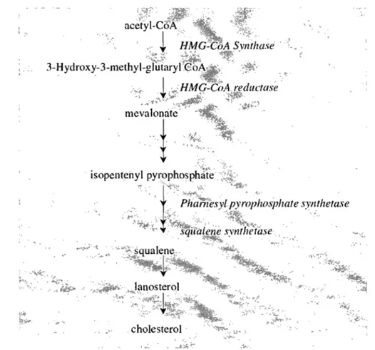
Although the structure of cholesterol suggests a complex biosynthetic pathway, the entire formation starts with the acetate precursor.
The first step in this pathway is the formation of 3-hydroxy-3-methylglutaryl-CoA (HMG-CoA) from acetyl-CoA and acetoacetyl-CoA, catalyzed by the enzyme HMG-CoA synthase. This is followed by the enzymatic action of HMG-CoA reductase to reduce HMG-CoA to mevalonate, wich represents the major rate-controlling step in cholesterol synthesis. Currently, HMG-CoA reductase IS
considered the rate-limiting key enzyme of the entire process. Mevalonate IS
converted into isopentenyl-5-pyrophosphate, followed by condensation to squalene and conversion into lanosterol, which is an immediate precursor of cholesterol. To convert lanosterol into cholesterol, the entire process involves 19 reactions (Goldstein and Brown, 1990).
In mammalians, HMG-CoA reductase is a glycoprotein of ~ 1 00 kDa. It shows an amino-terminal domain containing seven hydrophobic regions able to anchor the protein in the endoplasmic reticulum membrane, and a carboxy-terminal catalytic domain, which projects into the cytosol (Liscum et al., 1983). Virtually, every tissue and organ can synthesize cholesterol from acetyl-CoA (Dietschy and Siperstein, 1967; Spady and Dietschy, 1983), but the liver and intestine are considered as the major sites (Taylor et al., 1960; Dietschy and Gamel, 1971; Turley et al., 1981).
mevaJonàtè'
,.
~
'.t '"
Figure 2. Summary of cholesterol biosynthesis (Adapted from (Charlton-Menys and Durrington, 2008). Cholesterol is an essential molecule in many animaIs, inc1uding humans, but is not required in the mammalian diet. AlI cells can synthesize it from simple precursors. (Adapted from Lehninger et al., 2002)
HMG-CoA reductase is subject to both short and long-tenn controls (Goldstein and Brown, 1990). Long-tenn effects are mediated through alterations in its rate of synthesis and degradation. Short-tenn actions involve allosteric effects and alterations in its state of phosphorylation. Expression of HMG-CoA reductase and other enzymes in the cholesterogenic pathway is transcriptionally controlled by the sterol regulatory binding protein SREBP-2. Both expression and activity of the enzyme are rapidly reduced under conditions of high intracellular sterol concentrations (Nakanishi et al., 1988).
After biosynthesis and/or uptake by the enterocyte, cholesterol is mainly esterified at C3 with fatty acids to form cholesteryl ester, a reaction catalyzed by acyl-coenzyme A:cholesterol acyltransferase 2 (ACAT-2) (Joyce et al., 1999). Two ACAT genes (acat-l and acat-2) have been identified in mammals; the two enzymes may function in distinct and complementary manners (Chang et al., 1997; Farese, 1998; Chang et al., 2001; Rudel et al., 2001). In skin cells, macrophages, adrena1 cells and CHO cells, ACAT-1 is the major isoenzyme and constitutes 90% or more of the total cellular ACAT activity. In intestinal mucosal cells, ACAT -2 is the major isoenzyme. ACAT-2 may be allosterically regulated by cholesterol (Chang et al., 2000; Liu et al., 2005).
The objective of cholesterol esterification is the storage of cholesterol as cholesteryl esters in cytoplasmic lipid droplets. Cholesteryl esters can be hydrolyzed when necessary and the esterification/hydrolysis cycle provides cells with short-term buffering capacity for cholesterol. In contrast to cholesterol, plant sterols being poor substrates for ACAT-2, are not efficiently esterified and remain in majority unesterified. (Field and Mathur, 1983; Joyce et al., 1999; Temel et al., 2003). This reflects the main difference between plant sterols and cholesterol in all reactions taking place in the enterocyte. Cholesteryl esters can subsequently be secreted into the lymph after their packaging into chylomicrons, to ultimately reach the liver (Chang et al., 2006).
2.3. Cholesterol Biosynthesis Regulation: SREBP
Cholesterol biosynthesis is controlled by a family of transcription factors of the helix-loop-helix family designated Sterol Regulatory Element Binding Proteins (SREBPs). SREBPs activate the expression of more than 30 genes required for the synthesis of cholesterol, fatty acids, triglycerides and phospholipids, and are thus considered key regulators of cholesterogenesis and lipogenesis (Brown and Goldstein, 1997; Horton and Shimomura, 1999; Edwards et al., 2000; Sakakura et al.,2001).
SREBPs are encoded by two genes, srebp-l and srebp-2. Alternative promoter usage and alternative splicing of SREBP-l drive the production of two isoforms, SREBP-Ia and SREBP-Ic. The 29 additional amino acids present in the SREBP-Ia NH2 terminus are enriched in acidic residues and might be responsible for the higher transcriptional activity of SREBP-I a, compared with that of SREBP-I c. SREBP-Ic was initially cloned in rats and called adipocyte determination and differentiation factor-l (ADD 1) (Rosen and Spiegelman, 2000).
SREBP-I a has been mainly studied in celllines, showing a strong expression, while in animal tissues its expression is re1atively weak. SREBP-I c, highly expressed in liver, is the key regulator of lipogenesis and enhances transcription of genes encoding acetyl-CoA carboxylase (ACC), fatty acid synthase (FAS), stearoyl-CoA desaturase 1 (SCD-l) and glycerol-3-phosphate acyltransferase (GPAT), all important enzymes in the lipogenic pathway. SREBP-2, predominantly expressed in celllines, is also present in the liver and adipose tissue, but overall has a rather weak expression in animal tissues. It activates cholesterol synthe sis by inducing expression of genes encoding enzymes that catalyze the various steps in cholesterol synthesis, including HMG-CoA reductase (Desvergne et al., 2006).
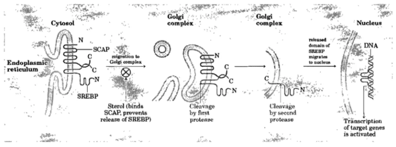
SREBPs are synthesized as inactive precursors of ~125-kDa, bound to the endoplasmic reticulum (Hua et al., 1995). In order to reach the nucleus and act as transcription factors, their NH2-domains must be cleaved. One protein required for the transfer of SREBP to the nucleus is an escort protein designated SREBP cleavage activating prote in (SCAP). A schematic representation of SREBP activation is shown in Figure 3.
Sterol (bincl. SCAP, prevents release of SREBP) Cleavage by!'irst p~teàse Cleavàge hy second prot-ease
[
~ .~~
Transcription of target genes is activatedFigure 3. SREBP activation. Sterol regulatory element-binding proteins are embedded in the ER when first synthesized, in a complex with the protein SREBP cleavage-activating prote in (N and C represent the amino and carboxyl termini of the proteins). When bound to SCAP, SREBPs are inactive. When sterol levels decline, the complex migrates to the Golgi complex, and SREBP is cleaved by two different proteases in succession. The liberated amino-terminal domain of SREBP migrates to the nucleus, where it activates transcription of sterol-regulated genes. (Adapted from Lehninger et al., 2001)
Low membrane cholesterol levels lead to the transport of SCAP/SREBP to the Golgi membrane where activation of the site 1 serine protease results in a first cleavage. A second enzyme, the site 2 metalloproteinase, completes the maturation of SREBPs and releases the 68-kDa NH2-terminal domain of SREBP from the membrane (Edwards et al., 2000). This fragment contains a basic helix loop helix (HLH) leucine zipper domain, which functions as a transcription factor upon translocation into the nucleus. The mature forms of SREBPs bind to elements initially characterized as featuring an enhancer sequence called E-box that is recognized by members of the HLH transcription factor family. SREBPs also bind to sites related to the direct repeat TCANCCAC (Horton et al., 2002).
Deletion of SREBP-l (eliminating both SREBP-la and SREBP-lc) or SREBP-2 leads to partial or fully embryonic lethality, respectively. In contrast, specific deletion of the SREBP-l c transcript is not lethal, suggesting an important role of SREBP-la and SREBP-2 in embryonic development. The SREBP maturation
process via membrane cholesterol sensing is consistent with their important role in cholesterol homeostasis (Sundqvist and Ericsson, 2003). SREBP-2 is mainly involved in cholesterol metabolism. SREBP-lc has an important implication in fatty acid synthe sis, whereas SREBP-l a is involved in cholesterol and fatty acid synthesis (Desvergne et al., 2006).
3. Intestinal Cholesterol Transport
Next to endogenous synthe sis, dietary intake provides the other main source of cholesterol in mammals. Only a fraction of dietary cholesterol is absorbed in the small intestine, with large interindividual variations in humans. Reported values range from 25% to 85% (Sehayek et al., 1998; Bosner et al., 1999). In contrast, only small amounts of plant sterols are absorbed (Lu et al., 2001b; Turley and Dietschy, 2003).
Recent studies suggest that cholesterol absorption is a protein-mediated process. In support to this hypothesis, cholesterol uptake by apical membranes in vitro follows a second-order reaction kinetics changing to a low-affinity first-order kinetics mechanism upon proteolytic digestion of proteins on the surface of the brush-border membranes (Thurnhofer and Hauser, 1990). The recent discovery of inhibitors (that selectively block cholesterol absorption at very low doses) and their binding to intestinal mucosa in a specific and saturable manner, supports the protein-mediated cholesterol absorption hypothesis (Hemandez et al., 2000).
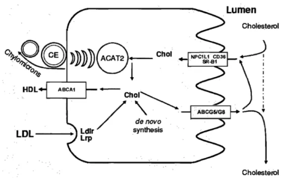
Various transporters, inc1uding fatty acid translocase/c1uster determinant 36 (FAT/CD36), scavenger receptor c1ass B type l (SR-BI) and Niemann Pick Cl-Likel (NPCILl) may be involved in cholesterol uptake, while the ATP Binding Cassette transporter family, inc1uding several cholesterol carriers (ABCAl, ABCBl, ABCG5/G8), may act as efflux pumps favoring cholesterol export out of absorptive cells into the lumen or basolateral compartments (Levy et al., 2007).
Intestinal cholesterol absorption consists on a multistep process that can be controlled at several leve1s. A schematic overview of the absorption process is shown in Figure 4. Cholesterol
~~~8 >>>>>~8-'
-
Chol Qo-Ol),r!
HDL -Chol~/
\ .
~
-1 ..
ÀBCG5IG8 denovo LDL - - -... a Ldlrsynthesis .. Lrp CholesterolFigure 4. Schematic overview of cholesterol transport in enterocytes. ABCAl/G5/G8, ABC-transporters Al,G5 and G8; ACAT2, acyl-coenzyme A:cholesterol acyltransferase-2; CE, cholesteryl ester; Chol, cholesterol; HDL, high density lipoprotein; LDL, low density lipoprotein; LDLr, receptor; LRP, LDL-receptor related prote in; NPClLl, Niemann-Pick Cl like 1, CD36, c1uster determinant 36; SR-BI, scavenger receptor c1ass B type I. (Adapted from (Levy et al., 2007)
After cholesterol is taken up by the enterocyte, it is esterified by the action of acyl-coenzyme A:cholesterol acyltransferase 2 (ACAT2) (Lee et al., 2000). Cholesteryl esters are packed into chylomicrons and subsequently secreted via the lymph to the circulation (Wang and Carey, 2003).
In addition, studies have shown that enterocytic cholesterol can also be transferred to lipid-poor apoA-I through the action of intestinal ABCAI (Brunham et al., 2006). The HDL partic1es are directly excreted into the circulation and contribute for approximately 30% to the steady-state plastna HDL pool in mice (Brunham et al.,
2006). However, abcar/- mice do not show reduced cholesterol absorption (McNeish et al., 2000; Drobnik et al., 2001), indicating that this process is not of regulatory relevance for mass cholesterol absorption. Non esterified cholesterol and plant sterols can be excreted from the enterocyte back into the intestinal lumen. This process is facilitated by the ABC half-transporters ABCG5 and ABCG8 (Berge et al., 2000; Igel et al., 2003). Overexpression of these transporters in mice increases excretion of cholesterol into the lumen and limits net cholesterol absorption (Yu et al., 2002b).
3.1. Cholesterol Transporters
Considerable effort has been spent over the past several years on identifying the cholesterol transporter(s) in enterocyte membranes. This section focuses on recent progress and explanatory findings associated to various pro teins that are potentially involved in cholesterol transport.
3.1.1. Scavenger Receptors CD36 and SR-BI
Several cell surface glycoproteins, inc1uding CD36 and SR-BI are designated as scavenger receptors and contribute to the uptake of modified lipoproteins (Ades et al., 1992; Calvo et al., 1995; Acton et al., 1999; Nakata et al., 1999; Husemann et al.,2001).
3.1.1.1. CD36
FAT/CD36, (fatty acid translocase/c1uster determinant 36) an 88-kDa membrane glycoprotein, is found in several cell types, inc1uding platelets, monocytes, macrophages and endothelial cells where it facilitates cellular uptake of long-chain fatty acids. In humans and mice, CD36 is also expressed in intestinal epithelial cells. (Ohgami et al., 2001; Nicholson and Hajjar, 2004).
CD36 has been reported to be a multifunctional receptor recognizing severalligands inc1uding OxLDL (Nakata et al., 1999; Nozaki, 1999), thrombospondin (Simantov and Silverstein, 2003), collagen (Femandez-Ruiz et al., 1993; Kopprasch et al., 2004), Plasmodium jalciparum-infected erythrocytes (Beeson and Brown, 2004) and anionic phospholipids (Ryeom et al., 1996; Bottcher et al., 2006). The importance of CD36 in fatty acid absorption is well established, since CD36 deficiency leads to abnonnallipid processing in enterocytes.
The potential role of CD36 in cholesterol absorption is reported in studies showing enhanced cholesterol uptake from micellar substrates by CD36-transfected COS-7 cells compared to mock-transfected cells (van Bennekum et al., 2005). The CD36-mediated cholesterol uptake properties in the transfected cells are similar to that observed with SR-BI-transfected cells as well as those observed with brush-border membrane vesic1es prepared from wild-type mice (van Bennekum et al., 2005). The importance of CD36 in mediating cholesterol absorption was demonstrated in a study that showed a significant reduction of cholesterol transport from the intestinal lumen to the lymph in null mice (Nauli et al., 2006). Interestingly, the CD36-facilitated cholesterol uptake process is similar to that observed for SR-BI-mediated cholesterol uptake in their sensitivity to ezetimibe inhibition (van Bennekum et al., 2005).
3.1.1.2. SR-BI
SR-BI, the scavenger receptor c1ass B type l, an 82-kDa protein, is highly expressed in the small-intestine brush border membrane where it facilitates the uptake of dietary cholesterol from either bile salt micelles or phospholipid vesic1es (Hauser et al., 1998). SR-BI is able to bind with fair but different affinities to HDL, LDL, and modified (acetylated or oxidized) LDL. On the other hand, HDL and LDL appear not to share the same binding sites (Gu et al., 2000).
The participation of SR-BI in cholesterol absorption was suggested by studies that showed that SR-BI cDNA-transfected cells displayed increased cholesterol uptake from micellar substrates compared with mock-transfected cells with low SR-BI expression (Altmann et al., 2002; van Bennekum et al., 2005). Moreover, the increase in cholesterol uptake by SR-BI-transfected cells showed a sensitive effect to ezetimibe inhibition in both of these studies.
Despite the strong evidences suggesting that SR-BI may participate in cholesterol absorption on the surface of brush-border membranes, its role in cholesterol absorption in a physiological environment remains controversial. Since scarbr/-mice, with a disruption of the sr-bi gene, efficiently absorbed cholesterol, in similar fashion to wild-type mice (Altmann et al., 2004), whereas cholesterol absorption was increased in transgenic mice with SR-BI-specific overexpression in the intestine (Bietrix et al., 2006).
3.1.2. NPCILl
Niemann-Pick Cl-like 1 prote in (NPClLl), a l5l-kDa prote in, is expressed predominantly in the gastrointestinal tract with peak expression in the proximal jejunum (Altmann et al., 2004; Davis et al., 2004). In situ hybridization and immunohistochemistry analysis of the jejunum revealed discrete NPClLl localization to the epithe1iallayer lining the luminal space along the crypt-villus axis (Altmann et al., 2004). Furthermore, our group (Sane et al., 2006) was able to assign NPClLl to the brush-border membrane with the use of cell fractionation and high-resolution immunoelectron microscopy. The protein was also found to be located in subcellular compartments of the human enterocyte, inc1uding lysosomes and mitochondria (Sane et al., 2006).
NPClLl has 50% ammo acid homology to NPCl, which is defective in the cholesterol storage disease Niemann-Pick type C (Carstea et al., 1997). A strong support for the essential role that NPC 1 L 1 plays in intestinal cholesterol absorption
lies in the following examples. Mice deficient in NPCILI lack the ability to absorb cholesterol and exhibit prevailing protection against the rise in plasma and hepatic cholesterol associated with the administration of high cholesterol diets (Altmann et al., 2004; Davis et al., 2004; Davis et al., 2007). Genetic modifications of NPCILI in cultured intestinal cells alter cholesterol uptake (Palmer et al., 1995; Yu et al., 2006; Yamanashi et al., 2007). Reduced expression in NPCILI was found associated with reduced sterol absorption, low-density lipoprotein-cholesterol (LDL-C) levels (Cohen et al., 2006) and LDL-C response to ezetimibe therapy (Hegele et al., 2005; Simon et al., 2005; Wang et al., 2005). Inactivation ofNPCILI led to defects in cholesterol transport, variations in key regulatory sterol enzymes (HMG-CoA reductase and ACAT), and high gene expression of SREBP, which suggest key roles for NPCILI in cholesterol homeostasis (Sane et al., 2006).
3.1.3. ATP-Binding-Cassette (ABC) Transporters
ABC-transporters compnse a large family of membrane proteins that mediate transport of a variety of compounds across cellular membranes against concentration gradients at the cost of ATP. Most ABC-transporters contain two transmembrane and two ATP-binding domains. Genes that encode for the ABC transporters are conserved from bacteria to mammals. They play critical roles in the active transport of a wide range of molecules across cellular membranes. Each ATP-binding domain contains the conserved Walker A and Walker B motifs that are involved in ATP binding and hydrolysis. Each transmembrane domain consists of six membrane-spanning a-helices. Sorne ABC-transporters contain only a single transmembrane and a single ATP-binding domain and are therefore called "half-transporters". These half-transporters assemble either as homo di mers or heterodimers to create a functional transporter (Dean et al., 2001; Borst and Elferink, 2002; Tusnady et al., 2006). Three specific ABC transporters of relevance for this study are discussed below.
3.1.3.1. ABCAI
ABCA1, a full-sized transporter, is probably the most extensively studied transporter of the ABC superfamily. ABCA1 is expressed in virtually aIl organs and tissues, with a high expression in hepatocytes, enterocytes and macrophages (Luciani et al., 1994; Wellington et al., 2002). The ABCA 1 protein is located on the basolatera1 surface of intestinal cells, indicating that this protein does not have a direct role in cholesterol absorption from the intestinal lumen. CUITent studies indicate that ABCA 1 expression in the basolatera1 membrane is crucial for intestinal secretion of HDL, which accounts for ~30% of HDL production in the body (Attie, 2007). ABCA1 mediates the efflux of cholesterol and phospholipids ABCG5/G8 to lipid-poor apoA-I and to pre-p-HDL. Therefore, ABCA1 has a crucial role in HDL formation, as evidenced from the discovery of mutations in the ABCA 1 gene in Tangier disease (Bodzioch et al., 1999; Brooks-Wilson et al., 1999; Rust et al., 1999). Tangier patients, as weIl as mice lacking ABCA1, are characterized by an almost complete absence of HDL and accumulation of cholesteryl esters in various tissues.
However, it is important to note that ABCA1, present in other tissues promotes reverse cholesterol transport to the li ver for biliary excretion (Vaisman et al., 2001). Therefore, ABCA 1 may indirectly impact on cholesterol absorption through the modulation of lipid composition in bile and the intestinal lumen, as evidenced by the moderately lower cholesterol absorption in abcal-I-) mice.
3.1.3.2. ABCG5 and ABCG8
Two groups (Berge et al., 2000; Lee et al., 2001) independently identified the two adjacent genes ABCG5 and ABCG8, in a head-to-head configuration that encode transporters, highly expressed in the liver and intestine. Unlike other ABC transporter genes that encode proteins with 12 transmembrane domains, ABCG5 and ABCG8 separetely encode a protein with 6 transmembrane domains, and the
heterodimerization of the two encoded proteins is required for transport activity (Graf et a1., 2002).
ABCG5 and ABCG8 are localized at the apical brush border membranes of enterocytes and the canalicular membranes of hepatocytes. These transporters constitute an efficient efflux pump system for cholesterol and plant sterols transporting out of intestinal cells back into the intestinal lumen and from hepatocytes into the bile. These efflux processes thus contribute to the regulation of intestinal absorption and biliary secretion of cholesterol and plant sterols (Yu et al., 2002a; Yu et al., 2002b; Yu et al., 2003; Klett et al., 2004).
Mutations in either one of these genes cause sitosterolemia, an error of metabolism characterized by the accumulation of plant sterols in the body due to a decreased ability for their hepatobiliary secretion (Berge et al., 2000; Lee et a1., 2001; Lu et a1., 2001a). Accordingly, mice deficient for ABCG5 and/or ABCG8 are characterized by accumulations of plant sterols in blood and organs. These mice have a reduced secretion and a clearly enhanced intestinal absorption of plant sterols, whereas absorption of cholesterol is not affected (Yu et a1., 2002a; Klett et aL, 2004; Plosch et al., 2004).
However, overexpression of ABCG5 and ABCG8 in transgenic mice causes a poor fractional intestinal cholesterol absorption besides the increased hepatobiliary cholesterol secretion (Yu et aL, 2002b). These findings have established ABCGS and ABCG8 as key transporters that regulate excretion of cholesterol and plant sterols from the body.
4. Transcription Factors
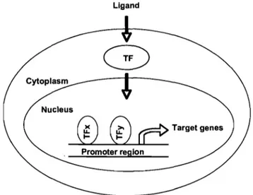
One type of metabolic regulation relies on the transcriptional regulation mechanism, which affects the level of expression of key enzymes and/or proteins and is effective on a long time scale. Transcriptional control requires specific signaIs, called
transcriptional factors, to be transduced to the cell nucleus where defined sets of genes are targeted.
Transcription factors are soluble proteins that are able to bind to DNA. Their binding to promoter sites of genes influences the transcription of these genes, leading to an up- or down-regulation of their expression. Sorne transcription factors need to be activated by ligands before they are targeted to the nucleus (Des vergne et al., 2006). A schematic model of this type of transcriptional regulation is given in Figure 5.
Ligand
Nucleus
00
Promoter reglonFigure 5. General schema of gene regulation by tranSCrIptIOn factors. TF, transcription factor. (Adapted from Bandsma et al., 2004)
Several food components and derivatives thereof may act as ligands for specifie transcriptional factors, providing means to adapt gene expression patterns in response to environmental (i.e., dietary) signaIs. The distinct roles of certain factors important in transcriptional control of genes involved in cholesterol, lipid and bile acid metabolism made them highly interesting as potential phannacological targets for prevention or treatment of certain diseases.
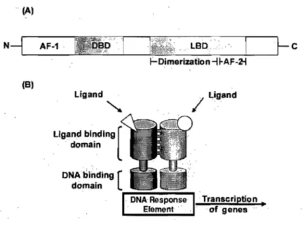
Transcriptional factor proteins exhibit a characteristic structure with regions of conserved sequence and function (Figure 6A) (Francis et al., 2003). The proteins contain an NH2-terminal region that harbors a ligand-independent transcriptional activation function (AF-1). Adjacent is the DNA binding domain (DBD), containing two highly conserved zinc finger motifs that target the receptor to specifie DNA sequences. This is followed by a region that permits protein flexibility to allow simultaneous receptor dimerization and DNA binding. Close to the COOH-terminal region is the usually highly conserved ligand-binding domain, and the last part of the receptor contains a ligand-dependent activation function (AF-2) (Chawla et al., 2001; Germain et al., 2006). Transcriptional factors can bind to DNA response elements in the promotor regions of their target genes as monomers, homodimers or as heterodimers with the Retinoid X Receptor (RXR) (Figure 6B) .
.. (A' (B, Ligand
,
1
Ligand Ligand binding [ . domain DNAbi~ding [ domain l'"''''''''~~ ... ....,...
---
...
. ofgenes ... . Transcription. .c
Figure 6. Schematic structure of transcriptional factors. (A) Organisation of a transcriptional factor. AF, activation function; DBD, DNA-binding domain; LBD, Ligand-binding domain. (B) Heterdimerization and DNA-binding of transcriptional factors. (Adapted from Edwards et al., 2000)
4.1. The Liver X Receptor (LXR)
The liver X receptor is a major player in regulating cholesterol metabolism. Two LXR isotypes have been identified in mammals, i.e., LXRa (NRIH3), which is predominantly expressed in the liver, kidney, intestine, adipose tissue, and macrophages, and LXRp (NR 1 H2) with a more ubiquitous expression pattern (Willy et al., 1995; Peet et al., 1998). These two isotypes, LXRa and LXRP, share 77% ami no acid identity in their DBD and LBD and are highly conserved between rodents and human.
LXR endogenous activators are oxysterols, i.e., cholesterol metabolites (Janowski et al., 1996; Forman et al., 1997; Lehmann et al., 1997; Janowski et al., 1999). As such, they participate in the cholesterol sensing processes and regulate important aspects of cholesterol and fatty acid metabolism (Tontonoz and Mangelsdorf, 2003).
LXRs heterodimerize with RXR to bind to their DNA response element, formed from a direct repeat of two hexamers related to the sequence AGTTCA, separated by four nuc1eotides. Mono-oxidized derivatives of cholesterol are strong LXR ligands. The most potent are 22(R)-hydroxycholesterol, 24(S)-hydroxycholesterol, and 24(S),25-epoxycholesterol, which activate both LXRa and LXRp. Little lS
known about the sterol hydroxylases that produce th~se metabolites, but it lS
assumed that oxysterol concentrations parallel those of cholesterol (Desvergne et al., 2006). Importantly, oxysterols are found at micromolar concentrations in tissues that express high levels of LXRa or· LXRp. Activation of the heterodimer can also be triggered by RXR ligands (Field et al., 2004). LXRa and LXRp null mutant mice have been generated and confirm the important role of these receptors, and more particularly that of LXRa in cholesterol homeostasis.
When LXRa becomes activated, the LXRalRXR complex induces transcription of target genes amongst which are ABCAl, ABCG5/G8. Activation of LXRa thus results in elevated levels of HDL-cholesterol, reduced intestinal cholesterol
absorption, increased hepatobiliary cholesterol secretion and increased neutral sterol 10ss via the feces. In this way, LXR activation has anti-atherogenic actions, as has been substantiated in a mouse model that is susceptible to develop atherosclerosis (ldl-r -/) treated with a synthetic LXR agonist, T090131773 (Goodwin et al., 2008). However, besides stimulation of cholesterol disposaI, activation of LXR by synthetic ligands like T090 1317 also leads to increased lipogenesis, hypertriglyceridemia through the production of larger VLDL partic1es and hepatic steatosis in rodents (Grefhorst et al., 2002). Therefore, general LXRa activation can only be used as an atheroprotective therapy if the undesirable effects on lipogenesis can be eliminated by the development of selective LXR modulators.
4.2. Peroxisome Proliferator-Activated Receptors (PP AR)
pp ARs were the first nuclear receptors identified as "sens ors" rather than classic hormone receptors. They are molecules that control a variety of genes in several pathways of lipid metabolism (De Vos et al., 1995). Three mammalian peroxisome proliferator-activated receptors (PPARs) have been cloned in Xenopus, rodents, and humans: PPARa (NRIC1), PPARp/ô (NRIC2) and PPARy (NRIC3). Two PPARy isoforms, PPARyl and PPARy2, are splice variants in their NH2-terminal domain. They are aIl activated by polyunsaturated fatty acids and eicosanoids and are therefore considered to be fatty acid sens ors (Willson et al., 2000). AlI PP ARs play important roles in the control of lipid and glucose metabolism and have been involved in obesity-related metabolic diseases, such as hyperlipidemia, insulin resistance, and coronary artery disease. pp ARs, which recognize and bind a variety of fatty acids, regulate mcst of the pathways linked to lipid metabolism (Michalik et al., 2003).
pp ARa is most highly expressed in tissue with high activity levels of lipid catabolism, e.g., liver, brown adipose tissue, and skeletal and heart muscle and was recognized to be responsive to fibrates, which are widely used drugs for treatment of hyperlipidemia (Issemann et al., 1993; Schonfeld, 1994). Activated PPARa
increases p-oxidation of fatty acids, thereby stimulating energy production and preventing lipid accumulation. Under the physiological fasting condition, pp ARa becomes activated by the free fatty acids re1eased from adipose tissue, after which hepatic p-oxidation is induced in order to provide energy to peripheral tissues (Kersten et al., 1999).
PPARy is mainly expressed in the adipose tissue and is involved in adipocyte proliferation. PPARy1 is mainly expressed in adipose tissues but is also detected in the colon, spleen, retina, hematopoeitic celIs, and skeletal mùscle. PPARy2 has been found mainly in the brown and white adipose tissue. PPARy is critical for the maintenance of glucose homeostasis and is a molecular target of thiazolidinediones (TZDs), a class ofinsulin-sensitizing drugs (Ros en and Spiegelman, 2001).
pp ARp has an ubiquitous expression pattern and appears to serve several functions. Besides its role in lipid metabolism, it is involved in skin biology, energy homeostasis and inflammatory processes (Michalik et al., 2003). pp ARp was reported as an important factor in the regulation of glucose metabolism and insulin sensitivity (Lee et al., 2006).
The ligand binding pocket of pp ARs is much larger than that of the other nuclear receptors and relatively easily accessible. The DNA binding domain is extremely weIl conserved. The less conserved NH2-terminal region bears a ligand-independent activation domain, at least in PPARa and PPARy (Xu et al., 2001a; Xu et al., 2001b; Xu et al., 2002; Xu et al., 2004).
pp ARs bind to DNA as heterodimers with RXR, on pp AR response elements (PPRE) comprising direct repeats of two hexamers closely related to the sequence AGGTCA and separated by one nucleotide. The five nucleotides that flank the 5'-end of this core sequence are also important for the efficiency of pp ARa:RXR binding (Desvergne et al., 2006).
The first molecules to be recognized as pp ARa activators, and later characterized as ligands, belong to a group of molecules that induce peroxisome proliferation in rodents, thus explaining the name of peroxisome proliferator activated receptor given to this receptor. This diverse group of substances includes, for example, sorne plasticizers and herbicides. More interestingly, various fatty acids, more particularly unsaturated fatty acids, and sorne eicosanoids mainly derived from arachidonic acid and linoleic acid, bind to PPARa, -~, and -y with varying affinities (Desvergne et al., 2006). In addition to being activated by fatty acids, pp ARa responds to fibrates that are hypolipidemic drugs, and pp ARy responds to thiazolidinediones that are insulin sensitizers, demonstrating their potential as drug targets. In the process of transcriptional regulation, ligand-bound pp ARs recruit coactivators, most likely organized in large complexes (Surapureddi et al., 2002). Co factor recruitment may be pp AR isotype specifie and may ensure the specificity of target gene activation. In addition to pp AR ligand binding, pp ARs can also be activated by phosphorylation of serines located in the AlB domain, and the pp AR:RXR heterodimer can be activated by RXR ligands (Desvergne et al., 2006).
4.3. The Retinoid X Receptor
Retinoid X receptors (RXRs; NR2B 1) play an important role in nuclear receptor signalling, as they function as general partners for a variety of nuclear hormone receptors that bind as heterodimers to DNA. There are three isotypes of RXR, a, ~,
and y, and several isoforms for each of them (Chambon, 1996). Each isotype and isoform has its specifie expression pattern. However, each single tissue contains one or several forms of RXR.
Almost aIl the nuclear receptors are active as heterodimers with RXR. The important part that RXR may play is further emphasized by the fact that RXR is itself a nuclear receptor that can be activated by specifie ligands. PPAR:RXR and LXR:RXR are permissive heterodimers: RXR can bind its own ligand, in the
absence of a ligand for its partner and can thereby activate the transcription of the heterodimer target genes (Desvergne et al., 2006).
In addition to the various heterodimers for which RXR is an obligatory partner, RXR can form homodimers. The in vivo relevance of these homodimers is still under study. RXR can be activated by 9-cis-retinoic acid, an isomer of all-trans-retinoic acid (Heyman et al., 1992). Retinoids are used for treatment of dermatological disorders and certain cancers and a common side-effect of this treatment is dyslipidemia (Farol and Hymes, 2004), indicating that retinoids and RXR are involved in the regulation of lipid metabolism.
Deletion of the RXRa gene in the liver allowed the identification of the most affected pathways (Wan et al., 2000). As expected, many PPARa-mediated functions were altered and the activity of LXR and FXR was also compromised, suggesting that the absence of RXRa cannot be compensated by RXR~ and RXRy in the liver.
5. Glucose metabolism
Carbohydrates are a main source of energy and can be stored in the form of starch in plants and glycogen in animaIs. Carbohydrates are also part of the structural framework of both DNA and RNA and form structural elements' in cell walls of bacteria and plants. An important group of carbohydrates comprises the monosaccharides of which glucose is an example. Glucose is primordial for the generation of energy. Monosaccharides are aldehydes or ketones with two or more hydroxyl groups, that can be described by the formula (CH20)n. Glucose
metabolism is tightly regulated in humans and animaIs to guarantee a sufficient glucose supply to glucose-dependent organs. The brain is the organ that is most dependent on an adequate supply of glucose, since it can only use ketone bodies as an alternative energy source and this only to a limited extent. Carbohydrates are transported to and from various tissues through the blood compartment. Glucose can
enter the blood via two routes, i.e., dietary glucose derived from the intestine and glucose production by the liver and the kidney. During fasting, the organism will solely depend on the production of glucose, mainly by the liver. Glucose can be produced directly through gluconeogenesis from various substrates, such as certain amino acids, lactate and glycerol. The liver is also able to produce glucose indirectly through phosphorylation of glycogen, the storage form of glucose. This process is called glycogenolysis (Rothman et al., 1991; Hellerstein et al., 1997). Glucose can also be taken up first by the blood, phosphorylated by glucokinase to form phosphate (G6P) and then be secreted again after dephosphorylation by glucose-6-phosphatase (G6Pase). This process is called glucose cycling (Jens sen et al., 1990).
5.1. Physiological interactions between carbohydrates and lipid metabolism
Carbohydrate metabolism and lipid metabolism are linked in many ways. First of all, mammals are capable of tuming glucose into fat. Glucose is degraded, through glycolysis, into acetyl-CoA, which is the precursor for fatty acid synthesis. On the other hand, fat cannot be tumed into glucose by mammals, because the enzyme system for this conversion is lacking. Evidence was generated in the sixties by the group of Dr. Randle evidencing that fat oxidation inhibits glucose oxidation, by interference at multiple levels (Randle et al., 1963). The key enzyme in this inhibitory process is
pyruv~te
dehydrogenase, which catalyzes the oxidative decarboxylation of pyruvate leading to the formation of acetyl-CoA. It was found that free fatty acids (FF A) increase concentrations of acetyl-CoA as well as of citrate, important in the citric acid cycle (Randle et al., 1963). Acetyl-CoA was found to decrease pyruvate dehydrogenase allosterically and citrate was found to inhibit phosphofructokinase, an enzyme involved in glycolysis. This whole process came to be known as the glucose-fatty acid cycle or Randle cycle. More recently, the group of Dr. Robert Wolfe provided data to indicate the opposite phenomenon (Sidossis and Wolfe, 1996). Using a hyperinsulinemic-hyperglycemic clamp technique they found that elevated glucose concentrations inhibited fatty acidoxidation. This effect might be due to increased intracellular malonyl-CoA levels. Malonyl-CoA is produced from acetyl-CoA and is the first step in fatty acid synthesis, i.e. de novo lipogenesis. Increased glycolysis produces more pyruvate leading to increased acetyl-CoA production, which in turn will lead to more malonyl-CoA. Malonyl-CoA is known for its inhibitory effect on carnitine-palmitoyl transferase l, an enzyme catalyzing the binding of camitine to long-chain fatty acids, a necessary step for entry into mitochondria and subsequent oxidation. Lipids and carbohydrates do not only influence each other in terms of oxidation but also in their synthetic processes. It has been known for sorne time that glucose is capable of promoting de nova lipogenesis (Groen et al., 2001). However, a high glucose intake probably does not promote hepatic synthesis of quantitatively important amounts of fatty acids in humans with a western dietary lifestyle (Hellerstein et al., 1996). It was found that the regulation of hepatic de novo lipogenesis is, at least partly, controlled by specific transcription factors. Evidence however shows that SREBP-1 and 2 can partially compensate each other, as SREBP-l knockout mice showed elevated levels of SREBP-2 and increased cholesterol synthesis rates (Shimano et al., 1997). Glucose is able to induce lipogenesis indirectly by inducing insulin secretion. Insulin has long been known for its lipogenic activity (Beynen et al., 1979). Two groups separately found that insulin has an additional effect by enhancing SREBP-lc gene expression and the abundance of the protein in the endoplasmic reticulum (Moon et al., 1999; Shimomura et al., 1999; Azzout-Marniche et al., 2000). The carbohydrate responsive element binding prote in (ChREBP) (Foufelle et al., 1998; Koo et al., 2001), is also involved in transcriptional regulation of lipogenesis. ChREBP is induced in situations characterized by high glucose concentrations (Koo et al., 2001; O'Callaghan et al., 2001; Yamashita et al., 2001). ChREBP itselfwas found to activate gene expression of both pyruvate kinase and acetyl-CoA carboxylase (Kawaguchi et al., 2001; Koo et al., 2001; O'Callaghan et al., 2001; Yamashita et al., 2001).
Hepatic very-low density lipoprotein (VLDL) secretion to plasma is also a process in which insulin is a primary factor. Insulin, after secretion in response to a rise in
plasma glucose concentration, regulates VLDL-triglyceride secretion, either directly by influencing the rate of apoB synthesis, or indirectly via its effect on the supply of FFA to the liver (Sparks and Sparks, 1994b; Aarsland èt al., 1996; Sidossis et al., 1998). The acute effects of insulin on regulation of VLDL secretion differ from its chronic effects. Acutely, insulin inhibits hepatic VLDL secretion (Sparks and Sparks, 1994b), whereas chronic exposure to insu lin has an stimulatory effect (Zammit et al., 1999). In addition to the regu1ation of lipid synthesis and secretion by carbohydrates and insulin, lipids might also promote gluconeogenesis. FF A stimulate hepatic glucose production. However, fasting, a situation with increased FF A availability, is well-known to inhibit hepatic glucose production (HGP) (Rothman et al., 1991; Neese et al., 1995).
Another level of metabolic regulation by FF A might be re1ated to the transcription factor pp ARa. pp ARa has also been suggested to induce phosphoenolpyruvate carboxykinase PEPCK gene expression (Kersten et al., 1999). pp ARa knockout mice suffer from fasting induced hypoglycemia, indicating a possible role in control of hepatic glucose production (Kersten et al., 1999). Apart from pp ARa, evidence exists that other transcription factors are involved in regulation of glucose metabolism. Glucokinase expression is activated by hepatic nuclear factor-4a (HNF-4a) (Roth et al., 2002). Glucose, through activation phosphorylationl dephosphorylation of ChREBP, influences transcription of pyruvate kinase (Kawaguchi et al., 2001). Glucose-6-phosphatase expression is also found to be mediated by transcriptional mechanisms as well as by breakdown of mRNA (Massillon, 2001).
In summary, transcriptional regulation is a form of metabolic regulation that is important for all metabolic routes of glucose. One must realize that it is likely that more transcription factors playing an important role in carbohydrate metabolism will be found in the future.
5.2. Pathophysiology of carbohydrate and lipid metabolism in diabetes
Diabetes means "excessive urination". The name diabetes mellitus was given to patients with excessive urine production in combination with a honey-flavored taste of the urine, caused by urinary glucose excretion. Diabetes mellitus today comprises a group of metabolic disorders characterized by chronic hyperglycemia. Currently, three types of diabetes mellitus are known: diabetes mellitus type 1, caused by an autoimmune-driven destruction of pancreatic ~-cells; diabetes mellitus type 2 (DM2), or non-insulin dependent diabetes mellitus as it mistakenly is also known. The third group is called maturity-onset diabetes of the young (MODY), which is a group of genetic diseases caused by mutations in numerous genes such as glucokinase and insu lin promo ter factor 1 (McGarry, 2002). DM2 is the most common disorder, accounting for more than 90 percent of cases, whose incidence is still growing in the western world even in children. The development of DM2 is in almost all cases caused by an overconsumption of food in relation to the energy expenditure and has become an epidemic disease in western societies. The primary event leading to full-blown DM2 is the deve10pment of insulin resistance, although discussion remains. Fat accumulation in muscle, liver and other tissues have been thought to induce insulin resistance (McGarry, 2002). Sorne researchers consider defective insulin secretion by the pancreas, instead of insulin resistance, to be primary in the development of DM2 (Ferrannini, 1998). It is, however, clear that insu lin resistance can precede clinically detectable DM2 by more than ten years (Lillioja et al., 1988), underscoring the importance of insulin resistance in the etiology of this disease. DM2 is associated with hyperglycemia and hyperlipidemia. Hyperinsulinemia occurs in the early stages of the disease when the pancreatic ~
cells try to compensate for the insulin resistance by increasing insulin secretion. As the disease progresses, pancreatic ~-cell failure develops giving rise to the full-blown DM2 phenotype.
Hypertriglyceridemia (Yoshino et al., 1996). Increased levels ofVLDL particles and small, dense LDL particles and decreased levels of HDL particles are commonly found (Reaven et al., 1993), giving rise to an atherogenic lipid profile.
Increased VLDL secretion might result from the decreased sensitivity to the inhibitory effects on this process of insulin directly as studies in animal models of diabetes and diabetic humans have shown (Lewis et a1., 1993; Sparks and Sparks,
1994a; Bourgeois et al., 1995). Increased VLDL secretion in DM2 might also be caused by insu lin indirectly through modulation of the supply of FF A to the liver. Increased FF A flux by modulation of hormone sensitive lipase, which is observed in insulin resistant states, has been suggested to enhance VLDL secretion by the liver. A number of studies have shown a diminished ability of insulin to suppress FF A rate of appearance in DM2 patients (Lewis et al., 2002). There is strong evidence that elevated FF A levels are associated with increased VLDL production in healthy humans (Lewis et al., 1995; Lewis, 1997). Overall, consensus practically exists that increased FF A flux to the liver is an important cause of overproduction of VLDL triglycerides by the liver in DM2. Decreased clearance of triglycerides from the blood in DM2 patients is related to impaired lipolysis of VLDL-triglycerides. Since this process is mediated by lipoprotein lipase, which is an insulin-senstivie enzyme, insulin resistance can lead to decreased levels of lipoprotein lipase. Multiple studies have shown decreased triglyceride clearance (Kissebah et al., 1982; Howard et al., 1983), although this has not been conclusive (Blades and Garg, 1995; Yost et al., 1995).