Protective Signaling of Oxytocin in an in vitro Model of
Myocardial Ischemia - Reperfusion
by
Araceli Gonzalez Reyes
Department of Biomedical Sciences Faculty of Medicine
Master's Thesis presented to the Faculty of Medicine in partial fulfilment of the requirements for the degree of
Master of Science Biomedical Sciences
December 2012
Université de Montréal
Faculté des études supérieures et postdoctorales
Ce mémoire intitulé:
Protective Signaling of Oxytocin in an in vitro Model of Myocardial Ischemia - Reperfusion
Présenté par: Araceli Gonzalez Reyes
A été évalué par un jury composé des personnes suivantes:
Dr. John Chan, président-rapporteur Dr. Marek Jankowski, directeur de recherche Dre. Jolanta Gutkowska, co-directrice de recherche
Résumé
Introduction : La prévention de la mort de cellules cardiaques contractiles suite à un épisode d'infarctus du myocarde représente le plus grand défi dans la récupération de la fonction cardiaque. On a démontré à maintes reprises que l'ocytocine (OT), l'hormone bien connue pour ses rôles dans le comportement social et reproductif et couramment utilisée dans l’induction de l’accouchement, diminue la taille de l'infarctus et améliore la récupération fonctionnelle du myocarde blessé. Les mécanismes de cette protection ne sont pas totalement compris.
Objectif : Étudier les effets d'un traitement avec de l'ocytocine sur des cardiomyocytes isolés en utilisant un modèle in vitro qui simule les conditions d'un infarctus du myocarde.
Méthodes : La lignée cellulaire myoblastique H9c2 a été utilisée comme modèle de cardiomyocyte. Pour simuler le dommage d'ischémie-reperfusion (IR), les cellules ont été placées dans un tampon ischémique et incubées dans une chambre anoxique pendant 2 heures. La reperfusion a été accomplie par la restauration du milieu de culture régulier dans des conditions normales d'oxygène. L'OT a été administrée en présence ou en absence d'inhibiteurs de kinases connues pour être impliquées dans la cardioprotection. La mortalité cellulaire a été évaluée par TUNEL et l'activité mitochondriale par la production de formazan pendant 1 à 4 heures de reperfusion. La microscopie confocale a servie pour localiser les structures cellulaires.
Résultats : Le modèle expérimental de l'IR dans les cellules H9c2 a été caractérisé par une diminution dans la production de formazan (aux alentours de 50 à 70 % du groupe témoin, p < 0.001) et par l'augmentation du nombre de noyaux TUNEL-positif (11.7 ± 4.5% contre 1.3 ± 0.7% pour le contrôle). L'addition de l'OT (10-7 a 10-9 M) au commencement de la reperfusion a inversé les effets de l'IR jusqu'aux niveaux du contrôle (p < 0.001). L'effet protecteur de l'OT a été abrogé par : i) un antagoniste de l'OT ; ii) le knockdown de l'expression du récepteur à l'OT induit par le siRNA ; iii) la wortmannin, l'inhibiteur de phosphatidylinositol 3-kinases ; iv)
KT5823, l'inhibiteur de la protéine kinase dépendante du cGMP (PKG); v) l'ODQ, un inhibiteur du guanylate cyclase (GC) soluble, et A71915, un antagoniste du GC membranaire. L'analyse confocale des cellules traitées avec OT a révélé la translocation du récepteur à l'OT et la forme phosphorylée de l'Akt (Thr 308, p-Akt) dans le noyau et dans les mitochondries.
Conclusions : L'OT protège directement la viabilité des cardiomyocytes, lorsqu'elle est administrée au début de la reperfusion, par le déclenchement de la signalisation du PI3K, la phosphorylation de l'Akt et son trafic cellulaire. La cytoprotection médiée par l'OT implique la production de cGMP par les deux formes de GC.
Abstract
Introduction: The prevention of the death of contractile cardiac cells following an episode of myocardial infarction represents the largest challenge in the recovery of myocardial function. Oxytocin, the hormone best known for its roles in reproduction and social behaviour and used commonly for the induction of parturition, has been repeatedly demonstrated to decrease the infarct size and to ameliorate the functional recovery of the injured myocardium. The mechanisms for this protection are incompletely understood.
Objective: To study the effects of oxytocin treatment on isolated cardiomyocytes using an in vitro model simulating the conditions of a myocardial infarction.
Methods: The cardiomyoblastic cell line H9c2 was used as a model of cardiomyocyte. For IR injury, the cells were placed in ischemic buffer and incubated in an anoxic chamber for 2 hours. Reperfusion was achieved by restoring cell media under normoxic conditions. OT was administered in the presence or absence of enzyme inhibitors. Cell death was evaluated by TUNEL and mitochondrial activity by formazan production during 1-4 hours of reperfusion. Confocal microscopy served for localization of cell structures.
Results. The experimental model of IR in H9c2 cells was characterized by decreased formazan production (at the range of 50-70% of normoxic control, p < 0.001) and by the increased number of TUNEL-positive nuclei (11.7±4.5 vs. 1.3±0.7% in normoxic control). The addition of OT (10-7 to 10-9 M) at the onset of reperfusion reversed the effects of IR to the control levels (p < 0.001). The protective effect of OT was abrogated by: i) an OT antagonist, OTA and siRNA-mediated OT receptor knockout; ii) the phosphatidylinositol 3-kinases inhibitor wortmannin; iii) the cGMP-dependent protein kinase (PKG) inhibitor, KT5823. Soluble guanylate cyclase (GC) inhibitor ODQ and particulate GC antagonist A71915 only partially blocked the protective effects of OT. Confocal analysis of OT-treated cells revealed translocation
of OT receptor and the phosphorylated form of Akt (Thr 308, p-Akt) into the nucleus and mitochondria.
Conclusions: OT directly protects cardiomyocyte viability if administered at the onset of reperfusion by triggering signaling of Pi3K, Akt phosphorylation and its cellular trafficking. OT-mediated cytoprotection involves cGMP production by both forms of GC.
Table of Contents
Résumé ... iii
Abstract ...v
List of Tables ... xii
List of Figures... xiii
List of Abbreviations ... xv
Acknowledgements ... xx
Dedication ...xxi
I. INTRODUCTION ...1
I.1. Myocardial ischemia - reperfusion injury ...1
I.1.1. Pathophysiology of ischemia - reperfusion injury ...1
I.1.2. Cardioprotection by ischemic pre- and postconditioning ...4
I.1.3. Cardioprotective signaling involved in pre- and postconditioning ...5
I.1.3.1. Reperfusion injury salvage kinase (RISK) Pathway ...6
I.1.3.1.1. Pi3K/Akt ...6
I.1.3.1.2. eNOS-cGMP/PKG ...8
I.1.3.2. Survivor activating factor enhancement (SAFE) pathway ...9
I.1.3.3. Mitochondria as the target for IRI and postconditioning ... 10
I.1.3.3.1. Mitochondrial ATP-sensitive K+ channels (mitoKATP) and PKC... 10
I.1.3.3.2. Signalosome hypothesis of cardioprotective signaling ... 11
I.2. Oxytocin System ... 12
I.2.1. Oxytocin receptors ... 14
I.2.2. OT/OTR in cytoprotective signaling ... 15
I.2.3. OT as a cardioprotective hormone ... 16
I.2.4. Atrial natriuretic peptide ... 21
I.2.5. Cardiovascular effects of OT and OTR gene knockouts ... 23
I.2.5.1. OT knockout ... 23
I.2.5.2. OTR knockout ... 24
I.3. The use of isolated cardiomyocytes for the study of cardioprotective signaling: advantages and limitations. ... 24
I.4. Hypothesis and Aims ... 26
I.4.1. Hypothesis ... 26
I.4.2. Aims ... 26
II. MATERIALS AND METHODS ... 27
II.1. Cell Culture ... 27
II.1.1. H9c2 myoblastic cell line from rat embryonic heart ... 27
II.1.2. Isolation of rat adult ventricular cardiomyocytes ... 28
II.2. Simulated Ischemia - Reperfusion ... 30
II.3. Cell viability and DNA fragmentation ... 33
II.4. Antagonists and Inhibitors ... 34
II.5. siRNA-mediated knockdown of OTR ... 36
II.6. Immunofluorescence and confocal microscopy ... 37
II.6.1. Image capture and processing ... 39
II.7. Intracellular ROS production ... 40
II.8. Western Blot ... 40
II.8.1. Protein extraction ... 40
II.8.2. Protein quantification ... 41
II.8.3. Electrophoretic migration of protein samples... 41
II.8.3.1. Sample preparation ... 41
II.8.3.2. Preparation of SDS polyacrylamide gels ... 42
II.8.4. Transfer of proteins to nitrocellulose membrane ... 44
II.8.4.1. Ponceau red transfer verification, blocking and incubation with antibodies ... 44
III. RESULTS... 47
III.1. Standardization of Experimental Conditions ... 47
III.1.1. H9c2 cells are a good model of cardiomyocyte in simulated ischemia-reperfusion experiments. ... 47
III.1.2. Viability of H9c2 cells ... 49
III.1.3. Simulated ischemia-reperfusion (IR) protocol ... 50
III.1.4. siRNA-mediated knockdown of OTR ... 51
III.1.4.1. Transfection efficiency determined using AF555-labelled negative control siRNA sequence ... 51
III.1.4.2. Confirmation of OTR knockdown ... 54
III.2. Oxytocin protects cardiomyocyte viability ... 55
III.2.1. Pre-treating H9c2 cells with oxytocin protects them from death after simulated ischemia-reperfusion. ... 55
III.2.2. Oxytocin protects cell viability optimally if administered at the onset of reperfusion ... 58
III.2.2.1. OT administered at reperfusion protects optimally at concentrations ranging from 1 - 250 nM ... 61
III.3. The protection afforded by OT is mediated by oxytocin receptor ... 62
III.3.1. OTA concentration-dependently blocks the protective effect of OT. ... 62
III.3.2. OTR knockdown reverses the protective pattern observed by OT treatment on cell death ... 63
III.3.3. A high concentration of vasopressin, administered at reperfusion, exerts a mild protection of metabolic viability in H9c2 cells. ... 64
III.3.4. An antagonist to vasopressin receptors V1a and V2 protects viability only in the absence of oxytocin ... 65
III.4. Mechanisms of oxytocin-induced cytoprotection ... 67
III.4.1. Oxytocin decreases the formation of reactive oxygen species (ROS) in cardiomyocytes exposed to IR. ... 67
III.4.2. OT treatment in normoxic conditions causes an increase in intracellular ROS
production. ... 68
III.4.3. OT preconditioning of H9c2 cells protects them from doxorubicin-induced cytotoxicity ... 70
III.4.4. Pi3K-Akt-eNOS signaling is involved in OT-mediated protection from simulated ischemia - reperfusion ... 72
III.4.4.1. Pi3K inhibitor Wortmannin blocks the protective effect of Oxytocin ... 72
III.4.4.2. OT stimulation causes Akt phosphorylation (Thr308) and co-localization with mitochondria in the perinuclear region ... 73
III.4.4.3. OT treatment causes eNOS phosphorylation (Ser1177) and nuclear co- localization with p-Akt (Thr308). ... 80
III.4.5. The cGMP/PKG pathway mediates OT-induced cardioprotection ... 83
III.4.5.1. cGMP-dependent protein kinase (PKG) activity ... 83
III.4.5.2. Guanylate cyclase activity ... 84
IV. DISCUSSION ... 87
IV.1. Experimental Models... 88
IV.1.1. H9c2 cells are a good in vitro model of oxytocin signaling in cardiomyocyte ischemia - reperfusion ... 88
IV.2. OT protects cardiomyocyte survival ... 89
IV.2.1. OT preconditioning directly protects cardiomyocyte survival... 89
IV.2.2. OT exerts an optimal protective effect if administered only at reperfusion ... 90
IV.2.3. The protection induced by OT is mediated by OTR ... 91
IV.2.3.1. OTR knockdown reverses the protective pattern seen by OT treatment. ... 92
IV.3. Mechanisms of OT - induced cardiomyocyte protection ... 93
IV.3.1. OT prevents the intracellular formation of reactive oxygen species after 1 and 2 hours of reperfusion ... 93
IV.3.2. OT treatment under normoxic conditions causes a short - lived burst in ROS production ... 93
IV.3.3. OT protects from doxorubicin - induced mitochondrial toxicity ... 94
IV.4. Signalisation involved in OT-mediated cardiomyocyte protection ... 95
IV.4.1. Pi3K/Akt signaling ... 95
IV.4.2. OT causes phosphorylated Akt (Thr308) to co-localize with mitochondria in or around the nucleus ... 96
IV.4.3. OT mediates cardioprotection through eNOS phosphorylation and cGMP-PKG signaling. ... 97
IV.5. Signalosome hypothesis ... 100
V. CONCLUSIONS... 102
List of Tables
Table 1. Oxytocin-mediated cardioprotection in myocardial ischemia - reperfusion... 19
Table 2. Reagents employed in cell culture... 27
Table 3. Cell culture media used for ARVCM isolation and culture ... 29
Table 4. Krebs-Henseleit (KH) buffer used for perfusion, with and without Ca2+... 29
Table 5. Perfusion Digestion Solution prepared in KH buffer ... 30
Table 6. Incubation Digestion Solution prepared in KH buffer ... 30
Table 7. Ischemic Buffer composition ... 31
Table 8. Antagonists and Inhibitors used ... 35
Table 9. siRNA sequences used ... 37
Table 10. Solutions used in immunofluorescent staining ... 38
Table 11. Commercial products used in immunofluorescence ... 38
Table 12. Antibodies and sera used for immunofluorescence ... 39
Table 13. RIPA buffer composition ... 41
Table 14. 5X Denaturing reducing Laemmli protein loading dye ... 42
Table 15. Solutions used in SDS-PAGE preparation ... 43
List of Figures
Figure 1. Pathophysiology of Myocardial Ischemia - Reperfusion injury ...3
Figure 2. Neurophysin - Oxytocin. ... 13
Figure 3. Schematic model of the human OTR spanning the membrane. ... 14
Figure 4. Simulated Ischemia - Reperfusion (IR) protocol. ... 32
Figure 5. H9c2 cells express cardiomyocyte proteins and receptors of the oxytocin system. ... 48
Figure 6. Formazan production is linearly related to H9c2 seeding density up to 20,000 cells/cm2. ... 49
Figure 7. Experimental simulated ischemia-reperfusion (IR) protocol. ... 50
Figure 8. H9c2 viability after various times under hypoxic conditions. ... 51
Figure 9. Optimization of siRNA-mediated knockdown of OTR. ... 53
Figure 10. siRNA-mediated knockdown of OTR ... 54
Figure 11. Preconditioning cardiomyocytes with 1 nM OT beginning 15 minutes prior to the onset of ischemia and maintained throughout the simulated ischemia-reperfusion (IR) experiment prevents cell death. ... 57
Figure 12. OT protects cardiomyocyte viability optimally if administered exclusively at reperfusion. ... 60
Figure 13. Concentration -response of oxytocin administered at reperfusion. ... 61
Figure 14. OT antagonist OTA concentration-dependently abolishes the protective effect of OT treatment at reperfusion. ... 62
Figure 15. OTR signaling is necessary for OT-induced protection of viability. ... 64
Figure 16. The role of vasopressin system in OT-mediated protection. ... 66
Figure 17. OT treatment prevents ROS formation following simulated ischemia-reperfusion. ... 68
Figure 18. Intracellular ROS production after stimulation of H9c2 cells with OT. ... 70
Figure 20. The phosphatidylinositol-3-OH kinase (Pi3K)-Akt pathway inhibitor, Wortmannin
blocks OT-mediated protection in a concentration - dependent manner. ... 73
Figure 21. OT treatment causes Akt phosphorylation and accumulation in and around the nuclei of H9c2 cells. ... 75
Figure 22. OT treatment causes p-Akt phosphorylation in a structure within the nuclei. ... 76
Figure 23. OT treatment causes p-Akt phosphorylation in a structure within the nuclei. ... 77
Figure 24. OT treatment causes p-Akt phosphorylation in a structure around the nuclei... 78
Figure 25. H9c2 cells subjected to hypoxia and treated with OT at reperfusion period display Akt phosphorylation and p-Akt and Cox IV translocation in the perinuclear region. ... 79
Figure 26. OT causes p-eNOS accumulation in the nuclei of H9c2 cells. ... 81
Figure 27. Co-localization of phosphorylated eNOS and phosphorylated Akt in H9c2 cells stimulated with 62.5 nM of OT. ... 82
Figure 28. cGMP-dependent protein kinase (PKG) inhibitor KT-5823 blocks the protective effect of OT (62.5 nM) treatment at reperfusion. ... 84
Figure 29. Inhibition of cGMP production blocks the protection induced by OT treatment at reperfusion. ... 86
Figure 30. Proposed protective signaling triggered by OT in cardiomyocytes. ... 99
List of Abbreviations
A: Adenine AF: Alexa Fluor
AMP: adenosine monophosphate AMPK: AMP-activated protein kinase ANOVA: analysis of variance
ANP: atrial natriuretic peptide APS: ammonium persulfate AR: adrenergic receptors
ARVCM: Adult rat ventricular cardiomyocytes ATP: adenosine triphosphate
AVP: arginine vasopressin Bcl-2: B-cell lymphoma 2 BNP: brain natriuretic peptide BSA: bovine serum albumin C - cytosine
Cai : intracellular calcium
CaM: Ca2+- Calmodulin CaMKK: CaM kinase kinase
cGMP: cyclic guanosine 3', 5’-monophosphate CHD: coronary heart disease
CM: cardiomyocyte
CM-H2DCFDA: 5-(and-6)-chloromethyl-2',7'-dichlorodihydrofluorescein diacetate, acetyl ester
CNP: C-type natriuretic peptide DAG: 1,2-diacylglycerol
DAPI: 4',6-diamidino-2-phenylindole DMEM: Dulbecco's modified eagle medium
DMSO: dimethylsulfoxide DNA: deoxyribonucleic acid Dox: Doxorubicin
EDTA: ethylenediaminetetraacetic acid ER : endoplasmic reticulum
Erk1/2 : extracellular-regulated kinases 1 and 2 FBS: fetal bovine serum
eNOS: endothelial nitric oxide synthase G : guanine
GAPDH: glyceraldehyde-3-phosphate dehydrogenase GLUT: glucose transporter
GPCR: G-protein coupled receptor GSK: glycogen synthase kinase
HEPES: 4-(2-hydroxyethyl)-1-piperazineethanesulfonic acid HR: heart rate
IgG: immunoglobulin G IL : interleukin
i.p. : intraperitoneal
IP3 : inositol trisphosphate IPC: ischemic pre-conditioning IR: simulated ischemia - reperfusion IRAP: insulin regulated aminopeptidase i.v.: intravenous
JAK: Janus kinase
KRH: Krebs-Ringer HEPES
KT-5823 : (2,3,9,10,11,12-hexahydro 10R-methoxy-2,9-dimethyl-1-oxo-9S,12R-epoxy-1H-diindolo[1,2,3-fg:3’,2’,1’-kl]pyrrolo[3,4-i][1,6]benzodiazocine-10-carboxylic acid, methyl ester) L: liter
LV: left ventricular
MAP: mean arterial pressure
MAPK: mitogen-activated protein kinase MI: myocardial infarction
min: minute(s)
mitoKATP: mitochondrial ATP-sensitive K+ channels
mPTP: mitochondrial permeability transition pore; mRNA: messenger RNA
mTOR: mammalian target of rapamycin
MTS: [3-(3,5-dimethylthialzol-2-yl)-5-(3 carboxymethoxypheny)-2-(4-sulphophenyl)-2H-tetrazolium]
NAC: N-acetylcysteine NHE: Na+/H+ exchanger NCE: Na+/Ca2+ exchanger NO: nitric oxide
NP: natriuretic peptide
NPR: natriuretic peptide receptor O2: molecular oxygen
ODQ : 1H-1,2,4- oxadiazolo [4,3-a] quinoxalin-1-one OT : oxytocin
OTA: oxytocin antagonist OTKO: oxytocin knockout OTR: oxytocin receptor
OTRKO: oxytocin receptor knockout p-Akt: phosphorylated Akt
p-eNOS: phosphorylated eNOS PBS: phosphate buffered saline PC: ischemic postconditioning
pGC: particulate guanylate cyclase Pi3K phosphatidylinositol-3-kinase
PIP2 : phosphatidyl inositol 4,5-bisphosphate PIP3: phosphatidylinositol-3,4,5-triphosphate PKC: protein kinase C
PKG: cGMP-dependent protein kinase PLB: phospholamban
PLC-β: phospholipase C type β
PMSF : phenylmethanesulphonylfluoride RAAS: renin-angiotensin-aldosterone system RIPA: radio immunoprecipitation assay RISK: reperfusion injury salvage kinases RNA: ribonucleic acid
ROS: reactive oxygen species rpm: revolutions per minute RT: room temperature
RT-PCR: Real time polymerase chain reaction s.c.: subcutaneous
SDS: sodium dodecyl sulfate
SERCA: sarcoplasmic reticulum Ca2+ ATPase SEM: standard error
sGC: soluble guanylate cyclase IR: simulated ischemia - reperfusion siRNA: small interfering RNA
STAT: signal transducer and activator of transcription T: thymine
TBS: tris buffered saline TCA: trichloroacetic acid
TGF: transforming growth factor TNF: tumor necrosis factor Tris: trisaminomethane
TUNEL: Terminal deoxynucleotidyl transferase-mediated dUTP nick end-labeling V1a: vasopressin receptor type 1a
Acknowledgements
I would like to thank my supervisors, Dr. Marek Jankowski and Dr. Jolanta Gutkowska for so generously accepting me into their laboratory and for their continued support and encouragement throughout the course of my master's project. I thank them for sharing their knowledge and enthusiasm with me. Thank you for believing in me and inspiring me to improve myself.
I would also like to thank all the members of the laboratory, in particular Christophe Fadainia, Dominique Genest and Dr. Eric Plante, whose encouragement and moral support have been invaluable to me; Dr. Bogdan Danalache and Dr. Alexandre Popov for so generously sharing their incredible expertise, and Dr. Ahmed Menaouar and Julien Durand for both moral and technical support.
I extend a special thank you to my family, including my new Canadian family, whose love and support have allowed me to thrive. Lastly I thank my loving husband, whose constant love, patience and inspiration mean the world to me.
Dedication
I dedicate this work to my parents, whose love, inspiration and support have always encouraged me to aim high and pursue my dreams. I am eternally grateful.
I. INTRODUCTION
I.1. Myocardial ischemia - reperfusion injury
Coronary heart disease (CHD) is the leading cause of death in the world1. A serious manifestation of CHD is cardiac ischemia, caused by the interruption of blood flow to the cardiac tissue and an inadequate O2 supply to the heart. Acute ischemia as in the case of acute
myocardial infarction (MI) is treated by the rapid restoration of blood flow through reperfusion strategies. Although reperfusion is necessary for cardiac rescue, the abrupt restoration of blood flow results in ischemia-reperfusion injury of the myocardium2,3. The ensuing cell death is triggered by several factors including cellular and mitochondrial calcium overload, oxidative stress, reduction in nitric oxide (NO) production, and inflammatory response. The loss of contractile cardiac cells, cardiomyocytes, remains the largest challenge to functional recovery from an episode of acute MI1. Even with current reperfusion strategies, 10% of acute MI patients die and 25% progress to heart failure1, with 7 million deaths each year directly attributable to MI around the world4.
I.1.1. Pathophysiology of ischemia - reperfusion injury
Mitochondrial damage is a major factor triggering the loss of myocardial cells5. Under normal physiological conditions myocardial tissue produces more than 90% of its ATP by mitochondrial oxidative phosphorylation6. During ischemia cardiomyocytes rely on anaerobic glycolysis, leading to intracellular acidosis7,8. This acidosis causes a stimulation of the activity of the
sarcolemmal Na+/H+ exchanger (NHE-1) to extrude excess H+ ions from the cytosol, with concomitant Na+ overload of the cytosol8. The excess of intracellular Na+ in turn causes an increase in intracellular Ca2+ due to the action of the Na+/Ca2+ exchanger (NCE)8. The depletion of ATP during ischemia prevents the activity of pumps such as the Na+/K+ ATPase, as well as active Ca2+ excretion which prevents the re-establishment of normal cellular ionic homeostasis5,8,9. The intracellular Ca2+ overload causes calpain activation, apoptotic cascades, mitochondrial permeability transition pore (mPTP) opening and hypercontracture. However, the intracellular acidosis present during ischemia causes a decrease in the affinity of troponin to Ca2+ and phosphofructokinase, which hinders the functioning of the myocardial contractile apparatus7 preventing hypercontracture. Similarly, cell death during ischemia is prevented as a lower intracellular pH inhibits the formation of the mitochondrial permeability transition pore.
Lethal reperfusion injury arises from the rapid normalization of physiological conditions after a period of ischemia. As the extracellular pH normalizes it causes a massive efflux of H+ from the cell and influx of Na+, causing the Na+/Ca2+ exchanger to operate in reverse mode and further increase intracellular Ca2+ overload. Furthermore, there is an increase in reactive oxygen species (ROS) production in the first minutes of reperfusion as O2 is re-introduced into damaged
mitochondria9. High intracellular [Ca2+] and ROS trigger the opening of the mitochondrial permeability transition pore (mPTP)5,10. The mPTP is a large conductance pore that forms on the mitochondrial membrane, releases mitochondrial proteins into the cytosol and triggers cell death11 by apoptosis and necrosis. Sustained opening of mPTP is considered the irreversible point in the commitment of a cell to death10.
Figure 1. Pathophysiology of Myocardial Ischemia - Reperfusion injury. During ischemia, the reduced O2 supply causes an increase in the rate of glycolysis, generating H+ and lactate and
decreasing intracellular pH (pHi). The Na+/H+ exchanger, NHE, overloads the cytosol with Na+ as the excess H+ are extruded, causing the reversal of the Na+/Ca2+ exchanger, which extrudes excess Na+ but overloads the cytosol with Ca2+. The use of ATP-dependent pumps is limited by lack of ATP. However, the intracellular acidosis inhibits mPTP opening. During reperfusion, abrupt restoration of the extracellular pH causes an even more pronounced efflux of H+ from the cell, aggravating further the mechanisms outlined above. As the pHi is normalized, and a ROS burst is created as O2 is reintroduced, the inhibition of mPTP opening is lifted and lethal
I.1.2. Cardioprotection by ischemic pre- and postconditioning
A significant reduction in infarct size is observed when the myocardium is exposed to brief intermittent periods of ischemia and reperfusion, both at a time prior to ischemia, known as ischemic preconditioning (IPC), or immediately after reperfusion, known as ischemic postconditioning (PC). For the first time, Murry et al. reported that four cycles of brief intermittent ischemia before the onset of a sustained episode of ischemia followed by reperfusion reduced infarct size to 25% of that seen in control animals12. They termed this phenomenon ischemic preconditioning (IPC) and much work has been devoted ever since to identify the mechanisms involved in this phenomenon. Intracellular signaling pathways that are activated during IPC and create a conditioned phenotype are responsible for cell protection and the reduction in infarct size13. Adenosine, bradykinin and opioids acting via their respective G-protein coupled receptors (GPCRs) can mimic the IPC response, which is thought to converge on protein kinase C (PKC) activation, likely via the opening of mitochondrial ATP-sensitive K+ channels14 and prevention of mPTP opening13. The signaling cascades recruited by IPC appear to be active during the reperfusion phase and mediate IPC-induced protection at reperfusion15.
More recently, a cardioprotective effect of performing an intermittent ischemia protocol at the onset of reperfusion following a prolonged ischemic episode in anesthetized dogs was reported by Zhao et al.16. They termed this effect ischemic postconditioning (PC) and it was shown to be equally effective as IPC at reducing final infarct size. Approximately 50% of the final infarct size is believed to be preventable through conditioning strategies1.
Two mechanisms of cardioprotection by PC have been proposed: passive mechanisms, in which the protection results from a gradual restoration of the pre-ischemic conditions rather than an abrupt reperfusion, and active, in which cardioprotective signaling mechanisms are triggered within the cardiomyocytes17. Active mechanisms include the recruitment and activation of kinase-mediated signaling pathways within the injured cardiomyocytes, such as the RISK and SAFE pathways (see section I.1.4. below).
I.1.3. Cardioprotective signaling involved in pre- and postconditioning
IPC is believed to act by activating pro-survival kinases at reperfusion15,18. Several signaling pathways have been shown to be activated by PC, and inhibitors of key kinases in these pathways can block the protective effect of IPC and PC. The reperfusion injury salvage kinase (RISK) pathway includes pro-survival kinases such as phosphoinositide 3-kinases (Pi3K)/Akt, AMP-activated protein kinases (AMPK) and extracellular signal-regulated kinases 1 and 2 (Erk ½), whose signalisation is thought to converge on the activation of cGMP-dependent protein kinase (PKG) and protein kinase C (PKC) ε, causing the inhibition of prolonged mPTP opening8. Similarly, the survivor activating factor enhancement (SAFE) pathway involving Janus kinase (JAK) and signal transducer and activator of transcription (STAT) signaling can provide protection separately from the RISK pathway8. The degree of crosstalk between the RISK and SAFE pathways is presently unclear, but both are believed to ultimately protect mitochondrial function19. The pharmacological manipulation of these pathways represents a more clinically - applicable alternative to ischemic conditioning20.
I.1.3.1. Reperfusion injury salvage kinase (RISK) Pathway
I.1.3.1.1. Pi3K/Akt
Pro-survival kinases known to be involved in PC include phosphatidylinositol 3-kinases (Pi3K), which causes the phosphorylation and activation of Akt8. Pi3Ks are a family of intracellular signal-transducing lipid kinases with a wide variety of roles in the regulation of cellular processes. Three classes of Pi3K (Class I, II and III) have been identified based on the specificity of the lipid substrate they phosphorylate21.
Akt kinase, also known as protein kinase B, is known to be a critical kinase at the core of cellular signal integration in survival, growth and proliferation22. Akt activity is modulated by its phosphorylation status at residues Thr308 and Ser47323. Accordingly, Akt activity is regulated through the activity of intracellular phosphatases and kinases that can directly or indirectly alter its phosphorylation status.
Upon a variety of stimuli including GPCR activation, class I Pi3Ks phosphorylate phosphatidylinositol-4,5-biphosphate (PIP2) and convert it to phosphatidylinositol-3,4,5-triphosphate (PIP3) at the inner surface of the plasma23 and nuclear24 membranes. PIP3 binds intracellular enzymes with a pleckstrin homology (PH) domain25, such as Akt and phosphoinositide-dependent kinase 1 (PDK-1). PH domains are conserved protein sequences of approximately 100 amino acids which are present in a wide range of proteins and mediate their interaction with other intracellular components such as proteins and lipids26,27. The interaction of PIP3 with Akt and PDK-1 allows for the PDK-1-mediated phosphorylation and activation of Akt at Thr30823. For full activation, Akt must also be phosphorylated at a second residue, Ser473,
which is thought to be mediated at the plasma membrane by mammalian target of rapamycin (mTOR) protein complex 228. Upon activation, Akt accumulates in different cellular compartments, including mitochondria29,30 and nuclei31,32.
Akt phosphorylation and activation reduces cardiomyocyte death from ischemia-reperfusion injury and regulates glycogen synthesis, glucose transport, glycolysis, protein synthesis, cell enlargement, mitochondrial integrity and cell death28,33. However, studies demonstrating negative effects of constitutive and sustained active Akt expression in cardiomyocytes, such as hypertrophy, suggested that there could be an important spatial and temporal regulation of Akt activation28.
It was recently discovered that Pi3K activation resulted in a rapid accumulation of Akt in the mitochondria in neuroblastoma and embryonic kidney cell lines, preventing the release of apoptotic proteins from the mitochondria34. It appears that translocation of phosphorylated (activated) Akt to the mitochondria preserves mitochondrial integrity35. In mice hearts, translocation of phosphorylated Akt from the cytosol to the mitochondria was associated with ATP-dependent mitochondrial K+ channel (mitoKATP) opening36. Similarly, insulin stimulated
phosphorylation and translocation of Akt to the mitochondria of mice cardiomyocytes37, and constitutively active mitochondria-targeted Akt over-expression prevented cell death in rat neonatal cardiomyocytes29. A mitochondria-targeted constitutively active Akt protected mitochondrial integrity as indicated by the prevention of the loss of mitochondrial membrane electrochemical gradient in cardiomyocytes exposed to H2O2 or Doxorubicin29.
There exist several known mitochondrial targets for Akt such as Bcl-2 family members22 and hexokinase II38 whose phosphorylation by Akt favours survival. Other downstream targets of Akt are nitric oxide (NO) synthase (NOS)22 isoforms (such as mitochondrial NOS)39, which become active upon Akt - mediated phosphorylation, as well as glycogen synthase kinase (GSK) and caspase-9, both of which become inactive through phosphorylation by Akt30.
I.1.3.1.2. eNOS-cGMP/PKG
Activation of the cGMP/PKG pathway plays a critical role in the pro-survival signaling triggered by IPC and PC. The second messenger cyclic guanosine 3', 5’-monophosphate (cGMP) is generated by soluble or particulate guanylyl cyclases (sGC or pGC). pGC are natriuretic peptide receptors (NPR), whereas sGC is a cytoplasmic heterodimeric haemoprotein activated by nitric oxide (NO) and carbon monoxide (CO). The subcellular localization of cGMP generation and accumulation plays a role in determining its effect40.
An important mediator of the effects of cGMP is cGMP-dependent protein kinase (PKG). PKG inhibits NHE activity41, delaying normalization of intracellular acidosis during reperfusion which is unfavourable for mPTP opening. Other downstream targets of PKG include the opening of mitoKATP14. Inhibition of PKG activity with inhibitor KT-5823 blocks the cardioprotective effect
of insulin42, of B-type natriuretic peptide and of NO43.
Additionally, nitric oxide (NO) production is known to be a mediator of IPC19, and exogenous NO can induce pharmacological postconditioning44 independently of PKG activity through
S-nitrosylation of protein thiol groups45. The phosphorylation-based activation of eNOS (at residue Ser1177) is known to be involved in the protection by IPC, PC and pharmacological conditioning19.
I.1.3.2. Survivor activating factor enhancement (SAFE) pathway
Activation of the JAK/STAT pathway is necessary for the infarct-reducing effects of ischemic preconditioning46 and plays important roles in postconditioning8. In the canonical model of JAK/STAT signaling the binding of cytokines, such as tumor necrosis factor alpha (TNFα), to plasma membrane cytokine receptors causes dimerization of the receptors. JAK proteins associated to these receptors trans-phosphorylate each other upon dimerization and become activated, allowing them to phosphorylate the cytoplasmic part of the associated cytokine receptor at specific tyrosines. STAT proteins can then specifically bind the phosphotyrosine motifs on the receptors, and get phosphorylated by JAKs. Phosphorylated STAT proteins dimerize and translocate to the nucleus, where they regulate the transcription of target genes47. TNFα activation in the heart is necessary for the induction of cardioprotection by IPC, and TNFα treatment mimics IPC independently of RISK pathway activation48. TNFα is believed to trigger signaling through the activation of STAT-3 proteins48 which cause the inactivating phosphorylation of glycogen synthase kinase 3β (GSK3β) and Bad, a pro-apoptotic protein, as well as an increase in Bcl-2 and a decrease in bax expression48. Cardioprotective STAT-3 signaling confers cellular protection in a timeframe that indicates actions independently of but concomitant to its transcription factor activities48.
I.1.3.3. Mitochondria as the target for IRI and postconditioning
As mentioned above, cardiomyocytes are highly reliant on oxidative phosphorylation. The mitochondrial permeability transition pore (mPTP) is a large conductance channel of unclear identity that forms across the mitochondrial membranes, collapses the mitochondrial membrane potential and allows for the release of mitochondrial proteins to the cytosol, which trigger caspase cascades and cell death8,10. Under normal physiological conditions, the mPTP remains closed, and it is well-established that mPTP opening occurs during reperfusion after myocardial ischemia11,49. The conditions that favour mPTP opening at reperfusion are a burst in ROS production when oxygen is re-introduced into damaged mitochondria, high intracellular and intra-mitochondrial Ca2+ levels and the normalization of the intracellular pH, since acidosis inhibits its opening8. Since mPTP opening is viewed as the irreversible commitment of a cell to death, prevention of its opening is seen as the key endpoint of cardioprotection10. Indeed, mPTP prevention is viewed as the effector of pre- and postconditioning10.
Activation of the reperfusion injury salvage kinases (RISK) pathway at reperfusion is thought to prevent mPTP opening8,50. Similarly, the opening of mitochondrial ATP-dependent K+ channels (mitoKATP) has been linked to the prevention of mPTP opening, and mitoKATP channels are
modulated by known cardioprotective kinases of the RISK pathway such as PKG14 and Akt36.
I.1.3.3.1. Mitochondrial ATP-sensitive K+ channels (mitoKATP) and PKC
The opening of mitoKATP channels causes an influx of K+ and water into the mitochondrial
MAPK19 and phosphorylation of phospholamban (PLB) at Ser16, which modulates the sarcoplasmic reticulum Ca2+ ATPase (SERCA) and normalizes Ca2+ handling, preventing hypercontracture and opening of the mPTP8. The molecular identity of mitoKATP remains
unclear, but they are known to be causally involved in IPC and PC19.
I.1.3.3.2. Signalosome hypothesis of cardioprotective signaling
The question remains as to how the activation of cytosolic kinases (such as those in the RISK pathway) could rapidly modulate the appearance of the mPTP in the mitochondrial membranes. To address this problem, Garlid et al.51,52 proposed the Signalosome hypothesis of cardioprotective signaling. This hypothesis proposes that GPCR signaling at the plasma membrane results in the caveolar assembly of vesicles containing various kinases, forming signaling complexes that are then translocated intracellularly to the mitochondria and phosphorylate receptors on the outer mitochondrial membrane. This triggers the phosphorylation of a mitochondrial pool of protein kinase C-ε which in turn phosphorylates and opens mitoKATP
channels51. These signalosomes are formed upon various cardioprotective stimuli, such as IPC, PC, bradykinin; they can be isolated and when added to untreated hearts can cause mitoKATP
opening and prevention of mPTP formation. These 100-140 nm vesicles contain caveolin 3, Akt, PKG, eNOS and GC51.
I.2.
Oxytocin System
Oxytocin (OT) is a small peptide known to mediate a wide variety of physiological functions, both as a hormone and as a neurotransmitter, from childbirth and lactation to socialization and appetite modulation53. Clinically, oxytocin analogues are widely used in the induction of labour during childbirth and for the prevention of post-partum hemorrhage. Synthetic analogues of oxytocin are amongst the most commonly used pharmaceuticals in the world and are listed in the World Health Organization's list of essential medications54.
Initially discovered by Dale in 1906 for its uterotonic and milk ejection effects, OT was the first peptide hormone whose structure was elucidated in 195355. OT is highly conserved in all animal species with the amino acid sequence Cys-Tyr-Ile-Gln-Asn-Cys-Pro-Leu-Gly-NH2 and a disulfide
bridge between Cys residues 1 and 656. In mammals, OT is primarily synthesized in the supraoptic and paraventricular nuclei of the hypothalamus. OT is transcribed as a long precursor protein, preprooxytocin, of 160-170 amino acid residues. This preprohormone includes the signaling peptide and the OT carrier protein neurophysin I which is synthesised from the same gene as OT. OT is then processed and transported by neurophysin I to the posterior pituitary and stored in neurosecretory granules at high concentrations (0.1 M) until its release into circulation56 in a pulsatile manner57. The intermediate form of OT is the tri-aminoacid extended OT-Gly-Lys-Arg (OT-GKR) and has recently been shown to also exert significant biological activity58.
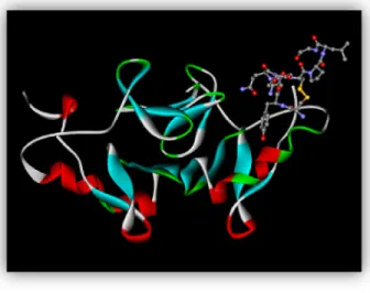
Figure 2. Neurophysin - Oxytocin. Larger ribbon structure depicts neurophysin; stick-and-ball structure depicts oxytocin. Taken from Wikimedia Commons.
Traditionally, OT has been considered a female reproductive hormone although the observations that OT is released in men and women to the same degree and through the same stimuli have revealed OT to be involved in many processes distinct from reproduction in both men and women. OT plays a role in the regulation of food intake, stress responses, immune modulation, cardiovascular regulation and in complex social behaviours such as bonding and relationship maintenance56,59.
OT is related to a second neurohypophyseal hormone, arginine vasopressin (AVP). AVP is also a nonapeptidic hormone differing from OT in residues 3 and 8 (Cys-Tyr-Phe-Gln-Asn-Cys-Pro-Arg-Gly-NH2). AVP induces vasoconstriction, water retention and corticotrophin release60,
whereas OT causes natriuresis, vasodilatation, decreases in blood pressure, negative cardiac inotropic and chronotropic effects61, and decreases cortisol levels62.
I.2.1. Oxytocin receptors
Only one receptor specific for OT has been discovered so far. This receptor, OTR, is a member of the class I family of G protein - coupled receptors (GPCR) with seven trans-membrane domains56.
Figure 3. Schematic model of the human OTR spanning the membrane. The amino acid residues in black circles have been proposed as OT docking sites, and the red bars represent docking sites of OT-GKR. Figure taken from Danalache et al., 201058.
The regulation of OTR expression is species-specific56. OTR expression has been described in a variety of tissues and cell types such as myometrial smooth muscle cells, myoepithelial cells of
mammary glands, vascular endothelium, kidney epithelium, hypothalamus, anterior pituitary, astrocytes, osteoblasts, adipocytes56 and, recently, cardiomyocytes63. Additionally, OT can trigger signalisation via vasopressin receptors V1 and V2a but with lower affinity64.
The OTR gene is composed of a 17 kb region and contains four exons and three introns, one of which (intron 3) is believed to be involved in OTR transcriptional suppression. The promoter region of the OTR gene contains interleukin response elements as well as estrogen response elements in rats, mice and humans56. It is believed that the OTR promoter is constitutively active. Introns 1 and 3 are probably involved in methylation-based suppression of OTR expression57.
OTR, like other GPCRs, undergoes a rapid desensitization upon OT stimulation (>60% decrease in 5-10 minutes of OT stimulation of Hek293 cells) that is based on receptor internalization56. OTR is associated to caveolae on the plasma membrane.
I.2.2. OT/OTR in cytoprotective signaling
OT has been shown to protect various tissues and cell types from ischemia-reperfusion injury. After the initial report that OT administration had anti-inflammatory, pro-survival effects on rat musculocutaneous flaps65, Tugtepe et al.66 showed that OT, given before the onset of renal ischemia and again before reperfusion was started, improved kidney function and decreased damage to the tissue and oxidative stress through decreases in neutrophil infiltration and fibrosis. Similarly, in a rat model of hepatic ischemia-reperfusion OT (500 µg/kg, i.p.) given 24 and 12 hours before an ischemic insult and again just prior to the onset of reperfusion alleviated hepatic
damage, fibrosis and neutrophil infiltration67. More recently, OT (1 µM administered during the ischemic insult) was shown to protect immature hippocampal cell cultures in vitro from loss of viability resulting from oxygen-glucose deprivation, and this effect was fully blocked by the use of an OT antagonist68.
I.2.3. OT as a cardioprotective hormone
Following the observation by our laboratory that OT is a cardiovascular hormone63,69, a cardioprotective effect of OT has been revealed. OT stimulates release of the cardioprotective atrial natriuretic peptide (ANP) from isolated dog atria in an OTR-mediated mechanism that appears to be dependent on the action of intrinsic parasympathetic postganglionic neurons63. OT was also shown to have transient negative inotropic and chronotropic effects on perfused isolated dog right atria in a mechanism independent of ANP release and mediated by nitric oxide (NO) production and acetylcholine release at cardiac parasympathetic postganglionic neurons61.
Ondrejcakova et al. 70 showed for the first time that in isolated rat hearts, a 25 minute perfusion with OT preceding the induction of an ischemic insult reduced infarct size by 66% as compared with control hearts. In the same study the elimination of the negative chronotropic effect of OT by the electrical stimulation of heartbeat eliminated the infarct reduction, which led the authors to conclude that OT- induced protection of ischemic myocardium is dependent upon its negative chronotropic effects. Kobayashi et al.71 demonstrated in a rabbit model of ACUTE MI that daily subcutaneous treatments with OT (10 mg/kg, s.c.) immediately following reperfusion
significantly reduced infarct size, had pro-angiogenic and anti-fibrotic effects and increased the activation of pro-survival kinases Akt, Erk1/2 and STAT3, as well as eNOS; anti apoptotic Bcl-2 was up-regulated in the ischemic myocardium following OT treatment, indicating potential anti-apoptotic effects. Interestingly, Akt, Erk1/2, eNOS and STAT3 have all been previously shown to be mediators of cardiac pre- and postconditioning8.
Further supporting a cardioprotective role for OT, the expression of OT and its receptor was strongly induced both in the heart and hypothalamus of ovariectomized and sham-operated rats following exercise training, indicating a possible involvement of OT in exercise’s cardioprotective effects72, and down-regulated by 40-50% in the injured left ventricle of mice73 and rats74 following MI. Jankowski et al. found that this down-regulation following MI could be reversed by administration of OT (25 ng/kg/h, s.c.) given before the onset of ischemia and during one week of reperfusion in a rat model of myocardial infarction74. In the same study, OT was revealed to reduce the expression of pro-inflammatory cytokines (TNFα, IL-1β and IL-6) produced by MI back to the levels seen in sham-operated rats; OT could also reduce immune cell infiltration (especially of neutrophils), induce eNOS production and normalize ANP mRNA expression in the scar area, and had pro-proliferative, anti-apoptotic and anti-fibrotic effects on injured hearts. OT was found to decrease IL-6 secretion from macrophages and endothelial cells in a dose-dependent manner, and to attenuate superoxide production in aortic endothelial and smooth muscle cells, monocytes and macrophages75. OT was also shown to induce glucose uptake in vitro in neonatal rat cardiomyocytes under conditions of chemical hypoxia by a pathway involving Pi3K76.
Work by Alizadeh et al.77 showed that the infarct size reduction in the anesthetized rat heart induced by a pre-treatment with OT (0.03µg/kg i.p.) 25 minutes prior to ischemia was eliminated by the co-treatment with an inhibitor of the opening of mitoKATP channels, which have been
proposed as the end-effectors of cardioprotective ischemic pre- and postconditioning78. Interestingly, the same group found that the protective effects of ischemic preconditioning (IPC) were completely eliminated by the administration of an antagonist specific to the OT receptor at the start of IPC, suggesting a role for endogenous OT in cardiac ischemic preconditioning79. No reports exist on the role of endogenous OT signaling in postconditioning.
Table 1. Oxytocin-mediated cardioprotection in myocardial ischemia - reperfusion. Ref. Myocardial Infarction model Ischemia - Reperfusion Protocol Dose and mode of OT administration Timing of administration Effect 70 Isolated perfused rat heart 25 min ischemia; 120 min reperfusion
90 µg/L Perfused with OT for 25 min before ischemia
infarct size by 66% (p < 0.01) Improved LV functional recovery
71 Rabbit MI 30 min ischemia; 2 and 14 days reperfusion 10 mg/kg/day s.c. Immediately after reperfusion and once per day for 5 days after MI
infarct size, fibrosis
Normalized LV diastolic and systolic dimensions, ejection fraction, fractional shortening, +dP/dt , heart rate as well as systolic and diastolic blood pressure on day 14 post-MI Increased activating phosphorylation of anti-apoptotic Akt, Stat3, eNOS and Erk ½ on day 2 and 14 post-MI
expression of VEGF, Bcl-2
expression of CD-31 positive microvessels in infarct border zone 74 Rat MI Partial ischemia for 7 days 25 and 125 ng/kg/h s.c.
Prior to ischemia and then for 3 to 7 days of reperfusion
Cell proliferation in infarct one (measured by PCN expression)
apoptosis, fibrosis in remote myocardium
neutrophil, macrophage and T lymphocyte infiltration inflammatory cytokine TNF and IL-6
77,80– 82 Rat MI 25 min ischemia; 120 min reperfusion 0.03 mg/kg i.v.and 0.03µg/kg i.p. 10 or 25 min prior to induction of ischemia
infarct size to nearly 50% of control
severity of ventricular arrhythmias, tachycardia and fibrillation
CK-MB in blood
OT effect blocked by:1. mitoKATP blocker
5-hydroxydecanoate; 2. OTR antagonist; 3. mPTP opener atractyloside; 4. NOS inhibitor L-NAME; 5. PKC inhibitor chelerythine; 6. ROS scavenger N-acetylcysteine.
83 Isolated perfused rat heart 30 min ischemia; 120 min reperfusion 10-12 to 10-10 M 5 min prior to reperfusion and continued for 25 min
infarct size, CK-MB and malondialdehyde at concentrations of 810-12, 10-11 and 210-11 M
I.2.4. Atrial natriuretic peptide
Atrial natriuretic peptide (ANP) is a 28 amino acid peptide that was first isolated from atrial muscle and identified to elicit potent diuretic and natriuretic effects84. Other natriuretic peptides identified include brain natriuretic peptide (BNP) and C-type natriuretic peptide (CNP). ANP and BNP are predominantly synthesized in the heart, CNP in the central nervous system85. Three known natriuretic peptide receptors have been identified: natriuretic peptide receptor A (NPR-A), B, and C. NPR-A and -B act as membrane-bound guanylate cyclases that upon stimulation signal intracellularly through the generation of cyclic guanosine monophosphate (cGMP); NPR-C is considered a clearance receptor because it internalizes and removes natriuretic peptides from the circulation85,86. ANP preferentially acts via NPR-A to stimulate cGMP production85.
ANP is synthesized as a 126 amino acid peptide pro-ANP and stored in dense secretory granules in the atria87. ANP secretion is mainly induced by mechanical stretching of the atria88. Cardiac ischemia and endothelin also stimulate, whereas NO inhibits, ANP secretion88. Importantly, elevated concentrations of oxytocin (10-6 M) have been reported to elicit ANP release from isolated perfused female rat hearts63.
The immediate effect of ANP is to increase diuresis (water excretion) and natriuresis (sodium excretion) by inhibiting Na+ and water re-absorption in the kidney, antagonising the effects of the renin - angiotensin - aldosterone system (RAAS)85. ANP acts as an endothelium-independent vasodilator and inhibits sympathetic nerve activity89. Furthermore, ANP can act as an autocrine and paracrine factor and directly stimulate cardiomyocytes mostly through NPR-A signaling85.
Treatment of cardiomyocytes with ANP antagonises the hypertrophic response to norepinephrine90 and phenylephrine as well as in basal conditions85 via the generation of cGMP. The importance of ANP-NPR-A signaling is demonstrated by the lethality of the NPR-A knockout in mice, at 6 months of age, due to congestive heart failure or aortic dissection91.
I.2.4.1. ANP in myocardial ischemia-reperfusion
The action of ANP opposes several detrimental effects of myocardial reperfusion injury. Plasma ANP levels are increased in patients with acute MI89, and infusion of ANP in patients with MI causes a decrease in plasma concentrations of aldosterone, angiotensin II and endothelin, and inhibits cardiomyocyte cell death89. ANP also decreases macrophage and neutrophil-mediated inflammation. In isolated cardiomyocytes, ANP causes an elevation of cGMP and the nuclear accumulation of phosphorylated Akt31, which is an key kinase with multiple cardioprotective effects in the context of myocardial ischemia-reperfusion (see section I.1.3.1.1). ANP has an anti-proliferative effect on cardiac fibroblasts86, helping to decrease fibrosis. Treatment of isolated hearts with ANP reduced infarct size via the NO - cGMP - mitoKATP pathway and
significantly improved the post-ischemic recovery of cardiac output and coronary flow85. A clinical trial with acute MI patients receiving an intravenous infusion of ANP (0.025µg/kg/min) immediately after reperfusion and during 3 days showed a significant decrease of 14.7% in infarct size and a 5% improvement in left ventricular ejection fraction 6-12 months after acute MI92.
I.2.5. Cardiovascular effects of OT and OTR gene knockouts
Mouse models of both OT and OTR knockout mice have been developed; these mice are viable and display no clear developmental changes, but are incapable of nursing their offspring93,94 and display clear metabolic changes95–97.
I.2.5.1. OT knockout
OT-/- (OTKO) mice were developed by Young et al.98 who reported that these mice present a mild hypotension. In this initial study baseline heart rate (HR) was unchanged, but intrinsic HR (HR following adrenergic and cholinergic blockade) was significantly higher in OTKO mice99. Vagal tone did not differ significantly, but OTKO mice showed lower levels of sympathetic activity, leading to a higher sympathetic reserve99. OTKO mice presented an increase in baroreflex gain99. Furthermore, OTKO mice consume higher quantities of salt, but not water, under overnight fluid deprivation100, in normal conditions101, and following fluid depletion induced by polyethyleneglycol102. Others found a slight decrease in BP and HR in OTKO mice102 and central stimulation with angiotensin II caused a similar increase in BP in both OTKO and control groups102. Mean arterial blood pressure (MAP) and heart rate (HR) averages over 24 hours were significantly lower in OTKO mice; upon induction of chronic shaker stress for 3 and 7 days, OTKO mice presented an increase in average MAP in OTKO, but not control, mice103. Basal levels of plasma corticosterone remained unchanged in OTKO mice compared to control, but stress-induced corticosterone increase was less pronounced in OTKO mice103.
OTKO mice displayed an unaltered expression level and distribution of oxytocin receptors (OTR)104. Vasopressin (AVP) can elicit physiological effect via the activation of OTR. It was therefore proposed that the lack of overt deficiencies induced upon knockout of the OT gene could be partially compensated by the direct stimulation of OTR by AVP96,104.
I.2.5.2. OTR knockout
There were no changes to the overall health, sensorimotor functions or anxiety behaviour in OTRKO mice94. OTRKO females displayed normal parturition96,105, mating and litter size105, but lacked the milk let-down reflex and maternal behaviour96,105, and the pregnant myometrium did not contract upon AVP stimulation105. Furthermore, OTRKO mice displayed increased obesity with increased white adipose tissue and adipocyte enlargement with no differences in food intake, as well as the inability to regulate internal temperatures upon cold exposure96. The expression of α2A adrenergic receptors (AR) was markedly increased, while β3 AR expression decreased in brown adipose tissue96.
I.3. The use of isolated cardiomyocytes for the study of
cardioprotective signaling: advantages and limitations
The myocardium is composed of a variety of cell types, including fibroblasts, endothelial cells, vascular smooth muscle and macrophages. Cardiomyocytes represent between 70-80% of the myocardial volume; however, the relative number of cardiomyocytes in the myocardium is a lot
lower, estimated between 35 and 55% of all cells, with fibroblasts representing up to 27% of myocardial cells106. Thus, interventions such as ischemic conditioning are likely to affect the variety of cells present in the myocardium, and the cardioprotective phenotype triggered is likely the result of the interplay between the different cells' response to protective stimuli4 through interactions and paracrine signaling between them. For example, using a transgenic mice model that only expressed erythropoietin (EPO) receptor in endothelial and hematopoietic cells, Teng et
al. reported that the acute infarct-sparing effect of erythropoietin was mediated by endothelial
cells107. On the other hand, ischemic preconditioning of isolated cardiomyocytes has been reported to protect their viability108,109, indicating an endogenous protective response of cardiomyocytes independently of other cell types.
In order to discern the specific intracellular signalisation elicited upon cardiomyocytes by cardioprotective stimuli, isolated cardiomyocytes studies are of great importance4. Furthermore, isolated cardiomyocytes in culture allow for a detailed study of the intracellular structures involved in cardiomyocyte survival in the context of ischemia-reperfusion injury109.
I.4. Hypothesis and Aims
I.4.1. Hypothesis
1. Oxytocin directly triggers protective signaling in cardiomyocytes during the acute phase of ischemia - reperfusion.
2. This protective signaling is mediated by the interaction of oxytocin with its receptor, OTR, that is expressed by cardiomyocytes.
3. Oxytocin causes the phosphorylation - dependent activation of pro-survival kinases involved in cardiomyocyte salvage from ischemia - reperfusion injury, such as those termed reperfusion injury salvage kinases (RISK), including Pi3K-Akt and cGMP-dependent protein kinase, PKG.
4. Oxytocin also induces protection indirectly through the release of ANP from intracellular stores, causing activation of natriuretic peptide receptor A (NPR-A) and subsequent cGMP-dependent cardioprotective signaling.
I.4.2. Aims
To establish an in vitro model that adequately simulates the damage induced by ischemia - reperfusion on isolated cardiomyocytes.
To study the detailed cellular effects of oxytocin signaling on cardiomyocytes during the acute phase of ischemia - reperfusion injury.
II.1. Cell Culture
II.1.1. H9c2 myoblastic cell line from rat embryonic heart
Commercially available H9c2 rat embryonic cardiac myoblasts were purchased from ATCC and grown in Dulbecco’s modified Eagle medium (DMEM) supplemented with 10% (v/v) fetal bovine serum (FBS, GIBCO Lot. 755216) and 1% (v/v) penicillin/streptomycin (Gibco 15140) at 37°C and 5% CO2 containing 4 mM L-glutamine, 4500 mg/L glucose, 1 mM
sodium pyruvate, and 1500 mg/L sodium bicarbonate. Cells were subcultured at 70-80% confluence to preserve the myoblastic phenotype.
For the majority of experiments, cells were seeded on the preceding day at a density of 104 cells/cm2 and allowed to attach overnight. For experiments where conditions required longer incubation times (e.g. siRNA treatments for 48 hours), the cells were seeded at 5,000 cells/cm2 to maintain the confluence below 80%. All cells were maintained in a humidified incubator (Sanyo MCO-17AC) at 37°C containing 5% CO2.
Table 2 summarizes the reagents used in routine care and culture of cells.
Table 2. Reagents employed in cell culture
Product Company Reference
Dulbecco’s modified Eagle medium (DMEM) ATCC 30-2002 (Lot 30201224)
Fetal Bovine Serum (U.S.) GIBCO 26140
Penicillin/Streptomycin GIBCO 15140-122
Trypsin 0.25%/EDTA 0.1% Multicell 325-043-EL
cardiomyocytes (ARVCMs) in order to validate our use of H9c2 as a model of adult cardiomyocyte. A non-perfusion cell isolation procedure (Cellutron #ac-7018) was attempted repeatedly, but gave unsatisfactory cell yields and very high mortality of the cardiomyocytes in culture. A perfusion - based isolation procedure was followed instead.
The chest cavity of anesthetized female adult Sprague-Dawley (250-290 g) rats was surgically opened; the ascending aorta was cut and cannulised. The heart was then excised, the atria were removed with scissors, and the ventricles were perfused with a KH-Ca2+ solution at 37°C for 5 min, followed by a 20 minute perfusion (Gemini PC-1 perfusion pump controller) with a digestion solution containing collagenase, hyaluronidase and BSA. The ventricles were then transferred to a Petri dish containing a second digestion solution, with trypsin and DNAse I, cut longitudinally with new blades, and incubated with light agitation at 37°C for 10 minutes. Cell suspensions were filtered using a syringe attached to a Swinnex filter to remove debris. After a light centrifugation (1 min, 1000 rpm) to separate fibroblasts, the remaining cell suspension, containing an assortment of cell types present in the adult ventricle, was purified for cardiomyocytes by sequential washes and decanting onto a 10 mL cushion of 6% BSA (3 g BSA in 50 mL M199 media). After allowing cells to settle based on density, myocytes accumulated at the bottom of the BSA cushion, with non-myocytes present in the upper phase. The myocyte phase was re-suspended in M199 media with 10% FBS, counted and plated at a density of 105 cells in each well of a laminin-coated 12-well plate (25,000 cells/cm2). When necessary, cells were pipetted using cut plastic micropipette tips to minimize mechanical damage to the cells.
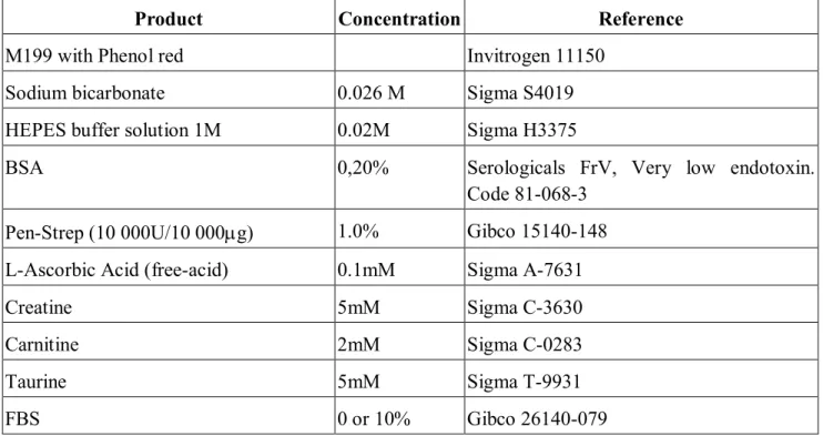
Table 3. Cell culture media used for ARVCM isolation and culture
Product Concentration Reference
M199 with Phenol red Invitrogen 11150
Sodium bicarbonate 0.026 M Sigma S4019
HEPES buffer solution 1M 0.02M Sigma H3375
BSA 0,20% Serologicals FrV, Very low endotoxin.
Code 81-068-3 Pen-Strep (10 000U/10 000g) 1.0% Gibco 15140-148 L-Ascorbic Acid (free-acid) 0.1mM Sigma A-7631
Creatine 5mM Sigma C-3630
Carnitine 2mM Sigma C-0283
Taurine 5mM Sigma T-9931
FBS 0 or 10% Gibco 26140-079
Table 4. Krebs-Henseleit (KH) buffer used for perfusion, with and without Ca2+
Product Final
Concentration
Reference
NaCl (MW 58.44) 118mM ACL S-2838
KCl (MW 74.6) 4.7mM Fisher Scientific P-217
MgSO4*7H2O (MW 246.48) 1.19mM Acros Organics
213115000
NaHCO3 (MW 84) 25mM Sigma S4019
Dextrose (MW 180.2) 11mM ACP D-0660
KH2PO4 (MW 136.1) 1mM Fisher Scientific P-285
CaCl2 100x 100mM * J.T. Baker
Perfusion Digestion Solution Final Concentration Reference
Collagenase 1 mg/mL Worthington S4B7097
205u/mg
Hyaluronidase 0.3 mg/mL Roche 10 106 500 001
BSA 1 mg/mL Serologicals FrV, Very
low endotoxin. Code 81-068-3
Table 6. Incubation Digestion Solution prepared in KH buffer
Final Concentration Reference
DNAse I ≤ 0.13 mg/mL Sigma DN-25
Trypsin ≤ 0.13 mg/mL Difco 215240
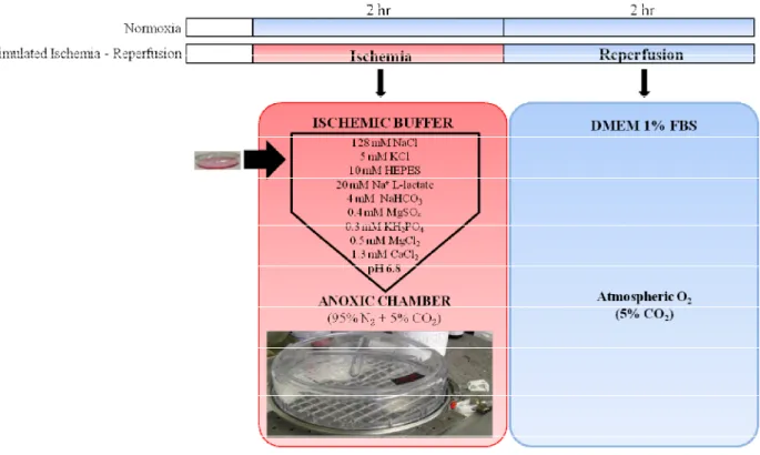
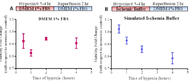
II.2. Simulated Ischemia - Reperfusion
Sub-confluent plates were placed in warm ischemic buffer in a sealed chamber (Billups-Rothenberg Modular Incubator Chamber, California, USA) saturated with 95% N2 + 5% CO2
for 2 hours at 37°C. Table 7 presents the composition of the ischemic buffer.
Figure 4 presents the treatment schedule for the simulated ischemia-reperfusion (IR) experiments. After 2 hours of ischemia, the plates were quickly processed and reperfusion of all conditions was performed within 3 minutes of the opening of the anoxic chamber. Reperfusion was simulated by the replacement of the ischemic buffer with DMEM supplemented with 1% FBS with or without oxytocin, under normoxic conditions (5% CO2),
reperfusion media (Figure 4). All controls were treated with the appropriate concentrations of vehicle. Additionally, OT antagonist OTA was administered 15 minutes prior to the induction of ischemia and maintained throughout the experiment.
Table 7. Ischemic Buffer composition
Product Concentration (mM) NaCl 128 KCl 5 MgSO4 0.4 KH2PO4 0.3 MgCl2 0.5 CaCl2 1.3 HEPES 10 Na L-Lactate 20 NaHCO3 4 pH 6.8
Figure 4. Simulated Ischemia - Reperfusion (IR) protocol. Sub-confluent monolayers of H9c2 cells were covered in warm ischemic buffer and placed inside an anoxic chamber for 2 hours at 37°C. The cells were "reperfused" with pre-equilibrated warm DMEM containing 1% FBS under normal atmospheric oxygen conditions and further incubated for 2 - 4 hours.
Cell viability was measured by the conversion of the tetrazolium salt MTS [3-(3,5-dimethylthialzol-2-yl)-5-(3 carboxymethoxypheny)-2-(4-sulphophenyl)-2H-tetrazolium] into a formazan product that is soluble in culture medium (Promega CellTiter Aqueous 96 # G3580) according to manufacturer's instructions. 20 µL of MTS were added to 100 µL cell culture medium in each well of a 96-well plate (Corning Costar #3595) where cells were grown. Absorbance at 490 nm was measured using a Symantek plate reader. Blank absorbance values resulting from the cell-free oxidation of MTS were subtracted from all experimental values.
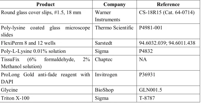
TUNEL (Terminal deoxynucleotidyl transferase-mediated dUTP nick end-labeling) assay was performed using DeadEnd™ Fluorometric TUNEL System (Cat. No. G3250, Promega, Montreal, QC, Canada) according to the manufacturer’s protocol. Briefly, cells were grown on poly-L-lysine - treated cover slips in 6 well plates and after the proper treatment, cells were fixed with a solution containing 6% formaldehyde and 2% methanol in PBS, pH 7.4, for 10 min at RT and permeabilized with 0.2% Triton X-100 in PBS, pH 7.4. Cells were incubated in the TUNEL reaction mixture for 1hr in the dark at 37°C, washed and mounted on Prolong Gold anti-fade reagent with DAPI (Cat. No. P36931, Invitrogen, Montreal, QC, Canada).
Cells were visualized using an inverted light fluorescent microscope (Olympus IX51) and images were captured at 10 and 20X magnifications for analysis using a digital charge-coupled device camera (Q IMAGING #Q23774). The quantification of TUNEL-positive