HAL Id: hal-00980086
https://hal.archives-ouvertes.fr/hal-00980086
Submitted on 17 Apr 2014
HAL is a multi-disciplinary open access
archive for the deposit and dissemination of
sci-entific research documents, whether they are
pub-lished or not. The documents may come from
teaching and research institutions in France or
abroad, or from public or private research centers.
L’archive ouverte pluridisciplinaire HAL, est
destinée au dépôt et à la diffusion de documents
scientifiques de niveau recherche, publiés ou non,
émanant des établissements d’enseignement et de
recherche français ou étrangers, des laboratoires
publics ou privés.
Mössbauer spectroscopy study of MgAl2O4-matrix
nanocomposite powders containing carbon nanotubes
and iron-based nanoparticles
Pierre Coquay, Eddy de Grave, Robert E. Vandenberghe, Charles Dauwe,
Emmanuel Flahaut, Christophe Laurent, Alain Peigney, Abel Rousset
To cite this version:
Pierre Coquay, Eddy de Grave, Robert E. Vandenberghe, Charles Dauwe, Emmanuel Flahaut, et
al.. Mössbauer spectroscopy study of MgAl2O4-matrix nanocomposite powders containing carbon
nanotubes and iron-based nanoparticles. Acta Materialia, Elsevier, 2000, vol. 48, pp. 3015-3023.
�10.1016/S1359-6454(00)00063-X�. �hal-00980086�
To link to this article : DOI:10.1016/S1359-6454(00)00063-X
URL:
http://dx.doi.org/10.1016/S1359-6454(00)00063-X
This is an author-deposited version published in:
http://oatao.univ-toulouse.fr/
Eprints ID: 10687
To cite this version:
Coquay, Pierre and De Grave, Eddy and Vandenberghe, Robert E. and Dauwe,
Charles and Flahaut, Emmanuel and Laurent, Christophe and Peigney, Alain and
Rousset,
Abel
Mössbauer
spectroscopy
study
of
MgAl2O4-matrix
nanocomposite powders containing carbon nanotubes and iron-based
nanoparticles. (2000) Acta Materialia, vol. 48 (n° 11). pp. 3015-3023. ISSN
1359-6454
O
pen
A
rchive
T
oulouse
A
rchive
O
uverte (
OATAO
)
OATAO is an open access repository that collects the work of Toulouse researchers
and makes it freely available over the web where possible.
Any correspondence concerning this service should be sent to the repository
administrator:
[email protected]
MOÈSSBAUER SPECTROSCOPY STUDY OF MgAl
2O
4-MATRIX NANOCOMPOSITE POWDERS CONTAINING
CARBON NANOTUBES AND IRON-BASED
NANOPARTICLES
P. COQUAY1{, E. DE GRAVE1, R. E. VANDENBERGHE1, C. DAUWE1, E. FLAHAUT2, CH. LAURENT2, A. PEIGNEY2 and A. ROUSSET2
1NUMAT, Department of Subatomic and Radiation Physics, University of Gent, Proeftuinstraat 86,
B-9000 Gent, Belgium and2CIRIMAT, UMR CNRS 5085, Laboratoire de Chimie des MateÂriaux
Inorganiques, Universite Paul-Sabatier, 31062 Toulouse cedex 4, France
AbstractÐMaterials involved in the catalytic formation of carbon nanotubes are for the ®rst time systema-tically studied by MoÈssbauer spectroscopy between 11 K and room temperature. Mg1ÿxFexAl2O4 x 0:1, 0.2, 0.3, 0.4) solid solutions are transformed into carbon nanotubes±Fe/Fe3C±MgAl2O4composite powders
by reduction in a H2±CH4 gas mixture. The oxides are defective spinels of general formulae
Mg1ÿx2Fexÿ3a2 Fe32aqaAl32 O42ÿ:Ferromagnetic a-Fe, ferromagnetic Fe3C and a g-Fe form, the latter poss-ibly corresponding to a g-Fe±C alloy, are detected in the composite powders. An attempt is made to corre-late these results with the microstructure of the powder. It seems that the nanoparticles, which catalyze the formation of the carbon nanotubes, are detected as Fe3C in the post-reaction MoÈssbauer spectroscopy
analysis.
ReÂsumeÂÐDes mateÂriaux impliqueÂs dans la formation catalytique de nanotubes de carbone sont pour la premieÁre fois systeÂmatiquement eÂtudieÂs par spectroscopie MoÈssbauer entre 11 K et la tempeÂrature ambiante. Des solutions solides Mg1ÿxFexAl2O4 x 0:1, 0.2, 0.3, 0.4) sont transformeÂes en poudres com-posites nanotubes de carbone±Fe/Fe3C±MgAl2O4par reÂduction dans un meÂlange gazeux de H2et de CH4.
Les oxydes sont des spinelles lacunaires de formule geÂneÂrale Mg2 1ÿxFexÿ3a2 Fe
3
2aqaAl32 O42ÿ:Du Fe-a ferro-magneÂtique, du Fe3C ferromagneÂtique et une forme de Fe-g, cette dernieÁre correspondant probablement aÁ
un alliage Fe±C-g, sont deÂtecteÂs dans les poudres composites. Des correÂlations sont faites entre ces reÂsultats et la microstructure de la poudre. Il semble que les nanoparticules qui catalysent la formation des nano-tubes de carbone sont deÂtecteÂes comme du Fe3C dans l'analyse par spectroscopie MoÈssbauer des produits
obtenus apreÁs la reÂaction.
Keywords:Carbon nanotubes; Iron; Carbides; MoÈssbauer eect
1. INTRODUCTION
Since the report by Iijima [1] on carbon nanotubes (hereafter denoted as CNTs), many laboratories
around the world have been studying this new form of carbon which presents high expectations for future applications such as light-weight composite materials, hydrogen storage and lithium-ion bat-teries. Methods of synthesis have been reviewed by Journet and Bernier [2]. Several of these involve nanometric metal particles as catalysts. Laurent et
al. [3] have surveyed the various mechanisms pro-posed for nanotube nucleation and growth from such particles. In particular, the catalytic decompo-sition of hydrocarbons or the disproportionation of CO on metal particles (generally based on Fe, Co or Ni) lead to Iijima-type CNTs, as opposed to
hol-low carbon ®bers, when the catalyst particles are suciently small [4±9]. Some works [8, 10, 11] have shown that the active particles are smaller than c. 6 nm in diameter. Catalytic methods are promising owing to the possibilities of up-scaling but one di-culty is to obtain the nanometric particles at the relatively high temperature (usually higher than 8008C) required for the formation of CNTs.
Works by some of the present authors [5, 10±15]
{To whom all correspondence should be addressed. Tel.: +1-32-92646569; fax: +1-32-92646697; E-mail: pierre. [email protected] (P. Coquay).
have shown that single-wall nanotubes (SWNTs) and small multi-wall nanotubes (MWNTs) are advantageously prepared by the selective reduction of oxide solid solutions in H2±CH4 gas mixtures.
The reduction of a solid solution between a non-reducible oxide such as Al2O3, MgAl2O4 or MgO
and one or more transition metal oxide(s) produces very small transition metal (Fe, Co, Ni and their alloys) nanoparticles at a temperature usually higher than 8008C. The decomposition of CH4over
the freshly formed metal nanoparticles prevents their further growth and thus results in the very strong proportion of SWNTs and small MWNTs compared with other forms of carbon.
CNTs±Fe/Fe3C±MgAl2O4 powders were prepared
by reduction in H2±CH4 atmosphere of
Mg1ÿxFexAl2O4 x 0:1, 0.2, 0.3, 0.4) solid sol-utions. Results on the synthesis and microstructure of these powders have been published earlier [15] but detailed information on the Fe species that could be responsible for the formation of the CNTs
is lacking. The present paper reports a study by
57Fe MoÈssbauer spectroscopy of the oxide solid
sol-utions and of the corresponding reduced powders.
2. EXPERIMENTAL
The Mg1ÿxFexAl2O4 x 0:1, 0.2, 0.3, 0.4) solid solutions were prepared by the combustion route as described elsewhere [15]. Brie¯y, the appropriate amounts of the desired metal nitrates (Mg, Al, and Fe) were mixed in stoichiometric proportions with urea and dissolved in a minimum amount of water. The solutions were then placed in a furnace pre-heated at 6008C, where the autocatalytic exothermic redox reaction takes place within minutes, produ-cing the oxide powder. These powders were then attrition-milled. In the next step, the oxide solid sol-utions obtained were reduced in a H2±CH4gas
mix-ture (18 mol% CH4) for 6 min at 10708C, giving
rise to the CNTs±Fe/Fe3C±MgAl2O4 composite
powders. For the sake of brevity, the samples will be referred to according to the following example: the solid solution Mg0.9Fe0.1Al2O4 will be named
Mg9Fe1 and the corresponding composite powder Mg9Fe1R, where R stands for ``reduced''.
The 57Fe MoÈssbauer spectra were recorded with 57
Co (Rh) sources in a conventional time-mode spectrometer with constant-acceleration drive and a triangular reference signal. Accumulation of the data was performed in 1024 channels until a back-ground of at least 106 counts per channel was
reached. The spectrometer was calibrated by collect-ing at room temperature (RT) the spectrum of a standard a-Fe foil and the isomer-shift values quoted hereafter are with reference to this standard. The spectra of the nanocomposite powders were analyzed assuming symmetrical components with a Lorentzian line shape, while those of the oxide pow-ders were ®tted with a quadrupole-shift distribution
where each subspectrum is composed of Lorentzian lines. Most of the spectra were recorded at 80 K and at RT to detect a possible evolution of the Fe phases. In addition, selected specimens were exam-ined at dierent temperatures between 11 K and RT.
3. RESULTS AND DISCUSSION
3.1. Oxide solid solutions
Previous analyses [15] have revealed that only a spinel phase is detected in the X-ray diraction (XRD) patterns. The MoÈssbauer spectra of the oxide solid solutions were recorded at 80 K. The corresponding parameters are reported in Table 1 and a typical spectrum is shown in Fig. 1(a).
The spectra were ®tted with two distributed doublets, characteristic of ferrous and ferric iron. A slightly better ®t is obtained in the case of Mg9Fe1 by adding a linear correlation between the isomer shift and the quadrupole splitting in the distribution for both doublets, as it was previously shown for similar oxides [16]. This correlation does not give a better result for the powders containing more iron and thus was not used in the corresponding ®ts. For these powders, the isomer-shift values given in Table 1 are average values. In both distributions of quadrupole splittings two main components may be recognized [Figs 1(b) and (c)]. Since the spinel lat-tice has octahedral and tetrahedral sites, the two peaks in the distribution pro®les for both the Fe2+ and Fe3+components could indicate that both
cat-ions are distributed among these two lattice sites. However, this is dicult to ascertain due to the in-accuracy of the calculated quadrupole-splitting dis-tributions, and hence possible artefacts. The presence of Fe3+ in the lattice of MgAl2O4
indi-cates that the obtained products are defective spi-nels, whose formulae can be written as Mg1ÿx2Fexÿ3a2 Fe32aqaAl32 O42ÿ 0:1RxR0:4).
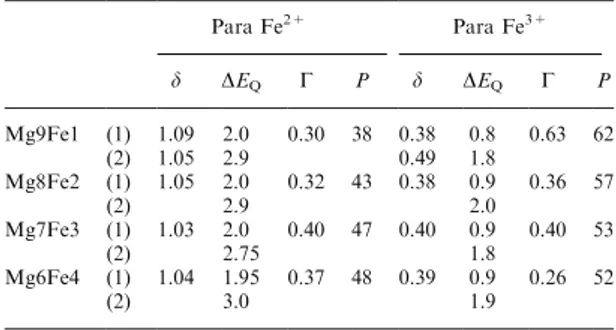
Table 1. 80 K MoÈssbauer parameters of the Mg9Fe1, Mg8Fe2, Mg7Fe3 and Mg6Fe4 oxide powders. Para: paramagnetic; d: iso-mer shift (mm/s); DEQ: quadrupole splitting (mm/s); G: line width
(mm/s) of elemental doublet; P: proportion (%); (1) and (2) refer to the two components which are recognized in the respective
probability distribution pro®les Para Fe2+ Para Fe3+ d DEQ G P d DEQ G P Mg9Fe1 (1) 1.09 2.0 0.30 38 0.38 0.8 0.63 62 (2) 1.05 2.9 0.49 1.8 Mg8Fe2 (1) 1.05 2.0 0.32 43 0.38 0.9 0.36 57 (2) 2.9 2.0 Mg7Fe3 (1) 1.03 2.0 0.40 47 0.40 0.9 0.40 53 (2) 2.75 1.8 Mg6Fe4 (1) 1.04 1.95 0.37 48 0.39 0.9 0.26 52 (2) 3.0 1.9
3.2. Nanocomposite powders
XRD-pattern analysis of the composite powders [15] has revealed the presence of a-Fe and Fe3C in
addition to the spinel matrix. However, no g-Fe could be resolved, possibly due to the superposition of its peaks with those of the major phases. Scanning electron microscopy (SEM) observations [15] have shown that the metal-oxide grains are uni-formly covered by a web-like network of CNTs
bun-dles, several tens of micrometers long. A typical SEM image referring to powder Mg8Fe2R is rep-resented in Fig. 2(a). It has been observed in trans-mission electron microscopy (TEM) [Fig. 2(b)] that the bundles are made up of SWNTs and small MWNTs with a diameter close to 4 nm. Carbon-coated particles of an unidenti®ed nature (metallic iron, iron±carbon alloy or iron carbide) in the 5± 20 nm size range were found at the surface of the CNTs, as opposed to inside the CNTs, and were
therefore not thought to be related to the processes of CNTs formation. In contrast, smaller particles
were occasionally found inside the tube tips. It is also important to note that the reduction of the fer-rous and ferric ions produces particles inside the spinel grains.
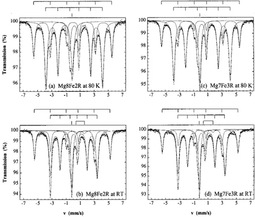
The characteristic parameters of the MoÈssbauer spectra recorded at RT and at 80 K are reported in Tables 2 and 3, respectively. Typical spectra are shown in Fig. 3. The ®ts consist of four patterns: one doublet, two sextets and one singlet. The doub-let at RT is characteristic of an Fe2+ phase. However, it could not be reasonably adjusted in the spectra at 80 K. The larger sextet is due to ferro-magnetic a-Fe particles and the narrower one is characteristic of ferromagnetic Fe3C (cementite).
Since Fe3C has two inequivalent crystallographic
sites [17], the MoÈssbauer parameters of the sextet
accounting for Fe3C correspond to the average of
the two Fe-site parameters. The values obtained at RT and at 80 K are in good agreement with those
Fig. 1. 80 K MoÈssbauer spectrum of the Mg9Fe1 oxide powder (a) and quadrupole-splitting distributions of the Fe2+(b) and Fe3+(c) doublets.
Fig. 2. SEM (a) and TEM (b) images of the Mg8Fe2R nanocomposite powder.
Table 2. RT Mo Èssbauer parameters of the Mg9Fe1R, Mg8Fe2R, Mg7Fe3R and Mg6Fe4R nanocomposite powders. Para: paramagnetic; ferro: ferromagnetic; Hhf : hyper®ne ®eld (kOe); d : isomer shift (mm/s); D EQ : quadrupole splitting (mm/s); 2 eQ : quadrupole shift (mm/s); G : line width (mm/s); P : proportion (%) Para Fe 2+ Ferro a -Fe Ferro Fe 3 C Non-ferro Fe d D EQ G P d Hhf 2 eQ G P d Hhf 2 eQ G P d G P Mg9Fe1R 1.19 0.85 0.69 2.5 0 331 0 a 0.28 47 0.18 205 0.026 0.34 27.5 ÿ 0.089 0.32 23 Mg8Fe2R 1.09 1.19 0.48 2 0 332 0 a 0.27 28 0.18 206 0.022 0.28 56 ÿ 0.085 0.33 14 Mg7Fe3R 1.15 1.36 0.49 2 0 331 0 a 0.28 24 0.18 206 0.019 0.29 58 ÿ 0.092 0.32 16 Mg6Fe4R 1.16 1.46 0.34 1.5 0 331 0 a 0.26 21.5 0.18 206 0.027 0.27 52 ÿ 0.095 0.28 25 aFixed parameter. Table 3. 80 K Mo Èssbauer parameters of the Mg9Fe1R, Mg8Fe2R, Mg7Fe3R and Mg6Fe4R nanocomposite powders. Ferro: ferromagnetic; Hhf : hyper®ne ®eld (kOe); d : isomer shift (mm/s); 2 eQ : quadrupole shift (mm/s); G : line width (mm/s); P : proportion (%) Ferro a -Fe Ferro Fe 3 C Non-ferro Fe d Hhf 2 eQ G P d Hhf 2 eQ G P d G P Mg9Fe1R 0.12 342 0 a 0.31 44 0.30 247 0 0 0.34 31.5 0.037 0.58 24.5 Mg8Fe2R 0.12 343 0 a 0.29 27.5 0.31 248 0 0 0.32 59 0.031 0.51 13.5 Mg7Fe3R 0.11 342 0 a 0.28 21.5 0.31 247 0 0 0.31 62.5 0.016 0.40 16 Mg6Fe4R 0.11 340 0 a 0.29 21 0.31 247 0 0 0.31 54 0.017 0.36 25 aFixed parameter.
reported by Bi et al. [18] for Fe3C nanoparticles.
Regarding the singlet, the negative value of the iso-mer shift at RT suggests a g-Fe phase.
The MoÈssbauer spectra of Mg9Fe1R and Mg6Fe4R were recorded at various temperatures between 11 K and RT (Tables 4 and 5, respect-ivelyÐFig. 4). Again, the weak Fe2+doublet could not be resolved at low temperatures. However, even using a larger velocity scale, no new sextet could be detected at these temperatures. This paramagnetic oxide phase could re¯ect the presence of Fe2+ ions
distributed in the spinel lattice, indicating that the starting oxide solid solutions are not fully reduced under the chosen conditions. The obtained MoÈssbauer parameters are in agreement with this conclusion and the expected values of the isomer shift and quadrupole splitting at low temperatures suggest that this weak doublet is completely obscured by the more intense pattern of the Fe3C
sextet. Considering this, no signi®cant evolution of the proportions of the dierent Fe phases can be detected. Nevertheless, referring to the proportions of the Fe phases at RT (Table 2) where the Fe2+ doublet is clearly resolved, relative errors of 22:5%, 24% and 21:5% are estimated for the proportions of a-Fe, Fe3C and the singlet,
respect-ively. These considerations based on the variations of the proportion values reported in Table 4 reason-ably include the uncertainties of the ®ts.
A considerable broadening of the singlet at low temperatures is clearly observed. For these tempera-tures, a narrow sextet was used instead of a singlet to ®t the spectra, yielding a more adequate repro-duction of the central part of the spectra. This could indicate that the corresponding Fe-species undergo a paramagnetic±antiferromagnetic trans-formation with a transition temperature at about 75 and 50 K for Mg9Fe1R and Mg6Fe4R, respect-ively. This also suggests a g-Fe phase since it is known that paramagnetic g-Fe shows an antiferro-magnetic coupling below 80 K [19]. The hyper®ne ®elds (Tables 4 and 5) are in good agreement with the saturation ®eld 2725 kOe reported by Crowell and Walker for small antiferromagnetic g-Fe±Ni particles [20].
The present results are to be compared with those resulting from a study of Fe±MgAl2O4
pow-ders prepared by reduction in an atmosphere of pure H2 [16]. Obviously no carbides, nor CNTs,
were formed in those powders. MoÈssbauer spec-troscopy analysis has revealed two phases: the sex-tet characteristic of ferromagnetic a-Fe is detected at all temperatures between 9 and 298 K and a sing-let appears above 50 K. The singsing-let was assigned to paramagnetic a-Fe in the form of very small nano-particles with a lowered Curie temperature. It was furthermore shown that the latter particles were intragranularly dispersed in the spinel matrix.
Fig. 3. 80 K (a) and RT (b) MoÈssbauer spectra of the Mg8Fe2R nanocomposite powder and 80 K (c) and RT (d) spectra of the Mg7Fe3R nanocomposite powder.
Table 4. Mo Èssbauer parameters of Mg9Fe1R between 11 K and RT. Para: paramagnetic; ferro: ferromagnetic; antiferro: antiferromagnetic; Hhf : hyper®ne ®eld (kOe); d : isomer shift (mm/s); D EQ : quadrupole splitting (mm/s); 2 eQ : quadrupole shift (mm/s); G : line width (mm/s); P : proportion (%) Para Fe 2+ Ferro a -Fe Ferro Fe 3 C Antiferro/para g-Fe ± C T d D EQ G P d Hhf 2 eQ G P d Hhf 2 eQ G P d H hf 2 eQ G P 11 ± ± ± ± 0.13 343 0 a 0.34 45 0.31 252 0 0 0.35 31.5 0.043 18.5 0 a 0.56 23.5 30 ± ± ± ± 0.12 343 0 a 0.34 44.5 0.31 252 0 0 0.35 31.5 0.038 17 0 a 0.57 24 50 ± ± ± ± 0.13 343 0 a 0.35 44.5 0.31 251 0 0 0.37 31 0.038 15 0 a 0.57 24.5 75 ± ± ± ± 0.12 342 0 a 0.36 45 0.30 248 0 0 0.36 30.5 0.040 10 0 a 0.54 24.5 100 1.01 2.03 0.52 1.5 0.11 342 0 a 0.34 45 0.30 246 0 0 0.36 30 0.030 ± ± 0.49 23.5 125 1.22 1.41 0.77 2 0.10 340 0 a 0.32 45 0.29 242 0 0 0.38 30 0.016 ± ± 0.41 23 150 1.31 1.65 0.56 2 0.092 339 0 a 0.34 45 0.28 238 0 0 0.36 30 0.003 ± ± 0.40 23 175 1.18 1.24 0.66 2.5 0.078 338 0 a 0.33 44.5 0.26 235 0 0 0.42 28.5 0 ± ± 0.40 24.5 200 1.21 1.39 0.47 2 0.067 337 0 a 0.35 45 0.26 230 0 0 0.41 28.5 ÿ 0.018 ± ± 0.41 24.5 225 1.24 1.48 0.56 3 0.046 335 0 a 0.34 46 0.24 226 0 0 0.41 27 ÿ 0.032 ± ± 0.39 24 250 1.10 1.21 0.48 3 0.032 334 0 a 0.39 45 0.23 219 0.019 0.43 28 ÿ 0.045 ± ± 0.39 24 275 1.07 1.03 0.52 3 0.017 332 0 a 0.36 47 0.21 213 0.018 0.41 26 ÿ 0.066 ± ± 0.39 24 295 1.13 1.13 0.51 3 0 331 0 a 0.36 48 0.20 206 0.016 0.38 25 ÿ 0.079 ± ± 0.40 24 aFixed parameter. Table 5. Mo Èssbauer parameters of Mg6Fe4R between 16 and 200 K. Ferro: ferromagnetic; antiferro: antiferromagnetic; para: paramagnetic; Hhf : hyper®ne ®eld (kOe); d : isomer shift (mm/s); 2 eQ : quadrupole shift (mm/s); G : line width (mm/s); P : proportion (%) Ferro a -Fe Ferro Fe 3 C Antiferro/para g-Fe ± C T d Hhf 2 eQ G P d Hhf 2 eQ G P d Hhf 2 eQ G P 16 0.12 341 0 a 0.42 21.5 0.31 251 0 0 0.35 53 0.026 9 0 a 0.46 25.5 30 0.12 341 0 a 0.41 22 0.31 250 0 0 0.36 52.5 0.023 7 0 a 0.44 25.5 50 0.12 341 0 a 0.38 22 0.31 249 0.01 0.37 53 0.019 6 0 a 0.40 25 75 0.11 340 0 a 0.44 22 0.31 247 0 0 0.36 53 0.020 ± ± 0.44 25 100 0.11 340 0 a 0.47 23 0.30 245 0 0 0.38 52 0.011 ± ± 0.43 25 125 0.10 339 0 a 0.39 22.5 0.29 242 0 0 0.36 53 0 ± ± 0.39 24.5 200 0.06 336 0 a 0.44 23 0.25 231 0.01 0.38 52 ÿ 0.035 ± ± 0.41 25 aFixed parameter.
It is proposed that the singlet in the current spec-tra, being present at temperatures as low as 11 K with an unchanged proportion, could re¯ect the for-mation of a g-Fe±C phase. Indeed, in addition to the nanosize of the particles [21], a small proportion of carbon added to iron (austenite) could also hin-der the martensitic transformation of g-Fe to a-Fe [22], which takes place at 9108C for pure bulk iron. The lower NeÂel temperature and the lower values of the hyper®ne ®eld observed for Mg6Fe4R com-pared with Mg9Fe1R (Tables 4 and 5) could be a consequence of slight dierences in carbon content. A comparison with the MoÈssbauer parameters reported by Bauer et al. [23] for g-Fe±C bulk ma-terials indicates that the hyper®ne interaction is characteristic of an iron atom with carbon atoms
farther away than the next-nearest neighbor pos-itions.
The absence in the present spectra of the para-magnetic a-Fe phase detected by QueÂnard [16] could indicate that at least a part of the corre-sponding intragranular particles are found in the form of g-Fe±C species in the present materials. A study on CNTs±Fe/Fe3C±Al2O3 composite powders
[10] has indeed shown that g-Fe (possibly g-Fe±C) species were located in the intragranular position. This suggests that a fraction of the carbon supplied by the decomposition of CH4 migrates inside the
lattice of the oxide matrix.
It has been shown [15] that the total carbon con-tent in the CNTs±Fe/Fe3C±MgAl2O4 composite
powders increases steadily with the proportion of
Fig. 4. MoÈssbauer spectra of the Mg9Fe1R (a) and Mg6Fe4R (b) nanocomposite powders measured at dierent temperatures.
iron and that the quantity of CNTs strongly
increases between Mg9Fe1R and Mg8Fe2R, reach-ing saturation for Mg7Fe3R (Fig. 5). The evolution between Mg9Fe1R and Mg8Fe2R could re¯ect the higher density of potentially active particles at the surface of the matrix grains upon the increase of the total iron content. For specimens with a higher iron content (Mg7Fe3R and Mg6Fe4R), this is balanced by an easier coalescence, and hence deacti-vation, of the surface particles.
The proportions of the dierent Fe phases pre-sent in the composite powders as revealed by the RT MoÈssbauer spectra (Table 2) are shown in Fig. 6(a). The proportion of Fe3C roughly doubles
from c. 30% to c. 60% between Mg9Fe1R and Mg8Fe2R and subsequently reaches saturation. The fraction of a-Fe follows an almost opposite evol-ution whereas the g-Fe±C proportion initially decreases and then increases again so that it is simi-lar for Mg9Fe1R and Mg6Fe4R. Figure 6(b) shows the amounts of the dierent Fe phases present in the samples. To obtain a reasonable approximation of the amounts from the proportions, the latter were multiplied by one, two, three and four for Mg9Fe1R, Mg8Fe2R, Mg7Fe3R and Mg6Fe4R, re-spectively. The relative errors were multiplied in the same way. The contents of both Fe3C and a-Fe
steadily increase with the iron content, the former one in a much more pronounced way. In contrast, the g-Fe±C content is the same for Mg9Fe1R and Mg8Fe2R and only increases for higher amounts of iron.
These results seem to indicate that the particles responsible for the nucleation and possibly the growth of the CNTsare found as Fe3C by
post-reac-tion MoÈssbauer spectroscopy analyses. The exact nature of the catalytic particle is not known but it is probably a very small carbon-containing Fe par-ticle in which some still poorly established driving forces make carbon atoms participate in the
for-mation of the CNTs. However, the continuous
increase of the Fe3C quantity [Fig. 6(b)] indicates
that some of the particles located at the surface of the spinel grains that were inactive for the for-mation of the CNTs also end up as cementite. The
continuous increase of the quantities of a-Fe and g-Fe±C could further indicate that such inactive par-ticles are detected as a-Fe and g-Fe±C in reac-tion MoÈssbauer spectra. The dierence in the post-reaction nature of the particles probably re¯ects the size distribution (5±20 nm) observed by TEM [15]. The relative thickness of the carbon coating com-pared with the size of a given particle could in¯u-ence its post-reaction nature.
4. CONCLUSIONS
Mg1ÿxFexAl2O4 x 0:1, 0.2, 0.3, 0.4) solid sol-utions were transformed into CNTs±Fe/Fe3C±
MgAl2O4composite powders by reduction in a H2±
CH4gas mixture. Materials involved in the catalytic
formation of carbon nanotubes were for the ®rst time systematically studied by MoÈssbauer spec-troscopy between 11 K and room temperature. The study has shown that the primary oxides are defec-tive spinels of general formulae
Fig. 6. Proportions of the dierent Fe phases present in the composite powders as revealed by the MoÈssbauer spec-tra at RT (a) and corresponding amounts of the dierent Fe phases in the powders (b). The amounts were obtained by multiplying the corresponding proportions by one, two, three and four for Mg9Fe1R, Mg8Fe2R, Mg7Fe3R and Mg6Fe4R, respectively. The lines are guides to the eye. Fig. 5. Carbon content (Cn) and quantity factor of carbon
nanotubes (DS ) in the composite powders. DS represents the surface area of carbon in the composite powder, which essentially corresponds to that of the CNTs. See Ref. [15]
Mg1ÿx2Fexÿ3a2 Fe32aqaAl32 O42ÿ 0:1RxR0:4). Dierent Fe phases have been detected in the com-posite powders: ferromagnetic a-Fe, ferromagnetic Fe3C and a Fe form that could correspond to a
g-Fe±C alloy. It has been attempted to correlate these results with the microstructure of the powder. This study suggests (i) that the particles that catalyze the formation of the CNTs are Fe3C in post-reaction
examinations, (ii) that the particles located inside the spinel grains are mostly in the g-Fe±C form, and (iii) that the inactive carbon-coated particles could end up as any of the a-Fe, Fe3C and g-Fe±C
forms.
AcknowledgementsÐThis work was partly funded by the Belgian National Programme of Inter-University Attraction Pole on Reduced Dimensionality Systems (PAI±IUAP P4/10) and by the Fund for Scienti®c Research, Flanders. The franco-belgian TOURNESOL program (project T99.006) is also gratefully acknowledged.
REFERENCES 1. Iijima, S., Nature, 1991, 354, 56.
2. Journet, C. and Bernier, P., Appl. Phys. A, 1998, 67, 1.
3. Laurent, Ch., Flahaut, E., Peigney, A. and Rousset, A., New J. Chem., 1998, 22, 1229.
4. Dai, H., Rinzler, A. G., Nikolaev, P., Thess, A., Colbert, D. T. and Smalley, R. E., Chem. Phys. Lett., 1996, 260, 471.
5. Peigney, A., Laurent, Ch., Dobigeon, F. and Rousset, A., J. Mater. Res., 1997, 12, 613.
6. Kong, J., Cassell, A. M. and Dai, H., Chem. Phys. Lett., 1998, 292, 567.
7. Cheng, H. M., Li, F., Sun, X., Brown, S. D. M.,
Pimenta, A., Marucci, A., Dresselhaus, G. and Dresselhaus, M. S., Chem. Phys. Lett., 1998, 289, 602. 8. Hafner, J. H., Bronikowski, M. J., Azamian, B. K., Nikolaev, P., Rinzler, A. G., Colbert, D. T., Smith, K. A. and Smalley, R. E., Chem. Phys. Lett., 1998, 296, 195.
9. Colomer, J. F., Bister, G., Willems, I., Konya, Z., Fonseca, A., Van Tendeloo, G. and Nagy, J. B., Chem. Commun., 1999, 1343.
10. Peigney, A., Laurent, Ch. and Rousset, A., J. Mater. Chem., 1999, 9, 1167.
11. Flahaut, E., Peigney, A., Laurent, Ch., Rousset, A., J. Mater. Chem., 2000, 10, 249.
12. Laurent, Ch., Peigney, A. and Rousset, A., J. Mater. Chem., 1998, 8, 1263.
13. Peigney, A., Laurent, Ch., Dumortier, O. and Rousset, A., J. Eur. Ceram. Soc., 1998, 18, 1995. 14. Flahaut, E., Govindaraj, A., Peigney, A., Laurent,
Ch., Rousset, A. and Rao, C. N. R., Chem. Phys. Lett., 1999, 300, 236.
15. Govindaraj, A., Flahaut, E., Laurent, Ch., Peigney, A., Rousset, A. and Rao, C. N. R., J. Mater. Res., 1999, 14, 2567.
16. QueÂnard, O., Doctoral thesis, Toulouse, 1997, p. 283. 17. Yakel, H. L., Int. metall. Rev., 1985, 30, 17.
18. Bi, X. X., Ganguly, B., Human, G. P., Huggins, F. E., Endo, M. and Eklund, P. C., J. Mater. Res., 1993, 8, 1666.
19. Weiss, R. J., Proc. Phys. Soc., 1963, 82, 281.
20. Crowell, J. M. and Walker, J. C., in Proc. 5th Int. Conf. on MoÈssbauer Spectroscopy, ed. M. Hucl and T. Zemcik. Czechoslovak Atomic Energy Commission Nuclear Information Center, Praha, 1975, p. 289. 21. Kachi, S., Bando, Y. and Higuchi, S., Japan J. appl.
Phys., 1962, 1, 307.
22. Ron, M., in ed. R. L. Cohen, Applications of MoÈssbauer Spectroscopy, Vol. II. Academic Press, New York, 1980, p. 335.
23. Bauer, Ph., Uwakweh, O. N. C. and Genin, J. M. R., Hyp. Int., 1988, 41, 555.