Université de Montréal
THE ROLE OF EPHB6 AND EPHRINBS IN
BLOOD PRESSURE REGULATION
par Zenghui Wu
Département de Sciences Biomédicales Faculté de Médecine
Thèse présentée à la faculté des études supérieures en vue de l’obtention du grade de
Philosophiae Doctor (Ph.D.) en sciences biomédicales
Décembre 2012
Université de Montréal Faculté des études supérieures
Cette thèse intitulée:
<<THE ROLE OF EPHB6 AND EPHRINBS IN BLOOD PRESSURE REGULATION>> Présentée par:
Zenghui Wu
a été évaluée par un jury composé des personnes suivantes: Dr Edward Bradley ……… Président du jury Dr Jiangping Wu ……… Directeur de recherche Dr Hongyu Luo ……… Directeur de recherche Dr Jonathan Ledoux ……… Membre du jury Dr Serge Lemay ……… Examinateur externe Dr Marie-Josée Hébert ……… Repésentante de la doyenne
Résumé
L’hypertension artérielle est le facteur de risque le plus important dans les maladies cardiovasculaires (MCV) et les accidents vasculaires cérébraux (AVC). L’hypertension artérielle essentielle est une maladie complexe, multifactorielle et polygénique. Même si on a identifié de nombreux facteurs de risque de l’hypertension artérielle, on ne comprend pas encore clairement les mécanismes qui la régissent.
Les kinases hépatocytes produisant l’érythropoïétine (Eph) constituent la plus grande famille des récepteurs tyrosine kinase qui se lient à des ligands de surface cellulaire appelés éphrines sur les cellules avoisinantes. On sait que les interactions de Eph et des éphrines sont essentielles aussi bien dans les processus de développement que dans le fonctionnement des organes et des tissus adultes. Cependant on n’a pas encore étudié la relation entre Eph/éphrines et l’hypertension artérielle.
Nous avons créé des modèles de souris knockout (K.O.) Ephb6-/-, Efnb1-/- et Efnb3-/- pour cette étude. Dans le modèle EphB6-/-, nous avons observé que les souris K.O. Ephb6 castrées, mais pas les femelles, ainsi que les souris mâles non castrées présentaient une tension artérielle élevée (TA) par rapport à leurs homologues de type sauvage (TS). Ceci suggère que Ephb6 doit agir de concert avec l’hormone sexuelle mâle pour réguler la TA. Les petites artères des mâles castrés Ephb6-/- présentaient une augmentation de la contractilité, une activation de RhoA et une phosphorylation constitutive de la chaîne légère de la myosine (CLM) lorsque comparées à celles de leurs homologues TS. Ces deux derniers résultats indiquent que la phosphorylation de CLM et de RhoA passe par la voie de signalisation de Ephb6 dans les cellules du muscle lisse de la paroi vasculaire (CMLV). Nous avons démontré que la réticulation de Efnbs mais non celle de Ephb6 aboutit à une réduction de la contractilité des CMLV. Ceci montre que l’effet de Ephb6 passe par la signalisation inversée à travers Efnb.
Dans le modèle Efnb1-/- conditionnel spécifique au muscle lisse, nous n’avons observé aucune différence entre Efnb1-/- et les souris de TS concernant la mesure de la TA dans des conditions normales. Cependant, la TA des souris K.O. Efnb1 lors d’un stress d’immobilisation est supérieure à celle des souris de TS. Dans les petites artères des souris K.O. Efnb1, le rétrécissement et la phosphorylation de CLM étaient élevés. In vitro, la contractilité et
l’activation RhoA de la CMLV des souris TS étaient augmentées quand leur Efnb1 était réticulé. Ces résultats corroborent ceux des souris KO Ephb6 et prouvent que l’effet de Ephb6 dans le contrôle de la TA se produit au moins par l’intermédiaire d’un de ses ligands Efnb1 dans les CMLV.
Dans le modèle Efnb3-/-, on a observé une augmentation de la TA et du rétrécissement des vaisseaux chez les femelles Efnb3-/-, mais non chez les mâles; l’échographie a aussi révélé une résistance accrue au débit sanguin des souris K.O. femelles. Cependant la mutation de Efnb3 ne modifie pas la phosphorylation de la CLM ou l’activation de RhoA in vivo. Dans l’expérience in vitro, les CMLV des souris femelles Efnb3-/- ont présenté une augmentation de la contractilité mais pas celle des souris mâles Efnb3-/-. La réticulation des CMLV chez les mâles ou les femelles de TS avec solide anti-Efnb3 Ab peut réduire leur contractilité.
Notre étude est la première à évaluer le rôle de Eph/éphrines dans la régulation de la TA. Elle montre que les signalisations Eph/éphrines sont impliquées dans le contrôle de la TA. La signalisation inverse est principalement responsable du phénotype élevé de la TA. Bien que les Efnb1, Efnb3 appartiennent à la même famille, leur fonction et leur efficacité dans la régulation de la TA pourraient être différentes. La découverte de Eph/Efnb nous permet d’explorer plus avant les mécanismes qui gouvernent la TA.
Abstract
Hypertension is the most important risk factor for the cardiovascular diseases (CVD) and strokes. The essential hypertension is a complex, multifactorial and polygenic disease. Although many hypertension risk factors have been identified, the comprehensive understanding of mechanisms remains elusive.
Erythropoietin-producing hepatocyte kinases (Ephs) are the largest family of receptor tyrosine kinases, which bind to cell surface ligands called ephrins on neighboring cells. Eph and ephrin interactions are known to be essential in developmental processes, as well as in functions of adult organs and tissues. However the relationship between Ephs/ephrins and hypertension has not been studied.
Ephb6-/-, Efnb1-/- and Efnb3-/-knockout mice models were established for this study. In the EphB6-/- model, we observed that the castrated Ephb6 KO mice but not female or uncastrated male mice presented heightened blood pressure (BP) compared to the wild type (WT) counterparts. This suggests that Ephb6 needs to act in concert with sex hormone to regulate blood pressure. Small arteries from castrated Ephb6-/- males showed increased contractility, RhoA activation and constitutive myosin light chain (MLC) phosphorylation compared to their WT counterparts. The latter two findings indicate that RhoA and MLC phosphorylation are in the signaling pathway of Ephb6 in vascular smooth muscle cell (VSMC). We demonstrated that, crosslinking of Efnbs but not Ephb6 resulted in reduced VSMC contractility. This indicates that the effect of Ephb6 is via reverse signaling through Efnbs.
In smooth muscle-specific conditional Efnb1-/- model, no difference was observed between Efnb1-/- and WTmice in BP measurement under a normal condition. However, the BP of Efnb1 KO mice during immobilization stress were higher than that of WT mice. In the small arteries from Efnb1 KO mice, the constriction and MLC phosphorylation were elevated. In vitro, the contractility and RhoA activation of WT VSMC were augmented when their Efnb1 was crosslinked. These results corroborate the findings from Ephb6 KO mice, and prove that the effect of Ephb6 in BP control is at least via one of its ligand Efnb1 in VSMC.
In the Efnb3-/- model the heightened BP and increased vessel constriction were observed in Efnb3-/-females but not males; the echography also revealed the increased blood flow resistance
RhoA activation in vivo. In in vitro experiment, VSMCs from Efnb3-/- female mice showed increased contractility but did not Efnb3-/- male mice. Crosslinking of VSMCs from WT males or females with solid anti-Efnb3 Ab can reduce their contractility.
Our study is the first to assess the role of Eph/ephrins in BP regulation. Eph/ephrins signalings are involved in the regulation of BP. The reverse signaling is mainly responsible for the elevated BP phenotype. Although the Efnb1, Efnb3 belongs to the same family, their function and effectiveness in the regulation of BP might be different. The discovery of Eph/Efnbs allows us to further explore the mechanism in BP.
INDEX
Résumé ... i
Abstract ... iii
List of figures and tables ... vii
List of abbreviations ... ix
Acknowledgements ... xiii
I. INTRODUCTION ... 1
Part I ... 2
Structure and function of Eph/ephrin family members... 2
1.1 Structure and signaling mechanism of Eph/ephrins... 3
1.2 Ephs/ephrins in nervous system ... 5
1.3 Ephs/ephrins in immune system ... 8
1.4 Ephs/ephrins in bone homeostasis ... 9
1.5 Eph/ephrin signaling in cancer... 10
1.6 Eph/ephrin in cardiovascular system ... 12
1.6.1 Eph/ephrin in angiogenesis ... 12
1.6.2 Eph/ephrins in VSMCs ... 14
Part II ... 15
Blood pressure regulation and hypertension ... 15
2.1 The physiology of vascular smooth muscle ... 15
2.2 Duality phenotypes of VSMC (contractile and synthetic phenotypes) ... 16
2.3 The renin-angiotensin-aldosterone system (RAAS) ... 18
2.4 Angiotensin II in RAAS... 18
2.5 Aldosterone in RAAS ... 20
2.5.1 Aldosterone and endothelial cells ... 20
2.5.2 Aldosterone and vascular smooth muscle cells ... 20
2.6 Calcium in VSMCs ... 21
2.7 NO and NOS in the cardiovascular system ... 22
2.10 Hormones in the cardiovascular system ... 25
Conclusion and remarks ... 28
Reference List ... 29
II. ARTICLES ... 56
Article 1 ... 57
Ephb6 Regulates Vascular Smooth Muscle Contractility and Modulates Blood Pressure in concert with Sex Hormones ... 57
Article 2. ... 102
A possible role of Efnb1- A lligand of Eph receptor tyrosine kinases in modulating blood pressure ... 102
Article 3. ... 138
Blood pressure modulation by Efnb3 protein in concert with sex hormones: evidence from gene knockout mice and blood pressure-associated single nucleotide polymorphisms in EFNB3 related genes in 69,395 human subjects ... 138
III. DISCUSSION ... 184
1. Signaling pathways to regulate VSMC contractility ... 185
2. The hypotheses of how Eph/Efnb in concert with testosterone regulate VSMC functions ... 186
3. Evidences supporting the hypothesis that the reverse signaling regulates the contractile phenotype ... 187
4. Efnbs regulate RhoA-ROCK-MLCP signaling pathway ... 187
5. The effect of Grip1 in VSMC ... 188
6. The role of EphB forward signaling in VSMC ... 188
7. The hypothesis that the effect of testosterone on VSMC is via its regulatory function on RhoA synthesis and activity ... 189
8. An attempt to reconcile some controversial findings in our study ... 190
9. A brief summary ... 191
10. Contribution to sciences and future research directions ... 192
List of figures and tables
INTRODUCTIONFigure 1 the family of Eph/ephrin proteins ... 3
Figure 2 structure of Eph/ephrin proteins ... 4
Figure 3 the signaling pathway of EphB3 /ephrinB1 in cell migration ... 11
Figure 4 the morphology of VSMC in different phenotypes ... 17
Figure 5 the expression level of contractile and synthetic protein marker ... 17
Figure 6 the Angiotensin system ... 19
Figure 7 the signaling pathway of Rho family ... 24
Figure 8 the distribution of Rho family ... 25
ARTICLE 1 Figure 1 Expression of Ephb6 and Efnbs in mouse VSMC ... 77
Figure 2 Contractility MLC phosphorylation and RhoA activation of mesenteric arteries from Ephb6 KO and WT mice... 79
Figure 3 the effect of sex hormones on BP and VSMC contraction of castrated Ephb6 KO mice. ... 81
Figure 4 Twenty-four-h urine catecholamine levels in Ephb6 KO mice. ... 83
Figure 5 Reverse but not forward signaling between Ephb6 and Efnbs dampens VSMC contractility. ... 84
Figure 6 Grip1 in the Efnb reverse signaling pathway in VSMC ... 86
Supplemental Figure1 Plasmid construct used to generate -actin promoter-driven Ephb6Ephb6 without its intracellular domain) Tg mice. ... 91
Supplemental Figure2 BP and HR of male and female Ephb6 KO mice. ... 92
Supplemental Figure3 Plasma and serum hormone levels in Ephb6 KO mice. ... 93
Supplemental Figure4 Castrated Ephb6/KO mice present no BP increase. ... 94
Supplemental Figure 5 Ephb6 expression in the adrenal gland medulla according to ISH ... 95
Supplemental Figure 6 Expression of EFNBs, Ephb6, AR, AT1, and Grip1 in VSMC and endothelial cells ... 96
ARTICLE 2
Figure 1 Generation of mice with SMC-specific deletion of Efnb1 ... 126
Figure 2 Decreased contractility of Efnb1-engaged VSMC ... 127
Figure 3 Early Efnb1-mediated signaling events in VSMC stimulated by PE and AngII ... 129
Figure 4 MLC phosphorylation and RhoA activation in VSMC ... 131
Figure 5 Grip1 in the Efnb1 reverse signling pathway ... 132
Figure 6 GTP-associated RhoA expression and vasoconstriction of Efnb1 KO mesenteric arteries ... 134
Figure 7 Blood pressure and heart rate of Efnb1 KO mice ... 135
Figure 8 Normal urine and plasma hormone levels in Efnb1 KO and WT mice ... 137
ARTICLE 3 Figure 1 Efnb3 deletion in vascular cells in Efnb3 KO mice ... 168
Figure 2 BP and HR of Efnb3 KO mice ... 169
Figure 3 Contractility of mesenteric arteries from Efnb3 KO mice ... 170
Figure 4 VSMC contractility is influenced by Efnb3 deletion and reverse signalling ... 171
Figure 5 Normal 1-adrenoreceptor (AR) expression and Ca++ flux in Efnb3 KO VSMC ... 173
Figure 6 MLC, MLCK and MLCP phosphorylation of small arteries and VSMC from WT and KO mice ... 175
Figure 7 Grip1 in the Efnb reverse signalling pathway in VSMC ... 176
Figure 8 BP-related hormone levels in Efnb3 KO mice ... 177
Table I Association of SNPs in the EPHB6/EFNB system with BP phenotypes in 69,396 human subjects ... 165
Table II Echographic analysis of CO, carotid artery resistance and left ventricle mass of KO and WT mice ... 166
Supplementary figure1 LocusZoom plots of –log10 p-values (left hand vertical axis) from IBPC meta-analysis for specific queried genes ... 179
DISCUSSION Figure 1 the contractile signaling pathway of VSMC... 185
List of abbreviations
Ab Antibody
ACE Angiotensin-converting enzyme
Akt Protein kinase B
AMPAR AMPA receptor
Ang II Angiotensin II
AP Arterial pressure
ApoER2 Apolipoprotein E receptor 2
AT1 Angiotensin receptor type 1
AT2 Angiotensin receptor type2
ATIP1 AT2R interacting protein-1
ATRAP AT1R-associated protein
BM Bone marrow
BM progenitors Bone marrow progenitors
BP Blood pressure
CDC42 Cell division control protein 42 homolog
CFNS Craniofrontonasal syndrome
cGMP Cyclic guanosine 3’, 5’-monophosphate
CIL Contact inhibition of locomotion
COX2 Cyclo-oxygenase 2
CRC Colorectal cancers
CS Cyclic stretch
CVD Cardiovascular disease
CXCL Chemokine (C-X-C motif) ligand
DP Diastolic pressure
GEPR G protein-coupled estrogen receptor
ENAC Epithelial sodium channel
eNOS Endothelial nitric oxide synthase
Eph Erythropoietin-producing hepatocellular
ERE Estrogen response element
ERK Extracellular signal-regulated kinases
ERs Estrogen receptors
FAK Focal adhesion kinase
FGF Fibroblast growth factor
FGFR1 Fibroblast growth factor receptor 1
GAP GTPase activating protein
GEFs Guanine nucleotide exchange factors
GEPR G protein-coupled estrogen receptor 1
GDI Guanosine nucleotide dissociation inhibitor
GPCRs G-protein-coupled receptors
GPI Glycosylphosphatidylinositol
GPI-ANCHOR Glycosylphosphatidylinositol anchor
GRIP 1 Glutamate receptor-interacting protein 1
Grb2 Growth factor receptor-bound protein 2
GTP Guanosine triphosphate
HR Heart rate
Hsp90 Heat shock protein 90
IgD Immunoglobulin D
IgG Immunoglobulin G
IP3 Inositol triphosphate
iNOS Inducible NOS
KO Knockout
LARG Leukemia associated Rho guanine nucleotide exchange factor
LECs Lymphatic endothelial cells
LTP Long term depression
MAP Mean arterial pressure
MAPK Mitogen-activated protein kinase
MET Mesenchymal-epithelial transition
MHC Major histocompatibility complex
miRNA MicroRNA
MLC20 Myosin light chain
MLCK Myosin light chain kinase
MLCP Myosin light chain phosphatase
MMP-2 Matrix metalloprotease 2
MYPT1 Myosin-targeting subunit 1
MR Mineralocorticoid receptor
NC Neural crest
NCC Neural crest cell
NMDA N-methyl-D-aspartate
NMDAR N-methyl-D-aspartate receptor
NO Nitric oxide
NOS Nitric oxide synthase
NSCLC Non-small-cell lung cancer
O2- Superoxide
ONOO- Peroxynitrite
PAK P21 protein-activated kinase
PARP Poly-ADP-ribose polymerase
PDAC Pancreatic ductal adenocarcinoma
PDGF Platelet-derived growth factor
PI3K Phosphatidylinositide 3-kinases
PLC Phospholipase C
PDZ Psd-95,DlgA and ZO1
PICK1 PKC-intracting protein 1
PRR Pro-renin receptor
PYK2 Protein tyrosine kinase 2
RAAS Rennin-angiotensin aldosterone system
RACK1 Receptor activated C-kinase 1
RANKL Receptor activator of NF-κB ligand
RAS Rennin-angiotensin system
RBD Receptor-binding domain
RELN Secreted glycoprotein reelin
ROS Reactive oxygen species
RNA Ribonucleic acid
RTK Receptor tyrosine kinase
RT-qPCR Reverse transcription quantitative polymerase chain reactioin
SAM Serile α-motif
SH2 Src Homology 2
sGC Soluble guanylate cyclase
SOC Store-operated Ca2+ channels
SR Sarcoplasmic reticulum
TCR T cell receptor
Tg Transgenic
TRP Transient receptor potential
VEGF Vascular endothelial growth factor
VEGFR Vascular endothelial growth factor receptor
VLDLR Very-low density lipoprotein receptor
VSMCs Vascular smooth muscle cells
Acknowledgements
I would like to express my sincere gratitude to my supervisor Dr. Jiangping Wu and Dr. Hongyu Luo for their scientific guidance and encouragement throughout my study and during the preparation of thesis.
I would like to thank all the colleagues in the lab for their kindly cooperation.
Thanks also extended to all my friends and those who always support me in different ways.
Finally, I would like to thank all of my family members for their understanding, patience, and their great support, which were critically important for me to complete this study.
Part I
Structure and function of Eph/ephrin family members
Receptor tyrosine kinases (RTKs) are trans-membrane glycoproteins 1, 2, which are the key components of signaling pathways involved in cell proliferation3, differentiation, migration and metabolism 4, 5. The activation of RTKs is due to the binding of their cognate ligands, by which they transduce the extracellular signal to cytoplasm through auto-phosphorylation process. The receptor tyrosine kinases are widely spread among different organs, and are involved in different kinds of cellular processes. More than 90 tyrosine kinase genes have been identified in human genome. 58 are cell surface receptors, which are divided into 20 subfamilies 6, 7.
The Eph receptors are the largest family of receptor tyrosine kinases8, which are successfully isolated from the cDNA of erythropoietin-producing hepatocellular carcinoma cell line9. Since then, the Eph receptors have been identified in affecting the physiological and pathological processes in many cell types and organs10, such as axon guidance, angiogenesis, tissue border formation, cell adhesion and cell migration 11-14. As the Eph receptor-interacting proteins15, ephrins also involve in many aspects of cell survival, differentiation, proliferation, and migration16.
The initial subdivision of Ephs into EphA and EphB classes was based on similarities in extracellular sequences8, and a corresponding binding preference to either the glycosylphosphatidylinositol anchored ephrin-A ligands or the three transmembrane ephrin-B ligands. Eph-ephrin interactions are promiscuous, but EphA members preferentially bind to ephrinAs, and EphBs bind to ephrinBs.
There are 16 members of Eph receptors identified in mammals17. EphA1-EphA10 bind to 5 ephrinAs; EphB1-EphB6 bind to 3 ephrinBs. As stated above, ten EphA (EphA1-A10) members promiscuously interact with 5 A-ephrins (EfnA1-A5) and six different EphBs (EphB1-B6) promiscuously interact with 3 B-ephrins (EfnbB1-B3). However, EphB2 can also bind to ephrinA5, and EphA4 binds to ephrinB2 18.
Figure 1 the family of Eph/ephrin proteins 1.1 Structure and signaling mechanism of Eph/ephrins
Like all RTKs, Eph receptors are type I trans-membrane protein. The functional domains can be divided into extracellular and intracellular region. The extracellular region contains ligand-binding globular domain of a highly conserved N-terminal sequence, which is both necessary and sufficient for ligand recognition and binding. The binding domain is followed by a cysteine-rich region and two fibronectin type III repeats19, which might be involved in receptor-receptor dimerization interactions or other proteins such as the NMDA receptors.
The cytoplasmic part of Eph receptors can be divided into four functional units: the juxtamembrane domain containing two conserved tyrosine residues; a classical protein tyrosine kinase domain, a serile α-motif (SAM) and a PDZ-domain-binding motif. The resolved structure of SAM domain (around 70 amino acids) indicates that Eph receptor could form dimers and oligomers. The PDZ-binding motif is located in the carboxy-terminal with 4-5 amino acid residues, which contains a consensus binding sequence that includes a hydrophobic residue (usually valine or isoleucine).
As ligands of Eph receptors, ephrins are membrane-anchored proteins or transmembrane proteins. EphrinAs have an extracellular Eph receptor-binding domain; and they directly attach to the cell surface without the intracellular tail. EphrinBs are transmembrane proteins. The extracellular Eph receptor-binding domain is located at its N-terminus, and about 3 amino-acid PDZ-binding motifs are at the carboxy terminus.
Figure 2 structure of Eph/ephrin proteins 20
As Ephs and ephrins are membrane proteins, they can only interact within short distance. Eph receptors are activated once they are bound by clustered ephrins on membrane between two neighboring cells, which express Ephs and ephrins respectively.
The first step in the formation of an Eph/ephrin “signaling cluster” is the monovalent interaction between Eph and ephrin in juxtaposed cell surface, which occurs with nanomolar affinity21. The Eph/ephrin complexes can progressively aggregate into larger
clusters, the size of which depend on the densities of Eph receptors and ephrins on the cell surface21.
The significant characteristics of Eph/ephrin signaling pathway is bi-directionality 22. The forward signals depend on Eph kinase activity in receptor-expressing cell, in which Eph/ephrin complex induces the initiation of Eph signaling through auto-phosphorylation, such as phosphorylation of juxtamembrane domain and downstream target proteins 23. The reverse signal depends on Src family kinases in eprhin-expressing cells 22.
Several years of studies have revealed that Ephs/ephrins have many effects on different aspects of cell activities and broad roles in normal physiology and in pathogenesis: such as angiogenesis 24, the development of neurons 25, cardiovascular development 26, 27 and tumorigenesis 28 et al.
1.2 Ephs/ephrins in nervous system
In the literature, many papers show that Eph/ephrin signaling plays an essential role during the process of synapse formation and neuronal plasticity29. It is known that Ephs are expressed in developing and mature nervous system, with classic functions of mediating axon guidance and target recognition30.
The roles of Eph receptors in spine morphogenesis have been widely studied. As small protrusions, the dendritic spines are located on the surface of dendrite, which receive the excitatory synapses and are responsible for synaptic transmission and long-term memory. N-methyl-D-aspartate (NMDA)-type glutamate receptors are essential for activity-dependent synaptic plasticity31 and memory formation32.
Eph forward signaling has effects on spine and synapse formation31. It is known that dynamic formation and retraction of spines is one of features underlying synaptic plasticity and possibly long-term memory 33.
Eph receptors expressed on dendrites are activated by ephrins, which are expressed on axons or astrocytes and regulate spine and synapse formation. Eph/ephrin complex participates in activity-induced long-term changes in synaptic strength34.
In hippocampus, several Ephs have been localized to dendrites and spines33, 34. The experiment of null mutant mice has clearly indicated a functionally redundant requirement of three EphB receptors in dendritic spine morphogenesis33. For example EphB2-deficient
mice show modestly reduced hippocampal long-term potentiation. In this model, EphB2 receptors associate with NMDA receptors at synaptic sites and play a role in synaptogenesis. Eph/ephrin complex induces the clustering of NMDA receptors and promotes morphogenesis of dendritic spines; in the meantime forward signaling can stabilize clustering of NMDAR to further enhance NMDAR-dependent Ca2+ flux 35. Although normal hippocampus synapse morphology still exists, the deletion of EphB2 drastically impairs long-lasting LTP and leads to extinguish LTD and depotentiation 33. EphB2 interacts with Syndecan-2 of trans-membrane heparin sulfate proteoglycan to induce mature spine morphology in mouse brain 36. EphB2 also plays an important role in the regulation of AMPAR, which mediates most fast excitatory synaptic transmission 37. Under the stimulation of ephrinB, the EphB2 co-clusters with Glu2 and Glu3 subunits of AMPAR, mediates the interaction between AMPAR binding protein and PKC-intracting protein 1 (PICK1) or GRIP1 through PDZ motif 38.
As mentioned before, EphB2 controls AMPA-type glutamate receptor localization through PDZ binding domain interactions and triggers presynaptic differentiation via its ephrin binding domain. However, not all the EphBs exhibit equal effects during the spine and synapse formation. Triple null mutation of EphB1-3 leads to synapse number reduction; double mutants (EphB1/EphB2, EphB1/EphB3, EphB2/EphB3) show obvious defects in spine formation; however, the EphB1/EphB2 mutant leads severe defects than other mutants in spine formation 39. The re-expression of EphB2 is sufficient to rescue phenotype and support normal function of spine in EphB2-/- mice.
In cultured hippocampal neurons, EphB receptors regulate spine morphology by modulating the activity of Rho family GTPases. This process involves the activation of focal adhesion kinase (FAK) 40, Src, Grb2 and paxillin, among which FAK is a non-receptor tyrosine kinase that is widely expressed in different cells and is typically activated after assembly of integrin-mediated focal adhesions. Spine morphogenesis is shown to depend on signaling of kalinrin/Rac1 and intersectin/Cdc42/N-WASP to promote filamentous actin formation 41. EphA binds and activates ephexin, which is a member of Dbl family of exchange factors that activate RhoA and suppress Cdc42 42. In the meanwhile, the EphBs appear to be linked to GEFs, such as the kalinrin and intersectin,
EphrinBs are also essential in regulation of neuronal migration and plasticity in brain during the process of neuronal migration and proper functional maintenance. The secreted glycoprotein reelin (RELN) guides migration of neurons by binding to two lipoprotein receptors, the very-low density lipoprotein receptor (VLDLR) and apolipoprotein E receptor 2 (ApoER2). Loss of reelin function in humans results in a severe developmental disorder lissencephaly and other neurological disorders such as Alzheimer’s disease and schizophrenia43. Compound mouse mutants (Reln+/-/EphrinB3-/- , Reln+/- /EphrinB2-/-) and triple EphrinB1, B2, B3 knockout mice show neuronal migration defects. Clustering of ephrinBs recruit and phosphorylate Dab1, which is necessary for the activation of Reelin signaling 44. Therefore, the lack of EphrinBs in neurons causes interruption between Reelin and Dab1, leading to a reduced phosphorylation of Dab1 then neuronal migration defects. In humans, mutations in ephrinB1 lead to the craniofrontonasal syndrome (CFNS) by disturbing the crest/mesoderm boundary formation 45. CFNS is an X-linked developmental disorder that different gender shows the different phenotypes: females have frontonasal dysplasia and cornal craniosynostosis (fusion of the coronal sutures); in males, hypertenlorism is the only typical manifestation. PDZ-dependent reverse ephrinB1 signaling is also critical for the formation of a major commissural axon tract and the corpus callosum46. The miR-124 of an abundant brain microRNA (miRNA) acts as a posttranscriptional effector to regulate ephrinB1 reverse signaling47 in development of neural tube48.
Plasticity is essential for maintaining memory and learning. AMPA receptors, which are the main transducers of rapid excitatory transmission in the brain, are pivotal in this process. EphrinB2 signaling is critical for the stabilization of AMPA receptors on cellular membrane. The lack of ephrinB2 enhances internalization of AMPA receptors on cell membrane and reduces synaptic transmission 49.
EphrinB3 regulates the morphogenesis of postsynaptic compartment by transducing the tyrosine phosphorylation/SH2-dependent and PDZ-dependent reverse signaling 50. It is reported that ephrinB3-/- mice exhibit peculiar hopping gaits, which are correlated to the loss of unilateral motor control. As a midline repulsive barrier, EphrinB3 prevents corticospinal tract axons from re-crossing once they enter the spinal gray matter51. EphrinB3 also induces axon pruning by binding the sytoplasmic SH2/SH3 adaptor protein
Grb4, which functions as a molecular bridge to connect activated tyrosine-phosphorylated intracellular domain of ephrinB3 with Dock180, Rac/Cdc 42, and PAK52.
1.3 Ephs/ephrins in immune system
Ephs and ephrins are widely expressed in lymphoid organs53-55 as well as lymphocytes 56. Study shows that Eph/Ephrin signaling is essential for thymus migration during the organogenesis and lymphocyte maturation 57, 58. Thymus organogenesis requires coordinated interactions of multiple cell types, such as neural crest (NC) cells, mesenchymal cells and thymic epithelial cells 59. EphB2-/-, EphB3-/-, EphB2-/-/EphB3-/- mice show significantly decreased thymic cellularity compared to WT mice 60
The formation, separation, and subsequent migration are critical for the development of thymus59. EphrinB2 plays an important role in correct position of thymus on NC-derived cells. The conditional mutation of ephrinB2in the NC-derived cell leads to abnormal anatomical location of thymus 57. However, deletion of ephrinB2 doesn’t disturb separation of thymus. EphA4 also influences the development of thymus through alternations of epithelial network development. The EphA4-/- mice show smaller thymuses compared to wild-type controls 54.
The migration of lymphoid progenitors from fetal liver or bone marrow (BM) into thymic parenchyma is the first step of intrathymic T cell development, and following steps are essential for immature thymocytes, which develop their proper function through distinct thymic epithelial environment 61. EphB2 regulates the response of BM progenitors to extracellular matrix (ECM) ligand and chemokine. Deletion of EphB2in BM progenitors exhibits a decreased migratory response to ECM ligands and chemokines (CXCL12, CXCL21, and CXCL25) 62. EphA1, EphA4 regulate the migration of CD4+ T lymphocyte and induce tyrosine phosphorylation of PYK2, which associates with Src family kinase members 63, 64.
Binding of ephrinA1 and EphA receptors stimulates migration on CD4+and CD8+ T cell 65, 66. However, ephrinA1 induced migration is mainly found in CD45RO+ memory subset cells 67. Stimulation of Eph receptors leads to migration involving activation of Lck, Pyk2, PI3K, Vav1 and Rho GTPase in CD8+ CCR7 T cells 67.
T cell receptor (TCR), located on the cell surface with a highly diverse repertoire of αβ chains, is responsible for recognizing antigens and bounding to major histocompatibility complex (MHC) molecules68. This recognition process is crucial for T cell differentiation and maturation 68. Studies have shown that Eph receptors modulate responses mediated by TCR 69. Under suboptimal condition, EphB6 cross-linked with EphB6 Ab leads to drastic T cell proliferation; EphB6+ T cell has stronger response to anti-CD3 and anti-CD28 activation than EphB6- T cell. EphB6 is aggregated and co-localized with TCR to enhance the following response through the activation of p38 MAPK signaling pathway69, 70. EphrinB1 and ephrinB2 are involved in thymocyte development and peripheral T cell differentiation 71, 72. EphB3/EphB6 induced reverse signaling of ephrinB1/ephrinB2 strongly suppresses Fas-induced apoptosis in Malignant T lymphocytes, in which the activation is associated with promotion of Akt activation and the inhibition of the Fas receptor-initiated caspase proteolytic cascade 73. EphrinB1/B2 also interacts with IL 7Rα to promote the IL7-indeuced internalization in T cell 74, 75.
EphA receptors inhibit TCR-induced (anti-CD3) apoptosis of CD4+CD8+ thymocytes in culture and selection process in vivo 76. The immobilized EphB2-Fc and EfnB1-Fc fusion protein, at certain concentration can modulate anti-CD3 Ab induced apoptosis of CD4+CD8+ thymocytes 58.
Eph/ephrin signaling is also involved in the regulation of B cell77. EphA4 and EphA7 are expressed on B cells 77. The activated human B lymphocytes express ephrinA4 78. EphrinB3 specifically binds to B lymphocytes in blood through the sulphated cell surface receptor 79, in which the binding of ephrinB3 and B cell induces migration of IgD- memory B cell subpopulation 79.
1.4 Ephs/ephrins in bone homeostasis
Bone homeostasis is strictly controlled by proper cellular communication between osteoclasts and osteoblasts 80, 81 and balance of bone formation and resorption 82, 83. The mutation of ephrinB1 in mice leads to severe defects, including the neural crest cell (NCC)-derived tissues, incomplete body wall closure, and abnormal skeletal patterning and embryonic lethality 84.
The bone formation and resorption is regulated by the EphB4/ephrinB2 bidirectional signaling pathway85. The osteoclasts express ephrinB2 and osteoblasts express the EphB4 respectively. Reverse signaling of ephrinB2 leads to the suppression of osteoclast differentiation by inhibiting the osteoclastogenic c-Fos-NFATc1 cascade, while the EphB4 forward signaling stimulates osteogenic differentiation 86. Osteoclast precursors87 express EphA2/ephrinA2, which is induced by receptor activator of NF-κB ligand (RANKL) 88, 89. EphA2/ephrinA2 up-regulates RhoA in osteoclast precursors to suppress osteoblast differentiation89.
1.5 Eph/ephrin signaling in cancer
Numerous studies implicate the role of Eph/ephrin signaling in cancer progression and neovascularization such as cancer cell migration and invasion, in which Eph/ephrins were thought to play an oncogenic role 3, 90, 91. In recent years, the aberrant expression of Eph and ephrin genes has been identified in a wide range of human tumors 92 such as colorectal cancers (CRCs), lung cancer, myeloid cancer, breast carcinoma cells 93and prostate cancer et al 94.
CRC is one of the most common cancers in the world. Strong relationship is observed between Ephs/ephrins and CRC. The mRNA level of EphA1 and EphA2 are highly elevated in CRC cell lines95 and down-regulated EphA1 expression in colorectal cancer is correlated with poor survival 96 indicating that down regulation of EphA1 correlates with invasion and metastasis in colorectal carcinomas 97.
Human gastric cancer has strong relationship with expression level of EphA4. The overexpression of EphA4 are observed in 55 tumor specimen 98, which correlates with overexpression of FGFR1. Patients with EphA4-positive cancers have significantly lower survival rate than the patients with EphA4-negative cancers 98.
EphA4/ephrinA signaling also regulates cancer cell proliferation in pancreatic ductal adenocarcinoma (PDAC) carcinogenesis and development. Suppressing expression of EphA4/ephrinA3 drastically attenuates PDAC cell viability. Conversely, overexpression of EphA4 increases growth rate in PDAC cells 99.
101. Interaction of EphB3/ephrinBs promotes MET through inactivation of CrkL-Rac1 signaling pathway: Overexpression of EphB3 inactivates both CrkL and Rac1; the knockdown of EphB3 activates CrkL by decreasing its phosphorylation. In CRCs, the expression level of EphB3 is decreased. Overexpression of EphB3 results in tumor suppression in HT29 CRC cell line 102.
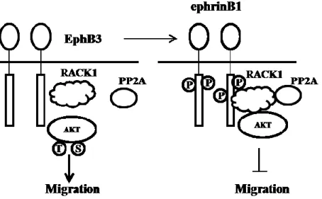
However, opposite results are also observed in other cancer cell line, such as non-small-cell lung cancer (NSCLC). In NSCLC non-small-cells, expression level of EphB3 is elevated comparing normal cells; the tyrosine phosphorylation of EphB3 is reduced; the migration is inhibited under the stimulation of ephrinB1-Fc/ephrinB2-Fc; the silencing of EphB3 shows suppression of tumor growth and metastasis in vivo 73, 103. The possible signaling pathway is that the activation of EphB3 forms the protein complex of RACK1, PP2A and Akt to inhibit cell migration; Silencing of EphB3 disassociates complex of AKT and RACK1, leading to cell migration103.
Figure 3 the signaling pathway of EphB3 /ephrinB1 in cell migration103
EphB4/ephrinB2 signaling has been linked to breast cancer. The breast cancer cells grow faster in EphB4ΔC-EGFP mouse xenograft model, where EphB4 kinase dominant negative form is overexpressed in recipients 104. Through the reverse signaling way, ephrinB2 increases blood content in tumor by enhancing the angiogenesis. EphB4 is expressed in human breast cancer; however its function is unclear. EphB4 induces migration of tumor cells and promotes tumor growth/survival. On the contrary, EphB4 also activates an antioncogenic Abl-Crk pathway in MDA-MB-435c breast cancer cells. EphB4 enhances
the phosphorylation of Crk on Tyr221 to inhibit breast cancer cell of viability, proliferation, motility, invasion and down-regulation of pro-invasive matrix metalloprotease (MMP-2)105.
In prostate cancer, ephrinB2 induces migration of Pc-3 prostate cancer cells and activates their Cdc42 by regulating the contact inhibition of locomotion (CIL), a process of stopping the continual locomotion of a cell in the same direction after collision with another cell 106. Through the Eph-Rho-ROCK pathway, Pc-3 prostate cancer cells switch from restrained state to invasive state107.
EphA2/ephrinA1 interaction also promotes tumor growth and metastasis of breast cancer cells 108. In a tumor xenografts mouse model with the MDA-MB-435 human breast cancer cells or KS1767 human Kaposi’s sarcoma cells, EphA2 shows continuous tyrosine phosphorylation throughout tumor vasculature 109. EphB2 is also important for the growth, migration and invasiveness in colon cancer cell. As the target gene of Wnt signaling pathway, EphB2 is related to early stage of colorectal cancer110.
1.6 Eph/ephrin in cardiovascular system
1.6.1 Eph/ephrin in angiogenesis
In vertebrate circulatory system, most of arteries and veins are formed in early stage of embryonic development 111. The remodeling and formation of vessels, through angiogenesis or the outgrowth of the new sprouts, perform complex cascade processes, in which Eph/ephrins are extensively involved 53, 112.
Angiogenesis is a highly orchestrated process that plays an essential role in physiological and pathophysiological development. Through angiogenesis, the pre-existing vessels sprout new vessels to complete the vascular formation and remodeling. EphB4/ephrinB2 is the first pair of genes which are found differentially expressed in arterial and venous endothelium113. According to the literature, EphB4/ephrinB2 plays an important role in embryonic vessel development and vascular remodeling 114. EphB4 marks venous endothelial cells and its ligand ephrinB2 marks arterial endothelial cells 115. Disruption of EphB4 or miss-expression of ephrinB2 results in intersomitic veins growing abnormally into the adjacent somatic tissue116.
Vascular endothelial growth factor (VEGF) signaling plays a crucial role in development and remodeling of blood vessels during embryogenesis117. VEGF is also involved in pathogenic angiogenesis, such as the recruitment and maintenance of tumor vasculature118. It is reported that VEGF can induce the expression of ephrinA1 in endothelial cells and subsequently activate EphA receptor signaling to promote angiogenesis through a juxtacrine mechanism. The soluble EphA2-Fc receptors inhibit VEGF-mediated endothelial cell survival, migration, sprouting, and assembly in vitro; however, it has no effect on FGF-induced angiogenesis 119.
Vascular endothelial growth factor receptor (VEGFR), the ligand of VEGF, is a receptor tyrosine kinase and a key regulator in blood vessel growth and homeostasis117. It is produced by endothelial, hematopoietic, and stromal cells. The VEGFR can be classified as three main subtypes, VEGFR-1, -2, and -3 120.
EphrinB2 regulates the internalization and signaling activity of VEGFR2. Through PDZ domain, ephrinB2 regulates VEGFR2 trafficking and controls endothelial tip-cell-mediated vessel sprouting121. EphrinB2 also regulates internalization of VEGFR3 in cultured lymphatic endothelial cells 122. However, the mutation of ephrinB2 only affects VEGFR but shows no effect on other angiogenic regulator receptors such as fibroblast growth factor receptors 123. Normal development of lymphatic vasculature needs the ephrinB2, in which PDZ motif plays the essential role124, 125. EphrinB2 regulates the remodeling of lymphatic vasculature through the interaction of PDZ-binding motif and PDZ-RGS3 and Dvl2 124, 125.
Slit/roundabout (Robo) proteins with three different members have emerged as key regulators of vascular remodeling and homeostasis126. The cooperation between Slit2 and ephrinA1 regulates a balance between angiogenesis and angiostasis. Slit2 stimulates angiogenesis through mRORC2-dependent activation of Akt and Rac GTPase. EphrinA1 down-regulates the activation of Slit2 and inhibits angiogenesis 127. EphrinB2 induces migration of endothelial cells by induction of Akt phosphorylation through the phosphorylatidylinositol-3 kinase pathway and promotes angiogenesis in adult vasculature 128. The dominant negative form of EphA2 inhibits capillary tube-like formation in human umbilical vein endothelial cells 109. EphA2 also promotes tight junction formation and impairs angiogenesis in brain endothelial cells 129.
1.6.2 Eph/ephrins in VSMCs
Eph/ephrin participates in the regulation of migration, spreading, contraction and attachment in VSMCs 130. EphrinA1 inhibits the activated Rac1 of Rac/PAK pathway to impair VSMC migration 131. EphrinB2 is involved in the spreading, migration 132 and attachment 133 of VSMC. During the blood-vessel-wall formation process, interaction of ephrinB2 and Crk-p130 (CAS) is required for directional migration and cell-matrix association in VSMC 53. Mice of ehprinB2 tissue-specific mutation in epithelium show vascular defects in skin, lung and gastrointestinal tract53.
EphA4 increases the activated RhoA via Vsm-RhoGEF to regulate VSMC contractility134. The EphB6 activated reverse signaling pathway regulates VSMC contractility through RhoA-ROCK-MLCP signaling pathway135, 136. In EphB6 knockout mice model, EphB6 knockout female and castrated male VSMCs show increased phosphorylated MLC and contractility135 and castrated male KO mice show elevated BP.
Part II
Blood pressure regulation and hypertension
Cardiovascular disease (CVD) is the lead cause of death worldwide, responsible for 30% of all death137. Increased blood pressure is the most-important risk factor for CVD. It is demonstrated that persistent hypertension is involved in stroke, ischemic heart diseases, kidney failure, and metabolic syndrome 138.
Hypertension is a medical condition, in which the blood pressure is chronically elevated According to “Reports of the joint national committee on prevention, detection, evaluation
and treatment”, the diagnosis of hypertension in adults will be made when the average of 2 or more diastolic BP measurements on at least 2 subsequent visits is over 90mmHg or when the average of multiple systolic BP readings on 2 or more subsequent visits is consistently over 140mmHg. Isolated systolic hypertension is defined as systolic BP >140mmHg and diastolic BP<90mmHg 139, 15% to 30% of adult population as well as more than half of elderly population suffer from high blood pressure in most countries. Normally, hypertension can be classified into two categories: the secondary hypertension and essential hypertension139. The secondary hypertension indicates that the elevated BP is a result of other conditions, such as reno-vascular disease 140, renal failure, pheochromocytoma, aldosteronism, et al. On the contrary, essential hypertension, also called idiopathic hypertension, accounting for 95% of all cases of hypertension, indicates that no specific medical cause is responsible for the elevated BP. Until now, the etiological of essential hypertension is still unclear. Genetic variations, gene malfunction, and disease conditions may be involved in essential hypertension, such as obesity and insulin resistance. Certain physiological status and life styles also involve in essential hypertension, such as high alcohol intake, high salt uptake, aging and stress139.
2.1 The physiology of vascular smooth muscle
In vasculature, small arteries and arterioles are main contributors to blood flow resistance in circulation141.Small arteries are composed of three layers: tunica adventitia, tunica media and tunica intima. The outer and inner layers are composed of mainly connective tissues and endothelial cells respectively; tunica media is composed of smooth muscle cells 142.
Two opposite effects tightly control homeostasis of VSMC tone: generation of force (contraction) and release of force (relaxation) 143, in which circulating neurotransmitters, hormones, and endothelium-1derived factors are involved.
Contraction and relaxation of smooth muscle cells are regulated by the phosphorylation and de-phosphorylation of myosin light chain (MLC20), which is controlled by myosin light chain kinase (MLCK) 144, 145 and myosin light chain phosphatase (MLCP) 146 separately. Upon stimulating of smooth muscle cell, Ca2+ is transiently elevated in the cytoplasm through extracellular fluid Ca2+ and intracellular stores. Ca2+ combines calmodulin to form the Ca2+/calmodulin complex, which binds and provokes MLCK to phosphorylate MLC20 at Ser 19 and Thr18 147. The activation of myosin ATPase promotes interaction of actin and myosin then leading to muscle contraction.
De-phosphorylated MLC20 is responsible for vessel relaxation, which is regulated by MLCP. MLCP is a heterotrimer, consisting of a catalytic subunit PP1cδ, a 20kDa subunit (M20) and Myosin-targeting subunit (MYPT1)148, 149. MLCP dephosphorylates MLC20 and induces VSMC relaxation.
2.2 Duality phenotypes of VSMC (contractile and synthetic phenotypes)
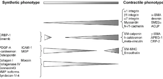
Under physiological condition, VSMC keep the quiescent “contractile” phenotype state and maintain appropriate vessel tone150. However VSMC shifts “contractile” to “synthetic” phenotypes 151 in response to certain circumstances such as stretch, injury, atherosclerosis and inflammation, in which VSMC enhances migration, proliferation and secretion but suppresses contraction 151. VSMC of synthetic phenotype changes morphology from “spindle” to “rhomboid” shape (Figure 4) and decreases expression level of contractile marker proteins (Figure 5), such as α-smooth muscle actin, smooth muscle myosin heavy chain, smoothelin-A/B152.
Figure 4 the morphology of VSMC in different phenotypes152
2.3 The renin-angiotensin-aldosterone system (RAAS)
The RAAS is essential in physiological and pathophysiological processes of cardiovascular system, and involved in the renal or non-renal complications of hypertension. Traditionally, RAAS plays the function of regulating renal sodium and water excretion 153, and body sodium-fluid balance154. With further understating of RAAS, its function is also recognized in endocrine systems and multiple hormonal systems, such as endocrine, paracrine and intracrine 155.
2.4 Angiotensin II in RAAS
Angiotensin II (Ang II), produced from the liver, is the active component of renin-angiotensin system (RAS)156. Ang II is the key effector to maintain systemic blood pressure through various mechanisms in cardiovascular and renal systems157. In classic RAAS, circulating renal-derived renin cleaves hepatic-derived angiotensinogen to form the deca-peptide angiotensin I (Ang I), which is converted by angiotensin-converting enzyme (ACE) to the Ang II. Besides the renin enzymetic pathway, Ang I and Ang II also can be produced through a nonrenin enzymetic pathway. For example the tonin and cathepsin convert angiotensinogen to Ang I then Trypsin, cathepsin and chymase convert Ang I to Ang II 158. As the cleavage product of Ang I, Ang II has potent effects on RAAS system. Ang II elevates blood pressure and retains salt and water in body. Ang II induces the secretion of aldosterone from zona glomerulosa of adrenal gland cortex. Ang II also stimulates vasoconstriction through AT1R-mediated activation of RhoA/Rho kinase-dependent myosin light chain (MLC) phosphorylation.
There are two subtypes of Ang II receptors, AT1 and AT2159. Both of them belong to the super-family of G-protein-coupled receptors (GPCRs). AT1 receptor is expressed ubiquitously and involved in most of the biological functions of Ang II, such as vasoconstriction, cardiac contractility, renal tubular sodium re-absorption, cell proliferation, vascular and cardiac hypertrophy, inflammatory responses, and oxidative stress160. In rodents, the AT1 can be classified into two isoforms, AT1a and AT1b. The AT1a is main activator of many signaling pathways in vascular smooth muscle cells. AT1R presents a basal activity in VSMC and may bind to GPCR kinases and β-arrestins. GPCR
protein (ATRAP) binds to AT1R and negatively regulates its signaling pathway. Inositol triphosphate (IP3) is the fast signaling factor in response to Ang II, leading to contraction of VSMCs 161, in which IP3 induces intracellular stores to release calcium and increases intracellular calcium concentration. AT1R is also involved in the inflammation process162. AT1R activates the NADPH oxidase, which generates superoxide anions and activates NF-κB in response to the induction of expression of pro-inflammatory molecules VCAM-1, MCP-1 and IL-6 163.
AT2R promotes different signaling pathway and works opposite to that of AT1R 164. AT2R is only highly expressed in fetal tissues164. Its expression dramatically decreases after birth and is restricted to a few organs mainly in cardiovascular system165. However, the AT2R is re-expressed in adult after cardiac and vascular injury. AT2R binds to AT2R-interacting protein-1 (ATIP1) to relax vascular smooth muscle cells through NO/cGMP signaling pathway 166.
Pro-renin receptor (PRR) 167 can be classified into three different molecular forms: a full-length integral TM protein (transmembrane), a soluble PRR, existing in plasma and urine, and a truncated form 168. The activation of PRR triggers the phosphorylation of mitogen-activated protein kinases (MAPKs), the intracellular signaling of extracellular signal regulated kinase 1/2 (ERK1/2) 169, up-regulation of cyclo-oxygenase 2 (COX2) and the activation of p38 MAPK/hsp27 170. PRR also regulates the Wnt/β-catenin signaling pathway of cell proliferation, migration and polarity171.
2.5 Aldosterone in RAAS
Numerous studies have shown that aldosterone, a steroid hormone, is widely involved in physiological and pathological processes in blood pressure regulation 172.
As downstream molecule of Ang II in RAAS, aldosterone is produced by the outer-section of adrenal cortex in adrenal gland in response to Ang II and affects renal distal tubules and collecting ducts of nephron173. Besides regulating salt homeostasis, aldosterone is now considered as a key factor of performing profibrotic and proinflammatory effects in cardiovascular diseases and metabolic diseases, such as hypertension, insulin resistance and dyslipidemia et al 174. Elevated level of aldosterone in patients with heart failure is related to a high risk of mortality175.
The mineralocorticoid receptor (MR), receptor of aldosterone, has two ligands, aldosterone and cortisol 176. MR is widely expressed on extra-renal tissues such as cardiomyocytes 177, 178, endothelial cells 179, 180 and VSMCs 181, 182. Through MR, aldosterone performs actions in renal distal convoluted tubules for fluid and electrolyte balance. The MR blockers (apironolactone and eplerenone) restrain action of aldosterone at the level of the receptor and effectively suppress blood pressure 173.
2.5.1 Aldosterone and endothelial cells
Endothelial cells are critical for vessel homeostasis 183. Aldosterone is broadly involved in physiological and pathophysiological process of endothelial cells. Aldosterone induces the early stage swelling and stiffness of endothelial cells by stimulating the epithelial sodium channel (ENAC) 184-186, ENAC decreases production of nitric oxide by down regulating endothelial nitric oxide synthase (eNOS) in endothelial cell 187, 188. Aldosterone also inhibits morphogenesis and angiogenesis of endothelial through down-regulating vascular endothelial growth factor receptor-2 (VEGFR-2) expression189.
2.5.2 Aldosterone and vascular smooth muscle cells
Aldosterone has been shown to induce vascular damage, proliferation 190. There are two ways to regulate VSMC by aldosterone. One is through MR, the other is through another proteins such as GPR30191.
proliferation of VSMCs through IGF-I signaling pathway, which enhances the VSMC proliferation under the stimulation of αVβ3 integrin 193, 194. Aldosterone modulates 12-/15-lipoxygenase (LO) atherogenesis in VSMC. VSMCs, treated with aldosterone, show elevated hydroxyeicosatetraenoic acid level and mRNA expression of platelet type 12-LO. In rat VSMC, uniaxial cyclic stretch (CS) promotes aldosterone synthesis and cytochrome p450 aldosterone synthase (CYP11B2) through ERK1/2 phosphorylation signaling 195. 2.6 Calcium in VSMCs
Plenty of ion channels are expressed in the plasma membrane of VSMCs, which form the walls of resistance arteries and regulate vascular tone196. Four types of K+ channels 197, 198, Ca2+ channel199 and two Cl- channels200 are expressed on the membrane of VSMC. Ca2+ is the key factor to trigger VSMC contraction196, 201. Four Ca2+ channels are shown as follows: the voltage-gated Ca2+ channels, receptor-operated Ca2+, the store-operated Ca2+ channels (SOC) and the Ca2+ permeable nonselective cation channels 202.
Voltage-gated Ca2+ channel is essential for influx of extracellular Ca2+ in vascular smooth muscles203. The channel is regulated by the membrane potential 204. Under physiological conditions, the channel is activated by depolarization and shut off by hyper polarization205. Voltage-gated Ca2+ channels can be classified into five different types, L-type, P-type, N-type, R-type and T-type channels. Through two basic modes of low-activity and high-activity of Ca2+ sparklets199, 206, 207, L-type Ca2+ channel primarily mediates Ca2+ influx in VSMC among five type channels 208.
The store-operated Ca2+ channels, being ATP and bradykinin sensitive, are highly selective for calcium 209. Store-operated Ca2+ channels are activated when intracellular calcium stores are empty 210. Store-operated Ca2+ channels are involved not only in the cell contraction but also proliferation211 and apoptosis 212. In smooth muscle cells, IP3-mediated Ca2+ release induces the mitogen or growth factor depletion of the sarcoplasmic reticulum (SR) 213 and results in the subsequent activation of transient receptor potential (TRP) cation channels that triggers Ca2+ entry into the cell. Platelet-derived growth factor (PDGF), an indicator in the development of pulmonary hypertension, enhances the store-operated Ca2+ entry by up-regulating STIM1/Orai1 through activation of Akt/mTOR211.
2.7 NO and NOS in the cardiovascular system
Nitric oxide (NO) is a gaseous molecule, which has broad functions in cardiovascular system214. NO works as a cofactor to interact with many molecules, such as NADPH, FMN, FAD, Calmodulin, Heme and tetrahydrobiopterin (BH4) to fulfill its functions. As a key signaling messenger, NO inhibits platelet aggregation and vascular smooth muscle proliferation to maintain vascular integrity. As an endothelium-derived relaxation factor, NO inhibits L-type Ca2+ channels and promotes Ca2+ re-uptake of SR to induce vasodilation in VSMC 215.
NO is produced by nitric oxide synthase (NOS). There are three distinct isoforms of NOS: inducible NOS (II), neuronal NOS (I) and endothelial NOS (III). NOS-III and NOS-I are constitutively produced by cardiomyocytes and NOS-II is induced under certain stress conditions216. Three isoforms are found in cardiac myocytes, VSMCs and vascular endothelial cells. Besides these three isoforms of NOS, there is a constitutively active NOS in mitochondria, which is the mtNOS 217, 218.
One of the most important functions for NO is to activate the soluble guanylate cyclase (sGC) signaling pathway via a complex interplay between NO and sGC of heme /non-heme sites 219. sGC, a heterodimer with α and β subunits acts as the biosensor of NO and leads to the production of intracellular cyclic guanosine 3’, 5’-monophosphate (cGMP), which is a ubiquitous intracellular secondary messenger, mediating VSMC proliferation, differentiation, cell growth, apoptosis, cellular mobility and contractility 220. In vasculature, NO up-regulates cGMP production, activates NO-dependent relaxation of vascular smooth muscles and leads to vasodilation 221. As a neurotransmitter in the autonomic nervous system, NO plays a role in the gastrointestinal tract, urinary tract, and mediates the smooth muscle relaxation of these tissues by increasing cGMP production.
NO can also be a modulator of oxidative stress in cells 222, where the NO interacts with superoxide (O2-) to form peroxynitrite (ONOO-). Large amounts of NO can directly inhibit mitochondrial complexes I, IV and activate poly-ADP-ribose polymerase (PARP) 223, lead to the depletion of cellular energy stores.
2.8 G proteins in the regulation of VSMCs
Guanine nucleotide regulatory proteins (G proteins) are a family of guanosine triphosphate (GTP)-binding proteins that play a key regulatory role as transducers in a variety of signal transduction systems. G proteins are heterotrimetric proteins, composed of three distinct subunits: α, β, γ 224.
Different sets of G proteins couple with their receptors to regulate the vasoconstriction through the phosphorylation of MLC via Ca2+/MLCK- or Rho/ROCK-mediated signaling pathways. A typical example is regulation of VSMCs through Gq-G11 and G12-G13225, 226. Gq/11 plays the role of maintaining basal tone in VSMC. Gq family induces the activation of β isoforms of phospholipase C (PLC) and forms inositol-1, 4, 5-trisphosphate, which leads to MLC20 phosphorylation through elevated Ca2+ concentration and complex of Ca2+ and calmodulin in the cytosol227. As one of RhoA-specific GEFs, P63RhoGEF selectively couples Gq/11to activation of RhoA in blood vessel to regulate Ca2+ sensitization 228. The injection of G12/13-expressing plasmids induces VSMC contraction within 3 hours, in which the signaling pathway might go through the Rho/ROCK pathway 226. In salt-induced hypertension mice, G12/13 activates RhoA/ROCK signaling pathway through Leukemia-associated Rho guanine nucleotide exchange factor (LARG) to regulate VSMC contractility 225.
2.9 The Rho/ROCK signaling pathway in VSMCs
Rho and Rac are two members of the Ras-related superfamily of small GTPases, which have diverse functions in eukaryotic cells, such as assembly of actin cytoskeleton 229, cell polarity and cell migration 230. GTPases are molecule switchers controlling complex cellular processes 231, 232. There is a cycle of Rho/Rac between two conformational states: they bind to GTP, they are in active state; they hydrolyze GTP to GDP and bind to it, they are in inactive state. The guanine nucleotide exchanger factors (GEFs) can switch GDP-bound Rho/Rac from inactive state to active GTP-GDP-bound state. The guanosine nucleotide dissociation inhibitors (GDI) work as inhibitors to maintain small GTPases in off-state and GTPase activating proteins (GAP) enhance GTP hydrolysis 233.
Figure 7 the signaling pathway of Rho family 233
Rho GTPases control signaling transduction pathways that link cell surface receptors to a variety of intracellular processes. There are evidences showing that Rho/Rho kinase signaling plays important physiological and pathological roles in cardiovascular system234, 235. The Rho family proteins are classified into five subfamilies: Rho, Cdc42, Rac, Rnd and RhoBTB subfamilies 236. More than 20 genes encoding Rho-like small GTPase proteins with a GTPase domain have been found in human229.
Figure 8 the distribution of Rho family 229
In VSMC, Rho not only directly interacts with MLC kinase which phosphorylates the regulatory MLC, but inhibits MLC phosphatase as well 237. Cdc42, Rac1 and RhoA are the most studied family members of Rho GTPase. They are well known as regulators of actin cytoskeleton, cell contractility, cell polarity, gene expression, microtubule dynamics and vesicular trafficking 238, 239. It is reported that recombinant RhoA increases Ca2+ sensitivity in permeable through the activation of ROCK, which inhibits MLCP and extends phosphorylation of MLC20 240 .
As specific ROCK inhibitor, Y-27632, can consistently suppresses the function of p160 ROCK, resulting in reduced Ca2+ sensitization in smooth muscle cells. It can thus reduce blood pressure in rat hypertensive model, in which p160 ROCK plays the role in formation of stress fibers in smooth muscle cell 235.
2.10 Hormones in the cardiovascular system
Catecholamines, produced by the medulla of the adrenal glands and postganglionic fibers of sympathetic nervous system are molecules, which include adrenaline, dopamine and
noradrenaline. Studies have shown that catecholamine bind to plasma proteins in bloodstream in response to stress 241. The different sympathetic reactivity is associated with elevated cardiovascular responses and plasma catecholamine 242. Increased catecholamine levels contribute to the elevated blood pressure in TRPM4-deficient and Ephb6-deficient mice 135, 243.
Endogenous estrogens are important blood pressure regulation molecules 244; however the mechanism is still unclear245. Normally estrogens are thought as a vasodilator to reduce vascular tone in various arteries 246, 247. Estrogens act via three different estrogen receptors (ERs) affecting both gene transcription and the rapid signaling pathways in a complex interplay. ERs can be classified into estrogen receptor α (ER α) 248, the estrogen receptor β (ERβ) and the GPR30.
ERs are ligand-activated transcription factors. Once binding to the estrogen, ERs change the conformation of their protein structure, which triggers their dissociation from 90-kDa heat shock protein to allow dimerization of receptors. The dimerized receptors then interact with estrogen response element (ERE), a specific regulatory DNA sequence present in the promoter region of target genes. As the transcriptional regulators, ERα and ERβ mediate vasodilation 249. Through ERβ, the estrogen rescues severe pulmonary hypertension in a rat model 250; on the other hand, ERβ-deficient mice show sustained systolic and diastolic hypertension 244. Estrogen also interacts with G protein on cell membrane. 17β-estradiol has been shown to modulate the process of vasodilation through the phosphatidylinositol 3-kinase-Akt pathway 251.
The G protein-coupled estrogen receptor 1 (GEPR), a cell membrane protein formally named GPR30, is widely expressed in the brain, heart, bone and vasculature 252. Several studies shown GPR30 is related to the expression of Bcl-2 253, cAMP generation 254, calcium mobilization 255 and PI3k activation 256. In kidney, GPR30 is highly expressed in renal tubules and renal epithelial cells 257. In Gper-/- mouse model, the male Gper-/- mice show the left ventricular dilatation and an elevation of diastolic pressure258, 259.
Testosterone, the main male sex steroid hormone, has been implicated in cardiovascular risks, such as increased arterial BP, left ventricular hypertrophy and myocardial infarction260, 261.
Testosterone regulates vascular remodeling by inducing VSMC migration through NADPH Oxidaseand c-Src pathways, in which testosterone promotes NADPH Oxidase expression and c-Src activation 262. However testosterone also promotes vessel relaxation by activating NO production in endothelial cells 263 as well as suppressing of vascular smooth muscle L type voltage-gated Ca2+ channels 264. In diabete rat model, testosterone showes the suppression of activated RhoA and mRNA level of ROCK1265.