FREEZE-DRIED CHITOSAN PLATELET-RICH PLASMA HYBRIDS FOR ROTATOR CUFF TEAR REPAIR
GABRIELLE DÉPRÉS TREMBLAY INSTITUT DE GÉNIE BIOMÉDICAL ÉCOLE POLYTECHNIQUE DE MONTRÉAL
THÈSE PRÉSENTÉE EN VUE DE L’OBTENTION DU DIPLÔME DE PHILOSOPHIAE DOCTOR
(GÉNIE BIOMÉDICAL) JUILLET 2017
UNIVERSITÉ DE MONTRÉAL
ÉCOLE POLYTECHNIQUE DE MONTRÉAL
Cette thèse intitulée:
FREEZE-DRIED CHITOSAN PLATELET-RICH PLASMA HYBRIDS FOR ROTATOR CUFF TEAR REPAIR
présentée par : DÉPRÉS TREMBLAY Gabrielle
en vue de l’obtention du diplôme de : Philosophiae Doctor a été dûment acceptée par le jury d’examen constitué de : Mme HOEMANN Caroline, Ph. D, présidente
M. BUSCHMANN Michael, Ph. D, membre et directeur de recherche M. NAZHAT Showan, Ph. D, P. Eng, membre
DEDICATION
ACKNOWLEDGEMENTS
This is the end; the end of my Ph. D journey, but the beginning of a new journey. Although it seems short, it was the most stimulating and challenging experience yet. This thesis embraces time and work from many valuable people. First, I would like to thank my director Dr. Buschmann for this wonderful experience. He gave me the chance to work in his laboratory, to perform and also to work on the project I really wanted. Moreover, his directions and effective discussions helped me built strong analytical and problem-solving skills and also to develop deep interest in the research field. Without his illuminating instructions, this thesis could not have reached this present form. I need to thank him for his encouragement, guidance, coaching, and most of all, his patience with me. Thank you for the trust you had in me.
I would also like to express my profound gratitude to my amazing supervisor, Anik Chevrier. Anik has been wonderful to me; she helped me every time I needed it, she listened to me, she guided me, she always took time for me no matter what and she walked me through all of the stages of my project. I was lucky to work with her, if it would not been for her I would
probably not have been here today and passionate about my project. I acknowledge your generosity and enthusiasm Anik. Thank you so much for the material, psychological and
intellectual support you provided me over my Ph. D journey. Special thank you go to professors Caroline Hoemann and, Leesa Galatz, and Showan Nazhar for being members of my thesis committee. I’m really thankful for all your assistance.
During that time, I also had, the opportunity to make incredible and loyal friends and to cross paths with so many other colleagues with whom I had the chance to share theories, fears, joys, doubts, concerns and achievements; Thank you Karim, Mohamad-Gabriel, Colleen, Daniel, Rasheh, Leili, David, Ashkan, Genevieve, Ousama, Etienne, and Chi-Yuan. This laboratory taught me much more than I could have learned from any books. I also need to recognize the contributions of my colleagues, Julie Tremblay, Jun Sun, Genevieve Picard, Vincent Darras, Marc Lavertu, Nicolas Tran-Khanh, Monica Nelea and Chi-Yuan Chang.
This work would also have been impossible without the support of my beloved family. My parents, Monique and Sylvain, have always been the source of my inspiration. They are always there for me: to support me, to help me, to push me and of course to love me. They are the reason why I succeed. Thank you both for your unconditional faith and love in me. There is also
my little sister, Bianca, whom has been wonderful to me. She always tries to make me feel better when something is not right: you are amazing little sis. I would like to also thank, the love of my life, Jean-Sebastien. Thank you for always being there for me. Thank you for being so patient with me. I love you with all my heart and I want you to know that your love is everything to me. Also thank you to my best friends, Nimara, Ines and Roxanne. You were amazing to me and still are. A big thank you to those organizations that have provided funding for my studies and research: CRSNG and Ortho Regenerative Technologies Inc.
RÉSUMÉ
Les déchirures de la coiffe du rotateur sont une des blessures musculo-squelettiques les plus répandues de l’épaule. Les techniques de réparation chirurgicales actuellement utilisées échouent dans environ 20 à 95% des cas dépendamment de l'âge, la taille, du tabagisme, du temps de guérison, de la qualité du tendon, de la qualité musculaire, de la réponse à la guérison et des traitements chirurgicaux. La majorité des déchirures chroniques des tendons surviennent principalement dans le supraspinatus, ce qui mène à des changements structurels tels que l'accumulation de gras, la perte de volume, le remodelage musculaire, la disparition de sarcomères et, parfois, une faiblesse musculaire profonde. Trouver un modèle animal similaire à l’humain est un défi de taille mais très utile pour améliorer notre compréhension des voies cellulaires et moléculaires impliquées dans la pathologie de la coiffe du rotateur. Les pathologies du tendon sont aggravées par son potentiel de guérison limité, attribué à la présence de changements dégénératifs et à une vascularité relativement faible. Le développement de nouvelles technologies pour traiter les déchirures de la coiffe du rotateur nécessite également des tests sur des modèles animaux afin d'évaluer l'innocuité et l'efficacité du traitement, avant d’effectuer des tests cliniques pour éventuellement améliorer les options de traitement thérapeutique. Il est donc important d'évaluer les modèles animaux utilisés pour la recherche des pathologies de la coiffe du rotator; Idéalement, ceux-ci présenteraient une dégénérescence des tendons, une atrophie musculaire et une infiltration de gras similaire à celle de l’humain. Notre premier but était donc de recenser les traitements cliniques actuels utilisés pour guérir la coiffe du rotateur, de décrire les nouvelles stratégies en cours de développement clinique et préclinique.
Nos résultats ont demontré qu'aucun animal n'a une anatomie comparable à celle des humains. Bien que les dernières techniques de sutures semblent augmenter le taux de guérisson des tendons, ceci n’a pas été traduit par une amélioration des résultats cliniques. Les patches de matrix extracellulaire n’ont pas démontrées de résultats prometteurs dans les essaies cliniques randomisés. Il n’existe encore aucune étude sur la réparation de la coiffe du rotateur utilisant des facteurs de croissances chez l’humain. L’utilisation de PRP en orthopédie est encore controversée et malheureusement aucunes des stratégies actuellement utilisées améliore la réparation des déchirures de la coiffe du rotateur. Une solution possible pourrait être l’utilisation d’implants de
chitosan-PRP. En résumé, plusieurs stratégies de réparation sont disponibles, mais d'autres essais cliniques sont nécessaires pour trouver le traitement optimal pour la réparation de la coiffe du rotateur.
Les implants de chitosane (CS)-PRP ont démontrés qu’ils pouvaient améliorer la réparation du ménisque, de la coiffe du rotateur et la réparation du cartilage dans des modèles précliniques. Cela nous a conduits à étudier les mécanismes d'action in vitro et in vivo des implants CS-PRP. Des formulations lyophilisées contenant 1% (p / v) de chitosane (80% désacétylé et masse moléculaire moyenne de 38 kDa), 1% (p / v) de trehalose, un lyoprotectant, et 42,2 mM de chlorure de calcium, l’activateur de caillot, ont été solubilisés dans du PRP. L'objectif de cette étude était d'étudier les mécanismes possibles par lesquels le chitosan inhibe la rétraction des caillots hybrides CS-PRP in vitro, caractériser l'effet du chitosan, du tréhalose et une combinaison sur l'activation plaquettaire et la sécrétion granulaire in vitro, caractériser le profil de libération de PDGF-AB et EGF à partir de caillots hybrides CS-PRP in vitro, et d’évaluer histologiquement la résidence, la bioactivité et la biodégradabilité des implants CS-PRP in vivo. Nos hypothèses de départ étaient que (1) le chitosane se lierait aux plaquettes d'une manière non spécifique inhibant l'agrégation plaquettaire dans les caillots hybrides et la rétraction du caillot médiée par les plaquettes; (2) le chitosane activerait les plaquettes et induirait la sécrétion des granules; (3) la libération de facteurs de croissances à faible point isoélectrique (chargé négativement à pH neutre), comme EGF, serait plus soutenue par les implants de CS-PRP que la libération de facteurs de croissances avec des points isoélectriques élevés (chargés positivement à pH neutre), tel PDGF-AB, en raison d'interactions électrostatiques avec le chitosane cationique; (4) Les implants CS-PRP résideraient beaucoup plus longtemps que le PRP seul in vivo, où ils induiraient le recrutement cellulaire et l'angiogenèse, mais seraient également dégradés à l’intérieur de 6 semaines.
Nos images confocales, SEM et TEM soutiennent notre première hypothèse selon laquelle le chitosan recouvre physiquement les plaquettes et autres composants du caillot sanguin pour inhiber l'agrégation plaquettaire, nécessaire pour la rétraction du caillot. Dans les caillots hybrides, le chitosane interfère physiquement avec la capacité des plaquettes à adhérer l'une à l'autre ainsi qu’au réseau de fibrine, exerçant ainsi des forces mécaniques. Nos deuxième et troisième objectifs visaient à déterminer si les plaquettes étaient activées dans les caillots
hybrides CS-PRP et, dans l'affirmative, comment les facteurs de croissances dérivés des plaquettes sont libérés des caillots hybrides CS-PRP. Conformément à notre troisième hypothèse, nous avons constaté que le chitosane induisait l'activation des plaquettes et la sécrétion des granules dans les suspensions cellulaires, même plus que l'ADP, un agoniste plaquettaire connu. Fait intéressant, l'incubation de la suspension cellulaire avec tréhalose et chitosane a légèrement diminué l'expression de Pac-1 et de p-sélectine par rapport à l'incubation avec du chitosane seul.
Même si les conditions d'essai dans le dosage de cytométrie en flux sont différentes du caillot hybride, nous avons prévu que les plaquettes dans les caillots hybrides CS-PRP seraient activées et libèreraient leur granules, ce qui a été déterminé par des tests ELISA. Notre troisième hypothèse de depart, était que le point isoélectrique des facteurs de croissance dérivés des plaquettes déterminerait comment les facteurs de croissance seraient libérés des hybrides CS-PRP. Le point isoélectrique du PDGF est de 9,8 et dans des conditions physiologiques, nous nous attendions à une répulsion ionique entre le PDGF-AB chargé positivement et le chitosane cationique qui provoquerait une libération rapide et courte. Pendant ce temps, on s'attendait à ce qu’EGF, avec un point isoélectrique de 4,6, se lirait au chitosane dans des conditions physiologiques et serait libéré de manière plus continue. Contrairement à cela, nous avons constaté que les caillots hybrides CS-PRP ont soutenu et augmenté la libération de PDGF-AB et d'EGF pendant 1 semaine in vitro, ce qui suggére que d’autres facteurs supplémentaires contrôlent leur libération dans ce système in vivo. Nous avons aussi constaté que les niveaux cumulatifs de PDGF-AB et EGF libérés dans le milieu de culture étaient plus élevés dans les caillots CS-PRP par rapport aux caillots PRP. En ce qui concerne la libération des facteurs de croissance, il est important de considérer la contribution de chaque type de cellule présente dans la préparation PRP. Les plaquettes sont les principaux contributeurs à la libération de facteurs de croissance du PRP et des corrélations positives ont déjà été trouvées entre les doses de plaquettes et la quantité de facteurs de croissance libérés, y compris PDGF-AB, TGF-β1, VEGF et EGF. Notre quatrième objectif était d'étudier les implants in vivo, et nous avons demontré qu'ils présentaient une résidence plus longue et une bioactivité plus élevée que le PRP.
En résumé, le chitosan enrobe physiquement les plaquettes, les cellules sanguines et les brins de fibrine dans les implants CS-PRP, ce qui inhibe l'agrégation plaquettaire, nécessaire pour la rétraction du caillot. Les plaquettes sont activées, granules sécrétées et des niveaux plus élevés de
PDGF-AB et d'EGF sont libérés à partir de caillots CS-PRP par rapport aux caillots PRP in vitro. Enfin, les implants CS-PRP résident pendant au moins 6 semaines après implantation sous-cutanée et induisent le recrutement cellulaire et la synthèse de tissue de granulation, confirmant une résidence plus longue et une bioactivité plus élevée par rapport au PRP in vivo.
L'objectif de la troisième étude était d'évaluer si les implants CS-PRP étaient capables d'améliorer la réparation des dechirures de la coiffe du rotateur dans un modèle de lapin. Des déchirures complètes ont été créées bilatéralement dans le tendon supraspinatus (SSP) des lapins blancs de Nouvelle Zéalande (n = 4 dans une étude de faisabilité pilote suivie de n = 13 dans une étude d'efficacité plus large), qui ont été réparés à l'aide de sutures transosseuses. Du côté traité, les implants CS-PRP ont été injectés dans les tunnels transosseux et dans le tendon lui-même, et la guérison a été évaluée histologiquement à des points temporels allant de 1 à 2 mois après la chirurgie. Nos hypothèses de départ étaient les suivantes: 1) Les implants CS-PRP induiraient le recrutement de cellules polymorphonucléaires (PMN) à des moments précoces après la chirurgie, 2) Les implants CS-PRP seraient dégradés 2 mois après la chirurgie et 3) Les implants CS-PRP amélioreraient la réparation des déchirures de la coiffe du rotateur grâce à une augmentation du recrutement cellulaire, de l'angiogenèse et du remodelage osseux.
L'un de nos objectifs était de déterminer la répartition de l'implant et d'évaluer la dégradation de l'implant au fil du temps. Un jour après la chirurgie, les implants CS-PRP résidaient à
l'intérieur du creux osseux, dans les tunnels latéraux et également sur les surfaces du tendon. Les implants CS-PRP ont également inhibé l'ossification hétérotopique du tendon SSP à 2 mois tout en favorisant la fixation du tendon SSP à la tête humérale grâce à un remodelage osseux accru à la tuberosité supérieure.
Les implants CS-PRP ont induit le recrutement de PMNs à des moments précoces après la chirurgie, soutenant notre première hypothèse. Contrairement à la deuxième hypothèse, la
dégradation des implants et les réactions inflammatoires associées étaient encore en cours dans 3 sur 9 épaules traitées à 2 mois. Les résultats ont partiellement soutenu notre troisième hypothèse selon laquelle CS-PRP améliorerait la réparation de la coiffe du rotateur, en améliorant la fixation du tendon SSP grâce à un remodelage osseux amélioré. De manière inattendue, de petites zones de tissu de granulation riche en neutrophiles entourant les tissus apoptotiques / nécrotiques étaient visibles dans 3 épaules traitées avec CS-PRP à 2 mois. La suppression de l'ossification
hétérotopique du tendon SSP (HO) par le traitement CS-PRP a été une découverte inattendue dans cette étude. Cette étude semble fournir des preuves que les implants CS-PRP sont sans danger et efficaces pour améliorer la réparation des déchirures de la coiffe du rotateur dans un petit modèle animal et que cela pourrait être traduit dans un contexte clinique.
L'objectif de la quatrième étude était d'étudier l'effet de l'utilisation d'implants de CS-PRP en conjonction avec des ancres de suture dans des modèles de déchirure de la coiffe du rotateur ovins aigus et chroniques et voir si cela pouvait améliorer la réparation de la coiffe du rotateur. Dans deux études de faisabilité, des déchirures unilatérales de pleine épaisseur ont été créées dans le tendon de l'infraspinatus (ISP) de brebis. Dans le modèle chronique (n = 4 brebis), les tendons ont été recouverts d’une membrane de silicone permettant une dégénération chronique pendant 6 semaines, tandis que les tendons ont été réparés immédiatement dans le modèle aigu (n = 4 brebis). Les tendons ISP transectés ont été rattachés à des ancres de suture et dans le cas des épaules traitées; Les implants composés de CS lyophilisé solubilisés dans du PRP autologue ont été appliqués en plus sur l'interface tendon-os et sur le site réparé.
Le modèle chronique a induit une dégénérescence et une rétraction importante du tendon et du muscle, ce qui a rendu la réparation beaucoup plus difficile que dans le modèle aigu. Le traitement par implants CS-PRP a induit le recrutement de cellules polymorphonucléaires à 2 semaines post-opératoires et a également amélioré l'organisation structurelle du tendon ISP à 3 mois. Le traitement a également augmenté le remodelage osseux et la croissance interne à
l'interface tendon-os à 3 mois, ce qui suggère qu'une fixation plus robuste pourrait être obtenue en combinant les implants CS-PRP et les ancres de suture. Ces études pilotes fournissent la première preuve que les implants CS-PRP peuvent améliorer la réparation des déchirures de la coiffe du rotateur dans de grands modèles animaux.
Notre hypothèse de départ a été partiellement soutenue par le fait que le traitement avec des sutures d'ancrage + CS-PRP a conduit à une amélioration de l'apparence structurelle du tendon, à un remodelage osseux et une croissance accrue à la jonction tendon-os. Nous avons constaté que recouvrir les tendons pendant 6 semaines avec des membranes de silicone de 5 cm empêchait probablement une diffusion adéquate d’éléments nutritifs et entraînait une mort cellulaire et une dégénérescence sévère du tendon lui-même, ce qui a rendu certains tendons non réparables. Bien que la dégénérescence n’ait pas été aussi marquée lorsque les tendons ont été
recouverts pendant 2 semaines avec une membrane de silicone de 5 mm, le rattachage à l'empreinte initiale aurait été difficile à atteindre puisque l'unité tendineuse avait considérablement rétracté. Nous avons constaté que les tissus cicatriciels abondants comblaient l'écart entre le tendon et la tuberosité après 2 et 6 semaines. À partir de maintenant, nous considérons que le modèle de réparation aiguë est plus cohérent et facilement reproductible que le modèle chronique.
Des cellules polymorphonucléaires (PMN) ont été observées dans le tissue de réparation du tendon de l'épaule traité avec des ancres + CS-PRP 2 semaines post-implantation. Le tendon traité avec des ancres a seulement montré de la chondrogénèse et l'expression de GAG dans le corps du tendon à 6 semaines, alors que ce n’était pas le cas avec le tendon traité avec les ancres + CS-PRP. De manière inattendue, la technique de réparation des ancrages + CS-PRP a entraîné un meilleur résultat structurel du tendon que les ancres seules à 3 mois, probablement par une modulation du moment de la séquence de guérison ou par un remodelage des tissues de réparation accru. Il n'y avait aucun effet délétère spécifique au traitement dans l'articulation de l'épaule, ce qui suggère que les implants CS-PRP sont sécuritaires. Les anomalies structurelles étaient visibles dans la plupart des glénoïdes, ce qui suggère que des contraintes plus importantes sont appliquées sur cette surface par rapport à la tête humérale dans le modèle des brebis.
L'infiltration de gras dans le muscle ISP a été induite dans les modèles à la fois chronique et aiguë, et aucun traitement n’a pu inverser cet effet. Une synovite transitoire légère était présente dans l'épaule traitée avec CS-PRP à 2 semaines, ce qui a été résolu à 6 semaines et 3 mois, une fois que le biomatériau a été dégradé.
En résumé, les techniques de développement pour augmenter la réparation de la coiffe du rotateur restent cliniquement pertinentes. Les défis techniques associés au modèle de réparation chronique chez les brebis rendent le modèle aïgu plus préférable pour les études futures. Ces deux études pilotes fournissent la première preuve que les implants CS-PRP améliorent la réponse de guérison chez les grands modèles animaux de réparation de la coiffe du rotateur, en partie grâce à un remodelage osseux accru au tissu de réparation du tendon et à l'interface osseuse sous-jacente.
ABSTRACT
Rotator cuff tears are the most common musculoskeletal injury occurring in the shoulder. Current surgical repair fails to heal in 20 to 95% of cases depending on age, size, smoking, time of repair, tendon quality, muscle quality, healing response, and surgical treatments. The majority of chronic tendon tears occurs mostly in the supraspinatus and ultimately leads to structural changes such as fatty accumulation, loss of volume, muscle remodeling, subtraction of sarcomeres, and sometimes, profound muscle weakness. Finding the right animal model is challenging but critically important to improve our understanding of the cellular and molecular pathways involved in rotator cuff pathology. These problems are worsened by the limited healing potential of injured tendons, attributed to the presence of degenerative changes and relatively poor vascularity of the cuff tendons. Development of new techniques to treat rotator cuff tears also requires testing in animal models to assess safety and efficacy prior to clinical testing to improve therapeutic treatment options. Hence it is important to evaluate appropriate animal models for rotator cuff research with degeneration of tendons, muscular atrophy and fatty infiltration similar to humans. Our first purpose was to review current clinical treatments and new repair strategies under development both clinically and pre-clinically.
Our findings showed that none of the animals have anatomy comparable to humans. Although the latest suture techniques seem to somewhat increase the rate of tendon healing, this has not been translated into improved clinical and functional outcomes. Extracellulaire matrix (ECM) patches have not shown promising results in randomized clinical trials and scaffolds still need clinical studies. Still no study exists on rotator cuff repair using growth factors in humans. Platelet-rich plasma (PRP) use in orthopedics is still controversial and none of these strategies enhance rotator cuff tear repair. One possible effective technique could be using chitosan-PRP implants. In summary, several repair strategies are available but further clinical trials are needed to find the optimal treatment for rotator cuff repair.
Chitosan (CS)-PRP implants have been shown to improve meniscus and cartilage repair in pre-clinical models. This has led us to investigate in vitro and in vivo mechanisms of action of CS-PRP implants. Our second purpose was to investigate possible mechanisms by which chitosan inhibits retraction of CS-PRP hybrid clots in vitro, characterize the effect of chitosan,
trehalose and a combination of both on platelet activation and granule secretion in vitro, characterize the release profile of PDGF-AB and EGF from CS-PRP hybrid clots in vitro, and histologically assess the residency, bioactivity and biodegradability of CS-PRP implants in vivo. Our starting hypotheses were that (1) chitosan would bind to platelets in a non-specific way inhibiting platelet aggregation in hybrid clots and platelet-mediated clot retraction; (2) chitosan would activate platelets and induce granule secretion; (3) the release of growth factors with low isoelectric point (negatively charged at neutral pH), such as EGF, would be more sustained from CS-PRP hybrids than the release of growth factors with high isoelectric points (positively charged at neutral pH), such as PDGF-AB, due to electrostatic interactions with cationic chitosan; (4) CS-PRP implants would reside longer than PRP in vivo, where they would induce cell recruitment and angiogenesis, but would be degraded within 6 weeks.
Confocal, SEM and TEM images supported our first hypothesis that chitosan physically coats platelets and other components of the blood clot to inhibit platelet aggregation, needed for clot retraction. In the hybrid clots, chitosan physically interferes with the ability of the platelets to adhere to each other and the fibrin network, hence exerting mechanical forces. Our second and third aims were to investigate whether platelets are activated in CS-PRP hybrid clots and, if so, how platelet-derived growth factors are released from CS-PRP hybrid clots. Consistent with our third hypothesis, we found that chitosan induces platelet activation and granule secretion in cell suspensions, even more so than ADP, a known platelet agonist. Interestingly, incubation of cell suspension with trehalose along with chitosan slightly decreased expression of Pac-1 and p-selectin compared to incubation with chitosan alone.
Even though test conditions in the flow cytometry assay are different than in the hybrid clot system, we expected platelets within the CS-PRP hybrid clots to be activated and release their granule content, and this was ascertained by ELISA assays. Our third starting hypothesis was that the isoelectric point of platelet-derived growth factors would determine how growth factors would be released from our CS-PRP hybrids. The isoelectric point of PDGF is 9.8 and, under physiological conditions, we expected ionic repulsion between positively charged PDGF-AB and cationic chitosan to result in burst release. Meanwhile, EGF, which has an isoelectric point of 4.6, would be expected to bind to chitosan under physiological conditions and be released in a more sustained manner. In contrast to this, we found that CS-PRP hybrid clots sustained and increased
release of both PDGF-AB and EGF for 1 week in vitro, which suggested that additional factors are controlling their release in this system. We found that cumulative levels of PDGF-AB and EGF released in the culture medium were higher in the case of CS-PRP clots compared to PRP clots. With regard to growth factor release, it is important to consider the contribution of each cell type present in the PRP preparation. Platelets are the main contributors to growth factor release from PRP and positive correlations were previously found between platelet doses and the amount of released growth factors including PDGF-AB, TGF-1, VEGF and EGF. Our fourth aim was to investigate the implants in vivo, and they were shown to exhibit longer residency and higher bioactivity than PRP.
In summary, chitosan physically coats platelets, blood cells and fibrin strands in CS-PRP hybrid clots, thus inhibiting platelet aggregation, which is required for clot retraction. Platelets are activated; granules secreted and higher levels of PDGF-AB and EGF are released from CS-PRP hydrid clots compared to CS-PRP clots in vitro. Finally, CS-CS-PRP implants reside for at least 6 weeks post-implantation subcutaneously and induce cell recruitment and granulation tissue synthesis, confirming a longer residency and higher bioactivity compared to PRP in vivo.
Our third purpose was to assess whether CS-PRP implants were capable of improving rotator cuff tear repair in a rabbit model. Complete tears were created bilaterally in supraspinatus (SSP) tendons of New Zealand White rabbits (n=4 in a pilot feasibility study followed by n=13 in a larger efficacy study), which were repaired using transosseous suturing. On the treated side, CS-PRP implants were additionally injected into the transosseous tunnels and the tendon itself, and healing was assessed histologically at time points ranging from 1 day to 2 months post-surgery. Our starting hypotheses were that: 1) CS-PRP implants would induce recruitment of polymorphonuclear cells (PMN) at early time points post-surgery, 2) CS-PRP implants would be degraded by 2 months post-surgery, and 3) CS-PRP implants would improve transosseous rotator cuff repair through an increase in cell recruitment, angiogenesis and bone remodeling.
One of our objectives was to determine implant distribution and assess implant degradation over time. At 1 day post-surgery, CS-PRP implants were resident inside the bony trough, in the lateral tunnels and also adhered to tendon surfaces. CS-PRP implants inhibited
heterotopic ossification of the SSP tendon at 2 months while also favoring attachment of the SSP tendon to the humeral head through increased bone remodelling at the greater tuberosity.
CS-PRP implants induced PMN recruitment at early time points post-surgery, supporting our first hypothesis. In contrast to the second hypothesis, implant degradation and associated inflammatory reactions were still ongoing in 3 out of 9 treated shoulders at 2 months. Results partially supported our third hypothesis that CS-PRP would improve rotator cuff tear repair, since treatment improved SSP tendon attachment through increased bone remodeling. Unexpectedly, small areas of neutrophil-rich granulation tissue surrounding apoptotic/necrotic tissues were visible in 3 CS-PRP treated shoulders at 2 months. The suppression of SSP tendon heterotopic ossification (HO) by CS-PRP treatment was an unexpected finding in this study. The bony trough was incompletely healed in some rabbits at 2 months. This study seems to provide evidence that CS-PRP implants are safe and effective in improving rotator cuff tear repair in a small animal model, and that this could potentially be translated into clinical setting.
Our fourth purpose was to investigate the effect of using CS- PRP implants in conjunction with suture anchors in chronic and acute ovine rotator cuff tear models and see if it can improve rotator cuff repair. In two subsequent pilot feasibility studies, unilateral full-thickness tears were created in the infraspinatus (ISP) tendons of mature female Texel-Cross sheep. In the chronic model (n=4 sheep), the tendons were capped with silicone and allowed to degenerate to chronic stage for 6 weeks, while the tendons were immediately repaired in the acute model (n=4 sheep). Transected ISP tendons were reattached with suture anchors and, in the case of treated shoulders; implants composed of freeze-dried CS solubilized in autologous PRP were additionally applied to the tendon-bone interface and on top of the repaired site.
The chronic defect model induced significant tendon degeneration and retraction, which made repair more challenging than in the acute defect model. Treatment with CS-PRP implants induced recruitment of polymorphonuclear cells at 2 weeks post-operatively and improved ISP tendon structural organization at 3 months. Treatment also increased bone remodeling and ingrowth at the tendon-bone interface at 3 months, suggesting that a more robust attachment could be achieved by combining CS-PRP implants with suture anchors.
Our starting hypothesis was supported in that treatment with anchors + CS-PRP implants led to improved tendon structural appearance and increased bone remodeling and ingrowth at the tendon-bone junction. We found that capping the ISP tendons for 6 weeks with 5-cm silicone tubes likely prevented proper nutrient diffusion and led to cell death and severe tendon degeneration, which rendered some tendons unrepairable. Although degeneration was not as marked when the tendons were capped for 2 weeks with 5-mm silicone length, reattachment at the footprint would have been difficult to achieve since the tendon-muscle unit had significantly retracted. We found that abundant scar tissues were bridging the gap between the capped tendon and the tuberosity after 2 weeks and 6 weeks. As of now, we consider the acute repair model to be more consistent and easily reproducible.
Polymorphonuclear (PMN) cells were observed in the tendon repair tissue of the shoulder treated with anchors + CS-PRP for 2 weeks. The tendon treated with anchors only showed chondrogenesis and GAG expression within the tendon body at 6 weeks, while the tendon treated with anchors + CS-PRP did not. Unexpectedly, the anchors + CS-PRP repair technique resulted in better tendon structural outcome than anchors only at 3 months, possibly through a modulation of timing of the healing sequence or through increased repair tissue remodeling. There were no treatment-specific deleterious effects in the shoulder joints, suggesting that CS-PRP implants have high safety. Structural abnormalities were visible in most glenoids, suggesting that greater stresses are applied on that surface compared to the humeral head in sheep. Fatty infiltration of the ISP muscle was induced in both chronic and acute models, and no treatment was able to reverse that effect. Mild transient synovitis was present in the shoulder treated with CS-PRP at 2 weeks, and this was resolved at the later 6 weeks and 3 months time points, once the biomaterial was degraded.
In summary, developing techniques to augment of rotator cuff repair remains clinically relevant. The technical challenges associated with the chronic repair model in the sheep make the acute model more preferable for future studies. These pilot studies provide the first evidence that CS-PRP implants improve the healing response in large animal models of rotator cuff repair, partly through increased bone remodeling at the tendon repair tissue and underlying bone interface.
TABLE OF CONTENTS
DEDICATION……… ... III ACKNOWLEDGEMENTS ... IV RÉSUMÉ………….. ... VI ABSTRACT……… ...XII TABLE OF CONTENTS ... XVII LIST OF TABLES ... XXII LIST OF FIGURES ... XXIII LIST OF SYMBOLS AND ABBREVIATIONS... XXX LIST OF APPENDICES ... XXXVI
CHAPTER 1 INTRODUCTION ... 1
1.1 General objective ... 4
CHAPTER 2 LITERATURE REVIEW ... 7
2.1 Basic biology of tendons ... 7
2.2 Tendon and enthesis development ... 12
2.3 Tendon and enthesis healing ... 14
2.4 Rotator Cuff Anatomy and Pathology ... 16
2.5 Surgical Treatments of Rotator Cuff Tears ... 18
2.6 Animal Models of Rotator Cuff Repairs ... 22
2.6.1 Rat ... 22
2.6.2 Rabbit ... 24
2.6.3 Sheep ... 25
2.7 Technologies under Development for Rotator Cuff Repair ... 28
2.7.1 Platelet-rich plasma ... 28
2.7.2 Tendon Patches & Grafts ... 31
2.7.3 Cells ... 33
2.7.4 Scaffolds, Cells and Combinations ... 35
2.7.5 Growth Factors & Cytokines ... 38
2.8 Chitosan ... 40
2.8.1 Chitosan/whole blood hybrids for cartilage repair ... 49
2.8.2 Rationale for using chitosan-PRP hybrids in rotator cuff repair ... 52
CHAPTER 3 ORGANIZATION OF ARTICLES ... 53
CHAPTER 4 ARTICLE 1: ROTATOR CUFF REPAIR: A REVIEW OF SURGICAL TECHNIQUES, ANIMAL MODELS, AND NEW TECHNOLOGIES UNDER DEVELOPMENT ... 55
4.1 Introduction ... 57
4.2 Rotator Cuff Anatomy and Pathology ... 58
4.3 Surgical Treatments of Rotator Cuff Tears ... 59
4.4 Animal Models of Rotator Cuff Repair ... 60
4.5 Technologies Under Development for Rotator Cuff Repair Augmentation ... 63
4.5.1 Extracellular Tendon Patches ... 63
4.5.2 Scaffolds ... 64
4.5.3 Stem Cells ... 65
4.5.4 Growth Factors & Cytokines ... 66
4.5.5 Platelet-rich plasma ... 67
4.6 Conclusions ... 70
CHAPTER 5 ARTICLE 2 :CHITOSAN INHIBITS PLATELET-MEDIATED CLOT RETRACTION, INCREASES PLATELET-DERIVED GROWTH FACTOR RELEASE, AND INCREASES RESIDENCE TIME AND BIOACTIVITY OF PLATELET-RICH PLASMA IN VIVO……… ... 91
5.1 Introduction ... 93
5.2 Materials and methods ... 95
5.2.1 Preparation of freeze-dried chitosan formulations ... 95
5.2.2 Preparation of platelet-rich plasma (PRP) ... 96
5.2.3 Solubilization of freeze-dried chitosan formulations in platelet-rich plasma (PRP) . 96 5.2.4 Assessment of clot retraction ... 97
5.2.5 Confocal fluorescent and spinning disk microscopy imaging ... 97
5.2.6 Scanning electron microscopy (SEM) imaging ... 98
5.2.7 Transmission electron microscopy (TEM) imaging ... 98
5.2.8 Preparation of cell suspension and flow cytometry ... 99
5.2.9 Quantification of growth factors released from clots ... 99
5.2.10 Subcutaneous implantation ... 100
5.2.11 Statistical analysis ... 101
5.3 Results ... 102
5.3.1 Clot retraction and platelet aggregation are inhibited in chitosan-PRP hybrid clots102 5.3.2 Chitosan coats clot components in chitosan-PRP hybrid clots ... 104
5.3.3 Chitosan induces platelet activation and granule secretion in cell suspension ... 106
5.3.4 Chitosan-PRP hybrids provide a greater and sustained release of PDGF-AB and EGF over time ... 107
5.3.5 Chitosan-PRP hybrids reside for at least 6 weeks in vivo and induce cell
recruitment….. ... 108
5.4 Discussion ... 109
5.5 Summary ... 114
CHAPTER 6 ARTICLE 3: FREEZE-DRIED CHITOSAN-PRP IMPLANTS IMPROVE SUPRASPINATUS TENDON ATTACHMENT IN A TRANSOSSEOUS ROTATOR CUFF REPAIR MODEL IN THE RABBIT ... 123
6.1 Introduction ... 126
6.2 Materials and methods ... 128
6.2.1 Rotator cuff tear model, surgical repair and study design ... 128
6.2.2 Preparation of freeze-dried chitosan formulations ... 132
6.2.3 Preparation of platelet-rich plasma (PRP) ... 132
6.2.4 Solubilization of freeze-dried chitosan formulations in PRP and injection of CS-PRP implants ... 132
6.2.5 Specimen collection and histological processing ... 133
6.2.6 Micro CT analysis ... 138
6.2.7 Statistical analysis ... 138
6.3 Results ... 139
6.3.1 CS-PRP implants adhered to SSP tendon tissue and were resident within the bony trough and lateral tunnels at 1 day post-surgery ... 139
6.3.2 CS-PRP implants induced recruitment of polymorphonuclear (PMN) cells from day 1 to day 14 post-surgery ... 141
6.3.3 CS-PRP implants inhibited heterotopic ossification of SSP tendon tissue at 2 months… ... 142
6.3.5 CS-PRP implants induced bone remodeling at the greater tuberosity at 2 months.. 149
6.3.6 CS-PRP treatment was safe ... 151
6.4 Discussion ... 153
CHAPTER 7 ARTICLE 4: FREEZE-DRIED CHITOSAN-PLATELET-RICH PLASMA IMPLANTS FOR ROTATOR CUFF TEAR REPAIR: PILOT OVINE STUDIES ... 166
7.1 INTRODUCTION ... 168
7.2 MATERIALS AND METHODS ... 170
7.2.1 Preparation of freeze-dried chitosan formulations ... 170
7.2.2 Preparation of platelet-rich plasma (PRP) ... 171
7.2.3 Experimental study design and surgical technique ... 171
7.2.4 Specimen collection and histological processing ... 175
7.3 RESULTS ... 176
7.3.1 The chronic repair model was more challenging than the acute repair model ... 176
7.3.2 CS-PRP implants induced recruitment of polymorphonuclear cells to the ISP tendon at 2 weeks and improved ISP tendon structural organization at 3 months ... 179
7.3.3 CS-PRP implants increased bone remodeling at the ISP tendon-bone junction ... 181
7.3.4 CS-PRP implants did not induce any treatment-specific deleterious effects in the shoulder joint ... 183
7.4 DISCUSSION ... 186
7.5 CONCLUSION ... 190
CHAPTER 8 GENERAL DISCUSSION ... 200
CHAPTER 9 CONCLUSION AND RECOMMENDATIONS ... 211
BIBLIOGRAPHY ... 217
LIST OF TABLES
Table 2-1: Zones of fibrocartilaginous entheses ... 12 Table 2-2: Advantages and disadvantages of different animal models for rotator cuff repair. ... 27 Table 6-1: Design of Study 1-Pilot feasibility study. ... 129 Table 6-2: Design of Study 2-Larger efficacy study. ... 129 Table 6-3: Microscopic scoring of SSP tendon. ... 134 Table 6-4: Microscopic scoring of SSP enthesis. ... 136 Table 7-1: Design of the chronic repair study. ... 173 Table 7-2: Design of the acute repair study. ... 174 Table 9-1: Clinical studies comparing arthroscopic versus open rotator cuff repair. ... 254 Table 9-2: Clinical studies comparing different suturing techniques. ... 261 Table 9-3: Rotator cuff rabbit repair models ... 269 Table 9-4: Rotator cuff sheep repair models ... 279 Table 9-5: Clinical studies comparing PRP study in Rotator cuff repair. ... 287
LIST OF FIGURES
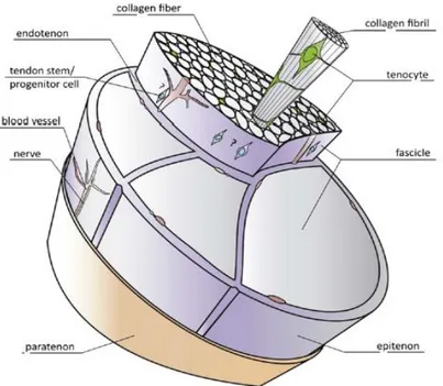
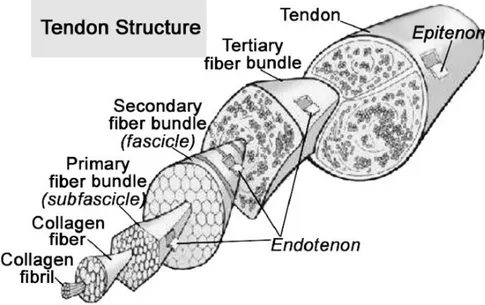
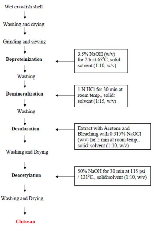
Figure 2-1: Schematic drawing of a basic tendon structure (Docheva, Muller et al. 2015). ... 7 Figure 2-2: Anatomy of a normal tendon (Sharma and Maffulli 2005). ... 8 Figure 2-3: Chemical structure of chitin ... 41 Figure 2-4: Chitin extracted from shellfish from which chitosan is made by N-deacetylation
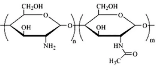
(Ahmadi, Oveisi et al. 2015). ... 43 Figure 2-5: Chitosan flow production (Ibrahim and Zairy 2015). ... 44 Figure 2-6: Schematic representation of chitosan structure (Dash, Chiellini et al. 2011). ... 45 Figure 2-7: Schematic illustration of chitosan’s versatility: At low pH, chitosan’s amine groups
are protonated conferring polycationic behaviour to chitosan. At high pH, chitosan’s amines are deprotonated and reactive (Dash, Chiellini et al. 2011). ... 46 Figure 5-1: After clotting for 1 hour at 37oC, clot retraction and serum expression (expressed as
% clot mass lost) was greater from PRP clots compared to CS-PRP hybrid clots (a). Data are presented as mean (diamond) and median (line) of n=7 clots from 3 different donors; Box: 25th and 75th percentile; Whisker Box to the most extreme point within 1.5 interquartile range. The Mixed model task in SAS Enterprise Guide 7.1 and SAS 9.4 was used to compare the different groups using sample (CS-PRP vs PRP) as a fixed effect and donor as a random effect. * p < 0.05 compared to PRP. Platelet aggregates were smaller in CS-PRP hybrid clots (b, d & f) compared to PRP clots (c, e & g), as shown by spinning disk microscopy images (b & c), confocal microscopy images (d & e) and 3-D stacks of confocal microscopy images (f & g). An Alexa-647 fibrinogen tracer was added to allow imaging of fibrin-covered platelets and fibrin in white. A Rhodamine-chitosan tracer was added to allow imaging of chitosan in red in d) and orange in f). ... 103 Figure 5-2: Chitosan (CS) appeared to be coating cellular and fibrous elements in scanning
electron microscopy (SEM) images of CS-PRP clots (a & b) while the fibrin (F) network was readily visible in PRP clots (c & d). Chitosan (CS) was found in the space between cellular elements and also at the surface of erythrocytes (E), platelet aggregates (P) and
fibrin (F) in transmission electron microscopy (TEM) images of CS-PRP clots (e & f). Erythrocytes (E) were tightly packed and platelet aggregates (P) were large in TEM images of PRP clots (g & h). ... 105 Figure 5-3: a) Flow cytometric analysis of PAC-1 (a) and p-selectin (b) staining of cell
suspension (unactivated in red). As expected, incubation with ADP (20 µm in green) and chitosan (in dark blue), both known platelet agonists, led to platelet activation (a) and granule secretion (b). In contrast, incubation with trehalose alone (in yellow) had no effect on platelet activation (a) or granule secretion (b). Interestingly, incubation with chitosan and trehalose simultaneously (in pale blue) decreased the intensity of both fluorescent signals compared to incubation with chitosan alone (in dark blue). ... 106 Figure 5-4: PDGF-AB (a) and EGF (b) cumulative release in culture medium was greater for CS-PRP clots compared to CS-PRP clots. Data are presented as mean (circle) and median (line) of n=13 clots from 6 different donors; Box: 25th and 75th percentile; Whisker: Box to the most extreme point within 1.5 interquartile range. The Mixed model task in SAS Enterprise Guide 7.1 and SAS 9.4 was used to compare the different groups with post-hoc analysis to look at pair-wise differences using sample and time as fixed effects and donor as a random effect. * p < 0.05 compared to PRP. Insets show total amount of growth factors released. ... 108 Figure 5-5: Formulations containing 1% (w/v) chitosan (CS Mn 38-43kDa and DDA 80-85%)
with 1% (w/v) trehalose and 42.2 mM CaCl2 were solubilized in autologous PRP and injected subcutaneously in rabbits, where they were found to be resident and induce cell recruitment at two weeks (a-c), four weeks (d-f), and six weeks (g-i) post-implantation. Invasion of the CS-PRP implants by host cells was accompanied by granulation tissue (GT) synthesis and formation of new blood vessels (BV). In contrast, recalcified PRP implants were completely degraded within a few days (not shown since the implant was absent). Outlines in a, d & g show where higher magnification images b, c, e, f, h & i were acquired. ... 109 Figure 6-1: a-d) Surgical procedure. A complete surgical tear was created in the supraspinatus
(SSP) tendon of the rotator cuff, as close as possible to the insertion site (a). Two 3.0 prolene sutures were pre-placed through the bony trough, the lateral tunnels (b) and the
tendon itself in a modified Mason-Allen pattern. In the case of treated shoulders, the CS-PRP mixture (150 µL) was injected into the bony trough prior to tightening the sutures (c), and it flowed out of all lateral tunnels. Sutures were tightened to attach the tendon to the humeral head (d). The CS-PRP mixture (150 µL) was then injected at the repaired insertion site and into the tendon itself. e) Schematic representation of the surgical model. f) Area in red is the region of interest (ROI) that was set over the greater tuberosity and used for micro-CT analysis. ... 130 Figure 6-2: a to h) Safranin O/Fast Green-stained paraffin sections of shoulder treated with
transosseous suturing + CS-PRP after 1 day. Polymorphonuclear cells (PMNs) were recruited to the bony trough (b), the lateral tunnels (c), to the endomyseal SSP muscle space (d) and to the SSP tendon (h). In some histological sections, needle tracks containing CS-PRP implant were visible within the SSP tendon (e & f). Note that stump of the tendon was not fully debrided in these samples (a and e). i to k) A rhodamine-chitosan tracer was used to image chitosan with epifluorescence in bright red. At one day post-surgery, chitosan-PRP hybrid implant was found adhering to the SSP tendon surface (i) and in the bony trough (j & k). ... 140 Figure 6-3: Safranin O/Fast Green-stained paraffin sections of shoulder treated with transosseous
suturing + CS-PRP (a to d) or suturing only (e to h) after 7 days. Residual structurally normal SSP tendon tissue was apparent and gaps were present at the tear site in all samples (a, b, e & f). Polymorphonuclear (PMN) cells were abundant in the granulation tissue of the CS-PRP treated shoulder only (c vs g). New bone was forming at the lateral aspect of the cortical bone in both groups (example shown in d). Chondrogenesis was observed in the SSP tendon of the control sutured shoulder only (h). Outlines in a & e show where higher magnification images were acquired. ... 142 Figure 6-4: Safranin O/Fast Green-stained paraffin sections of intact shoulders (a to d), and test
shoulders treated with transosseous suturing + CS-PRP (e to h) or suturing only (i to l) after 2 months, showing best and worst overall tendon scores for all groups. SSP tendon structure was altered in all surgically treated shoulders with several tendons displaying a highly cellular and vascular phenotype (e, f, i & j). Inflammatory PMN-rich tissue was present in 3 out 9 shoulders treated with transosseous suturing + CS-PRP at 2 months (g & h).
Heterotopic ossification within the SSP tendon was observed in 5 out 9 shoulders treated with transosseous suturing (k & l). Data in m are presented as mean (circle), median (line); Box: 25th and 75th percentile; Whisker: Box to the most extreme point within 1.5 interquartile range. * p < 0.05 compared to intact. # p < 0.05 compared to sutures group. & p < 0.05 compared to sutures + CS-PRP group. ... 145 Figure 6-5: Safranin O/Fast Green-stained paraffin sections of intact shoulders (a to d), and test
shoulders treated with transosseous suturing + CS-PRP (e to h) or suturing only (i to l) after 2 months, showing best and worst overall enthesis scores for all groups. The original tendon stump was often observed in surgically treated shoulders (e, f, i & j). In the best repair cases, fibrocartilage formation and partial restoration of the tidemark were observed at the enthesis (f & j). In the worst repair cases, gaps were present at the tendon-bone interface (h & l), although treatment with transosseous suturing + CS-PRP decreased such instances (m). Data in m are presented as mean (circle), median (line); Box: 25th and 75th percentile; Whisker: Box to the most extreme point within 1.5 interquartile range. * p < 0.05 compared to intact. # p < 0.05 compared to sutures group. ... 148 Figure 6-6: Polarized light microscopy images of SSP entheses. In the best cases, wave-like
structures and alignment of the collagen fibres parallel to the long axis of the SSP tendon were visible (a, c & e). In the worst cases, very little collagen alignment was apparent in the fibrous tissue adjacent to the bone (b, d & f). ... 148 Figure 6-7: Micro-CT of intact (a) and surgically treated shoulder at 1 day (b), and test shoulders
treated with transosseous suturing + CS-PRP (c to g) or suturing only (d to h) after 2 months. Incomplete repair of cortical bone at the lateral aspect of the humerus (c & d) and lateral bone formation (e & f) were present in all shoulders, regardless of treatment. Incomplete repair of the bone troughs was observed bilaterally in 5 rabbits (g & h). In half the treated shoulders, bone remodeling was highly stimulated by chitosan-PRP treatment (compare panel c to d), which led to increases in bone surface (j) and connectivity (k). Data in i, j & k are presented as mean (circle), median (line); Box: 25th and 75th percentile; Whisker: Box to the most extreme point within 1.5 interquartile range. * p < 0.05 compared to intact. # p < 0.05 compared to sutures group. & p < 0.1 compared to intact. ... 150
Figure 6-8: Safranin O/Fast Green-stained paraffin sections of intact shoulders (a to d), and test shoulders treated with transosseous suturing + CS-PRP (e to h) or suturing only (i to l) after 2 months, showing best and worst OOCHAS scores for humeral head and glenoid articular surfaces (a to l). Some structural abnormalities such as GAG depletion, fissures, cell changes and thinning of the articular cartilage were occasionally observed in both humeral head (b, f & j) and glenoid (d, h & l) surfaces, although average histological scores were not significantly different from intact (m). Data in m are presented as mean (circle), median (line); Box: 25th and 75th percentile; Whisker: Box to the most extreme point within 1.5 interquartile range. ... 152 Figure 6-9: Safranin O/Fast Green-stained paraffin sections of SSP muscles from intact shoulders
(a & b), and test shoulders treated with transosseous suturing + CS-PRP (c & d) or suturing only (e & f) after 2 months, showing best and worst scores for fatty infiltration. Surgical treatment induced fatty infiltration of SSP muscle after 2 months (g). Data in g are presented as mean (circle), median (line); Box: 25th and 75th percentile; Whisker: Box to the most extreme point within 1.5 interquartile range. * p < 0.05 compared to intact. ... 153 Figure 7-1: Chronic tear model and repair with CS-PRP. Full-thickness rotator cuff tears were
created in the infraspinatus (ISP) tendon of the shoulder close to the enthesis (a, b) and capped with 5 cm length of silicon (c) in 4 sheep. At 6 weeks after surgery, the tendons were macroscopically abnormal (d) and 2 tendons were found to be irrepairable. One tendon was repaired with 4 suture anchors in the suture bridge configuration. One tendon was repaired with one suture anchor + CS-PRP. The sutures were pre-placed in a Mason-Allen pattern and a first injection of 0.5 mL CS-PRP was applied at the debrided bone interface (e). The anchor was inserted to tighten the sutures and an additional 0.5 mL CS-PRP implant was applied on top of tendon at the repaired site and also under the tendon. Macroscopic appearance of an ISP tendon capped for 2 weeks with 5 mm length of silicon (f). ... 177 Figure 7-2: Acute tear model and repair with CS-PRP. Full-thickness rotator cuff tears were
created in the infraspinatus (ISP) tendon of the shoulder close to the enthesis in 4 sheep (a, b). All tendons were immediately repaired with 4 suture anchors in a suture bridge configuration. In 2 out of 4 sheep, repair was augmented with CS-PRP. The first row of anchors were inserted and the sutures were passed (c) and a first injection of 0.5 mL
CS-PRP was applied at the debrided bone interface (d). The second row of anchors were inserted to tighten the sutures (e) and an additional 0.5 mL CS-PRP was applied on top of tendon at the repaired site and also under the tendon (f). ... 178 Figure 7-3: Safranin O/Fast Green stained paraffin sections of the ISP tendons in the chronic
repair model. Intact control tendons were organized in bundles, as expected (a to c). Bundle organization was still apparent in areas of untreated tendons at chronic stage, while other areas were disorganized, hypercellular and vascularized or hypocellular (d to i). The tendon of the shoulder treated for 2 weeks with suture anchors was mostly disorganized, hypercellular and vascularized , with a small hypocellular area (j to l). The tendon of the shoulder treated with suture anchors + CS-PRP was mostly disorganized, hypercellular and vascularized , with a small area organized in bundles and another area rich in polymorphonuclear cells (m to o) ... 180 Figure 7-4: Safranin O/Fast Green-stained paraffin sections and polarized light microscopy (d, h,
l, p & t) images of the ISP tendons in the acute repair model. Intact control tendons were organized in bundles, as expected (a to d). At 6 weeks post-surgery, the tendons were mostly composed of a disorganized and vascular fibrous repair tissue in both groups (e to l). Chondrogenesis and GAG expression were apparent in the anchors only group at 6 weeks (e&f). At 3 months post-surgery, the tendon in the anchors only group contained aligned tissue, expressed high levels of GAG, and had a small area organized in bundles (m to p). In contrast, the tendon in the anchors + CS-PRP group was mostly organized in bundles with a smaller area of tendon-like repair tissue (m to t). ... 180 Figure 7-5: Safranin O/Fast Green stained paraffin sections of the ISP tendon entheses in the
chronic repair model. Intact controls had normal entheses consisting of 1) unmineralized fibrocartilage, 2) tidemark, 3) mineralized fibrocartilage and 4) bone, as expected (a to c). The tidemark was still recognizable 2 weeks after defect creation (d), but not at longer time points or after repair (g, j & m). Scar tissue was growing above the entheses in the untreated chronic defects (d&g), suggesting that some spontaneous repair can occur even without any treatment in this model. Integration of the scar tissue with the underlying bone was achieved through bone remodeling and ingrowth into the scar tissue (f, i, l & o). Treatment with anchors + CS-PRP increased the area of remodeling bone (compare o to l). ... 182
Figure 7-6: Safranin O/Fast Green stained paraffin sections of the ISP tendon entheses in the acute repair model. Intact controls had normal entheses consisting of 1) unmineralized fibrocartilage, 2) tidemark, 3) mineralized fibrocartilage and 4) bone, as expected (a to c). Scar tissue was growing superior to the entheses from 6 weeks (d to i) to 3 months (j to o) post-surgery. Integration of the scar tissue with the underlying bone was achieved through bone remodeling and ingrowth into the scar tissue (d to o). This was more apparent in the anchors + CS-PRP group (compare g&m to d&j). The site of anchor insertion was apparent in some sections (* in panel j). ... 183 Figure 7-7: Safranin O/Fast Green stained sections of humeral head and glenoid articular surfaces
from intact controls (a&b), from the chronic defect model (c to f) and from the acute defect model (g to j). The humeral articular surfaces were all structurally normal but showed signs of GAG depletion (a, c, e, g & i). Mild structural abnormalities were observed at the center of some glenoid articular surfaces, including GAG depletion, hypercellularity, cell cloning and fissures (k to m). These were apparent in all treatment groups as well as the intact controls. ... 184 Figure 7-8: Hematoxylin and Eosin (a, d & e) and Safranin O/Fast Green (b & c) stained paraffin
sections of muscle biopsies from intact control (a), from the chronic defect model (b & c) and from the acute defect model (d & f). Fatty infiltration was not prevented by any treatment ... 185 Figure 7-9: Hematoxylin and Eosin (a, d & e) and Safranin O/Fast Green (b & c) stained paraffin
sections of synovial biopsies from intact control (a), from the chronic defect model (b & c) and from the acute defect model (d & f). There was mild synovitis and increased cell infiltration in the chronic model treated with anchors + CS-PRP for 2 weeks (c). ... 186
LIST OF SYMBOLS AND ABBREVIATIONS
a-PRP Activated Platelet-Rich Plasma
Ac Acetyl
ADP Adenosine Diphosphate ADSC Adipose Derived Stem Cell
AMSC Adipocyte Mesenchymal Stem Cell aP-PRP Activated Pure Platelet-Rich Plasma
ASES American Shoulder and Elbow Surgeons score bFGF Beta-Fibroblast Growth Factor
BM Bone Marrow
BM-MSC Bone Marrow Mesenchymal Stem cell BMAC Bone Marrow Aspirate Cells
BMP-13 Bone Morphogenic Protein BMSC Bone Marrow Stem Sell BS Bone Surface area BSA Bovine Serum Albumin BV Bone Volume
BV Blood Vessels CaCl2 Calcium Chloride cAMP Cyclic AMP CH3COOH Acetic Acid Col Collagen
Col-II Type II collagen Col-X Type X collagen
CS Chitosan
CS-GP Chitosan-Glycerol Phosphate CS-PRP Chitosan-Platelet-Rich Plasma
CT Computed Tomography
DDA Degree of Deacetylation DNA Deoxyribonucleic acid DR Double-Row
ECM Extracellular Matrix EGF Endothelial Growth Factor
ELISA Enzyme-Linked Immunosorbent Assay FC Fibrocartilage
FD Freeze-Dry
FG Fast Green
FGF Fibroblast Growth Factor FITC Fluorescein Isothio-Cyanate g Gram GAGs Glycosaminoglycans GF Growth factor Glc D-glucosamine GlcN D-glucosamine GlcNAc N-acetyl-D-glucosamine GP Glycerol Phosphate GPIIb-IIIa Glycoprotein 2ß-3α
GT Granulation Tissue H&E Hematoxylin and Eosin H2O Water
H2SO4 Sulfuric Acid HA Hyaluronic Acid HA Hyaluronic Acid HCl Hydrochloric Acid HCL Hydrochloric Acid HCOOH Formic Acid HNO3 Nitric Acic
IGF Insulin Growth Factor Ihh Indian Hedgehog
IL Interleukin ISP Infraspinatus
JOA Japanese Orthopaedic Association KCL Potassium Chloride
kDa Kilodalton kg Kilogram kV Kilovolt
L-PRF Leukocyte-Rich Platelet-Rich Fibrin L-PRP Leukocyte-Rich Platelet-Rich Plasma M Molar
MA Masson-Allen MgCl2 Magnesium Chloride
mL Millilitre mm Millimeter
MMP Matrix Metalloproteinase MRI Magnetic Resonance Imaging MSC Mesenchymal Stem Cell MW Molecular Weight
Na2HPO4 Sodium Phosphate Dibasic NaCl Sodium Chloride
NaHCO3 Sodium Carbonate
NBF Normal Buffered Formalin NZW New Zealand White
P-PRF Pure Platelet-Rich Fibrin P-PRP Pure platelet-rich plasma
PDGF Platelet Derived Growth Factor PGE2 Prostaglandin E2
PLGA Polyglycolic acid PLLA Poly-L-Lactic acid PMN Polymononuclear cells PRF Platelet-Rich Fibrin
PRGF Platelet Rich in Growth Factors PRP Platelet-Rich Plasma
PTH Parathyroid Hormone
PTHrP Parathyroid Hormone related Peptide RBC Red blood cell
RCT Rotator Cuff Tear
rhPDGF Human recombinant Platelet-Derived Growth Factor Runx Runt-related transcription factor
Saf-O Safranin-O SC Stem Cells SCP Subscapularis SCX Scleraxis
SEM Scanning Electron Microscopy SIS Small Intestine Submucosa
SPCHT Porous Chitosan Sponges Scaffold SR Single-Row
SSP Supraspinatus
TDSC Tendon-Derived Stem Cells
TEM Transmission electron microscopy TGF Transforming Growth Factor
TIMP Tissue inhibitors of metalloproteinase TM Teres minor
TNF Tumour Necrosis Factor TOE Transosseous-Equivalent TP Tricalcium Phosphate
TSPC Tendon Stem Cell progenitor Cell
UCLA University of California, Los Angeles score v/v volume/volume
w/v Weight by volume α Alpha
β Beta µ Micro µL Microlitre
LIST OF APPENDICES
APPENDIX A – CLINICAL STUDIES COMPARING ARTHROSCOPIC VERSUS OPEN ROTATOR CUFF REPAIR ... 254 APPENDIX B – CLINICAL STUDIES COMPARING DIFFERENT SUTURING
TECHNIQUES. ... 261 APPENDIX C – ROTATOR CUFF RABBIT REPAIR MODELS ... 269 APPENDIX D – ROTATOR CUFF SHEEP REPAIR MODELS ... 279 APPENDIX E- CLINICAL STUDIES COMPARING PRP STUDY IN ROTATOR CUFF
CHAPTER 1
INTRODUCTION
More than 28 million Americans are affected by musculoskeletal injuries, estimated to cost more than $254 billion each year (Praemer, Furner et al. 1999). Rotator cuff tears are among the most common injuries occurring in the shoulder and are often seen in older athletes of overhead sports, like tennis or basketball (Yamamoto, Takagishi et al. 2010). It is a widespread crisis, causing high rate of morbidity and inability in workplaces and sports (Lehman, Cuomo et al. 1995, Yanke and Chubinskaya 2015). Rotator cuff tears are associated with structural and architectural alterations of the musculotendinous unit, such as tendon retraction, fatty infiltration and muscular atrophy (Antoniades, Scher et al. 1979, Borges, Borchard et al. 2007, Periayah, Halim et al. 2014).
Cuff tears result in shoulder pain, stiffness, weakness and loss of motion (Trent, Bailey et al. 2013). It usually starts as an acute tendinopathy with progressive degeneration leading to a partial thickness tear and eventually full rupture (Neer, Craig et al. 1983). The shoulder joint can still function with minimal pain in spite of a rotator cuff tear by using the deltoid and the scapular stabilizing muscles (Feeley, Gallo et al. 2009), however limited function of upper extremities will impair the ability to carry out basic activities (Nho, Brown et al. 2009, Yamamoto, Takagishi et al. 2010). Rotator cuff injury is the second most common musculoskeletal pathology after lower back pain (Picavet and Schouten 2003) and is the most common shoulder condition that patients seek therapy for (Gomoll, Katz et al. 2004, Riley 2004).
Cuff tears may cause irreversible changes in the structural and physiological properties of the shoulder, causing intolerable chronic pain (Funakoshi, Majima et al. 2006) and severe functional disability, as well as compromise joint mechanics leading to degenerative changes (Aurora, McCarron et al. 2007). Degenerative changes in the structure and composition of the tendons make healing very difficult. After surgical repair, failure rate ranges between 20 to 95% (Galatz, Ball et al. 2004) due to tendon degeneration, hypo vascularization (Cohen 1985, Jackson 2007, Gulotta, Kovacevic et al. 2009, Longo, Franceschi et al. 2009, Lui, Zhang et al. 2010, Longo, Forriol et al. 2011, Ahmad, Howard et al. 2012), muscle atrophy, fatty infiltration of tendon and muscle and a lack of tendon-to-bone integration (Gartsman 1997, Totani, Cumashi et al. 1998, 2005). Tendon vascularization is mostly compromised at the junction zones and sites of
torsion, friction or compression (Sharma and Maffulli 2006), which reduces the chances of adequate self-repair (Chahla, Dean et al. 2016).
Patients with re-tears usually experience some pain relief but continue to have lower functional outcomes (Heldin and Westermark 1999, Galatz, Ball et al. 2004). Rotator cuff insertion site, also known as the enthesis, is never completely reformed after surgical reattachment (Gulotta and Rodeo 2009, Chung, Kim et al. 2014). The overall structure, composition, and organization of a normal insertion site do not regenerate, especially, the calcified cartilage zone (Gerber, Schneeberger et al. 1999). Instead, healing occurs through fibro vascular disorganized scar tissue formation (Pencev and Grotendorst 1988, Robertson, Maley et al. 1993), lacking strength, load-to-failure, and biomechanical tendon properties leading to increased risk of further damage and high re-tear rates (Galatz, Ball et al. 2004, Angeline and Rodeo 2012, Del Buono, Oliva et al. 2012) (Galatz, Ball et al. 2004, Boileau, Brassart et al. 2005, Galatz, Sandell et al. 2006, Angeline and Rodeo 2012, Del Buono, Oliva et al. 2012).
Current standard surgical treatment involves open or arthroscopic repair of torn tendons frequently using suture anchors in a variety of configurations (McElvany, McGoldrick et al. 2015). The goal of suturing repair is to increase initial fixation strength, mechanical stability and increase biological tendon-to-bone healing (Cole, ElAttrache et al. 2007), which most often fails. The current suturing techniques are thought to increase footprint contact area, which could improve the rate of healing but re-tear rates are still relatively high (Denard and Burkhart 2013). Considerable variation in surgical procedures exists, confirming the lack of a single reliable technique (McElvany, McGoldrick et al. 2015). It is recognized that current surgical treatments need improvement (Gerber, Fuchs et al. 2000, Galatz, Ball et al. 2004). Improving repair techniques could be achieved through biological or synthetic tendon grafts or through augmentation devices (Dwivedi, Chevrier et al. 2017, Ghazi zadeh, Chevrier et al. 2017), however success is limited. The current goal is to completely regenerate the fibrocartilaginous insertion zones through biological augmentation. A good cuff repair technique should decrease pain, increase strength as well as range of motion (Cole, ElAttrache et al. 2007). New techniques in tissue engineering have been postulated to increase rotator cuff tear repair, such as scaffolds, growth factors and cell seeding (Derwin, Badylak et al. 2010), but their uses are still preliminary.
Despite the advances in tissue engineering, rotator cuff tear repair still represents an enormous challenge.
Platelet-rich plasma (PRP) is a plasma fraction with a high platelet concentration (Deprés-Tremblay, Chevrier et al. 2017) that is obtained through whole blood centrifugation divided by weight particle. Activation of platelets in PRP releases several growth factors, such as PDGF, TGF-β, IGF, VEGF, and EGF (Sheth, Simunovic et al. 2012). PRP has been suggested to stimulate revascularization and enhance growth factors that could potentially increase tendon healing (Barber, Hrnack et al. 2011). Growth factor release at the injury site could eventually lead to cell proliferation, cell differentiation and angiogenesis (Sheth, Simunovic et al. 2012). PRP has been used to treat several soft tissue pathologies, however the results have been inconsistent, possibly due to its short half-life and high diffusibility (Li, Xu et al. 2014). Routine use of PRP to treat rotator cuff tears is not supported by current clinical evidence (Castricini, Longo et al. 2011).
Chitosan (CS), a biodegradable and biocompatible natural polymer, is obtained through chitin deacetylation (Muzzarelli 2009, Krueger, Wenke et al. 2012). Once injected, chitosan is slowly hydrolyzes by lysozymes and produces chito-oligomers, which favours correct deposition, assembly and orientation of collagen fibrils in ECM reformation in tissues (Muzzarelli, Mattioli-Belmonte et al. 1999). In the context of cartilage repair, implants of CS-glycerol phosphate (GP)/blood have previously been shown by our research group to increase cell recruitment, vascularization and bone remodeling (Chevrier, Hoemann et al. 2007, Hoemann, Sun et al. 2007), activate a beneficial phenotype of pre-wound healing macrophages (Hoemann, Chen et al. 2010) and enhance tissue repair integration through osteoclast activity (Chen, Sun et al. 2011), all of which are expected to also be beneficial for rotator cuff repair. More recently, we have developed freeze-dried formulations of CS that can be solubilized in PRP to form injectable CS-PRP implants that coagulate in situ. We have shown that residency time and bioactivity of CS-PRP implants are superior to that of PRP alone in vivo (Chevrier,
Hoemann et al. 2007). CS-PRP implants were tested in meniscus repair models and were shown
to induce cell migration and repair tissue synthesis, while PRP alone or wrapping the meniscus with a collagen membrane did not (Chevrier, Deprés-Tremblay et al. 2016, Ghazi zadeh, Chevrier et al. 2017). CS-PRP were also tested in a chronic cartilage repair model and were