ÉTUDE DES VARIATIONS NATURELLES DE LA PROTEINE VEGETALE ARGONAUTE 2 ET DE LA RESISTANCE VIRALE
par
Ayooluwa Adurogbangba
Thèse présentée au Département de Biologie en vue de l’obtention du grade de docteur ès sciences (Ph.D.)
FACULTÉ DES SCIENCES UNIVERSITÉ DE SHERBROOKE
A STUDY OF THE NATURAL VARIATIONS IN THE PLANT ARGONAUTE 2 PROTEIN AND VIRAL RESISTANCE
by
Ayooluwa Adurogbangba
Thesis submitted to the Department of Biology in fulfillment of the requirements for the degree of Doctor of Philosophy degree (Ph.D.)
FACULTÉ DES SCIENCES UNIVERSITÉ DE SHERBROOKE
Le 24 octobre 2019
le jury accepte la thèse de Madame Ayooluwa Adurogbangba dans sa version finale
Membres du jury
Professeur Peter Moffett Directrice de recherche
Département de biologie, Université de Sherbrooke
Professeur John Carr Évaluateur externe University of Cambridge
Professeur Pierre-Étienne Jacques Évaluateur interne
Département de biologie, Université de Sherbrooke
Professeure Pascale Beauregard Président-rapporteur
v SOMMAIRE
La neutralisation de l'ARN est un mécanisme majeur de défense antivirale constitutive chez les plantes. Les protéines Argonaute (AGO) et les protéines de type dicer (DCL) ont été identifiées comme étant les principaux acteurs de la voie de silençage de l'ARN. En ciblant l'ARN double brin issu d'une infection virale, les plantes peuvent utiliser le silençage de l'ARN en tant qu'outil antiviral efficace. Le génome de la plante modèle, Arabidopsis thaliana, code pour dix protéines AGO et quatre protéines DCL. Des études antérieures ont révélé que, même si toutes les AGO possèdent une capacité intrinsèque de cibler l'ARN viral en fonction du virus en question (Brosseau et Moffett, 2015), les AGO 1 et 2 sont les AGO les plus étudiées pour la défense contre les virus à ARN.
Les travaux effectués au cours de mes études ont porté sur la variabilité génétique présente dans le gène AGO2. Afin de déterminer le (s) rôle (s) de la variation génétique dans le gène AGO2 au cours d'infections virales, les polymorphismes présents dans le gène AGO2 dans des accessions d'Arabidopsis naturelles ont été évalués. Ces accessions étaient infectées par un Potexvirus, un Potyvirus et un Cucumovirus. Sur la base de travaux antérieurs dans notre laboratoire (Brosseau et al., 2019), nous avons classé ces accessions en tant qu'accessions de type Col-0 AGO2 ou en tant que accessions de type C24 AGO2. Les résultats de cette étude montrent que l'allèle AGO2 de type Col-0 est nécessaire à la défense contre potex et poty, alors que l'allèle AGO2 de type C24 ne confère pas de résistance aux infections virales. Bien que l'allèle C24 AGO2 ne soit pas important pour la résistance à l'infection virale, nous montrons que cet allèle est important pour les efforts de reproduction en l'absence de virus. Cela a révélé un compromis entre reproduction et défense. Étant donné que les deux allèles AGO2 sont présents à une fréquence élevée, l'allèle C24-AGO2, qui confère une sensibilité aux virus, a été retenu pour son avantage en matière de reproduction. Un résultat qui n'a jamais été rapporté pour un gène AGO.
Les variations interspécifiques de la protéine AGO2 ont également été explorées. En développant des plantes transgéniques de Nicotiana benthamiana et de tomates possédant une AGO2 d'Arabidopsis, on a évalué l'accumulation de PVX. Dans ces plantes transgéniques, AtAGO2 a
vi permis de limiter le mouvement et l'accumulation de PVX dans les feuilles systémiques. Nous rapportons que les plantes transgéniques étaient plus tolérantes à l'infection à PVX. En conclusion, les travaux présentés dans cette thèse étudient les variations naturelles présentes dans et leur importance pour la résistance aux virus. En outre, en utilisant une approche de résistance sans hôte, l’ingénierie de plantes résistantes aux virus était réalisable.
Mots-clés: ARN inhibant, plante, Argonaute, défense antivirale, variation naturelle, transgénis, tolérance virale.
vii ABSTRACT
RNA silencing is a major mechanism of constitutive antiviral defense in plants. Argonaute proteins (AGO) and dicer-like proteins (DCL) have been identified to be the major players in the RNA silencing pathway. By targeting double-stranded RNA derived from infection from viruses, plants are able to use RNA silencing as an efficient antiviral tool. The genome of the model plant, Arabidopsis thaliana (A. thaliana) encodes ten AGO proteins and four DCL proteins. Previous studies have revealed that although all AGOs possess an intrinsic ability to target viral RNA depending on the virus in question (Brosseau and Moffett, 2015), AGOs 1 and 2, are the most studied AGOs for defense against RNA viruses.
Work done during my studies, focused on the genetic variability present within the AGO2 gene. In order to determine the role(s) of genetic variation within the AGO2 gene during virus infections, the polymorphisms present in the AGO2 gene in natural A. thaliana accessions was assessed. These accessions were infected with a Potexvirus, a Potyvirus and a Cucumovirus. Based on previous work in our lab (Brosseau et al., 2019), we classified these accessions as Col-0-like AGO2 accessions or C24-like AGO2 accessions. Results from this study show that the Col-0-like AGO2 allele is necessary for defense against certain virus whereas the C24-like AGO2 allele does not confer resistance to virus infections. Although the C24 AGO2 allele is not important for resistance against virus infection, we show that the said allele is important for reproductive efforts in the absence of viruses. This revealed a trade-off between reproduction and defense. Since both AGO2 alleles are present at high frequency in nature, the C24-AGO2 allele, which confers susceptibility to viruses, has been retained due to its conferring a reproductive advantage. A result that has never been reported for an AGO gene.
The interspecies variations in the AGO2 protein was also explored. By developing transgenic N. bethamiana and tomato plants that express an A. thaliana AGO2 (AtAGO2) protein, the accumulation of PVX was monitored. In these transgenic plants, AtAGO2 helped to curtail the movement and accumulation of PVX in upper non-inoculated leaves. We report that the transgenic plants were more tolerant to PVX infection. In conclusion, work presented in this thesis
viii investigates the natural variations present in and how these variations are important for virus resistance. Also, by utilizing a non-host resistance approach, the engineering of viral resistant plants was achievable.
Keyword: RNA silencing, plant, Argonaute, antiviral defense, natural variation, transgenics, viral tolerance.
ix ACKNOWLEDGEMENTS
Truly, undertaking this Ph.D. has been a life changing experience for me. This degree would not be possible without the help, support and guidance of several people. Firstly, I would like to express my sincere appreciation to my supervisor, Dr. Peter Moffett. Without whose scientific guidance and supervisory role, this degree would be achievable. Peter, thank you for investing in my scientific career and for all the opportunities you gave me as your graduate student.
I gratefully acknowledge Dr. Pascale Beauregard and Dr. Pierre-Étienne Jacques for their constant feedback and their advisory role. Your insights have provided me with direction throughout this degree. Many thanks to Dr. John Carr for agreeing to be the external examiner of my thesis defense examination. I greatly appreciate the support and the collaborative work undertaken at Dr. Fernando García-Arenal’s lab at UPM, Madrid, Spain. To Dr. Sharon Regan: thank you for investing in me when that was all I needed.
I am indebted to my friends at the Moffett lab, both past and present. Many thanks to Chantal (brainstorming sessions will be missed), Teura, Guilherme, Zhenxing, Charles and Matt for your friendships. I am also grateful to the Agrophytoscience scholarship program for the numerous funds received during my research work.
I especially thank Toluwalase Bolaji, my husband and the love of my life. Tolu, you are the best outcome from these past 5 years. Thank you for your unwavering support during this degree. You have been extremely supportive and have made countless sacrifices to help me get through this phase. I am forever grateful to my parents, Dr. Mike and Dr. Moji Adurogbangba for their indispensable presence in my life. Thank you for giving me a solid foundation to build on. For the numerous financial supports and care packages you lavish me with, I am extremely thankful. Thank you for sacrificing ALL to immigrate to a foreign land, in order to expose us to a world of possibilities. I thank God for making you stewards of my life. Thank you both for selflessly encouraging me to explore novel directions in life and to seek my own destiny. I am very grateful to my siblings: Ibukun and Damola, Olaolu and Ehimare. I thank each of you for your care and support over the years. I thank Revd. Dr. and Mrs. Bolaji for their support and interest in my work. Lastly, I am grateful for the friendships that survived these 5 years. To each of you, it is a privilege to call you my friend.
x Table of Contents SOMMAIRE ...v ABSTRACT ... vii ACKNOWLEDGEMENTS... ix LISTS OF ABBREVIATIONS ... xv
LIST OF TABLES ... xviii
LIST OF FIGURES ... xix
CHAPTER 1 ...1
1.1 Plant Defense Responses. ...1
1.1.2 Introduction to RNA Silencing ...4
1.2.1 Origin and biogenesis of siRNAs and the role of RDRs. ...5
1.3 Dicer and Dicer-like proteins. ...7
1.4 Other players in the RNA silencing. ...9
1.5 RISC Complexes and ARGONAUTE Proteins. ... 10
1.6 Structure of AGO proteins. ... 10
1.6.2 Classification of AGO proteins ... 12
1.6.2.1 AGO 1/10. ... 12
1.6.2.2 AGO 2/3/7. ... 13
1.6.2.3 AGO5. ... 14
1.6.2.4 AGO4/6/8/9, RdDM and DNA Methylation. ... 15
1.7 DNA viruses and Methylation-mediated defense. ... 18
1.8 Detailed roles of AGO proteins in plant-pathogen interaction ... 19
1.8.1. RNA Silencing as a defense mechanism against viruses ... 19
1.8.2 WHICH AGOs ARE ANTIVIRAL? ... 21
xi
1.9 Viruses used in this study ... 30
1.9.1 Cucumber Mosaic Virus ... 30
1.9.2 Potato Virus X ... 31
1.9.3 Turnip Mosaic Virus ... 32
1.10 Purpose and Objectives of the projects... 34
1.10.1 Intra-species variation in Arabidopsis thaliana AGO2 alleles and viral infection ... 34
1.10.2 Differences between Arabidopsis thaliana AGO2 and Nicotiana benthamiana AGO2. ... 35
CHAPTER 2 ... 36
2. Genetic Variation in AGO2 determines susceptibility to virus infection and reveals opposing fitness trade-off between reproduction and resistance in a naturally occurring population. ... 37
2.1 ABSTRACT ... 38
2.2 Author summary... 38
2.3 INTRODUCTION ... 39
2.4 RESULTS ... 41
2.4.1 Two polymorphisms in AGO2 are widely distributed in natural populations. ... 41
2.4.2 Col-0 AGO2 confers resistance to PVX infection in wild accessions. ... 43
2.4.3 Susceptibility to TuMV is determined by polymorphisms in AGO2... 44
2.4.4 Resistance to CMV is not determined by AGO2 ... 47
2.4.5 C24-like AGO2 allele is associated with a reproductive advantage in the absence of virus infection. ... 48
2.4.6 Col-0 AGO2 confers a reproductive advantage in the presence of TuMV infection. ... 51
2.5 DISCUSSION ... 52
2.6 MATERIALS AND METHODS ... 55
2.6.1 Plant material and growth condition. ... 55
xii
2.6.3 Protein extraction and Western Blotting. ... 56
2.6.4 Sequencing and Polymerase Chain Reaction. ... 57
2.6.5 Quantification of CMV multiplication. ... 57
2.6.6 Quantification of life-history traits. ... 58
2.6.7 Statistical Analysis. ... 58 2.7 Acknowledgements ... 59 2.8 Contributions... 59 2.9 References ... 59 2.10. Supplemental Data ... 98 2.10.1 Supplemental Figures. ... 99 CHAPTER 3 ... 103 3.1 ABSTRACT ... 104 3.2 INTRODUCTION ... 105
3.3 MATERIALS AND METHODS ... 107
3.3.1 Generation and selection of transgenic plants. ... 107
3.3.2 Plant material and growth conditions and virus inoculation. ... 108
3.3.3 Plasmid Construction ... 108
3.3.4 Transient Expression Assays ... 109
3.3.5 Protein Extraction and Analysis ... 109
3.4 RESULTS ... 110
3.4.1 Generation of transgenic N. benthamiana and tomato plants. ... 110
3.4.2 Transgenic plants expressing AtAGO2 are less susceptible to PVX... 112
3.4.3 AtAGO2 does not confer tolerance to PlAMV ... 115
3.4.4 In tomato, AtAGO2 delays viral movement and confers tolerance to PVX. ... 117
xiii
3.6 References ... 121
3.7 Supplemental data ... 159
CHAPTER 4 ... 161
4.1Variation in AGO2: comparison with Natural variation in other viral defense-related genes. ... 161
4.2 Inter-specific differences in Plant AGO2-The future of transgenic viral tolerant plants. ... 163
4.3 How knowledge of AGO biology can enrich future transgenic approaches. ... 166
APPENDIX - SUPPLEMENTAL MANUSCRIPT ... 174
5. Natural variation in the Arabidopsis AGO2 gene is associated with susceptibility to PVX ... 175
5.1 Abstract ... 176
5.2 Introduction ... 177
5.3 Results ... 178
5.3.1 AGO2 proteins from different genera display specific antiviral activity ... 178
5.3.2 The Arabidopsis AGO2 gene displays a high degree of polymorphism ... 180
5.3.3 C24-like AGO2 alleles are strongly associated with systemic infection by PVX in Arabidopsis. ... 183
5.3.4 Validation of the effect of different AGO2 alleles and susceptibility to PVX in reciprocal inbred lines ... 185
5.3.5 C24 AGO2 is not a null allele ... 187
5.3.6 C24 AGO2 shows decreased antiviral activity against PVX compared to Col-0 AGO2. ... 190
5.4 Discussion ... 192
5.4.1 Susceptibility to PVX is common in Arabidopsis thaliana ... 192
5.4.2 High prevalence of polymorphisms in the AGO2 coding sequence ... 192
5.5 Conclusion ... 195
xiv
5.6.1 Plant material and growth conditions ... 195
5.6.2 Plasmid construction and transient expression ... 195
5.6.3 Virus inoculation ... 196
5.6.4 Protein extraction and analysis ... 196
5.6.5 Gene expression and DNA methylation analysis ... 197
5.6.6 Quantification of SA ... 197 5.7 References ... 198 5.8 Acknowledgements ... 238 5.9 Contributions... 238 5.10 Competing interests ... 238 5.11 Supplementary Figures ... 239 BIBLIOGRAPHY ... 245
xv LISTS OF ABBREVIATIONS
Abbreviations Full name
+ssRNA positive-sense single-strand RNA
AGO ARGONAUTE(S)
At Arabidopsis thaliana
BaMV Bamboo mosaic Virus
BMV Brome mosaic virus
bp base pair
CaLCuV Cabbage leaf curl virus
CaMV Cauliflower mosaic virus
cDNA complementary DNA
CMV Cucumber mosaic virus
CDS Coding DNA sequence
CP Coat protein
CRP Cysteine-rich protein
CymRSV Cymbidium ring spot tombusvirus
DCL Dicer-like
DNA Deoxyribonucleic acid
dpi day post-inoculation/ days post-infection DRB double-stranded RNA binding protein
dsRNA double-stranded RNA
ETI Effector triggered immunity
EV Empty vector
GFP Green fluorescence protein
HA Hemagglutinin
HEN1 Hua Enhancer
hcRNA Heterochromatic siRNA
xvi
HRP Horseradish peroxidase
MAMPs Microbe-associated molecular patterns
MgCl2 Magnesium Chloride MID Middle miRNA microRNA MP Movement protein Nb Nicotiana benthamiana Nt Nicotiana tabacum nt Nucleotide
NB-LRR Nucleotide-binding, leucine-rich repeats
OD Optical density
Prom Promoter
PAGE Polyacrylamide gel electrophoresis
PCR Polymerase chain reaction
PlAMV Plantago asiatica mosaic virus PTGS Post-transcriptional gene silencing
PTI PAMP-triggered immunity
PVX Potato virus X
RdDM RNA-dependent DNA Methylation
RISC RNA-induced silencing complex
RNA Ribonucleic acid
SDS Sodium dodecyl sulfate
SGS3 Suppressor of gene silencing 3
sRNA Small RNA
siRNA Small interfering RNA
TBSV Tomato bushy stunt virus
TCV Turnip crinkle virus
T-DNA Transfer DNA
TGB Triple gene block
TGS Transcriptional gene silencing
xvii
TuMV Turnip mosaic virus
UV ultraviolet
VRC Viral replication complex
VSR Viral suppressor of RNA silencing
xviii LIST OF TABLES
CHAPTER 1
Table 1.1 List of AGOs reported to have antiviral activities, as well as the wild-type virus and/or VSR-defectived mutant version (X) used in the correspondng study……24 CHAPTER 2
Table 2.1 Classification of individual Iberian Peninsula wild accessions………..43 Table 2.2 Mean values of rosette, inflorescence and seed weight of mock inoculated plants of Iberian A. thaliana accessions………...50 Table S2.1 List of the classification of the allometric groups and AGO2 allele distribution of the Iberian Peninsula wild accessions……….70 CHAPTER 3
Table 3.1 Primers list used in this study………..93 CHAPTER 4
Table 4.1 Summary of transgenic interference with factors affected during plant-virus interactions………...101
xix LIST OF FIGURES
CHAPTER 1
Figure 1.1 The zig-zag model of Plant innate immunity with all the four phases……….3
Figure 1.2 A Schematic representation of the general structure of AGO proteins………12
Figure 1.3 AGO proteins classifications and functions………18
Figure 1.4 Schematic representation of RNA silencing during viral infection………...20
Figure 1.5 Genome organization of CMV………...31
Figure 1.6 PVX genome………..32
Figure 1.7 Schematic diagram of the TuMV genome………..33
CHAPTER 2 Figure 2.1 Genetic variation in AGO2 of natural Iberian Peninsula wild accessions determines susceptibility to PVX……….…43
Figure 2.2 Defense against TuMV is determined by AGO2 allele………..46
Figure 2.3 Lack of correlation between AGO2 alleles and accumulation of CMV RNA…...47
Figure 2.4 Life history analyses of accessions and RILs supports a reproductive advantage for C24-like AGO2 alleles………50
Figure 2.5 Col-0 AGO2 confers a reproductive advantage in the presence of TuMV……….52
Figure S2.1 Origin of RILs and JAX1 identity of Iberian Peninsula wild accessions………...68
Figure S2.2 Wild accessions and TuMV infection………69
xx CHAPTER 3
Figure 3.1 Testing of AGO2 genomic clones using PVX∆TGB and PVX-WT………79 Figure 3.2 AtAGO transgenic plants showed reduced viral symptoms and viral accumulation
in upper non-inoculated leaves……….…82 Figure 3.3 WT and transgenic plants are susceptible to PlAMV……….84 Figure 3.4 AtAGO2 delays PVX movement and confers tolerance to PVX in tomato plants………..…85 Figure S3.1 Screening of N. benthamiana and tomato plants for the presence of AtAGO2 and NbAGO2………92 APPENDIX
Figure 5.1 AtAGO2, but not NbAGO2, shows anti-viral activity against PVX………111 Figure 5.2 Residue 33 of Arabidopsis AGO2 has undergone positive selection in natural populations………...113 Figure 5.3 Natural variation in the N-terminus of AGO2 correlates with susceptibility to PVX……….115 Figure 5.4 Exchange of AGO2 alleles between Col-0 and C24 changes susceptibility to
PVX……….117 Figure 5.5 C24 AGO2 retains anti-bacterial and methylation-related functions………119 Figure 5.6 Polymorphisms found in C24 AGO2 affect its antiviral activity in Arabidopsis………..121 Figure S5.1 C24 and Col-0 alleles are found in all eight Eurasian populations analyzed………...137 Figure S5.2 Alignment of the N-terminus of AGO2 of accessions tested in Figure 3………139 Figure S5.3 Phylogenetic analysis of Arabidopsis AGO2 sequences……….140
xxi Figure S5.4 Correlation between AGO2 alleles in RILS and susceptibility of Arabidopsis to PVX……….141 Figure S5.5 Alignment of the N-terminus of AGO2 proteins tested………...142 Figure S5.6 Polymorphisms found in C24 and C24-like AGO2 affect its antiviral activity in N. benthamiana.………....142 Figure S5.7 The difference in antiviral activity observed between the different alleles is also observed against PlAMV………..143
1 CHAPTER 11
GENERAL INTRODUCTION
1.1 Plant Defense Responses.
In nature, plants, like animals are attacked by microorganisms in their environment. Due to pathogen infection, an estimated 30% of crop production is lost both before and after harvesting (Jones et al., 2016). Therefore, protection against diseases and pests are vital for sustainable agriculture (Jones et al., 2016). These pathogens include bacteria, viruses, nematodes, insects and fungi. However, to deal with these challenges, plants have evolved a number of ways to combat these biotic stresses (Chisholm et al., 2006; Dempsey and Klessig, 2012; Jones et al., 2016; Wei et al., 2015). Plant defense against pathogens is based on a multi-layer immune system that includes pathogen (or microbial)-associated molecular pattern (PAMP)-triggered immunity (PTI), effector-triggered immunity (ETI), as well as a type of whole plant immunity known as systemic acquired resistance (SAR) (Chisholm et al., 2006; Dempsey and Klessig, 2012; Jones et al., 2016; Wei et al., 2015).
The immune system of plants plays a major role in inhibiting the growth of pathogens, therefore, an important aspect of the plant immune system is that each plant cell has the ability to recognize pathogens (Jones et al., 2016). In many cases, plant immune responses are initiated by the recognition of the invasion of pathogens through immune receptors (Boutrot and Zipfel, 2017). A first level of recognition is performed by a class of host cell surface transmembrane proteins called pattern recognition receptors (PRRs) (Jones and Dangl, 2006; Pieterse et al., 2012). PRRs recognize specific conserved structures on pathogens, often referred to as pathogen (or microbe) associated molecular patterns (PAMPs or MAMPS), which includes proteins (such as flagellum
1Some information in this chapter has been readapted and taken from: An original review article to be submitted to the Journal of Experimental Botany. Guilherme Silva Martins*, Ayooluwa Adurogbangba* and Peter Moffett (2019). What does it take to be antiviral? An Argonaute Centered Perspective in Plant Antiviral Defense. (*both authors contributed equally to the work).
2 and elongation factor Tu), carbohydrates (such as fungal chitin), lipopolysaccharides (Felix et al., 1999; Kunze et al., 2004; Albert, 2013) and other molecules that are essential for microbial survival. Once a PAMP is recognized, a cascade of defense responses is triggered (Nicaise et al., 2015), which leads to the prevention of further pathogen ingression (Cook et al., 2015; Nicaise et al., 2015). Plants show similar responses upon the recognition of diverse PAMPs. There is an activation of PRRs, which results in intracellular signaling and the modulation of gene expression within the host, thereby leading to defense responses that restrict microbial proliferation in the host. Also, break-down products of host molecules caused by wounding or infection, known as damage-associated molecular patterns (DAMPs), may induce PTI-like responses following interactions with host PRRs (Tang et al., 2007; Yamaguchi et al., 2010).
Pathogens deploy effectors that contribute to the virulence of the pathogen - proteins encoded by pathogens that interfere with PTI by preventing the detection of PAMPs or by suppressing downstream signaling, thereby enhancing pathogen proliferation within the host (Nicaise et al., 2015). ETI is activated upon the specific recognition of effector proteins by specific nucleotide-binding and leucine-rich-repeat proteins (NLR) proteins, which leads to an induction of a robust immune response. NLR proteins encode either an N-terminal coiled-coil (CC) or Toll-interleukin receptor (TIR) domain, a central nucleotide-binding (NB) and a C-terminal leucine-rich repeat (LRR) domain (McHale et al., 2006). How NLRs induce ETI is poorly understood (Lai and Eulgem, 2018). ETI often results in the development of a hypersensitive response (HR), a form of programmed cell death which, together with other defense reactions provide protection against pathogens (Cook et al., 2015; Jones et al., 2016; Stael et al., 2015). However, HR is not observed in all cases of ETI (Coll et al., 2011).
Plants encode resistant genes (R genes) that confer disease resistance to pathogens (Cook et al., 2015; Garner et al., 2016). The majority of R genes encode NLR proteins, which recognize, directly or indirectly, pathogen effectors. Historically, effectors recognized by R genes were called avirulence (Avr) factors as they make pathogens avirulent on plants carrying the R genes, whereas they contribute to virulence on plants without the cognate R gene (Liang Wu, Huan Chen, 2014). One could look at this model in a co-evolutionary context where natural selection drives pathogens to diversify their effectors to overcome R genes and plants will benefit from the advantage of
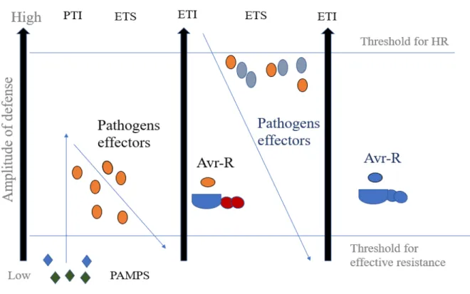
3 having expanded and diversified R gene repertoires, leading to the so–called zigzag model (Jones and Dangl, 2006). In the zig-zag model, there are four phases. In the first phase, PAMPs/MAMPs are recognized by PRRs, leading to PTI that alters further pathogen colonization (Figure 1.1). However, in phase 2, successful pathogens deploy effectors, which contributes to the virulence of pathogen. These effectors interfere with PTI, leading to effector-triggered susceptibility (ETS). In phase 3, an effector is recognized by an NLR protein, resulting to ETI and an HR is often observed at the infection site. Lastly, in phase 4, the pathogen avoids ETI by diversifying the recognized effector gene or by producing additional effectors (Jones and Dangl 2006) (Figure 1.1). Therefore, for successful invasion, pathogens need to suppress the plant innate immunity to cause diseases.
Figure 1.1: The zig-zag model of Plant innate immunity with all the four phases.
During phase 1, through PRR, the plant detects MAMPs/PAMPs (green and blue diamonds) and triggers PAMP-triggered immunity (PTI). In the second phase, effectors are delivered during successful invasion by the pathogens, leading to effector triggered susceptibility (ETS). For phase 3, there is a recognition of an effector (depicted in brown) by an NLR protein (blue and red), resulting in effector-triggered immunity (ETI). Phase 4 shows a situation wherein a pathogen has
4 lost the brown effector and maybe gained new effectors (blue). This leads to the avoidance of ETI but ETI can eventually be restored as the plant evolves new NLR alleles. Adapted from (Jones and Dangl, 2006).
1.1.2 Introduction to RNA Silencing
In plants, an additional defense mechanism, largely specific to antiviral defenses involves the RNA silencing pathway, which is based on 20- to 30- nucleotide RNAs. RNA silencing is a series of related processes in which small RNAs target nucleic acids for regulation in a sequence-specific manner (Fang and Qi, 2016). Small RNAs (sRNA) are key players in RNA silencing pathway and sRNAs are involved in plant development, the reprogramming of the genome and reproduction (Borges and Martienssen 2015). RNA silencing targets double stranded RNA (dsRNA) from endogenous and exogenous invading nucleic acids such as viruses and transposable elements (TE) (Borges and Martienssen 2015). The major classes of sRNAs are microRNAs (miRNAs) and small interfering RNAs (siRNAs) (Fang and Qi, 2016; Kim, 2005; Shamandi et al., 2015). miRNAs are involved in post transcriptional gene silencing (PTGS) through the cleavage of transcripts or the repression of translation and miRNAs also trigger the production of secondary siRNA transcripts (Fang and Qi, 2016; Kim, 2005). miRNAs are derived from precursor RNAs with partially double-stranded regions (Borges and Martienssen, 2015; Jouannet et al., 2012; Moreno et al., 2013). The miRNA pathway is initiated with the transcription of MIRNA genes by RNA polymerase II, forming a 5’capped and poly-A tailed pri-miRNA (Axtell, 2013). These miRNAs negatively regulate gene expression by pairing with the appropriate mRNA bases as part of a RISC complex, subsequently resulting in either RNA cleavage or an inhibition of protein translation (Axtell, 2013). RNA silencing can also be triggered by exogenous or endogenous dsRNA precursors, including dsRNA viral replication intermediates and local self-complementary double-stranded regions of viral genomes (Borges and Martienssen, 2015; Zhang et al., 2015), making RNA silencing a first line of antiviral immunity in plants (Ding and Voinnet, 2007; Dunoyer et al., 2006).
The mechanism of RNA silencing begins with long double-stranded RNA duplexes (Fang and Qi, 2016; Shamandi et al., 2015), and these duplexes are recognized by members of a protein family called dicer-like (DCL), proteins (Fang and Qi, 2016; Shamandi et al., 2015). These DCL proteins
5 cleave the dsRNA into sRNAs that are usually 21 to 24 nucleotides in length (Fang and Qi, 2016; Shamandi et al., 2015). Upon generation by DCLs, sRNAs are bound by a second family of endoribonucleases, the Argonaute (AGO) proteins, (Wang et al., 2019) which form the core of what are known as RNA-induced silencing complexes (RISC). AGO proteins allow RISC complexes to be guided by the sRNA to target RNA or DNA molecules through base-pair complementarity (Figure 1.3). Following sRNA-target binding, AGO proteins either cleave targeted RNA or repress its translation. Alternatively, in the case of DNA targeting, AGO proteins modify chromatin through recruitment of the RdDM machinery (Fang and Qi, 2016; Zhang et al., 2015). Although RNA silencing components are involved in a plethora of gene regulatory mechanisms, for this thesis, emphasis will be placed on the role of AGO proteins in defense against viruses.
Finally, in plants, RNA silencing also functions in association with DNA methylation and suppression of transcription (Carbonell and Carrington, 2015). This RNA silencing pathway occurs at the chromatin level and has been found to protect plant genomes against damage caused by transposons (Lario et al., 2013; Paulo et al., 2017) (see below for more details). The aforementioned silencing pathways are present in plants, however other organisms may have lost one or more of these pathways (Bologna and Voinnet, 2014; Borges and Martienssen, 2015; Moreno et al., 2013). For this thesis, emphasis will be placed on the cytoplasmic siRNA pathway due to the nature of the research projects to be discussed.
1.2.1 Origin and biogenesis of siRNAs and the role of RDRs.
Research has shown that siRNAs protect the genome in various ways, such as suppressing viruses that try to invade the cells, silencing transposable elements and repetitive elements in the genome, as well as silencing of aberrant transcripts and genes in the genome (Yu et al., 2016; Zhang et al., 2015). There are a number of molecular mechanisms involved in RNA silencing. Several of these pathways are explained in detail below. In addition to the sources of dsRNA described above, dsRNA may arise from the hybridization of sense and antisense mRNA transcripts (Di Serio et al., 2001). dsRNA molecules may also be synthesized by RNA-dependent RNA-polymerases (RdRPs/RDR) with or without initial priming. Priming is the synthesis of a short strand of RNA,
6 initiating polymerase-catalyzed synthesis of long dsRNA. RDRs are defined by the presence of a conserved RNA-dependent RNA polymerase catalytic domain (Willmann et al., 2011). There are 3 major clades of eukaryotic RDRs: RDRα, RDRβ and RDRγ. RDRα is reported to be found in fungi, plants and in animals, while RDRγ is only found in plants and RDRα is present in only animals and fungi (Wassenegger and Krczal, 2006). In A. thaliana, there are 3 types of RDRα: RDR1, RDR2, RDR6 and three types of RDRγ: RDR3, RDR4 and RDR5. While the RDRγ clade remains functionally uncharacterized in plants, its significance is based on its presence in many fungi and its involvement in transcriptional gene silencing (TGS) in the fission yeast Schizosaccharomyces pombe (Wassenegger and Krczal, 2006; Yoshikawa, 2013). One or more of the 6 RDR paralogs in plants are involved in strengthening silencing responses by the production of dsRNA via viral templates (Molnar et al., 2010; Qi et al., 2009). Initially, RDRs were studied due to their roles in antiviral plant defense and transgene silencing (Wang and Metzlaff, 2005), however, RDRs also have other molecular functions that include control of chromatin structure and the regulation of cellular gene expression (Wassenegger and Krczal, 2006). RDR1 is involved mostly in the amplification of exogenous, virus-induced small RNAs, making it a part of the plant antiviral RNA silencing system, in conjunction with DCL2 and DCL4 (Xiaoming Zhang et al., 2012). RDR2 plays a role in DNA methylation by converting the ssRNA produced by polymerase IV into dsRNA. RDR6 functions in both endogenous trans-acting siRNA biogenesis as well as antiviral silencing (Yoshikawa et al., 2005; Curaba et al., 2008; Garcia-Ruiz et al., 2010).
For RNA viruses to be replicated, virus-encoded RDRs are required (Pogany and Nagy, 2015, 2012). During an infection, RDRs act in concert with other viral and host cellular factors that play a role in RNA synthesis, RNA elongation, and other functions (Pogany and Nagy, 2015). In some viruses, RDR proteins are reported to be inactive in the cytoplasm to prevent the formation of viral dsRNA that could trigger RNA silencing (Pogany and Nagy, 2012). Therefore, the activation of viral RDRs is an important step for viral infection (Pogany and Nagy, 2012). In the initial stages of RNA virus infection, the plus stranded RNA is released from the virion and produces the viral RDR using the host translation machinery. The viral RDR then produces the minus strand using the plus strand RNA (Newburn and White, 2015; Verchot-Lubicz et al., 2010).
7 As a mechanism of amplification of antiviral response, plants generate secondary viral siRNA (vsi-RNA) that can spread systemically to neighboring cells via plasmodesmata (PD) and the phloem. The amplification of the silencing signal is mainly dependent on host RDR1/6 and suppressor of gene silencing 3 (SGS3) proteins. These steps start with the de novo synthesis of viral dsRNA with or without initial priming from “aberrant” viral RNA cleavage products. One strand of the vsiRNA can act like a primer for the dsRNA production by RDRs (Molnar et al., 2010; Verlaan et al., 2013; Wang et al., 2010). This secondary dsRNA is processed by DCL proteins to form secondary vsiRNA, thereby amplifying the antiviral RNA-silencing.
1.3 Dicer and Dicer-like proteins.
The enzymes responsible for producing siRNA and miRNA from dsRNA are Ribonuclease (RNase) III-like enzymes belonging to the Dicer family (Vickers et al., 2003).
A. thaliana encodes four specialized Dicer-like (DCL) proteins named DCL1, 2, 3, and 4 (Bellaoui et al., 2003; Vickers et al., 2003). While DCL1 processes fold-back precursors to generate miRNAs, (Bellaoui et al., 2003; Liu et al., 2009; Vickers et al., 2003) plant siRNAs are processed primarily by DCL2, DCL3, and DCL4 (Liu et al., 2009). These siRNAs are known as heterochromatin siRNAs (hetsiRNAs) (Xiaoming Zhang et al., 2012). Of all these small RNAs, the 24-nucleotide hetsiRNAs are the most abundant, as they play a role in transcriptional silencing of repetitive elements within the genome with the aid of RNA-directed DNA methylation (RdDM). The role of each DCL is well established and rather specific, however, redundancy between DCL functions has been proposed (Katsarou et al., 2016).
In A. thaliana, it has been shown that DCL1 is the only dicer protein that produces 21-nt miRNAs (Kurihara and Watanabe, 2004; Reinhart et al., 2002) and null mutants of dcl1 alleles are embryonic lethal (Blevins et al., 2006; Curtin et al., 2016; Tsuzuki et al., 2014.). DCL2, together with RDR6 and SGS3, has been shown to be involved in the cleavage of antisense transcripts and in the synthesis of 22-nt natural antisense- small interfering RNA (nat-siRNA) (Bellaoui et al., 2003; Liu et al., 2009; Vickers et al., 2003). DCL2 processes dsRNAs to produce 22-nt vsiRNA to be loaded into AGO proteins (Pumplin and Voinnet, 2013). DCL2 can function in antiviral defense in the absence of DCL4 and may play a redundant role in some respects (Xiaoming Zhang
8 et al., 2012) and 22-nt and 21-nt siRNAs are both required for optimal resistance against viral invasion (Gonzalez-Gaitan et al., 2004; Parent et al., 2015; Wang et al., 2011). The DCL4 protein is the major producer of 21-nt antiviral siRNA and endogenous siRNA such as tasiRNA and phasiRNA (Bouché et al., 2006; Qu et al., 2008; Yoshikawa, 2013). Indeed, studies have shown that in A. thaliana, maximal viral replication is achieved upon the mutation of both dcl2 and dcl4 genes (Gonzalez-Gaitan et al., 2004; Oa et al., 2010; Jaubert et al., 2011; Andika et al., 2015; Parent et al., 2015).
For certain viruses, such as cucumber mosaic virus (CMV) (Bouché et al., 2006; Fusaro et al., 2006), turnip crinkle virus (TCV) (Deleris et al., 2006 ) and cabbage leaf curl virus (CaLCuV), the above observation is valid (Blevins et al., 2006). However, some studies have shown situations where DCL2 and DCL4 appear to function differently. These studies include work done by (Mlotshwa et al., 2008), where DCL2 was identified as the protein responsible for secondary transitive siRNA synthesis, whereas DCL4 was shown to be involved in the production of primary siRNAs. Additional studies have also supported the above findings, demonstrating that DCL2-dependent 22-nt siRNAs do not contribute to CMV resistance (Wang et al., 2011). The inactivation of DCL4, induces a high level of viral replication, indicating that DCL4 is essential for intracellular antiviral silencing (Song and Rossi, 2017). While DCL2 can produce abundant 22-nt viral siRNAs in the absence of DCL4, these siRNAs are less efficient in mediating antiviral defense (Andika et al., 2015; Wang et al., 2011). Therefore, based on the above, it can be concluded that there could be a specialization of function of both DCL2 and DCL4 proteins.
DCL3 is reported to be important for defense against DNA viruses through DNA methylation, although the silencing suppressors of some RNA viruses interfere with DCL3 (Csorba et al., 2015). DCL3 produces 24-nt siRNAs derived from transposons and DNA repetitive elements (Daxinger et al., 2009; Priya Raja et al., 2014). These 24-nt siRNAs play an essential role in transcriptional gene silencing (TGS) by directing RdDM for the repression of transposon and DNA repeats in a TGS process (see below). Suppression of the expression of DCL3 is linked to the enhancement of systemic PTGS (Chen et al., 2018).
9 1.4 Other players in the RNA silencing.
Modifications are made to sRNAs upon processing. These modifications include 2’-O-methylation, 3’-uridylation or 2’-adenylation, and adenosine deamination (Jamous et al., 2011; Zust et al., 2011). In plants, sRNAs are 2’-O-methylated at the 3’-terminal by HUA ENHANCER 1 (HEN1) to prevent uridylation (a signal for degradation) by the enzyme nucleotidyl transferase HEN1 SUPPRESSOR 1 (HENSO1) (Jamous et al., 2011; Zust et al., 2011).
dsRNA-BINDING (DRB) proteins are involved in the biogenesis of miRNA or ta-siRNA in plants (Montavon et al., 2017). Two DRBs, Hyponastic Leaves 1 (HYL1) and DRB4, have been found to be required for the proper function of DCL1 and DCL4, respectively (Montavon et al., 2017). DRB4 has 2 dsRNA binding motifs (dsRBD1 and 2) in its N-terminus (Montavon et al., 2017). The interaction of DRB4 and DCL4 leads to the generation of 21-nt siRNAs including ta-siRNa, DCL4-dependent miRNAs or vsiRNAs from either endogenous or exogenous dsRNAs (Montavon et al., 2017). In addition, DRB4 has been found to be targeted by the suppressor of silencing encoded by the DNA virus, cauliflower mosaic virus (CaMV) (Qu et al., 2008). However, studies have shown that DRB4 may not be directly involved in siRNA production but it might be involved in the stabilization of 21-nt viral siRNA. This is evidenced by the finding that there is a significant decrease in 21-nt siRNAs in mutant plants despite only a slight increase in viral RNA levels in drb4 mutants (Montavon et al., 2017; F. Qu et al., 2008). DRB3 has also been shown to interact with DCL3 for defense against DNA viruses (Raja et al., 2014).
SGS3 is an RNA binding protein that binds RNA to prevent them from degradation before its conversion to dsRNA by RDR proteins (Okano et al., 2014). Previous studies have shown that AtSGS3 binds to, and stabilizes, RNA templates during the initiation of RDR6-mediated dsRNA synthesis (Li et al., 2017). SGS3, in cooperation with RDR6, has also been reported to have antiviral defense activities against DNA viruses (Li et al., 2017). In addition, A. thaliana SGS3 and RDR6 both co-localize in cytoplasmic granules called SGS3/RDR6-bodies (Li et al., 2017). Indeed, in A. thaliana, a lack of SGS3 increases susceptibility to some viruses, such as CMV, but not others, such as turnip mosaic virus (TuMV) and turnip vein clearing virus (TVCV) (Mourrain
10 et al., 2000). In contrast, during potyvirus infection, the silencing of SGS3 mRNA reduces the accumulation of viral RNA, as seen with potato virus A and soybean mosaic virus (Chen et al., 2015).
1.5 RISC Complexes and ARGONAUTE Proteins.
Primitive plants have been reported to encode only a few AGO proteins (Schuck et al., 2013; Vaucheret, 2008). The model plant A. thaliana encodes ten AGO proteins, designated AGO1 to AGO10, of which AGOs 1, 2, 4, 5, 7 and 10 have been shown, to varying degrees, to possess antiviral activities in certain contexts (Fang and Qi, 2016). The AGO protein family has expanded during plant evolution, leading to functional diversification. The specialization of AGO proteins in different pathways and biological processes is due to their intrinsic biochemical properties, spatiotemporal expression patterns as well as the protein and sRNA partners with which they interact. To successfully carry out their functions, AGO proteins have some key biochemical properties. Firstly, AGO proteins bind sRNAs in order to facilitate base pairing with complementary target RNAs (Fátyol et al., 2016; Hauptmann et al., 2015). Secondly, they possess RNaseH-like endonuclease through which they cleave target RNA (Fátyol et al., 2016; Hauptmann et al., 2015) and facilitate strand separations of fully complementary small RNA by passenger strand cleavage (Fátyol et al., 2016; Hauptmann et al., 2015). In addition, AGO proteins can amplify silencing responses by producing cleaved RNA fragments that can serve as new substrates for RDR. Lastly, AGO proteins may serve as platforms to which silencing cofactors may bind (Poulsen et al., 2013). It is known that in plants, animals, and fungi, AGO proteins bind to proteins containing Gly-Trp (GW) dipeptides (Fátyol et al., 2016; Hauptmann et al., 2015), which are often essential cofactors in RNA silencing (El-Shami et al., 2007; Poulsen et al., 2013).
1.6 Structure of AGO proteins.
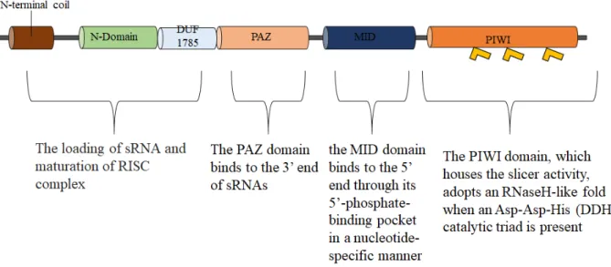
The known biochemical functions of AGO proteins are carried out in large part by 4 domains: a variable N-terminal domain, a conserved Piwi-Argonaute-Zwille (PAZ) domain, a domain in the
11 middle of the primary structure (MID), and a C-terminal (PIWI) domain (Mallory and Vaucheret, 2010; Poulsen et al., 2013) (Figure 1.2). The N-terminal domain is divided into 3 elements of primary structure, called the N-terminal coil, N domain, and a domain of unknown function 1785 (DU 1785), formerly known as linker 1 (Fátyol et al., 2016; Mallory and Vaucheret, 2010). sRNA binding involves the MID and PAZ domains (Poulsen et al., 2013). The PAZ domain binds to the 3’ end of sRNAs (Ma et al., 2004) while the MID domain binds to the 5’ end through its 5’-phosphate-binding pocket in a nucleotide-specific manner (Ma et al., 2004; Mallory and Vaucheret, 2010; Mi et al., 2008; Montgomery et al., 2008). The PAZ and MID domains are connected by a large piece of intervening sequence referred to as linker 2 (L2) (Ma et al., 2004). The PIWI domain, which possesses slicer activity, adopts an RNaseH-like fold with an Asp-Asp-His (DDH) catalytic triad (Baumberger and Baulcombe, 2005; Liu et al., 2004; Rivas et al., 2005; Song et al., 2004). The mechanisms of sRNA loading into AGO proteins occurs when siRNA duplexes are loaded into the AGO protein by the RISC-loading complex in an ATP-dependent reaction (Iwasaki et al., 2010). Once bound to the dsRNA/siRNA duplex, the AGO protein cleaves 1 of the strands, the passenger strand, an action that triggers dissociation of the latter from the complex, resulting in a mature RISC containing only the guide strand (Carthew and Sontheimer, 2009).
AGO proteins are classified into different clades based on phylogenetic and (sometimes) functional relationships: AGO 1/10, AGO 2/3/7 AGO4/6/8/9 and AGO5 (Fang and Qi, 2016; Kapoor et al., 2008; Manavella et al., 2012; Vaucheret, 2008). A study identified specific and conserved amino acid sequences (blocks) containing motifs of unknown function in AGO proteins (Rodríguez-Leal et al., 2016). This study also showed that AGO4/6/8/9 and AGO1/10 clades have the most conserved linear organizations of blocks while AGO2/3/7 and AGO5 clades have a more variable linear organization of the said blocks (Rodríguez-Leal et al., 2016). Furthermore, domains of unknown functions that show a predictable position within a conserved primary structure were identified. Domains A-1 and B-1 were identified for AGO2/3/7 clade, AGO4/6/8/9 clade is characterized by containing the A-2 and PIWI-1 domains. AGO5 clade possesses the A-3, DUF1785-3, PAZ-3, B-3 and PIWI-2 domains. Lastly, AGO2/3/7 clade showed a conserved
12 sequence of blocks that incudes A-4, PAZ-3, B-4, PWI-3 and DUF1785-2 domains (Rodríguez-Leal et al., 2016).
Figure 1.2. A Schematic representation of the general structure of AGO proteins. This figure displays the N-terminal domain, a domain of unknown function (DUF 1785) a conserved Piwi-Argonaute-Zwille (PAZ) domain, a domain in the middle of the primary structure (MID), and a C-terminal (PIWI) domain. Each domain is shown with an explanation of its function.
1.6.2 Classification of AGO proteins
1.6.2.1 AGO 1/10.
AGO1 is involved in the regulation of diverse miRNA genes (Vaucheret et al. 2004). AGO1 is thought to be the effector for most miRNAs and ta-siRNAs (Vaucheret et al. 2004; Baumberger & Baulcombe 2005; Qi et al. 2005; Mi et al. 2008) These miRNAs and ta-siRNAs guide AGO1 to regulate the stability and/or translation of mRNAs of genes involved in numerous developmental and physiological processes. In addition, ago1 mutants are more susceptible to some viruses (Morel et al., 2002; Qu et al., 2008; Takeda et al., 2008; Wang et al., 2011). AGO1 protein recruits
13 a limited number of miRNAs and is responsible for gene regulation in specific situations. AGO1 homeostasis is regulated by miR168 through miR168-AGO1-dependent slicing of AGO1 mRNA, leading to an increase in the accumulation of miR168 in response to elevated AGO1 levels (Vaucheret et al., 2006).
A. thaliana AGO10, is the closest homolog to AGO1 in protein sequence, however, it differs to AGO1 in expression patterns and in its developmental functions (Yu et al. 2017). In A. thaliana, it has been shown that in shoot apical meristem, AGO10 competes with AGO1 for specific miRNAs and counters its activities through sequestration (Zhu et al., 2011). AGO10 sequesters miR165/166 thereby releasing the negative regulation they exert on the HD-ZIP III gene family that regulates meristem development (Zhou et al., 2015; Zhu et al., 2011). AGO10 also regulates meristem development via the mediation of translational inhibition of multiple miRNA target genes (Brodersen et al., 2008; Mallory et al., 2009). AGO10 has also been reported to have a limited role in defense against TuMV (Garcia-Ruiz et al., 2015).
1.6.2.2 AGO 2/3/7.
Proteins of the AGO2/3/7 clade show clear phylogenetic relationships but are implicated in different phenomena (Figure 1.3). The AGO2 protein of A. thaliana is involved in antiviral defense and is required for resistance to multiple plant viruses (Garcia-Ruiz et al., 2015; Harvey et al., 2011; Jaubert et al., 2011; Wang et al., 2011; Xiuchun Zhang et al., 2012). Biochemical experiments revealed that AGO2 loaded with synthetic vsiRNAs can target viral RNAs for cleavage, which leads to the inhibition of viral replication (Schuck et al., 2013a). AGO2, which primarily binds to 21-nt to 22-nt sRNAs, has been shown to associate with certain miRNAs (Maunoury and Vaucheret, 2011) and to regulate the production of a small number of proteins, including MEMB12, which is involved in innate immunity against bacteria (Zhang et al., 2011). AGO2 has also been implicated in DNA double-strand break repair (Wei et al., 2012) and in DNA methylation (Pontier et al., 2012), but has been studied mostly for its role in antiviral defense against multiple plant viruses (see below).
14 AGO3 is phylogenetically closely related to AGO2 but appears to have different biological functions (Fátyol et al., 2016). AGO3 has also been reported to recruit 24-nt sRNA for RdDM as AGO3 bound 24-nt sRNAs overlapped with those bound to AGO4; AGO4 is a key AGO in the RdDM pathway (see below) (Zhang et al., 2016). A recent study using Bamboo mosaic virus (BaMV) showed that there was an increase in expression of AGO3 upon virus infection, while an ago3 mutant showed enhanced susceptibility to BaMV. This increase in AGO3 expression is Abscisic acid-mediated (Alazem et al., 2017).
AGO7 in A. thaliana is also known as ZIPPY. AGO7 binds 21-22 nt sRNAs and mediates tasiRNA biogenesis, which is important for development timing and leaf morphology (Adenot et al., 2006; Howell et al., 2007; Montgomery et al., 2008; Yoshikawa et al., 2013). AGO7 induces the secondary siRNA pathway where mi390-directed, AGO7 mediated cleavage of TAS3 transcripts leads to the biogenesis of tasiRNA species through the action of SGS3 and RDR6 (Adenot et al., 2006; Fahlgren et al., 2006; Howell et al., 2007; Montgomery et al., 2008). TAS3 tasiRNAs are known to target several AUXIN RESPONSE FACTOR (ARF) genes that are involved in the regulation of lateral organ development and developmental timing (Adenot et al. 2006; Fahlgren 2006 et al. 2006; Hunter et al. 2006. montogomery et al. 2008). AGO7 is also known to function with DRB4, which takes part in defense against viruses (Qu et al., 2008). Upon infection with mutant TCV∆CP-lacking its viral suppressor of RNA silencing (VSR), ago7 mutants showed higher accumulation of TCV∆CP (Qu et al., 2008). Aside the aforementioned example, AGO7 shows little involvement in defense against viruses.
1.6.2.3 AGO5.
AGO5 appears to be involved in specific developmental processes. In A. thaliana, AGO5 is expressed in megaspores and the somatic cells around megaspore mother cells as well as being expressed during all stages of flower and seed formation (Kapoor et al., 2008; Schmid et al., 2005). Consistent with this, AGO5 affects pigmentation in Glycine max seeds through the silencing of
15 chalcone synthase (Cho et al., 2017) and a semi-dominant ago5 mutant allele leads to a defect in the initiation of mega-gametogenesis (Tucker et al., 2012). Likewise, the mutation of MEL1, 1 of the 5 AGO5 clade members in rice, results in meiotic arrest, male sterility, and aberrant pollen mother cells (Nonomura et al., 2007). In addition, AGO5 may play a role in plant-microbe interactions as it appears to act in the establishment of nodules in legume-rhizobia interactions legumes (Reyero-Saavedra et al., 2017). AGO18 homologues are most closely related to the AGO5 and AGO1-containing clades and may also play important roles in development and virus defense (see below), although their activities have been less characterized than other AGO proteins (Zhang et al., 2015b).
1.6.2.4 AGO4/6/8/9, RdDM and DNA Methylation.
Although most AGO proteins are thought to function in the cytoplasm, members of the AGO4/6/8/9 clade (Xie & Yu 2015), function primarily in the nucleus. Members of this clade bind to 24-nt hc-siRNAs and target corresponding DNA sequences for RdDM through the recruitment of DNA methylation factors (Havecker et al., 2010; Zilberman et al., 2003). This results in epigenetic modifications leading to the silencing of transposons, retrotransposons and other repetitive elements, as well as controlling the expression of specific genes (Matzke et al., 2015). Proteins in the AGO4/6/8/9 clade show a high level of sequence conservation and are thought to function similarly, however they show distinct spatial-temporal expression patterns and are thought to act on different genetic elements at different times (Matzke et al., 2014; McCue et al., 2015), (Havecker et al., 2010; Mallory and Vaucheret, 2010).
RNA directed DNA methylation (RdDM), an epigenetic modification, plays a critical role in repressing transposons as well as in the regulation and maintenance of genome stability (Xie et al., 2004). Mediation of DNA methylation is performed by AGO4-bound hc-siRNAs through the RdDM pathway and catalyzed by Domains Rearranged Methyltransferase 2 (DRM2), which interacts with the Pol V complex. In this pathway, DCL3 produces 24-nt hc-siRNAs (Xie et al., 2004). The hc-siRNAs are exported into the cytoplasm, where they are loaded into AGO4, leading to an AGO4/siRNA complex. The AGO4/siRNA complexes are recruited to target loci through base-pairing with scaffold transcripts generated by RNA polymerase V (Pontier et a., 2005;
16 Mosher et al., 2008; Wierzbicki et al., 2009). This recruitment may be facilitated by the interaction of AGO4 with the GW/WG-rich (known as the ‘AGO hook’) extensions of NRPE1 (the largest subunit of Pol V) and some transcription elongation factors (Pontier et al., 2012; Pontes et al., 2006; He et al., 2009; Bies-Etheve et al., 2009). The AGO4/siRNA complexes lastly recruits DRM2 to methylate the target DNA (Duan et al., 2015; Li et al., 2006; P. Raja et al., 2014). Furthermore, in A. thaliana, in addition to 24-nt hc-siRNAs, RDR6-dependent 21-nt siRNAs which originate from trans-acting siRNA (TAS) loci have the ability to recruit AGO4, a process that directs DNA methylation at the TAS loci (Wu et al., 2012). 24 nt-long miRNAs (lmiRNAs), which are generated from the actions of DCL1, in conjunction with AGO4, have the ability to direct DNA methylation in trans at the targeted gene site, thereby resulting in TGS. DNA viruses accumulate in the nuclei of infected plants where AGO4 binds to the generated viral siRNA, forming a complex. The AGO4/siRNA complex is also exported into the cytoplasm and processed as described above. In addition to AGO4, given the close phylogenetic relationship of AGO6 and AGO9 to AGO4; AGO6 and AGO9 have also been reported to mediate de novo DNA methylation by recruiting 24-nt siRNAs (Nuthikattu et al., 2013; Pontier et al., 2012).
AGO4 is the major AGO protein involved in hc-siRNA action, that directs DNA methylation via the RdDM pathway (Li et al., 2006; Qi et al., 2009). RdDM mechanisms have evolved in part to silence foreign or repetitive DNA and, not surprisingly, this mechanism also functions against plant DNA viruses. However, despite the existence of several RdDM-associated AGO proteins, only AGO4 has been implicated in antiviral mechanisms against DNA viruses, likely because it has a broad and constitutive expression pattern. As would be expected, AGO4 inhibits DNA viruses through a methylation-mediated mechanism (P. Raja, Jackel, Li, Heard, & Bisaro, 2014; Priya Raja et al., 2010, Wang et al., 2019). Surprisingly, although AGO4 is best characterized for its function in the nucleus, it is also important in the defense against RNA viruses that have cytoplasmic replication strategies. Mutation of ago4 allows for a higher accumulation of tobacco rattle virus (TRV) and BaMV, although AGO2 seems to be the more important AGO in these cases (Alazem et al., 2017; Ma et al., 2015). AGO4 is also reported to be required for resistance to the bacterial pathogen, Pseudomonas syringae infection (Agorio & Vera 2007). It has been reported that the loss of function in RdDM pathway components, upstream or downstream of AGO4 affects resistance to P. syringae pv. tomato DC3000 infection (Agorio & Vera 2007). However, it is interesting to note that for viruses, loss of other RdDM pathway components does not impair
17 resistance to plantago asiatica mosaic virus (PlAMV) infections, suggesting that AGO4 most likely has a function in defense independent of its role in RdDM pathway (Brosseau et al. 2016). Interestingly, in the presence of PlAMV, the localization of AGO4 shifted from being primarily in the nucleus to being mainly in the cytoplasm and a nuclear-localization deficient mutant of AGO4 was not compromised in its antiviral activity (Brosseau et al., 2016). Normally, AGO4 binds to hcsiRNAs in the cytoplasm (Ye et al., 2012), upon which a nuclear localization signal (NLS) is exposed and the complex is imported into the nucleus. Thus, PlAMV infection results in either an inhibition of AGO4 import into the nucleus or an active re-localization to the cytoplasm. The mechanism behind this is not clear, however, it may represent a kind of counter-counter defense, wherein RNA silencing is countered by the PlAMV, but this may in turn result in a mobilization of AGO4 by localizing to where it is able to target the viral RNA. AGO4 is also required in plant defense against DNA viruses, via the RdDM pathway, suggesting that DNA methylation by AGO4 is a mechanism used by plants to defend against DNA viruses (see below). For example, it has been reported that the DNA of a beet curly top virus (BCTV) VSR-defective mutant extracted from recovered plants is hyper methylated and that host recovery requires AGO4 (Raja et al. 2008).
AGO6 mutation partially suppresses TGS and affects DNA methylation at several RdDM target loci (Zheng et al., 2007). Compared to AGO4, AGO6 differs in expression pattern; AGO6 is predominantly expressed in the shoot and root apical as well as in dividing cells (Eun et al., 2011; Zheng et al., 2007). Studies have demonstrated that 21-22-nt siRNA, produced via RDR6 during RNA silencing of mRNAs, get incorporated into AGO6 directly. This then guides AGO6 to chromatin containing transposable elements for RdDM (McCue et al., 2015; Nuthikattu et al., 2013). Previous work has also shown that for DNA methylation, AGO6 and AGO4 are required at different stages or loci of DNA methylation (Duan et al., 2015).
AGO8 was previous thought to be a pseudogene due to a computational analysis prediction that the coding sequence of AGO8 contains splicing-inducing frame shifts; suggesting the formation of a nonfunctional protein (Takeda et al., 2008). However, AGO8 does appear to be functional in some species (Pradhan et al., 2017). In addition, a recent study showed that pre-meiotic ovules of ago4 and ago9 mutant gametes overexpress AGO8, suggesting a possible role of AGO8 in
18 compensatory effect of gametophytic cell fate (Hernández-Lagana et al., 2016.) A. thaliana AGO9 is involved in controlling female gamete formation; by restricting specification of gametophyte precursors (Olmedo-Monfil et al., 2010). AGO9 was also shown to interact with 24-nt siRNAs derived from transposable elements and it silences transposable elements in female gametes in a non-cell autonomous manner (Olmedo-Monfil et al. 2010).
Figure 1.3. AGO proteins classifications and siRNA binding partners. Small RNA binding preference for AGO proteins and their downstream functions, using data from A. thaliana research. AGOs are colour coded and arranged with respect to their clades.
1.7 DNA viruses and Methylation-mediated defense.
In plants, the majority of studies that have investigated the role of RNA silencing during DNA virus infection uses geminiviruses (Inoue-Nagata et al., 2016). Geminiviridae is a large family of plant viruses that causes crop diseases of economic importance worldwide (Inoue-Nagata et al., 2016). Their genome consists of a circular single-stranded DNA (ssDNA) genome encapsulated in a twinned icosahedral particle and they replicate through a double-stranded DNA intermediate (dsDNA) (Inoue-Nagata et al., 2016). During infection by DNA viruses, DCL4 and DCL3-derived
19 vsiRNA initiate PTGS or TGS respectively (Csorba et al., 2015). DCL3-generated 24 nt vsiRNA following the HEN1-methylation are loaded into AGO4. While the silencing of RNA viruses predominantly takes place in the cytoplasm via PTGS mechanisms, silencing of DNA viruses occur in both the cytoplasm and the nucleus (Chellappan et al., 2005; Laufs et al., 1995).
Geminivirus-derived sRNAs of 21, 22 and 24-nt have been identified in infected hosts and all four DCLs in A. thaliana have been implicated in the production of geminivirus siRNAs (Priya Raja et al., 2014, 2008). A. thaliana methylation-deficient mutants are reported to be hypersusceptible to geminivirus infection. Although the viral dsRNA structures are accessible to all DCLs, a hierarchy exists between them and DCL3 has been reported to be crucial against DNA viruses (Akbergenov et al., 2006; Qu et al., 2008; Csorba et al., 2015). A. thaliana dcl3 mutant are unable to recover from geminivirus infection when compares to wild-type, dcl2 and dcl4 (Raja et al., 2014). Furthermore, studies of virus induced gene silencing (VIGS) triggered by CaLCuV, a geminivirus, have shown that DCL2 and 3, RDR6, HEN1 and SGS3 are required to convert geminivirus-derived transcripts into dsRNAs, which induces RNA silencing of host mRNAs (Akbergenov et al. 2006; Muangsan et al. 2004). In addition, a number of VSRs encoded by these geminiviruses are able to suppress PTGS (Buchmann et al., 2009). These include the AL2-like proteins from african cassava mosaic virus and tomato yellow leaf curl virus, P6 protein from cauliflower mosaic virus (CaMV) and L2 of BCTV (Raja et al., 2008).
1.8 Detailed roles of AGO proteins in plant-pathogen interaction
1.8.1. RNA Silencing as a defense mechanism against viruses
While RNA silencing is mostly involved in endogenous gene regulation, given its ability to degrade dsRNA and target homologous ssRNA, it also functions as a defense mechanism against virus infection (Figure 1.4). Most plant viruses have an RNA genome and/or generate dsRNA from replication intermediates, local self-complementary regions of the viral genome, or through the action of host RNA-dependent RNA polymerase (RDR) on viral RNA (vRNA) templates (Donaire et al., 2009; Qi et al., 2009; Szittya et al., 2010). Like endogenous targets, viral dsRNA can also
20 be processed by DCL proteins into viral small interfering RNAs (vsiRNA). As outlined above, RNA silencing encompasses multiple related gene-regulating phenomena and different RNA silencing components have undergone amplification and diversification, like AGO proteins. Many of these were initially identified in studies investigating transgene silencing and/or developmental regulation of endogenous gene expression (Adenot et al., 2006; Bohmert et al., 1998; Fagard et al., 2000; Lynn et al., 1999; Moussian et al., 1998; Zheng et al., 2007; Zilberman et al., 2003) and subsequently tested for the role the play in virus defense. Thus, it begs the question: have AGOs become specialized to target viruses in different plants and if so, which AGOs are able to do so.
Figure 1.4 Schematic representation of RNA silencing during viral infection. A Model for plant antiviral defense based on RNA silencing. Virus-derived dsRNA structures are recognized by DCL4 and DCL2, which leads to the generation of DCL-dependent vsiRNAs. The vsiRNAs are loaded into the RISC complexes, where AGO proteins are contained. The vsiRNA-loaded RISC complexes targets viral transcripts for cleavage. Aberrant RNAs are also generated, which are targeted by RDR1 and RDR6. The RDR-dependent secondary vsiRNAs target viral regions
21 that are distant from the sites of primary vsiRNA processing. Modified from (Willmann et al., 2011).
1.8.2 WHICH AGOs ARE ANTIVIRAL?
The study of antiviral RNA silencing in planta comes with inherent challenges since this requires the use of a virus and compatible host. However, if a virus can infect a given host, this usually means that it has overcome the host’s RNA silencing mechanisms, most likely through the action of its VSR. This experimental constraint can be overcome by using VSR-defective mutant viruses to infect mutant plants to determine what genetic ablations can compensate for the lack of VSR activity. This approach has been very informative but since viral proteins are often multi-functional, it is not possible for all viruses. Alternatively, one can use wild-type (WT) viruses and assess for increased virus accumulation in mutant plants. Table 1 lists studies showing genetic evidence for the involvement of different AGO proteins, as assessed by increased virus accumulation of either WT or VSR-defective viruses in mutant plants. Early genetic studies and hypotheses regarding antiviral RNA silencing focused on AGO1 due in part to the order in which different AGO proteins were characterized. Although many initial insights into RNA silencing phenomena came from the study of plant-virus interactions, most of the first genetic screens were designed to identify RNA silencing components affecting the post-transcriptional silencing of endogenous genes or transgenes. These led initially to the characterization of AGO4 and AGO1 (Bohmert et al., 1998; Zilberman et al., 2003). The first AGO protein shown to virus infection was AGO1, with the report that a hypomorphic ago1 mutant was hyper-susceptible to CMV (Morel et al., 2002). This was one of the only AGO mutants available at the time and subsequent studies reported that ago1 mutants permitted increased accumulation of brome mosaic virus (BMV), as well as VSR-defective variants of CMV and TCV, suggesting an important function for AGO1 in antiviral RNA silencing (Dzianott, Sztuba-Solińska, & Bujarski, 2012; Qu, Ye, & Morris, 2008). In Nicotiana benthamiana, silencing of AGO1 attenuates symptom recovery in plants infected with tomato ringspot virus (ToRSV) (Ghoshal and Sanfaçon, 2014). Likewise, AGO1 knockdown in rice (Oryza sativa) permits increased accumulation of rice dwarf phytoreovirus (RDV) and rice stripe virus (RSV) (Wu et al., 2015). Thus, AGO1 appears to play a role in multiple plant-virus interactions. However, it should be noted that studies with AGO1 are often confounded by the
22 severe developmental phenotypes exhibited by ago1 mutants and that AGO1 is involved in the regulation of large numbers of endogenous genes (Baumberger & Baulcombe, 2005; Bohmert et al., 1998; Kidner & Martienssen, 2005; Morel et al., 2002), some of which could indirectly affect virus infection.
The availability of mutants for all AGO-encoding genes in A. thaliana has allowed for systematic analyses to identify which AGO proteins have antiviral activity (Table 1). This approach has enabled multiple groups to identify AGO2 as being involved in resistance against a range of viruses, including, PVX, CMV, TCV, TRV, TuMV, PlAMV and BaMV, suggesting a broad involvement of AGO2 in defense against viruses (Jaubert et al. 2011; X.-B. Wang et al. 2011; Harvey et al. 2011; Cabonell et al. 2012; Ma et al. 2015; X. Zhang et al. 2012; Brosseau et al. 2016; Hernan Garcia-Ruiz et al. 2015; Alazem et al. 2017). Multiple studies have demonstrated a role for AGO2 in antiviral defense in N. benthamiana as well. When NbAGO2 is silenced by virus-induced gene silencing (VIGS), several Tombusviruses (TBSV, cymbidium ringspot virus [CymRSV], carnation Italian ringspot virus [CIRSV] and cucumber necrosis virus [CNV]) accumulate to higher levels (Odokonyero et al., 2015) and plants no longer recover from infection by a VSR-defective version of TBSV (Scholtof et al., 2011). Likewise, CRISPR-generated N. benthamiana ago2 knockout plants are more susceptible to PVX and TuMV. The same mutants also showed more symptoms when infected with TCV, VSR-deficient CIRSV and CymRSV (Ludman et al., 2017a). Given the similarities of AGO7 and AGO3 with AGO2, it might be predicted that these proteins might also be involved in antiviral defense. However, AGO7 has been implicated in plant-virus interactions only to a small extent. A. thaliana ago7 mutants show some increase in susceptibility to BaMV as well as attenuated versions of TCV-GFP and TuMV, albeit to a limited degree (Alazem et al., 2017; Garcia-Ruiz et al., 2015; Qu et al., 2008). In N. benthamiana, VIGS of AGO7 caused a subtle increase in susceptibility to TBSV, sunn-hemp mosaic virus (SHMV), and foxtail mosaic virus (FoMV) (Odokonyero et al., 2017). Likewise, AGO3 has only been shown to play a relatively minor role in curtailing infection of one virus, BaMV (Alazem et al., 2017). Thus, AGO2 appears to have evolved to play a major role in anti-viral defenses (Table 1.1).