DEPARTEMENT OF BIOCHEMISTRY
N°……/SNV/2021
THESIS
Presented by
Gheraibia Sara
For the fulfillment of the requirements for the degree of
DOCTORATE
3rdCycle
IN BIOLOGY
Special filed: BIOCHEMISTRY
TOPIC
ا
Isolation and characterization of bioactive extracts of Costus speciosus
and evaluation of their biological effects in vitro and in vivo.
Presented publically in: / / 2021.
JURY:
President : Bouriche Hammama Pr., FSNV, UFA, Sétif 1 Supervisor: Belattar Noureddine Pr., FSNV, UFA Sétif 1 Co-Supervisor: Mosaad A. Abdel-Wahhab Pr., NRC Cairo, Egypt Examiners : Amira Smain Pr., FSNV , UFA, Sétif 1
Derouiche Samir MCA., U. d'El-Oued
Laboratory of Applied Biochemistry
Université Ferhat Abbas
Sétif 1
Faculté des sciences
De la Nature et de la Vie
شابع ثاحرف تعهاج
فيطس
1
In the first, I would like to thank of all almighty God for giving the health, the patience, the power and the will for carry this thesis and complete my research.
First and foremost, the acknowledgment must be made to my supervisor Pr. Belattar Noureddine. I would like sincerely to thank him for all the time and effort he spent in assisting me in my work. Thank you for being an inspirational mentor and for motivating me to pursue relevant research.
I' m deeply indebted to Pr Mosaad Attia Abdel-Wahhab for his warm welcome, kind support, sincere guidance, and invaluable advice during my thesis.
Great thanks are also to Dr. Bensouici Chawki and Mr Diaf Youcef, for their valuable help, encouragement, plentiful advice and their co-operation during this work.
To Dr. Aziza A El-Nekeety , Dr. Marwa E. Hassan, Pr. Sekene H Abedel -Azeim and Dr .Nabila S Hassan for all their help. Working with them was a pleasure!
I would express my profound gratitude to Pr. Bouriche Hammama for accepting to preside my thesis defense committee. Many thanks go to Pr. Amira Smain , Dr. Derouiche Samir , for accepting to review my thesis manuscript and for their presence in defense committee .
I am so grateful to my family and friends for their support and for encouraging me to furither my studies. To my parents for their unconditional confidence which always sustains me. Thank you for always taking an interest in my research and for providing endless support while I pursue my goals.
Finally, I wish to express my sincere gratitude and appreciation to everyone who assisted and supported me throughout the course of this work.
List of publication
Gheraibia, S., Belattar, N., & Abdel-Wahhab, M. A. (2020). HPLC analysis, antioxidant and cytotoxic activity of different extracts of Costus speciosus against HePG-2 cell lines. South African Journal of Botany, 131, 222-228.
List of communication
1. Gheraibia, S Belattar, N. Profile des polyphénols de divers extraits costus indien et leur activité antioxydante vis a vis du DPPH.Poster. Congrés de biotechnologie et Valorisation de Bio-Ressources . 20 - 23 Mars 2018, Tabarka –Tunisie.
2. Gheraibia, S Belattar, N. Evaluation de l’activité antioxydante d’extrait méthanolique de costus marin.Poster. Congrés Bio-ressources et Economie Bleue et verte du 26 au 29 Avril
2018 à Hammamet –Tunisie.
3. Gheraibia, S Belattar, N. Etude effect du solvant et de temps d’extraction sur la teneur en composés phénolique et l’activite antioxydante de costus marin .Poster. journeé scientifique sur biotechnologies .16 avril 2018, M’sila-Algeria..
4. Gheraibia, S Belattar, N. influence de la granulometrie sur la cintique de l’activite antiradicalaire de costus indien et costus marin par la methode quencher-DPPH .Séminaire national biologie environnement et sante .08-09 Octobre 2018, Skikda-Algeria.
5. Gheraibia, S Belattar, N. Evaluation physico-chimique de quelque variéteés de miel produites dans différentes regions de la wilaya du Tébessa. Poster .Séminaire international sur l’agroalimentaire .16-17 Octobre 2018, Guelma-Algeria.
6. Gheraibia, S Belattar, N. antioxidant activity of methanol extract of costus marin .third africa international allelopathy congress. Poster .24-26 novembre 2018, blida-Algeria.
7. Gheraibia, S Belattar, N. Activité antioxydantes et propriétés physicochimiques de divers types de miels. Poster .Séminaire international des sciences alimentaires .15-16 Octobre 2018, Constantine –Algeria.
8. Gheraibia, S Belattar, N. Determination of Sun Protection Factor (SPF) of aqueous extract of Costus Indian by Ultraviolet Spectroscopy Method by Ultraviolet. Poster . The 2nd
International Conference on Bioanalysis: Food And Health . December 15th,201815-16
Octobre 2018, Mahdia Tunisie.
9. Gheraibia, S Belattar, N. Etude comparative des teneurs en polyphénols et en antioxydants totaux d’extraits éthanolique de costus indien et costus marin. Poster . The 2nd International
Conference on Bioanalysis: Food And Health . December 15th,201815-16 Octobre 2018,
Mahdia Tunisie.
10. Gheraibia, S Belattar, N. Analyse des criteres de qualite physicochimique de miel de la region de setif .Poster. Le premier colloque International Sécurité Alimentaire et Développement Durable en Milieu Semi-Aride. 8-10 décembre 2018 , Sétif, -Algeria.
11.Gheraibia, S Belattar, N. activite antioxydante de l’extrait aqueux de costus indien
.Poster. séminaire national de biodiversité, biologie médicale et ecotoxicologie
environnementale. 30-31 octobre 2019 , Skikda -Algeria.
12.Gheraibia, S Belattar, N. activite antioxydante et anticoagulante de l’extrait aqueux de costus indien .Poster. séminaire national de l’apport des biotechnologie sur la protection de l’environnement . 15-16 decembre 2019 , M’sila -Algeria.
13.Gheraibia, S Belattar, N. Evaluation l’activite antibactérienne de l’extrait méthanolique de costus marin. Poster .the third international symposium Medicinal Plant and Materials .25-77 February 2020, Tebessa-Algeria.
٘ذُٓنا ؾسقنا C. speciosus تطنا ٙف فٕطٕي ْٕ بًك ٙفٛظٔ ءاذغك خيذخزسًنا ؾسقنا عإَأ ٗنإ ًٙزُٚ دبجَ ٍُٛسنا ٍي فلاآ حذعن خٚذٛهقزنا خٚٔدلأا ٙف عسأ قبطَ ٗهع وذخزسٚٔ ٕ٘جُنا . ِذْ ذفذٓزسا ذٚذحر ٗهع فزعزنا خسارذنا ًنأ ٙنَٕبضٚلإأ ٙئبًنا ضهخزسًهن خٛنُٕٛفنا دبجكزًنا ٛ دبجُن ٙنَٕبض C. speciosus ذٚذحر كنذك ، خٛنُٕٛفنا دبجكزًنا ( ءادلاا ٙنبع مئبسنا ٙفارٕربئزكنا مٛهحزنا سبٓج خطسإث HPLC ) حدبؼًنا زٛصأزنا ، حذسكلأن دبؼًنا زٛصأزنا ىٛٛقرٔ ، أ ٌبؽزسهن لاا ضهخزسًهن ٙئبقٕنا رٔذنا ىٛٛقرٔ ؾهجزن ٌُٕٛنازٚشنا ٍع جربُنا ٘ذسكأزنا دبٓجلإا ذػ ٙنَٕبضٚ (ZN) ٙف ٌازئفنا ٌبك ٙنَٕبضٚلإا ضهخزسًنا ٙف لُٕٛفٛنٕجنأ ذَٕٕٚفنبفنا ٖٕزحي ٙنبًجإ ْٙ بٓٛهع مظحزًنا جئبزُنا ىْا ذَبكٔ. ّٛهٚ ٗهعلأا ضهخزسًنا ضهخزسًنا ىص لَٕبضًٛنا بًنا داسٔ ٙئ صلاخزسلاا ذقٔ حدبٚشث دبجكزًنا ِذْ ٍي جربُنا . دزٓظأ مٛهحر جئبزَ HPLC دٕجٔ 31 ضهخزسي مك ٙف بًجكزي شٛكززث كٛفبكنا غًحٔ ٍٛزٛسزٛكنا دبجكزي ذَبكٔ خفهزخي غًحٔ كٛجَزٛسنا غًحث خُٛغ خٛنٕحكنا دبظهخزسًنا ذَبك بًُٛث ، ٙئبًنا ضهخزسًنا ٙف خٛنبع كٛنبغنا غًحٔ ٍُٛٛجُٚربُنأ كُٛٛجٔرٕهكنا . غعث عي حذسكلأن دبؼيزٛصأر بٓن حزجزخًنا دبظهخزسًنا عًٛج ٌأ بؼٚا جئبزُنا ذحػٔأ بفلازخلاا ٍٛرٔربك بزٛث ربجزخا ءبُضزسبث خفٛفطنا د ،
reducing power ions, chelating assay دزٓظأ شٛح دبظهخزسًنا خٛنٕحكنا ٗهعأ بًؽبشَ ة 05 ٙئبًنا ضهخزسًنبث خَربقي ٪ . خٛطبخ ٙنَٕبضًٛنا ضهخزسًنا زٓظأ ّٕٚق ك دبؼً بٚلاخنا ذػ ٌبؽزسهن خَٛبؽزسنا HePG2 خٛطبخ زٓظأ ٘ذنا ٙئبًنا ضهخزسًنا ىص ،ٙنَٕبضٚلإا ضهخزسًنبث بًعٕجزي دزٓظأ .خفٛعػ جئبزَ ي ؾهجزهن حدبؼًنا خطشَلأا بٚزجخ دبظهخزسًنا ٌأ ٌازئفنا ٙف ٔا ٗنا ٘دؤر حدبٚس شٛح ؾهجزنا ٍيس ؾشًُنا ٙئشجنا ٍٛزسلاثٕجئزضنا ٍيس ظٕحهي مكشث (aPTT) ٍٛجئزصٔزجنا ذقٔٔ (TT) ضنا ذقٔٔ ٍٛجئز (PT) خَربقي خَربقًنا دبعًٕجًنبث . ٍع ىجبُنا ٘ذسكأزنا دبٓجلإا زٓظأ ZN ٙف خُٕٚعي حدبٚس ٗنإ ٌازئفنا ٙف لأا خٛؽبشَ دبًٚشَ ALT ، AST ، ALP ، خٛصلاضنا ٌْٕذنا ، لٔززسٛنٕكنا ، بٚرٕٛنا ، كٚرٕٛنا غًح ، ٍُٛٛربٚزكنا دبٕٚزسئ ، LDL ميبع ؽبشَ ، ف بفنأ ، ٙيرٕنا شخٕنا ،ٍٛرٔزث ٕزٛ TNFα ، interleukin-6 ذٚبْذنبَٕنبًنأ ٙهكنا ٍٛرٔزجنا ٙف زٛجك عبفخَبث بًثٕحظي ، ُْٗٛذنا عبًحلاأ ٍٛيٕجنلأا خؼفخُي ( خفبضكنا LD L ٖٕزحئ ) ( ِذسكلان حدبؼًن دبًٚشَلاا ٍي خٛهكنأ ذجكنا TAC, GPx, CAT ) شٛح لاا ضهخزسًنا عبطزسأ عًٛج ٙف بُسحر ساذحا ٙنَٕبضٚ خٛعٛجطنا ىٛقنا ِبجرا ٙف خسارذنا محي دبسبٛقنا َربقًنا دبَإٛحهن خ . ددأ ّهيبعًنا ثـ ZN ٙف سٔذح دازٛغر خٛجٛسَ حزٛجك ٙف ذجكنا ٗهكنأ ذزجصأ جئبزُنا ٌأ خجنبعًنا لاا ضهخزسًنبث ٙنَٕبضٚ ِدزفًث ىن ٍكٚ بٓن دازٛصبر ٙهع تٛكززنا ٙجٛسُنا ذجكهن ٗهكنأ . بًك دربشأ جئبزُنا ٌا خجنبعًنا خعزجث خٛنبع ذعُي دازٛغزنا خٛجٛسُنا ٙف ذجكنا ٗهكنأ ٌُٕٛنازٚشنا ٍع خجربُنا . َ ٌأ خسارذنا ِذْ ٍي ضهخزس دبظهخزسي رٔذج ؾسقنا خٛئبقٔ ضئبظخٔ خٛجٕنٕٛجنا خطشَلأن خٛنبع دبَبكيإ دزٓظأ ٗهكنٔ ذجكن ِبجر ةزكنا ٘ذسكبزنا سذحًنا خطسإث ٌُٕٛنازٚشنا ٔ ٙزنا ذطجررا بْإزحًث ٙنبعنا ٍي لُٕٛفٛنٕجنا ذَٕٕٚفلافنأ . لكلا تام ةيحاتفملا : ٘ذُٓنا ؾسقنا , لُٕٛفٛنٕجنا ,HPLC, حذسكلأن دبؼي , ٌبؽزسن دبؼي , دبؼي ؾهجزن , ٌُٕٛنازٚشنا , ٘ذسكأزنا دبٓجلإا , خٛهكنأ ذجكنا خٚبًح .
Abstract
Costus specious is a plant that belongs to Costus species, used as functional food as
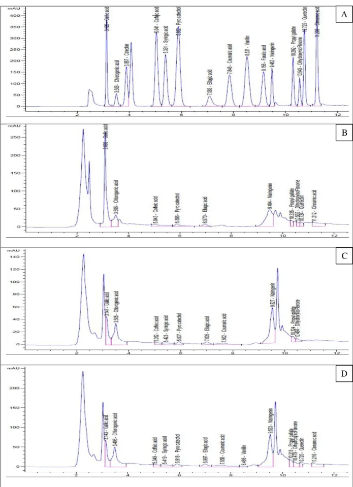
described in the Prophetic medicine and used widely in traditional medicines for many thousand years. This study aimed to identify the chemical profile of the phenolic compounds of the aqueous, ethanolic and methanolic extracts of C. speciosus under different extraction conditions, to determine the phenolic by HPLC, and evaluate, in vitro and in vivo, their antioxidant, anticoagulant and anticancer activities. The protective role of the ethanolic extract against zearalenone (ZN) as an inductor of oxidative stress in the liver and kidney in rats was also established. The obtained results indicated that : The content of total falvonoids and polyphenol in the ethanolic extract was the highest followed by the methanolic then the aqueous extracts and is was increased by increasing or extraction time. The HPLC analysis identified a total of 13 compounds in each extract with different concentrations. Quercetin, caffeic acid, and gallic acid were high in the aqueous extract, while the alcoholic extracts were rich in syringic acid, chlorogenic acid and naringenin. All the tested extracts have an appreciable antioxidant inhibiting effect with some slight variations except for beta-carotene test, reducing power ions, chelating assay where the extracts showed an activity higher than 50% compared to the aqueous extract. The methanolic extract showed a potential anticancer property against HePG2 cancer cell lines followed by the ethanol extract, then the water extract which showed a weak anticancer property. The in vitro and in vivo anticoagulant activities tests showed that the extracts prolonged significantly the activated partial thromboplastin time (aPTT), prothrombine time (TT) and thrombin time (PT) compared to the negative control. The oxidative stress induced by ZN in the rat showed a significant increase in serum ALT, AST and ALP activites ,level of creatinine, uric acid, urea, cholesterol, triglycerides, LDL, carcinoembryonic antigen, alpha-fetoprotein, TNFα, interleukin-6, malondialdehyde and a significant decrease in the content of serum TP, albumin, HDL, hepatic and renal TAC, CAT and GPx. Treatment of the stressed rats with the ethanolic extract resulted in improvement of all biochemical markers by restoring their values to normal. Histologically, the induced stress also caused significant tissue changes in the liver and kidneys. However, most of them were counteracted by ethanolic extract administration and the high dose was more effective than the low dose. In conclusion, the extracts of c. speciosus have a high potential biological activities and preventive and protective properties towards the liver and the kidneys these activities were linked to their high contents of polyphenols and flavonoids.
Keywords: Costus speciusus Phenolic Compounds, HPLC, Antioxidant, Anticoagulant, Anticancer, Zearalenone, Oxidative stress, Hepato-nephroprotectve.
Resumé
Costus speciosus est une plante qui appartient au genre Costus, c’est un aliment
fonctionnel qui a été décrit en médecine prophétique et qui est largement utilisé dans les médicaments traditionnelle depuis plusieurs milliers d'années. Cette étude vise a établir le profil chimique des composés phénoliques des extraits aqueux, éthanoliques et méthanoliques de C.
speciosus sous différents conditions d’extraction, à déterminer les composés phénoliques par
HPLC, en évaluant, in vitro et in vivo, leurs activités antioxydantes, anticoagulantes et anticancéreuse. Le rôle protecteur de l'extrait méthanolique contre le stress oxydatif induit par la zéaralénone (ZN) dans le foie et les reins chez le rat a été aussi établi. Les résultats obtenus indiquent que: La teneur totale en polyphénols et flavonoïdes dans l'extrait éthanolique est plus élevée que dans les extraits méthanoliques et aqueux et augmente avec le temps d'extraction. L'analyse par HPLC a permis d'identifier 13 composés dans chaque extrait à des concentrations différentes. La quercétine, l'acide caféique et l'acide gallique sont les principaux composés de l'extrait aqueux, tandis que les extraits alcooliques sont riches en acide syringique, en acide chlorogénique et en naringénine. Tous les extraits testés ont un effet antioxydant significatif avec de légères variations sauf pour les tests de bêta-carotène, du pouvoir réducteur et chélateur où les extraits alcooliques ont montré une activité supérieure de 50% par rapport à l'extrait aqueux. Les extraits méthanoliques et éthanoliques ont montré une propriété anticancéreuse potentielle contre la ligné cellulaire HePG2, tandis que l'extrait aqueux a présenté une faible activité. Les tests des activités anticoagulantes, in vitro et in vivo, des extraits ont montré que les extraits prolongent de manière significative le temps de thromboplastine partielle activée (aPTT), le temps de prothrombine (TT) et le temps de thrombine (PT) par rapport au témoin négatif. Le stress oxydatif induit par la ZN chez le rat a montré une augmentation significative de l'activité del’ ALT sérique, de l'AST et de l'ALP, de la teneur de la créatinine, de l'acide urique, de l'urée, du cholestérol, des triglycérides, du LDL, de l'antigène carcinoembryonnaire, de l'alpha-foetoprotéine, du TNFα, de l'interleukine-6, du malondialdéhyde et une diminution importante de la teneur en TP sérique, albumine, HDL, TAC hépatique et rénale, CAT et GPx. Le traitement des rats stressés avec l’extrait éthanolique a amélioré tous les marqueurs biochimiques en rétablissant leurs valeurs à la normal. Par ailleurs et sur le plan histologique, le stress induit a entrainé des modifications tissulaires importantes au niveau du foie et des reins. L’extrait éthanolique a exercé un effet préventif et protecteur du foie et des reins contre toute altération à la dose élevée.En conclusion, les extraits de C. speciosus possèdent un fort potentiel d'activités biologiques et des propriétés préventives et protectrices vis-à-vis du foie et des reins qui étaient liées à leurs teneurs élevées en polyphénols et des flavonoïdes.
Keywords: Costus specious, Polyphénols, Analyse HPLC, Antioxydantes, Anticoagulantes, Anticancéreuse, zéaralénone, stress oxydant, hepatonéphroprotecteur.
Lists of tables
Table 01 . Classification of coagulation factors………. 20
Table 02 . The chemicals, reagents and kits………. 32 Table 03 . HPLC analysis of the total polyphenols of the three extracts of C. speciosus.. 60
Table 04 . Total phenolic content (TPC) and total flavonoid content (TFC) the three
extracts of C. speciosus………. 63
Table 05 . Effect of treatment with C. speciosus extract and ZEN on the comet tail
Lists of figures
Figure 01. Costus speciosus………...………..…..………….. 09
Figure 02. Chemical structures of some C. speciosus active ingredients………...…....…. 10
Figure 03. Mitochondrial ROS production………... 12
Figure 04. Extrinsic, intrinsic, and common pathways of blood coagulation during hemostasis and thrombosis………..……...……… 22
Figure 05. Direct mechanisms involved in cancer-associated thrombosis……..…….…... 25
Figure 06. Indirect mechanisms promoting thrombosis in cancer………….…………... 26
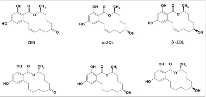
Figure 07. Chemical structures of zearalenone………... 27
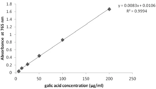
Figure 08. Standard curve of gallic acid used in the determination of total polyphénols in various plant extracts..………...……….. 34
Figure 09. Basic structure of flavonoides.…..………..………….... 35
Figure 10. Standard curve of rutin used in the determination of total flavonoids in various plant extracts………….………...…... 36
Figure 11. Reaction mechanism of 2,2-diphenyl-1-picrylhydrazyl (DPPH) with antioxidant………...………...………….... 38
Figure 12. The redox reaction between copper-neocuproine complex and trolox……….. 39
Figure 13. Malondialdehyde formation………..….………… 57
Figure 14. Percentage of yield of the three extract of C. speciosus after 24 and 36 h………...…. 59
Figure 15. HPLC chromatograms of the total polyphenols of (A) standard, (B) the aqueous,(C) the ethanol 70% and (D) the methanol 70% extracts of C. speciosus…….………..……….…. 61
Figure 16. Total antioxidant capacity of different extracts of C. speciosus………...…….….... 64
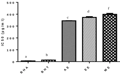
Figure 17. The IC50 values in the DPPH radical scavenging activity assay of the extracts……….………...………...…….… 65
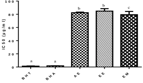
Figure 18. The IC50 values in the ABTS radical cation decolorization activity assay of the extracts……….………...…... 66
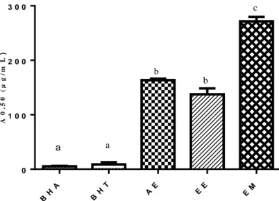
Figure 19. The A0.5 values in the Cupric reducing antioxidant capacity test of the extracts………..….. 67
Figure 20. The IC50 values in the β -carotene bleaching test antioxidant capacity of the extracts……….………...…….... 69
Figure 21. The IC50 values in the ferrous ions chelating test antioxidant capacity of the extracts…….………... 70
.
Figure 22. The A0.5 values in the Phenanthroline assay of the extracts….….………. 71
Figure 23. Relative viability of cells (%) of various extrats and standard ( DOX :
doxocubicin)………..……….……… 72
Figure 24. Cytotoxic activity of some compounds against HePG2 cell
line……….……….. 72
Figure 25. Activated thromboplastin time (aPTT) of various extracts of C speciosus.…………... 74 Figure 26. Prothrombin time (PT) of various extracts of C. speciosus……….….….. 75 Figure 27. Thrombin time (TT) of various extracts of C. speciosus…….……….…….. 76 Figure 28. in vivo Anticoagulant activity of ethanolic extract of C. speciosus evaluated by the
measurement of time of of activated partial thromboplastin time (aPTT), prothrombin
and thrombin time (TT)………..………... 77
Figure 29. Effects of ethanolic extract of C. speciosus on total body weight gain in rats treated
with ZN………….………..…...…..… 80
Figure 30. Effect of ethanolic extract C. speciosus on serum ALT activity in rats treated with
Zeralenone……….………...…...….... 81
Figure 31. Effect of ethanolic extract C. speciosus on serum AST activity in rats treated with
Zeralenone………...……...….… 82
Figure 32. Effect of ethanolic extract C. speciosus on serum ALP activity in rats treated with
ZN………..….… 83
Figure 33. Effects of ethanolic extract C. speciosus on total protein level in rats treated with
ZN………...……..………..… 84
Figure 34. Effects of ethanolic extract C. speciosus on albumin level in rats treated with
ZN………...………...……….… 85
Figure 35. Effects of ethanolic extract C. speciosus on urea level in rats treated with
ZN………...……….……... 86
Figure 36. Effects of ethanolic extract of C speciesus on uric acid level in rats treated with
ZN………..……….… 87
Figure 37. Effects of ethanolic extract of C speciesus on serum creatinine level in rats treated
with ZN………..………... 88
Figure 38. Effect of ethanolic extract of C speciesus on serum cholesterol level in rats treated
Figure 39. Effect of ethanolic extract of C speciosus on serum triglycerides level in rats treated
with ZN………….……….. 90
Figure 40. Effect of ethanolic extract of C speciosus on high-density lipoprotein cholesterol
level (HDL-CHL) in rats treated with ZN………….………..…………... 91
Figure 41. Effect of ethanolic extract of C. speciosus (LD) and (HD) on Low-density
lipoprotein cholesterol level (LDL-CHL) in rats treated with ZN………..…… 92
Figure 42. Effect of ethanolic extract of C speciosus on serum TNF-α level in rats treated with
ZN………...………....… 93
Figure 43. Effect of ethanolic extract of C. speciesus on serum in interleukin 6 (IL-6) level in
rats treated with ZN……….……….………….. 94
Figure 44. Effect of ethanolic extract of C. speciosus on serum in carceno embryonic antigen (CEA) level in rats treated with ZN………..…….. 95 Figure 45. Effect of ethanolic extract of C speciosus on serum in alpha feto protein (AFP) level
in rats treated with ZN………...………...……..…….... 96
Figure 46. Effect of ethanolic extract of C. speciosus on lipid peroxidation in the liver and renal
tissues of rats treated with ZN…...………...……..……….... 98
Figure 47. Effect of ethanolic extract of C. speciosus on hepatic and renal TAC of rats treated
with ZN………..………... 100
Figure 48. Effect of ethanolic extract of C. speciosus on hepatic and renal GPX of rats treated
with ZN………...….... 101
Figure 49. Effect of ethanolic extract of C. speciosus on hepatic and renal CAT activity of rats
treated with ZN……….………. 103
Figure 50. Effect of treatment with C. speciosus extract and ZN on the micronucleus formation
and cytotoxicity rate in rat bone marrow cells……...………..……... 105
Figure 51. Fluorescence photomicrographs of rat bone marrow cells treated with C. speciosus
extract and ZN showing (a) intact cells; (b-f) different patterns of comet tail
formation………...… 106
Figure 52. Photomicrograph of liver section………...… 109 Figure 53. Photomicrographs of liver section of the rats treated with ZN……….... 110 Figure 54. Photomicrographs of liver sections of rats treated with (a) ZN plus CSE (LD) and (b)
ZN plus CSE (HD)………...……….……….. 110
Figure 55. Photomicrographs of kidney section of (a) control rat, (b) rats treated with CSE (LD)
Figure 56. Photomicrographs of kidney sections of rats treated with ZN plus CSE (HD) showing
atrophy of the glomeruli (G), the tubules were fairly preserved (R), cellular regeneration (arrow) and swelling urinary space (V) ………... 112
Figure 57. Photomicrographs of kidney section of rats treated with ZN plus CSE (LD) showing
the improvement of cellular regeneration which is quite
prominent………... 112
Figure 58. Photomicrographs of kidney section of rats treated with ZN plus CSE (LD) showing
the expanded glomeruli with narrow urinary space and swelling in tubular epithelial cells with obliterated in the lumen (arrow)………... 113
List of abbreviations
ABTS 2, 2′-azino-bis (3-ethylbenzthiazoline-6-sulfonic acid)
AFP Alpha-fetoprotein Tumor Marker
ALP Alkaline phosphatase
ALT Alanine Aminotransferase
ANOVA Analysis of variance
AST Aspartate Amino Transferase
APTT Activated partial thromboplastin
BHT Butylatedhydroxytoluene
BHA Butylatedhydroxyanisole
CSE Costus speciosus ethanolic
DF Degrees of freedom
DPPH 2, 2-diphenyl-1-picryl-hydrazyl
EDTA Ethylene diamine tetra acetic acid
ELISA Enzyme-linked immuno-sorbent assay
FBS Fetal bovine serum
GGT Gamma Glutamyltransferase GPX Glutathion Peroxidse GPx Glutathione peroxidase GR Glutathione reductase GSH Glutathione GSH-PX Glutathione Peroxidase GSSG Glutathione disulfide
GAE Gallic acid equivalent
IL6 Interleukin6
HCC Hepatocellular carcinoma
HDL High-density lipoprotein cholesterol
HPLC High Performance Liquid Chromatography
IC50% Inhibitory concentration for 50% of activity
LDL Low-density lipoproteins
LPS Lipopolysaccharide
MDA Malondialdehyde
MN Micronucleus (MN)
MTT 3-(4,5-dimethylthiazol‐2‐yl)‐2,5‐diphenyltetrazolium bromide
MNPCE micronucleated polychromatic erythrocytes
NADPH Nicotinamide adenine dinucleotide phosphate
NO Nitric oxide
NOS Nitric oxide synthase
NCE normochromic erythrocytes
OTM Olive tail moment
PT Prothrombin time
PC Protein C
PCE polychromatic erythrocytes,
PPP Platelet poor plasma
ROS Rective oxygen species
RNS Reactive nitrogen species
TAC Total antioxidant capacity level
TG Triglycerides
TAC Total antioxidant capacity
TMB Thiobarbituric acid
TPP Tripolyphosphate
TPA Plasminogen activator
TNFα Tumor necrosis factor alpha
TM Tail moment
TFPI Tissue factor pathway inhibitor
VLDL Very low-density lipoproteins
WHO World Health Organization
List of contents
Introduction
Review of literature
1. Medicinal plant……….. 03
1.1 Traditional medicine……… 03
1.2. African traditional medicine………... 03
1.3. Drug discovery from medicinal plant………. 04
1.4. Plant secondary metabolite………..………... 04
1.4.1. Terpenes………. 05
1.4.2. Nitrogen containing compounds……… 05
1.4.3. Polyphenols………... 06
2.Costus speciosus...…………..……… 08
2.1.Traditional use of the Root …...……….. 09
2.2. Phytochemistry of Costus speciosus……… 10
3. Oxidative stress………..……...……….... 10
3.1. Forms of ROS and RNS…………..………. 10
3. 2. Sources of ROS and RNS………..……….. 11
3. 3. Molecular targets of free radicals..……….. 14
3.4. Relationship between Oxidative stress and diseases ………... 15
3.5.Defence system against oxidative stress………..….……. 16
4. Hepatocellular carcinoma………...………... 19 5.Blood Coagulation ……….………..……….. 20 5.1. Extrinsic Pathway………..……...… 20 5.2. Intrinsic Pathway………..……….... 21 5.3. Common Pathway………...………...……...… 21 5.4. Fibrinolytic system………...… 22
5.5.Haemostasis screening Assay……….... 23
5.6. Regulation of Coagulation……….... 23
5.7. Blood Coagulation Disorders………....………...… 24
3.8. Mechanisms of Cancer-Associated Thrombosis………..…. 25
6. Zeralenone………..………... 26
6.1. Occurrence of Zearolenone………..………... 26
6.2. pharmacokinetics of zearalenone………..……….. 28
6.3. Adverse effects and toxicity of zearalenone………..………. 28
6.3.1.Endocrine toxicity ………...………... 28
6.3.2. Hematotoxicity……….. 29
6.3.3. Reproductive and developmental toxicity………. 29
6.3.4. Immunotoxicity………...……….. 30
6.3.5. Cytotoxicity………... 30
6.4. Protective effect of plant against zearalenone toxicity ……..……… 30
Material and Methods
1. Material ……… 32 1.1. Chemicals ……….………...……….…. 32 1.2. Plant material………...………... 33 2. Methods………. 33 2.1. Plant extraction………...……… 33 2.2. Phytochemical Characterization………. 332.2.1. Determination of polyphenols of C speciosus extracts by HPLC………. 33
2.2.2. Determination of total phenolic content of C speciosus extracts………...…... 33
2.2.3. Determination of total flavonoid content of C speciosus extracts………..….. 34
2.3. Antioxidant activity of C speciosus extracts………... 35
2.3.1. Total antioxidant capacity of C speciosus extracts……….. 36
2.3.2. Free radical scavenging activity by DPPH assay………. 36
2.3.3. ABTS decolorization assay of C. speciosus extracts ……….. 37
2.3.4. Cuprac assay of C speciosus extracts……….…. 38
2.3.5. β-carotene bleaching assay of C speciosus extracts……… 38
2.3.6. Ferrous ions chelating assay of C speciosus extracts……….. 39
2.3.7. Phenanthroline method……… 39
2.4. Cytotoxicity assay of C speciosus extracts………..…... 40
2.5. Anticoagulant activity of C speciosus extracts………... 40
2.5.1. In vitro evoluation of C speciosus extracts……… 40
2.5. 1.1. Collection of blood and separation of plasma……… 40
2.5.1.2. Prothrombin Time (PT) test……….……… 41
2.5.1.3. Activated partial thromboplastin time (aPPT) test……….. 41
2.5.1.4. Thrombin time (TT)………. 42
2.5.2. In vivo evaluation of C speciosus extracts……… 42
2.5.2.1. Animals……… 42
2.5.2.2. Experimental design ……… 43
2.5.2.3. Anticoagulant assay ……… 43
2.5.2.4. Anticoagulant assay………. 43
2.6.Evaluation of the protective role of ethanolic extract against Zearalenone-induced hepato-nephro toxicity ……….. 43
2.6.1. Experimental design……….. 43
2.6.2. Sample collection………...…... 44
2.6.3. Determination of serum biochemical parameters………. 44
2.6.3.1. Determination of serum ALT……….………….…………. 45
2.6.3.2. Determination of serum AST………..………. 45
2.6.3.3. Determination of serum ALP………..………..……….. 46
2.6.4. Total Protein………...……... 46
2.6.5.Albumin………...……... 47
2.6.6. Determination of Creatinine levels……….………... 47
2.6.7. Determination of Urea……….………….. 47
2.6.8. Determination of uric acid……….……… 48
2.6.9. Evaluation of serum lipid profile………..………. 49
2.6.9.1. Determination of serum CHL………..………. 49 2.6.9.2.Determination of TG………. 49 2.6.9.3. Determination of HDL……….… 50 2.6.9.4. Calculation of LDL………..… 51 2.6.10. TNF-α level………. 51 2.6.11. Determination of IL-6……….. 51 2.6.12. CEA test………...……… 52 2.6.13. AFP level………. 53
2.6.14. Determination of antioxidants and oxidative stress parameters in liver and kidney homogenate ……….. 53
2.6.14.1. Lipid peroxidation……….. 53
2.6.14.2. Determination of TAC………... 54
2.6.14.4.Determination of CAT enzyme activity ……… 56
2.7.Histological examination of liver and kidney………. 56
2.8. Genotoxicity test……….……….. 57
2.8.1. Micronucleus (MN) assay………..………. 57
2.8.2. Single cell gel electrophoresis (comet) assay……….………. 57
2.8. Statistical analysis ………..………...…... 58
Resultats and Discusion
1. Extraction………..………...………. 592. HPLC analysis of the extracts……….………….. 59
3. Total phenolic content and Total flavonoid content …..………. 63
4. In vitro evaluation of antioxidant activity………. 64
4.1. Total antioxidant capacity assay……….……….... 64
4.2. Free radical scavenging assay by DPPH……….……… 65
4.3. ABTS+. radical cation decolorization assay………..………. 67
4.4. CUPRAC assay ………..……… 68
4.5. β-carotene bleaching assay……….……… 69
4.6. Ferrous ions chelating assay………..………. 70
4.7. Phenanthroline method……….….. 71
5. Cytotoxicity assay against HePG-2 cell lines……… 72
6. Anticoagulant activity……… ………….. 74
6.1. Activated partial thromboplastin time (aPPT) test………..…… 74
6.2.Prothrombin Time (PT) test………..….. 75
6.3. Thrombin time (TT)………..…. 75
6.4. In vivo Anticoagulant activity……… 76
7. Effect of ethanolic extract on the hepatotoxicity and nephrotoxicity induced by Zeralenone………..…. 80
7.1. Body Weight evolution………... 80
7.2. Serum biochemical parameters………..………. 81
7.2.1. Serum alanine aminotransferase (ALT)……… 81
7.2.2. Serum aspartate aminotransferase (AST)……….…………. 81
7. 2.3. Alkaline Phosphatase (ALP)……… 82
7.2.4. Serum protein profile……….……… 84
7.2.5. Serum albumin level……….………. 84
7.2.6. Serum Urea………..………….. 86
7.2.7. Serum uric acid……….………. 86
7.2.8. Serum creatinine………..………….. 87
7.3. Serum lipid profile……….. 88
7.3.1. Serum cholesterol (CHL)……….………….. 88
7.3.2.Triglycerides (TG)………..……… 89
7.3.3.High-density lipoprotein-cholesterol (HDL-CHL)……….……… 90
7.3.4. Low-density lipoprotein cholesterol (LDL-CHL)……….……… 91
7.4. Serum cytokines……….. 93
7.4.1. Tumor necrosis factor alfa (TNF-α) level in serum………..………. 93
7.4.2. Interleukin 6 (IL-6)………..………….. 93
7.4.3. The carceno embryonic antigen (CEA)………..…………... 94
7.4.4.Alpha fetoprotein level (AFP) as a tumor marker………..… 95
7.5. Tissue biochemical assay………...…. 97
7.5.2. Total antioxidant capacity (TAC)………...…….. 99
7.6. Antioxidant enzymes………..……… 100
7.6.1. Glutathione peroxidase activity (GPX)………. 100
7.6.2. Catalase Activity (CAT)………...………. 104
7.7. Genotoxicity test………..……... 104
7.7.1. Micronucleus findings………..………. 105
7.7.2. Comet assay findings………. 105
7.8.Histological studies ………. 108
Conclusion
1
Introduction
The human body has a complex system of natural enzymatic and non-enzymatic antioxidant defenses which counteract the harmful effects of free radicals and other oxidants. Free radicals are responsible for causing a large number of diseases including cancer (Kinnula and Crapo, 2004), cardiovascular disease (Singh and Jialal, 2006), neural disorders (Sas et al., 2007), Alzheimer’s disease (Smith et al., 2000), mild cognitive impairment (Guidi et al., 2006), Parkinson’s disease (Bolton et al., 2000), alcohol induced liver disease (Arteel, 2003), ulcerative colitis (Ramakrishna et al., 1997), aging (Hyun et al., 2006) and atherosclerosis (Upston et al., 2003). Bioactive compounds, such as polyphenols and the secondary metabolites flavonoids and proanthocyanidins, derived from plants have been associated with various health benefits (Vuong et al., 2014; Dailey and Vuong, 2015).
Phenolic compounds are commonly found in both edible and nonedible plants, and they have been reported to have multiple biological effects, including antioxidant activity (Alothman et al., 2009 ; Bonoli et al., 2004) . Crude extracts of fruits, herbs, vegetables, cereals, and other plant materials rich in phenolics are increasingly of interest in the food industry because they retard oxidative degradation of lipids and thereby improve the quality and nutritional value of food (Pérez et al.,2008). The importance of the antioxidant constituents of plant materials in the maintenance of health and protection from coronary heart disease and cancer is also raising interest among scientists, food manufacturers, and consumers as the trend of the future is moving toward functional food with specific health effects (Loliger, 1991). Therefore, many attempts have been made to extract and isolate bioactive compounds from plant materials for utilization in the food and pharmaceutical industries. As bioactive compounds range from very polar to very non-polar compounds, the extraction solvent plays an important role in extraction efficiency of bioactive compounds from the plant materials (Pinelo et al., 2005; Ye et al., 2015).
In Arabic civilization or tradition, prophetic medicine corroborated by clinical and epidemiological researches and evidences constitute a platform for mankind to cure their ailments (Sheikh et al., 2017; Hussain and Hussain, 2016). In this context, Costus plant species which include C speciosus is one the functional food cited in Prophetic medicine used widely in traditional medicines for many thousand years (Choudhary et al., 2015). Costus speciosus (family Costaceae) is an herbaceous plant widely growing in South East Asian countries such as India, Malaysia, Srilanka and Indonesia (Anonymous, 2007; Rani, 2012). The rhizome is used in the traditional system to treat bronchitis, fevers, dyspepsia,
2
inflammations, anaemia, rheumatism, lumbago and hiccough (El-Far et al., 2016 ; El-Far et al 2018). The natives of North-east India use the rhizome in urinary troubles, fever, headaches and also to dissolve kidney stones(Kumar, 2012; Jha et al., 2010)
The aims of this study are:
Studying the effects of different solvents and extractions time on the yield and of biological compounds of C speciosus.
Determination of the bioactive compounds in the extracts using HPLC.
Determination of totalpolyphénols and flavonoides contents in C speciosus extracts.
Evaluation of the antioxidant activity of C speciosus extracts using different assay (DPPH, ABTS, chelation of iron, Cuprac, β-carotene and Phenanthroline).
Evaluation in vitro of the anticancer activity of the extracts of C speciosus.
Evaluation in vitro and in vivo of the antithrombotic activity of C speciosus extracts. Evaluation of the therapeutic potential protective effects of C speciosus ethanolic
extract against zeralenone-induced oxidative stress, genotoxicity and histological changes in the liver and kidney of rats.
3 1. Medicinal plant
Throughout the ages, humans have relied on nature for their basic needs, for the production of food, shelter, clothing, transportation, fertilizers, flavours and fragrances, and medicines (Cragg and Newman, 2005). Plants have formed the basis of sophisticated traditional medicine systems that have been in existence for thousands of years and continue to provide mankind with new remedies. Although some of the therapeutic properties attributed to plants have proven to be erroneous, medicinal plant therapy is based on the empirical findings of hundreds and probably thousands of years of use. The first records, written on clay tablets in cuneiform, are from Mesopotamia and date from about 2 600 BC (Heinrich and Teoh, 2004). Among the substances that were used are oils of Cedrus species (cedar) and Cupressus sempervirens (cypress), Glycyrrhiza glabra (licorice), Commiphora species (myrrh) and Papaver somniferum (poppy juice), all of which are still in use today for the treatment of ailments ranging from coughs and colds to parasitic infections and inflammation. (Beissert and Schwarz, 2002)
1.1 Traditional medicine
Plants have been utilized as medicines for thousands of years (Samuelsson, 2004). These medicines initially took the form of crude drugs such as tinctures, teas, poultices, powders, and other herbal formulations (Samuelsson, 2004). The specific plants to be used and the methods of application for particular ailments were passed down through oral tradition. Eventually information regarding medicinal plants was recorded in herbal phamacopoeias (Balunas, 2005)
1.2. African traditional medicine
African traditional medicine in its varied forms is holistic, involving both the body and the mind. The healer typically diagnoses and treats the psychological basis of an illness before prescribing medicines to treat the symptoms. Well known African medicinal plants include Acacia senegal (gum arabic), Agathosma betulina (buchu), Aloe ferox (Cape aloes), Aloe vera (north African origin), Artemisia afra (African wormwood), Aspalanthus linearis (rooibos tea), Boswellia sacra (frankincense), Catha edulis (khat), Commiphora myrrha (myrrh), Harpagophytum procumbens (devil’s claw), Hibiscus sabdariffa (hibiscus, roselle), Hypoxis hemerocallidea (African potato), Prunus africana (African cherry) (Newman et al., 2000).
4
Madagascar has contributed Catharanthus roseus (rosy periwinkle) and has the potential of contributing more in view of the diversity of the flora and fauna (2000; Neuwinger, 2000).
1.3. Drug discovery from medicinal plant
Numerous methods used to acquire compounds for drug discovery include: isolation from plants and other natural sources synthetic chemistry combinatorial chemistry, and molecular modeling (Ley and Baxendale, 2002; Geysen et al., 2003; Lombardino and Lowe, 2004). Despite the recent interest in molecular modelling, combinatorial chemistry, and other synthetic chemistry techniques by pharmaceutical companies and funding organizations, the natural products, and particularly that of medicinal plants, remain an important source of new drugs, drug leads and chemical entities (Newman et al., 2003; Butler, 2004).
1.4. plant secondary metabolite
In plants, as a result of metabolic processes, many different kinds and types of organic compounds or metabolites are produced. These metabolites are grouped into primary and secondary metabolites. The primary metabolites like chlorophyll, amino acids, nucleotides, simple carbohydrates or membrane lipids, play recognised roles in photosynthesis, respiration, solute transport, translocation, nutrient assimilation and differentiation. The secondary metabolites also differ from primary metabolites in having a restricted distribution in the plant kingdom. That is, particular secondary metabolites are often found in only one plant species or a taxonomically related group of species, whereas the basic primary metabolites are found throughout the plant kingdom (Taiz and Zeiger, 2006). During the past few decades, experimental and circumstantial evidence has made it clear that many secondary metabolites do indeed have functions that are vital for the fitness of a plant producing them. The main roles are:
Defence against herbivores (insects, vertebrates)
Defence against fungi and bacteria
Defence against viruses
Defence against other plants competing for light, water and nutrients
Signal compounds to attract pollinating and seed dispersing animals
Signals for communication between plants and symbiotic micro-organisms
5
They have also provided an invaluable resource that has been used to find new drug molecules (Gurib-Fakim, 2006). Plant secondary metabolites can be grouped into three chemically distinct classes: terpenes, nitrogen containing and phenolics compounds.
1.4.1. Terpenes
Terpenes belong to the biggest class of secondary metabolites and basically consist of five carbon isoprene units which are assembled to each other (many isoprene units) by thousands of ways. Terpenes are simple hydrocarbons, while terpenoids are modified class of terpenes with different functional groups and oxidized methyl group moved or removed at various positions (Gonzalez-Burgos and Gómez-Serranillos, 2012). Terpenoids are divided into monoterpenes, sesquiterpenes, diterpenes, sesterpenes, and triterpenes depending on its carbon units. Most of the terpenoids with the variation in their structures are biologically active and are used worldwide for the treatment of many diseases. Many terpenoids inhibited different human cancer cells and are used as anticancer drugs such as Taxol and its derivatives. Many flavorings and nice fragrances are consisting on terpenes because of its nice aroma. Terpenes and its derivatives are used as antimalarial drugs such as artemisinin and related compounds. Meanwhile, terpenoids play a diverse role in the field of foods, drugs, cosmetics, hormones and vitamins (De Santana Souza et al.,2014; Maione et al ., 2013).
1.4.2. Nitrogen containing compounds
A large variety of plant secondary metabolites have nitrogen in their structures. Included in this category are such well-known antiherbivore compounds such as alkaloids and cyanogenic glycosides, which are of considerable interest because of their toxicity to humans and their medicinal properties. Most nitrogenous secondary metabolites are biosynthesised from common amino acids (Taiz and Zeiger, 2006)
Alkaloids
The term ‘alkaloid’ has been defined as a cyclic organic compound containing nitrogen in a negative oxidation state, which has limited distribution in living organisms (Taiz and Zeiger, 2006). Based on their structures, alkaloids are divided into several subgroups: non-heterocyclic alkaloids and non-heterocyclic alkaloids, which are again divided into 12 major groups according to their basic ring structure. Mescaline is an example of a non-heterocyclic
6
or pseudo-alkaloid, tetrandrine is an example of a bisbenzylisoquinoline alkaloid while solasodine is a triterpene alkaloid (Gurib-Fakim, 2006)
Cyanogenic glycosides
Perhaps the most obvious defence-related secondary metabolites are the cyanogenic glucosides (Vierheilig et al., 2000). They are not in themselves toxic but are readily broken down to give off volatile poisons when the plant is crushed. Cyanogenic glycosides release the well-known respiratory poisonous gas, hydrogen cyanide (Taiz and Zeiger, 2006)
1.4.3. Polyphenols
Polyphenols are natural compounds synthesized exclusively by plants, with chemical features related to phenolic substances and strong antioxidant properties. These molecules or classes of substances are mainly present in fruits, vegetables, green tea, and whole grains (Singla et al., 2019). Phenolic compounds are one of the most numerous and widely distributed group of aromatic compounds in the plant kingdom, with over 8000 phenolic structures currently known, of which more than 6000 are the flavonoids (Garcia-Salas et al., 2010; Tsao et al., 2010; Vladimir-Kneţević et al., 2012). From the chemical point of view, polyphenols are natural compounds with aromatic structures containing one or more aromatic rings with or without the vicinity of a heterocycle and which are grafted with hydroxyl, carboxyl, methoxyl and carbonyl functional groups.
Classes of polyphenols
There are three main classes of polyphenols: phenolic acids, flavonoids, and other phenolics.
Phenolic acids
Phenolic acids are a subclass of the larger phenolics category, occurring in food plants as esters or glycosides conjugated with other natural compounds such as flavonoids, alcohols, hydroxyfatty acids, sterols, and glucosides. Structurally, phenolic acids are phenols that possess one carboxylic acid moiety that can be directly attached either to the aromatic ring (benzoic acid derivatives) or attached to an alkyl residue (hydroxycinnamic acid derivatives). Although the basic skeleton remains the same, phenolic acids differ in the number and
7
position of the hydroxyl groups on the aromatic ring (Losada-Barreiro and Bravo-Díaz, 2017). The majority of phenolic acids are linked through ester, ether, or acetal bonds either to structural components of the plant (cellulose, proteins, lignin) or to larger polyphenols (flavonoids), or smaller organic molecules (glucose, quinic, maleic, or tartaric acids) or other natural products such as terpenes (Goleniowski et al., 2013).
Flavonoids
Flavonoids are the most studied class of polyphenols. Although they are low molecular weight secondary metabolites, their chemical diversity, size, three-dimensional shape, and physical and biochemical properties allow them to interact with multiple targets to influence biological activity in plants, animals, humans and microbes. Consequently, many therapeutic properties have been assigned to flavonoids (Francisco et al., 2014).
Flavonoids comprise the most abundant class of plant polyphenols with more than 9000 of identified chemical structures (Wang et al., 2018). They share a carbon skeleton of diphenyl propanes, two benzene rings (A and B) joined by a linear three carbon chain. This central chain usually forms a closed pyran ring (C) with one of the benzene rings. According to the degree of oxidation of the C ring, the hydroxylation pattern of the nucleus, and the substituent at carbon 3, the flavonoids can thus be classified into different subclasses: flavones, isoflavones, flavanols (catechins), flavonols, flavanones, anthocyanins and proanthocyanidins (Losada-Barreiro and Bravo-Díaz, 2017).
Other phenolics
There are at least two major classes of tannins: (i): hydrolyzable and nonhydrolyzable (also known as condensed) tannins and (ii): proanthocyanidins and procyanidins. Structurally, hydrolyzable and nonhydrolyzable tannins are richly hydroxlyated oligomers or polymers of hydroxybenzoic acids such as gallic acid or flavan-3-ols such as catechin, respectively. High-molecular-weight condensed tannins may contain 50 or more flavan-3-ols subunits attached by carbon-carbon bonds (Selma et al., 2009). They are highly astringent and noticeable in unripe fruits.
Stilbenes are well-known class of naturally occurring phytochemicals. They bear classical C6-C2-C6 structures with two hydroxyl groups on the A ring and one on the B ring. Stilbenes are characterized by a double bond connecting the phenolic rings. These compounds
8
are stress metabolites produced in response to fungal infection. Though known as plant defense compounds, stilbens have an enormous diversity of beneficial human health effects. One of the most relevant and extensively studied stilbene is resveratrol found largely in grapes (Martinez et al., 2014; Losada-Barreiro and Bravo-Díaz, 2017).
Lignans are found in all plants and show enormous structural diversity, with their molecular backbone consisting of two phenylpropane (C3-C6) units. The most lignans that are of special interest owing to their many powerful health benefits are tetrahydrofurofuran and sesamin (Martinez et al., 2014).
2. Costus speciosus
Costus speciosus is a succulent, vertical, everlasting, herbaceous, ornamental, tuberous stem, sub-woody at the base, stout crawling rhizomes growing up to 2.0 - 2.7 m tallness with long lanceolate leaves and essential white flowers (Dubey et al., 2010; Karthikeyan et al., 2012; Najma et al., 2012). The herb blossom by July and August, whereas the aerial parts lose during the cold weather (Rajesh et al., 2009; Nehete et al., 2010). Flowers of C. speciosus resemble crepe paper. C. speciosus occurs in the moist and wet evergreen areas of the Indo-Malayan region and Sri Lanka (EL-far et al., 2013).The herb has a red color fruit, whereas the seeds are black (Fig. 01) (Pawar and Pawar. 2014). Its classification is as follows:
Kingdon: Plantae Subkingdom: Tracheobinota Super Division: Spermatophyta
Division: Mangoliophyta Class: Liliopsida Sub Class: Zingiberidae
Order: Zingiberales Family: Costaceae
Genus: Costus
9 Figure 01. Costus speciosus
2.1.Traditional use of the Root
Ibn AI-Qayim stated in his book Al-Teb Al- Nabawi that costus contains different therapeutic advantages. It expels the phlegm and checks its excessive production and effective in common cold. It is good tonic for stomach and liver, increases the cutaneous circulation and removes the stains on the face if applied with honey. It is a good remedy for freckles if applied on face with vinegar and effectively used in cases of tetanus and intestinal parasites. Imam Zahbi said that it is useful in paralysis, an antidote for snake poison and its oil is effective in back pain. According to the authentic books of Al-Hadith, it is described
accompanied with the description of cupping. Anas Bin Malik narrated that the
Prophet stated “Out of those things which are being used by you for treatment,
the cupping and Qust Bahri are the best treatment”. (Sahih Bukhari 5371). This description might be interpreted to mean that if somebody fails to use to cupping, he may use the Saussurea lappa instead of cupping (Khan et al., 2001).
Hadith - Mustadrak-al-Hakim, Narrated Jabir Bin Abdullah narrates that
Prophet stated " If someone's child gets Azra (upper respiratory tract infection)
or headache then she should take Qust and after grinding it in water, apply it to the child" (Mustadrak-Al-Hakim). Azra is an ancient term, might be interpreted as tonsillitis, in which tonsils become swollen and painful with or without pus formation. Hence, this drug is especially effective against any type of tonsillitis and might be used in all types of phlegmatic diseases. It is effective in general weakness after diarrhea and cholera. The water extract of the root was used to wash the females’ internal organs after the menstruation.
10
2.2. Phytochemistry of Costus speciosus
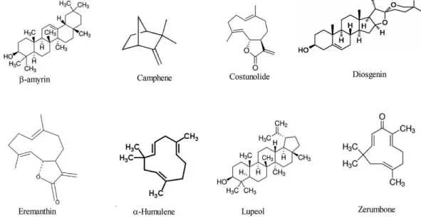
Phytochemical screening of C. speciosus detected the presence of alkaloids, glycosides, steroids, phenolic, (Rates, 2001; Singh et al., 2014 ) flavonoids, polyphenols, tannins, and β-carotene ( Kotebagilu et al., 2014) Diosgenin, β-sitosterol, furostanol saponins-costusosides, β-D-glucoside, prosapogenins, dioscin, gracillin, dihydrophytylplastoquinone, and α-tocopherolquinone were isolated from C. speciosus and have a wide variety of biological activities, (Duraipandiyan et al ., 2012; Lijuan et al ., 2011). Moreover, β-amyrin, camphene, costunolide, diosgenin, α-Humulene, lupeol, and zerumbone for anticancer activity were recognized (Santos et al., 2012; Zhang et al., 2012). The chemical structures of some actives principals isolated from different parts of C. speciosus are presented in figure (02).
Figure 02. Chemical structures of some C. speciosus active ingredients (El-Far et al., 2018)
3. Oxidative stress
Oxidative stress resulted from an imbalance between the formation of Reactive oxygen species (ROS)/ Reactive nitrogen species (RNS) and the impaired ability of an organism to detoxify these reactive intermediates or to repair the damage that they cause (Poprac et al., 2017).
3.1. Forms of ROS and RNS
All living aerobic multicellular organisms require molecular oxygen (O2) to survive
11
Reactive oxygen species (ROS) are small molecules derived from oxygen molecules including free oxygen radicals, such as superoxide (O2.-), hydroxyl (.OH), peroxyl (RO2⋅), and
alkoxyl (RO-) as well as hypochlorous acid (HOCl), ozone (O
3), singlet oxygen (1O2), and
hydrogen peroxide (H2O2), which are non-radicals. These non-radicals are either oxidizing
agents or easily converted into radicals. Nitrogen-containing oxidants, such as nitric oxide (NO.) peroxynitrite (ONOO.), nitrogen dioxide (NO2) are called reactive nitrogen species
(RNS) (Bedard and Krause. 2007; Pisoschi and Pop, 2015 ).
3. 2. Sources of ROS and RNS
Both endogenous and exogenous sources contribute to intracellular ROS/RNS levels.
Endogenous sources
Mitochondria
Most of the intracellular ROS are derived from mitochondria. The superoxide radicals are produced at two major sites in the electron transport chain, namely complex I (NADH dehydrogenase) and complex III (ubiquinone cytochrome c reductase). The transfer of electrons from complex I or II to coenzyme Q or ubiquinone (Q) results in the formation of
reduced form of coenzyme Q (QH2). The reduced form QH2 regenerates coenzyme Q via an
unstable intermediate semiquinone anion (Q-)in the Q-cycle. The formed ˙Q- immediately transfers electrons to molecular oxygen leading to the formation of superoxide radical. The generation of superoxide is non-enzymatic and therefore higher the metabolic rate, the greater is the production of the ROS (Finkel and Holbrook, 2000) as shown in figure (03).
The superoxide anion is converted to hydrogen peroxide by the action of mitochondrial superoxide dismutase (MnSOD). However, H2O2 can be detoxified by the Catalase (CAT)
and glutathione peroxidase (GPx). The other mitochondrial components which contribute to the formation of ROS include monoamino oxidase, aketoglutarae dehydrogenase, glycerol phosphate dehydrogenase and p66shc (Starkov et al., 2008).
12 Figure 03 . Mitochondrial ROS production (Alugoju et al., 2015)
Peroxisomes
In peroxisomes the respiratory pathway involves the transfer of electrons from various metabolites to the oxygen leads to H2O2 formation, but is not coupled to oxidative
phosphorylation to produce ATP instead free energy is released in the form of heat. The other free radicals produced in peroxisomes include H2O2, O2•- OH• and NO•. The -oxidation of
fatty acids is the major metabolic process producing H2O2 in the peroxisomes. Additionally,
the different peroxisomal enzymes such as acyl CoA oxidases, D-amino acid oxidase, L-α-hydroxy oxidase, urate oxidase, xanthine oxidase, D-aspartate oxidase have been shown to produce different ROS (Fahimi and Schrader, 2006).
Endoplasmic Reticulum
The enzymes of endoplasmic reticulum such as cytochrome p-450, b5 enzymes and diamine oxidase contribute to the formation of ROS (Gross et al., 2006). Another important thiol oxidase enzyme, Erop1p catalyses the transfer of electrons from dithiols to molecular oxygen results in the formation of H2O2 (Droge, 2002).
Exogenous sources
There are multiple external triggers that induce oxidative stress and have direct or indirect effects on responses. Air pollutants, tobacco smoke, ionizing and nonionizing radiations, foods and drugs, as well as xenobiotics can all contribute to oxidative stress. Chemical agents like quinones (Bolton et al., 2000), heavy metals such as lead, arsenic,
13
mercury, chromium, and cadmium; organic solvents; and pesticides are common exogenous sources of ROS (Yildirim et al., 2000).
Radiation and chemotherapy
Ionizing radiation, such as x-rays, neutrons, as well as α, , and γ rays, can all cause oxidative stress. α Particles have weak penetrative power, but the rest are very penetrating through the human body. Ionizing radiation can produce HO· by radiolysis of water or ROS via secondary reactions (Roessner et al., 2008).
Cancer chemotherapy is often accompanied by toxic side effects, and ROS generation by chemotherapeutic agents is the primary event leading to induced toxicity. This is evident by increased lipid peroxidation, and reduced antioxidant and tissue GSH levels during chemotherapy. Both radiation and chemotherapy induce systemic oxidative stress and reduce levels of vitamin E and beta-carotene in patients (Conklin, 2004).
Cigarette smoke
Cigarette smoke is another significant generator of ROS. It is comprised of more than 7,000 chemical compounds and oxidative agents, and tobacco smoke contains 1014-1016 free radicals per puff. The active chemicals include aldehydes, quinones, benzo(a)pyrene, epoxides, and peroxides . Cigarette smoke has a gas phase which contains ·NO, peroxyl
radicals, and carbon-centred radicals as well as a tar phase containing relatively stable polycyclic aromatic hydrocarbons and nitrosamines. In the presence of iron, tar semiquinone can generate HO· and H2O2 (Witschi.2005; Yang et al., 2008).
Foods and alcohol.
Ingested food can generate O2.- and H2O2 in the gastrointestinal tract. Humans ingest
macronutrients (carbohydrates, proteins, and fats), micronutrients (minerals and vitamins), food preservatives, as well as microorganisms. Dietary iron and also copper generate ROS by the Fenton reaction. Increased intake of Fe2+ generates ROS and RNS, lipid peroxidation, and oxidative stress, and its accumulation in tissues increases the risk of cancer and inflammation. Trans fatty acids in processed foods also generate ROS. Lipids from vegetable and animal origin, when heated in microwave ovens, generate free radicals (Fraga et al., 2010 ; Zapolska-Downar et al., 2005).
14
Drugs and xenobiotics
Many drugs and xenobiotics contribute to the formation of free radicals in the body. Anticancer drugs such as anthracyclines and analogs, mitoxantrone and other quinones, actinomycin D, enediynes such as bleomycin, chartreusins, elasmin A and related compounds can cause oxidative stress (Deavall et al., 2012).
3.3. Molecular targets of free radicals
Since these free radicals are highly reactive, they can damage all the three important classes of biological molecules including nucleic acids, proteins, and lipids (Droge, 2002).
Deoxyribonucleic Acid (DNA)
Both ROS/RNS can oxidatively damage the nucleic acids. The mitochondrial DNA ismore vulnerable to the ROS attack than the nuclear DNA, because it is located in close proximity to the ROS generated place. ROS, most importantly, the OH• radical directly reacts with all components of DNA such as purine and pyrimidine bases, deoxyribose sugar backbone (Dizdaroglu et al., 2002) and causes a number of alternations including single and double stranded breaks in DNA. The OH• radical abstracts hydrogen atoms to produce a number of modified puine as well as pyrimidine base by-products and DNA- protein cross links.
The RNS, most importantly, OONO - interacts with guanine to produce nitrative and oxidative DNA lesions such as 8-nitroguanine and 8-oxodeoxyguanosine respectively (Hiraku et al., 2010). 8-nitroguanine formed is unstable and can be spontaneously removed, resulting in the formation of an apurininc site .Conversely adenine can be paired with 8-nitroguanine during DNA synthesis resulting in a G-T transversions (Hofer et al., 2005). Accordingly, 8-nitroguanine is a mutagenic DNA lesion involved in carcinogenesis.
Ribonucleic acid (RNA)
ROS can attack different RNAs produced in the body. The RNA is more prone to oxidative damage than DNA, due to its single stranded nature, lack of an active repair mechanism for oxidized RNA, less protection by proteins than DNA and moreover these cytoplasmic RNAs are located in close proximity to the mitochondria where loads of ROS are produced. Indeed, RNA is subjected to more oxidative damage than DNA in humans (Hofer