Université de Montréal
Caractérisation morphologique, biochimique et physiologique des
protéines de jonction lacunaire, les connexines 46 et 50, dans les
cellules folliculo-stellaires TtT/GF de l’hypophyse antérieure
Par: Christopher Juan Garcia
Département de pathologie et biologie cellulaire, Faculté de médicine
Mémoire présenté à la Faculté de médicine en vue de l’obtention du grade de maîtrise ès sciences (MSc) en pathologie et biologie cellulaire, option biologie cellulaire.
Avril 2014
Université de Montréal
Morphological, biochemical and physiological characterization of
the gap junction proteins, connexins 46 and 50, in the
folliculo-stellate TtT/GF cells of the anterior pituitary gland
By: Christopher Juan Garcia
Department of Pathology and Cell Biology Faculty of Medicine
Thesis presented to the Faculty of Medicine as a requirement for the granting of the degree of Master of Science (MSc) in Pathology and Cell Biology, option: Cellular Biology
April 2014
Université de Montréal
Faculty of Medicine
Department of Pathology and Cell Biology
Thesis evaluation committee:
Professor Lucien Ghitescu – Committee president Professor Victor Gavino – Committee member Professor María Leiza Vitale – Research supervisor Professor R.-Marc Pelletier – Research co-supervisor
Résumé
Les cellules folliculo-stéllaires (FS) de l'hypophyse antérieure possèdent une forme étoilée et étendent de longues projections cytoplasmiques qui forment des pseudo-follicules entourant les cellules endocrines. Les cellules FS sont connectées entre elles par des jonctions lacunaires (des fois aussi connu sous le nom de jonction communicante) formant ainsi un réseau tridimensionnel continu. Un des rôles principaux des cellules FS est le maintien du microenvironnement de l'hypophyse antérieure, une activité qui est en partie réalisée par la sécrétion de divers facteurs de croissance et de cytokines. Ces messagers chimiques, y compris le bFGF, le VEGF, l’IL-6 et l’IL-1 contrôlent de nombreux processus cellulaires tels que l’expression des gènes d’hormones. Notre intérêt est de déterminer si la communication entre les cellules FS contribue à leur activité régulatrice. Dans notre étude, nous avons utilisé la lignée cellulaire TtT/GF qui partage de nombreuses caractéristiques morphologiques, physiologiques et biochimiques avec les cellules FS.
Les jonctions lacunaires/communicantes sont formées par l’association de deux connexons de cellules adjacentes qui unissent le cytoplasme des cellules connectées et permet la diffusion de petites molécules. Chaque connexon est formé par l’oligomérisation de six protéines connexine (Cx) de la famille , . Les connexons, intégrés dans la membrane d’une vésicule du cytoplasme, se migrent vers la membrane cellulaire où ils s’incorporent dans la couche bilipidique.
L’expression de la Cx43 ( ) par les cellules FS est régulée en réponse à des facteurs de croissance et des cytokines. Des changements dans le microenvironnement de l'hypophyse
antérieure causés par des molécules de signalisation sont susceptibles de modifier la Cx43, en particulier l’état de phosphorylation de la protéine. Ces modifications de la Cx43 peuvent ensuite déclencher des changements du comportement de jonctions lacunaires/communicantes formées par la Cx43, comme leur perméabilité et le renouvellement de la protéine Cx43.
Les tissus expriment généralement plus d’un type de connexine. Jusqu’aujourd’hui, la Cx43 est la seule connexine à avoir été identifiée dans les cellules FS. Le cristallin exprime les connexines : Cx43, Cx46 et Cx50. Leur expression est modulée par des facteurs de croissance. Notre hypothèse de travail a été de vérifier si la Cx46 et la Cx50 étaient exprimées par les cellules FS et si celles-ci contribuaient au rôle modulateur des cellules FS hypophysaires.
Dans cette étude, nous avons identifié et caractérisé la Cx46 et la Cx50 dans la lignée cellulaire TtT/GF. Nous avons identifié les produits de transcription de Cx46 et de Cx50 par la technique d’analyse northern blot (PCR). Par la suite, les protéines Cx46 et Cx50 ont été identifiées en utilisant des anticorps dans des analyses western blot. Par microscopie confocale, nous avons déterminé la co-localisation de la Cx46 avec certaines marqueurs d’organites : réseau trans-Golgien, endosomes précoces et lysosomes. La Cx50 co-localise avec des marqueurs du réticulum endoplasmique, du réseau cis-Golgien et des endosomes précoces. Un protocole d’isolation des membranes résistantes aux détergents non-ionique a révélé que la Cx46 et la Cx50 n’étaient pas associées à des radeaux lipidiques ni aux cavéoles. Cependant, la microscopie confocale a montré une co-localisation cytoplasmique de la Cx50 et de la flotilline-1.
Nous avons poursuivi l’étude sur la localisation de la Cx46 dans le noyau en utilisant une technique d’isolation des fractions enrichies en noyau. Nous avons établi que plusieurs isoformes de la Cx46 sont exclusivement associées au noyau. De plus, avec la microscopie confocale nous avons démontrée une co-localisation de la Cx46 avec un marqueur du nucléole/corps de Cajal.
Nous avons démontré un effet du bFGF sur l'expression temporelle de la Cx46 et de la Cx50. L’expression de la Cx46 diminue au cours de longues expositions au bFGF tandis que les niveaux de Cx50 augmentent de façon transitoire au cours du traitement. Dans une autre étude nous avons démontré des changements importants dans les niveaux de la Cx46 et de la Cx50 dans l’hypophyse antérieure des visons durant le cycle de reproduction annuel.
Notre étude démontre que les cellules FS expriment la Cx46 et la Cx50. Nous avons aussi établi que la Cx46 et la Cx50 sont localisées dans différentes structures sous-cellulaires, ce qui suggère des rôles différents dans les cellules FS pour ces protéines de jonction lacunaire/communicante. Il est possible que la Cx46 et la Cx50 ne jouent pas un rôle majeur dans la communication intercellulaire dans les cellules FS quiescentes. Nos résultats suggèrent que la Cx46 et la Cx50 peuvent avoir d'autres fonctions : des isoformes de la Cx46 peuvent contribuer à la biogenèse des ribosomes tandis que la Cx50 pourrait avoir un rôle dans la communication dans les cellules stimulées au bFGF. Nos études établissent une base pour des recherches futures.
Mots clés: Cx, Connexines, jonction lacunaire, jonction communicante, cellules folliculo-stellaires, TtT/GF, hypophyse antérieure, bFGF, vison.
Summary
The folliculo-stellate (FS) cells of the anterior pituitary are star-shaped and extend long cytoplasmic processes forming pseudo-follicles encircling hormone-secreting cells. Dispersed throughout the anterior pituitary gland, FS cells are joined to form a continuous three dimensional network through communicating gap junctions. One of the primary roles of FS cells is the maintenance of the anterior pituitary microenvironment, accomplished through the expression and secretion of various growth factors and cytokines. These chemical messengers, including bFGF, VEGF, IL-6 and IL-1 mediate a range of cellular processes such as hormone gene expression. Our aim is to study whether intercellular communication among FS cells contributes to the modulatory activity of the FS cells within the anterior pituitary gland. To pursue this, we use the TtT/GF cell line that shares many morphological, physiological and biochemical characteristics with FS cells.
Gap junctions are formed by the joining of two connexons/hemichanels from adjacent cells that link their cytoplasms allowing for the passive diffusion of small molecules. Connexons/hemichannels are themselves formed by the oligomerization of six connexin (Cx) proteins from the family , or , which then migrate into the lipid bilayer of the cell membrane.
FS cells express Cx43 ( -connexin), which is regulated in response to growth factors and cytokines. Changes in the anterior pituitary microenvironment due to signaling molecules results in modifications to Cx43, particularly in the phosphorylation status of the protein. Such
alterations yield alterations in the physiological behaviour of Cx43 gap junctions such as permeability and turnover.
Tissues generally express more than one connexin type and to date, Cx43 has been the sole connexin to be identified in FS cells. The ocular lens expresses the -connexins: Cx43, Cx46 and Cx50, which are modulated by growth factors that are also present in the anterior pituitary. Based on these facts, we hypothesize that Cx46 and Cx50 are also expressed by the FS cells and contribute to the FS modulatory role in the anterior pituitary gland.
In the present study, we have identified and characterized Cx46 and Cx50 in the TtT/GF cell line. We identified Cx46 and Cx50 transcripts through northern blots and identified the corresponding protein products using antibodies and western blot analyses. Through confocal microscopy, we determined that Cx46 co-localized with the organelle markers: trans-Golgi, early endosomes and lysosomes. Cx50 co-localized with markers for the ER, cis-Golgi and early endosomes. An isolation procedure using a non-ionic detergent we showed that neither Cx46 nor Cx50 were associated to lipid rafts or caveolae. However, confocal microscopy showed a cytoplasmic co-localization between Cx50 and flotillin-1.
We pursued a finding that localized Cx46 to the nucleus and using a nuclear isolation technique, demonstrated that several isoforms of Cx46 are exclusively located in the nuclear compartment. Furthermore, with confocal microscopy we found a co-localization of Cx46 with a nucleolus/coiled body marker.
We demonstrated an effect of bFGF on the temporal expression patterns of Cx46 and Cx50 and showed that Cx46 levels decreased over longer exposures to the growth factor while Cx50 levels transiently increased. Lastly, drastic changes were noted in an in situ study of Cx46 and Cx50 in the male and female mink anterior pituitary during the annual reproductive cycle.
Our study indicates that addition to Cx43, FS cells also express Cx46 and Cx50. We also demonstrated that Cx46 and Cx50 localize to different sub-cellular structures, suggesting different roles in the FS cells. While they may not play a major role in intercellular communication in quiescent FS cells, our results suggest that Cx46 and Cx50 may serve other functions: Cx46 isoforms may contribute to ribosome biogenesis and Cx50 may have communication-related responsibilities in stimulated cells. Importantly, our identification and characterization studies provide a foundation on which future studies can be built.
Key words: Cx, Connexin, gap junction, folliculo-stellate cells, TtT/GF, anterior pituitary, endocrinology, bFGF, mink.
List of tables
Table 1. Primary antibodies used in western blot and immunofluorescence Table 2. PCR primer pairs – nucleotide sequence and product information Table 3. Peroxidase-conjugated secondary antibodies used in western blot
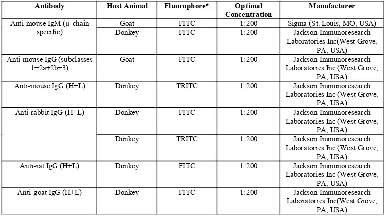
Table 4. FITC- and TRITC-conjugated secondary antibodies used in immunofluorescence
Table 5. Summary of co-localization studies between Cx46 and Cx50 with markers of cellular
List of figures
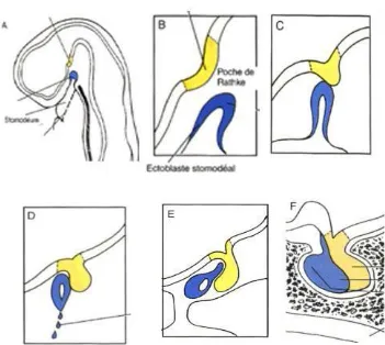
Figure 1. Embryological development of the pituitary gland
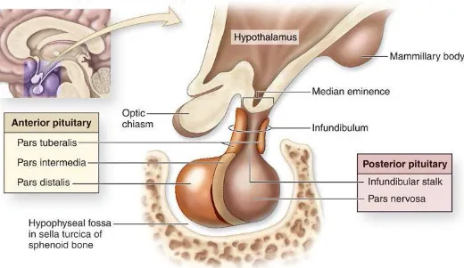
Figure 2. Pituitary gland anatomy and localization within the skull
Figure 3. Positive and negative feedback mechanisms of the hypothalamic-pituitary axis Figure 4. Table of human and mouse connexin gene and protein information
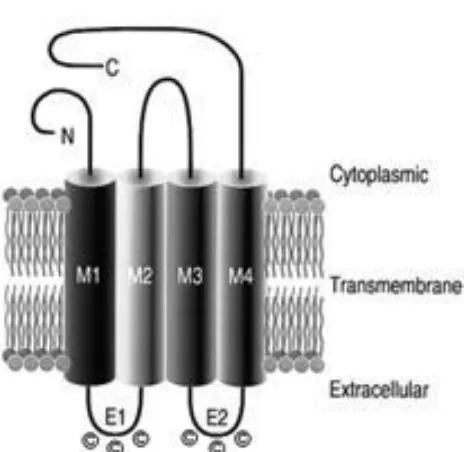
Figure 5. Schematic of connexin protein topology
Figure 6. Diagram of homo- and heteromeric connexons and homo- and heterotypic gap
junctions
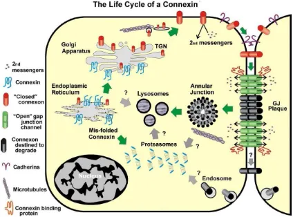
Figure 7. The life cycle of a connexin protein
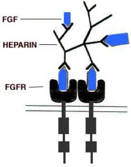
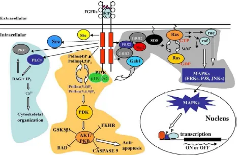
Figure 8. Illustration of FGF binding and receptor dimerization Figure 9. Intracellular signaling pathways activated by bFGF
Figure 10. Studies on the presence of Cx46 and Cx50 in the TtT/GF folliculo-stellate cell line
and in the mouse anterior pituitary
Figure 11. Confocal microscopy studies on the co-localization of Cx46 and cell organelles in
TtT/GF folliculo-stellate cells
Figure 12. Confocal microscopy studies on the co-localization of Cx50 and cell organelles in
TtT/GF folliculo-stellate cells
Figure 13. Studies on the presence of Cx46 and Cx50 in lipid rafts and caveolae in TtT/GF
folliculo-stellate cells
Figure 14. Immunofluorescence studies on the co-localization of Cx46 and Cx50 with lipid raft
and caveolae markers in TtT/GF folliculo-stellate cells
Figure 15. Studies on the presence of Cx46 isoforms in the nucleus of TtT/GF folliculo-stelate
Figure 16. Confocal microscopy studies on the co-localization of Cx46 and nuclear markers in
TtT/GF folliculo-stelate cells
Figure 17. Expression profiles of Cx46 and Cx50 in the TtT/GF folliculo-stellate cells in
response to bFGF treatment
Figure 18. Expression profiles of Cx46 and Cx50 in female and male mink anterior pituitary
List of abbreviations
3-D – 3-Dimensional
ACTH – Adrenocorticotropic hormone
ATP – Adenosine triphosphate
bFGF – Basic fibroblast growth factor C-terminal – Carboxyl-terminal Ca2+ – Calcium ion
cAMP – Cyclic adenosine monophosphate cDNA – Complementary DNA
CRH – Corticotropin-releasing hormone Cx – Connexin
DAG – 1,2-diacylglycerol
DIG – Detergent-insoluble glycolipid ER – Endoplasmic reticulum
FGF – Fibroblast growth factor
FGFR – Fibroblast growth factor receptor
FITC – Fluorescein-5-isothiocyanate FS Cells – Folliculo-stellate cells
FSH – Follicle-stimulating hormone
GAPDH – Glyceraldehyde-3-phosphate dehydrogenase GFAP – Glial fibrillary acidic protein
GH – Growth hormone
GHRH – Growth hormone-releasing hormone GnRH – Gonadotropin-releasing hormone
HPRT-1 – Hypoxanthine phosphoribosyl transferase-1 IL – Interleukin
IP3 – Inositol 1,4,5-triphosphate
kDa – kilo-Daltons
MEF – Mouse embryonic fibroblast MHCB – Myosin heavy chain IIB mRNA – Messenger RNA
NO – Nitric oxide
No-RT – No reverse transcriptase NPC – Neural progenitor cells N-terminal – Amino terminal
PACAP – Pituitary adenylate cyclase-activating peptide PBS – Phosphate buffered solution
PKA – Protein kinase A PKB – Protein kinase B PKC – Protein kinase C PLC – Phospholipase C PRL – Prolactin PI3 – Phosphatidylinositol-3 PIP3 – Phosphatidylinositol-3,4,5-triphosphate POD – Peroxidase
RER – Rough endoplasmic reticulum
SDS-PAGE – Sodium dodecyl sulfate polyacrylamide gel electrophoresis siRNA – Small interfering RNA
SUMO – Small-ubiquitin like modifier T4 – Thyroxine T3 – Triiodothyronine TGF- – TRH – Thyrotropin-releasing hormone TRITC – Tetramethylrhodamineisothiocyanate TSH – Thyroid-stimulating hormone
WGA – Wheat germ agglutinin
Dedication
The work contained within this manuscript is dedicated to my mother, Norma Garcia-Pastor and to my father, Arnaldo Garcia.
Words of gratitude
I am deeply grateful to my research supervisors Dr Maria Leiza Vitale and Dr R.-Marc Pelletier, who have given me the opportunity to conduct research under their supervision and who have also provided me with the training and skills that will serve me in all aspects of life. Working for them was an enriching, stimulating and rewarding experience.
Thank you to my colleagues Ahmed Barry and Dr Casimir Akpovi who, throughout the years, have helped me enormously with my research through insight and support. Being in the lab with both of you is something I looked forward to everyday. I am also thankful for the technical assistance that was provided to me by Dr Li Chen. Her willingness to share her expertise and knowledge helped me tremendously in completing my research project.
I am grateful for the financial assistance/scholarship that was awarded to me by the Faculty of Medicine allowing me to further pursue this research project. Furthermore, I acknowledge funding of the project by the NSERC (Natural Sciences and Engineering Research Council).
Table of contents
RESUME ...IV SUMMARY ...VII LIST OF TABLES ...X LIST OF FIGURES...XI LIST OF ABBREVIATIONS ...XIII DEDICATION ...XV WORDS OF GRATITUDE...XVI TABLE OF CONTENTS...XVII
1. INTRODUCTION... 1
1.1. THE PITUITARY GLAND... 1
1.1.1. Pituitary gland embryonic development ... 1
1.1.2. Anatomy and physiology ... 3
1.1.3. Morphology of adenohypophyseal cells ... 8
1.1.4. The folliculo-stellate (FS) cells ... 9
1.2. GAP JUNCTIONS ... 13
1.2.1. Gap junction structure ... 13
1.2.2. Gap junction and connexon/hemichannel functions... 16
1.2.3. The life cycle of a gap junction ... 19
1.2.4. Connexin46 and connexin50 ... 23
1.3. BASIC FIBROBLAST GROWTH FACTOR... 27
1.3.1. Ligand and receptor properties ... 27
1.3.2. Basic fibroblast growth factor in the anterior pituitary gland ... 31
1.4. EXPERIMENTAL MODELS ... 34
1.4.1. The TtT/GF folliculo-stellate cell line... 34
1.4.2. The mink as an animal model for research in endocrinology, reproduction and immunology ... 34
1.4.2. FS cells and intercellular communication in the mink anterior pituitary... 36
1.5. BASIS FOR OUR STUDY... 37
2. MATERIALS AND METHODS ... 38
2.1 ANTIBODIES AND PROBES FOR WESTERN BLOT AND MICROSCOPY STUDIES... 38
2.2. CELL CULTURE AND TISSUE PREPARATION ... 38
2.2.1. Cell lines and culture ... 38
2.2.2. Basic fibroblast growth factor treatment of TtT/GF cells... 39
2.2.3. Preparation of cell homogenate ... 39
2.2.4. Mouse ocular lens ... 40
2.2.5. Preparation of mink and mouse anterior pituitary tissue ... 40
2.3. LIPID RAFT ISOLATION ... 42
2.4. CELL FRACTIONATION –NUCLEAR FRACTION ISOLATION ... 43
2.4.1. Cell lysis... 43
2.4.2 Separation of nuclear fraction from cytoplasm and plasma membrane ... 43
2.4.3. Isolation of post-nuclear fraction... 43
2.4.4. Lysing of nucleus and sample preparation ... 44
2.5. ALKALINE PHOSPHATASE TREATMENT ... 45
2.6.2. RNA isolation and complementary DNA synthesis ... 46
2.6.3. PCR and Southern blot ... 47
2.7. WESTERN BLOT... 48
2.7.1. Protein dosage and sample preparation ... 48
2.7.2. SDS-PAGE ... 48
2.7.3. Transfer onto nitrocellulose membrane... 48
2.7.4. Blocking of non-specific sites and antibody incubation... 49
2.7.5. Densitometry of bands obtained in western blot... 49
2.8. IMMUNOFLUORESCENCE... 51
2.8.1. Preparation of cells ... 51
2.8.2. Fixation and permeabilization ... 51
2.8.3. Blocking and incubation with antibodies ... 52
2.8.4. Mounting and observation ... 52
3. RESULTS ... 58
3.1. IDENTIFICATION OF CX46 AND CX50 IN THE TTT/GFFOLLICULO-STELLATE CELL LINE AND MOUSE ANTERIOR PITUITARY... 58
3.1.1. Expression of Cx46 and Cx50 mRNA by the TtT/GF folliculo-stellate cell line ... 59
3.1.2. Protein expression of Cx46 and Cx50 in the TtT/GF folliculo-stellate cell line and mouse anterior pituitary ... 60
3.1.3. Expression of Cx46 and Cx50 in mouse embryonic fibroblasts ... 61
3.1.4. Phosphorylation status of Cx46 in the TtT/GF folliculo-stellate cell line... 61
3.1.5. Distribution of Cx46 and Cx50 in the TtT/GF folliculo-stellate cell line ... 62
3.2. ASSOCIATION OF CX46AND CX50WITH CELLULAR ORGANELLES IN THE TTT/GF FOLLICULO-STELLATE CELL LINE ... 68
3.2.1. Association of Cx46 with cellular organelles in TtT/GF folliculo-stellate cells... 68
3.2.2. Association of Cx50 with cellular organelles in TtT/GF cells... 69
3.3. CHARACTERIZATION OF CX46AND CX50.PRESENCE WITHIN MEMBRANE DOMAINS: LIPID RAFTS AND CAVEOLAE, OF TTT/GF FOLLICULO-STELLATE CELLS ... 79
3.3.1. Identifying the Detergent-insoluble glycolipid fractions isolated from TtT/GF folliculo-stellate cells via the presence of flotillin-1 and caveolin-1... 80
3.3.2. Association of Cx46 and Cx50 with lipid raft and caeolae markers... 81
3.4. INVESTIGATION OF THE NUCLEAR LABELLING OFCX46IN TTT/GF FOLLICULO-STELLATE CELLS... 88
3.4.1. Separation of different Cx46 immunoreactive bands by fractionation / nuclear isolation of TtT/GF folliculo-stellate cells ... 88
3.4.2. Association of nuclear Cx46 with the nuclear protein markers in the TtT/GF folliculo-stellate cells... 91
3.5. STUDIES ON THE PROTEIN EXPRESSION PROFILES OF CX46AND CX50 IN THE TTT/GFFOLLICULO-STELLATE CELL LINE IN RESPONSE TO BFGFTREATMENT ... 98
3.5.1. Protein expression profile of Cx46 in response of bFGF treatment of TtT/GF folliculo-stellate cells... 98
3.5.2. Protein expression profile of Cx50 in response of bFGF treatment of TtT/GF cells ... 99
3.6. STUDIES ON THE PROTEIN EXPRESSION PROFILES OF CX46AND CX50 IN THE ANTERIOR PITUITARY OF MALE AND FEMALE MINK THROUGHOUT THE ANNUAL REPRODUCTIVE CYCLE... 103
3.6.1. Expression patterns of Cx46 and Cx50 in the female mink anterior pituitary gland ... 104
3.6.2. Expression patterns of Cx46 and Cx50 in the male mink anterior pituitary gland ... 105
3.6.3. Variations in the mink serum levels of the anterior pituitary hormones: PRL, FSH and LH ... 105
4. DISCUSSION ... 110
4.1. PRESENCE OF CX46AND CX50IN THE TTT/GFFOLLICULO-STELLATE CELLS OF THE ANTERIOR PITUITARY GLAND... 111
4.2. INTRACELLULAR TRAFFICKING OF CX46 AND CX50IN THE TTT/GF FOLLICULO-STELLATE CELLS ... 117
4.3. PRESENCE OF CX46AND CX50WITHIN THE LIPID RAFTS AND CAVEOLAE OF TTT/GF FOLLICULO-STELLATE CELLS ... 120
4.4. THE PRESENCE OF CX46 ISOFORMS WITHIN THE NUCLEI OF TTT/GFFOLLICULO-STELLATE CELLS ... 125
4.5. STUDIES ON THE RESPONSE OF CX46 AND CX50TO BFGFTREATMENT OF TTT/GFFOLLICULO-STELLATE CELLS ... 130
1. INTRODUCTION
1.1. The pituitary gland
Located at the base of the skull embedded within the sphenoid bone, the pituitary gland is a critical relay station for endocrine functions in vertebrates. The gland is in intimate physical and functional association with the hypothalamus, from which it receives and relays chemical messages. This close relationship of both the neural and endocrine systems is demonstrated not only anatomically, by the presence of neural tissue in this endocrine gland but also by the physiological influence of hypothalamic secreting factors on the function of the pituitary gland.
1.1.1. Pituitary gland embryonic development
The adult pituitary gland results from the fusion of two separate tissues during embryogenesis, the adenohypophysis develops in the embryo starting from oral ectodermal tissue. The hypophyseal placode is the first primitive structure to develop from the oral ectoderm. As it grows, it folds upon itself dorsally to form Rathke’s pouch, the succeeding structure in this developmental process. Rathke’s pouch grows towards the evaginating infundibulum (forming the neurohypophysis), eventually becoming a distinct structure as it separates from the oral ectoderm (Rizzoti and Lovell-Badge 2005). Adenohypophyseal progenitor cells will differentiate into hormone secreting cells. As its name implies, the neurohypophysis, develops from the neural ectoderm during a time frame concurrent with that of the adenohypophysis. The developmental events begin with an evagination from the floor of the diencephalon eventually forming a funnel-shaped process known as the infundibulum that will extend towards Rathke’s pouch (Figure 1).
The sphenoid bone will develop around the gland creating the sella turcica in the process (Drouin 2011).
Figure 1. Embryological development of the pituitary gland (Larsen 2003).
Cell signalling pathways and transcription factors play critical roles in pituitary organogenesis and mediate the anatomical events that occur throughout this process. The early formation of Rathke’s pouch, for example, is highly dependent on the pituitary homeobox factors, Pitx1 and -2 (Rizzoti and Lovell-Badge -2005). The combined disruption of both transcription factors results in the cessation of adenohypophyseal development at a very early stage (Drouin 2011). Similarly, the differentiation of progenitor cells into hormone-secreting cells is also reliant on signalling pathways and transcription factors. For instance, the Notch signalling pathway regulates the expression of transcription factors that retain the multipotency of pituitary progenitor cells and must therefore be silenced to induce their terminal differentiation. In the
eventuality that the activity of Notch pathway proteins is sustained, multiple lineages will fail to fully differentiate (Zhu, Wang et al. 2007).
1.1.2. Anatomy and physiology
The adenohypophysis and neurohypophysis formed during embryogenesis also serve as the two major anatomical divisions in the gland. The adenohypophysis is subdivided into the pars
distalis, pars tuberalis and pars intermedia. The pars distalis occupies the anterior-most portion
of the gland while the pars tuberalis wraps around the infundibulum; together, these two subdivisions form the anterior pituitary. Dorsal to the pars distalis is the pars intermedia, which contains a number of cysts amassing to form Rathke’s cleft, an embryological remnant of the lumen of Rathke’s pouch. The neurohypophysis occupies the posterior portion of the gland and is continuous cranially to the hypothalamus. It is divided to include the median eminence, the infundibulum and the pars nervosa, with the later being the sole component of the posterior pituitary gland (Mizeres 1981, Junqueira, Carneiro et al. 1986) (Figure 2).
Figure 2. Pituitary gland anatomy and localization within the skull (McKinley and O'Loughlin
2012).
Being an endocrine organ, the vascularisation of the pituitary gland is essential for its functioning. The gland as a whole, receives its blood from the superior and the inferior hypophyseal arteries, the latter of which supply the neurohypophysis. The superior hypophyseal arteries feed the adenohypophysis and also form the hypophyseal-portal vascular system, a vascular network critical to the proper functioning of this portion of the gland. This system begins with the right and left superior hypophyseal arteries that will spread out to form the primary capillary plexus within the median eminence; a location where they are in close association with the axon terminals of the neuro-secretory parvicellular neurons. The capillaries form portal vessels that first run along the pituitary stalk then divide again a secondary capillary plexus within the anterior lobe (Junqueira, Carneiro et al. 1986). This provides a “natural” route for trophic/inhibitory hypothalamic hormones to be released into the primary plexus, travel down the portal vessels and arrive at the secondary plexus where the capillaries surround the target
endocrine cells. Ultimately, blood from both the adenohypophysis and neurohypophysis drains into dural sinuses via venous vessels (Junqueira, Carneiro et al. 1986).
The synthesis and release of the hormones, adrenocorticotropic hormone (ACTH), thyroid-stimulating hormone (TSH), follicle-thyroid-stimulating hormone (FSH), lutenizing hormone (LH), growth hormone (GH) and prolactin (PRL), is the primary function of the anterior pituitary gland and is a process rigorously controlled by the hypothalamus and systemic factors. The neurosecretory neurons of the hypothalamus synthesize and release hypophysiotrophic hormones that will subsequently trigger or inhibit anterior pituitary hormone synthesis and secretion. This process begins with the synthesis and storage of hypophysiotrophic hormones by hypothalamic neurosecretory cells. Upon stimulation, these factors will be released from the axonal endings of the neurons in the median eminence into the primary capillary plexus of the hypophyseal-portal vascular system where they are carried to the secondary plexus in the pars distalis. Some hypophysiotrophic hormones are: thyrotropin-releasing hormone (TRH), dopamine, growth hormone-releasing hormone (GHRH), somatostatin, gonadotropin-releasing hormone (GnRH) and corticotropin-releasing hormone (CRH). Each physiotrophic hormone exerts a stimulatory or inhibitory effect on a specific hormone-secreting cell type in the anterior pituitary gland and several (TRH and GnRH) will affect more than one cell type (Brook and Marshall 2001) (Figure 3).
Feedback loops that ultimately directly or indirectly act on the anterior pituitary also modulate hormone synthesis and release in this gland. As a result of the action of anterior pituitary hormones, target glands will synthesize and secrete their own hormones that, apart from
exhibiting a primary physiological role, return to the hypothalamo-pituitary axis to reduce (negative feedback) or increase (positive feedback) the secretion of the hypothalamic or pituitary factor that initially resulted in their release. Thyroxine (T4) and triiodothyronine (T3) are synthesized and released by the thyroid gland in response to TSH. Once released into circulation, aside from their systemic effect on metabolism, T4 and T3 act in a negative feedback manner at the level of the hypothalamus by reducing the secretion of TRH. The thyroid hormones also act further down the hypothalamo-pituitary axis, by reducing the sensitivity of thyrotrophs to TRH, thereby decreasing the secretion of TSH (Brook and Marshall 2001, Rhoades and Bell 2009). An example of positive feedback can be observed in the female for a brief period just prior to ovulation. LH secretion from the anterior pituitary will result in an increased ovarian production of oestrogen that then influences the pituitary gonadotrophs by increasing production of LH. In this particular physiological state, LH stimulates its own production and secretion via the ovary and estrogen hormones (Heffner and Schust 2010).
Figure 3. Illustration of positive and negative feedback mechanisms of target organ factors on
In addition to the hypophysiotrophic hormones, anterior pituitary secretory cells are also strongly influenced by a wide variety of paracrine messengers that are synthesized and secreted directly within the gland. These are present in a variety of chemical forms including: small molecules (Ex: nitric oxide (NO) and adenosine), and an assortment of proteins that include cleavage -subunit of glycoproteins LH, FSH and TSH) and peptides (follistatin and vasoactive intestinal peptide (VIP)). The general effect on hormone secretion of many of these paracrine messengers is similar to that of the hypophysiotrophic hormones, in that secretion is either decreased or increased in response to a particular messenger. For instance, adenosine (secreted by folliculo-stellate cells) has been shown to decrease FSH secretion from gonadotrophs (Yu, Kimura et al. 1998). Furthermore, paracrine messengers exert their effects on hormone secretion by other means including: increasing endocrine cell proliferation (mitogenic effect) (ex: insulin-like growth factor I (IGF-I) and basic fibroblast growth factor (bFGF)), hormone-secreting cell hyperplasia (galanin) and stimulation of endocrine cell differentiation (common -subunit of glycoproteins LH, FSH and TSH). The fact that the production of these factors is assumed by the anterior pituitary hormone-secreting cells or the folliculo-stellate cells (to be discussed in subsequent sections), suggests a local control mechanism amongst adenohypophyseal endocrine cells (Schwartz 2000).
The secretion of the neurohypophyseal hormones oxytocin and vasopressin occurs through a mechanism different to that of the adenohypophysis. The cell bodies of the neurosecretory cells producing oxytocin and vasopressin are respectively located in the paraventricular and supraoptic nuclei of the hypothalamus, while their axons descend through the infundibulum into
the pars nervosa. After synthesis in the cell bodies, oxytocin and vasopressin will be stored in axonal vesicles awaiting their release triggered by specific stimuli (Brook and Marshall 2001).
1.1.3. Morphology of adenohypophyseal cells
The major cell types in the anterior pituitary are the granular, hormone secreting cells. Of these, the somatotrophs are the most abundant accounting for close to 50% of anterior pituitary cells. In humans, they are most abundant in the lateral portion of the anterior lobe, are of medium size and are spherical or oval in shape. Lactotrophs, which constitute 10 – 25% of anterior pituitary cells are scattered throughout the anterior lobe but are mostly concentrated in areas close to the posterior lobe. They are generally seen in two forms: angular or elongated of small to medium size, or large and of polyhedral shape. Corticotrophs are primarily located in the central region of the anterior lobe and overall represent 10 – 15% of anterior pituitary cells. They exhibit an oval shape and are of medium to large size. The thyrotropes are the least abundant cell type in the anterior pituitary, representing less than 10% of all cells in the gland; they are large and oval or irregular in shape and localize to the anteromedial area of the pars distalis. Gonadotrophs, accounting for 15 – 20% of anterior pituitary cells, can be found throughout the pars distalis where they adjoin capillaries and are frequently in close association with lactotrophs, furthermore, they also localize to the pars tuberalis (Imura 1985, Heaney and Melmed 2004). Another, non-endocrine cell type can also be found in the gland, the folliculo-stellate cells, which are the topic of the current study.
1.1.4. The folliculo-stellate (FS) cells
Initially described with the electron microscopy study by Rinehart and Farquhar (1953), the FS cells account for 5 – 10% of all anterior pituitary cells and are thoroughly distributed through the
pars distalis. They are star-shaped (hence their name) with long cytoplasmic processes forming
pseudo-follicles by surrounding endocrine cells. Unlike the hormone-secreting cells of the gland, FS cells are agranular. Other morphological aspects of these cells include the presence of an abundance of type-III vimentin intermediate filaments in their cytoplasm (Cardin, Carbajal et al. 2000). FS cells were initially believed to originate from the ectoderm of the oral cavity, implying that they are a derivative of Rathke’s pouch cells and share a common progenitor with their endocrine hormone-secreting neighbours. However, it was later established that FS cells express the neuroglial cell-specific marker GFAP (Glial fibrillary acidic protein), implying a neuroectodermal origin to the FS cells (Inoue, Couch et al. 1999). Another marker that had facilitated the identification of FS cells is S-100, a calcium-binding protein not expressed by anterior pituitary endocrine cells but typically expressed by FS cells (Nakajima, Yamaguchi et al. 1980).
In vivo, FS cells have been shown to alter their morphology in response to different hormonal
environments. Cardin et al, (2000) have described two distinct S-100 positive cell types (I and II) in the mink (Mustela vison) that are also suspected to carry out different physiological activities. The type-I cell was characterized by a stellate shape and were most prevalent during physiological periods characterized by high serum and anterior pituitary PRL levels and by low FSH and LH levels. The type-II cell typically lacked cytoplasmic projections and exhibited a rounded morphology. Furthermore, their presence was most noted during periods when serum
and anterior pituitary PRL levels were low and FSH and LH levels were high. It has been proposed that the endocrine milieu of the anterior pituitary could influence the transformation of one cell type to the other (Cardin, Carbajal et al. 2000). Similarly, studies of the equine anterior pituitary gland throughout its annual breeding cycle have confirmed a morphological change in the FS cells (Henderson, Hodson et al. 2008).
While the FS cells have a variety of functions in the anterior pituitary, it is largely accepted that they are responsible for the maintenance of the anterior pituitary microenvironment. FS cells were initially ascribed the function of pituitary scavengers involved in the phagocytosis and degradation of apoptotic cells and extracellular debris (Devnath and Inoue 2008). However, the major function of FS cells is the secretion of a variety of growth factors and cytokines that act in both autocrine and paracrine manners. These chemical messengers include bFGF, Vascular endothelial growth factor (VEGF), Interleukin-6 (IL-6) and IL-1 which mediate a plethora of functions ranging from immune system modulation to hormone gene regulation (Allaerts and Vankelecom 2005, Herkenham 2005).
In addition, several potential functions have also been attributed to FS cells. The first of these is based on the close physical association of FS cell extensions and pituitary capillaries and suggests that FS cells act as an intermediary in the transport of nutrients and oxygen from capillaries to endocrine cells, a function similar to that of astrocytes in the central nervous system (Inoue, Couch et al. 1999, Inoue, Mogi et al. 2002). Furthermore, FS cells were found to express Pitx-1, a transcription factor common to all pituitary cells. This discovery led to the hypothesis that FS cells may in fact be pituitary stem cells with the capacity to differentiate into endocrine
cells of the adenohypophysis (Tremblay, Marcil et al. 1999, Devnath and Inoue 2008, Drouin 2011). FS cells have also been shown to express adenohypophyseal-hormone receptors, specifically: the receptors for Thyroid-Stimulating Hormone (TSHR), Growth Hormone (GHR)
and Adenocorticotropin (ACTHR). This finding may further implicate the FS cells in a local,
paracrine-driven control mechanism (Brokken, Leendertse et al. 2004).
The manner in which FS cells are organized within the anterior pituitary is also a critical feature with regard to intercellular communication throughout the gland. Studies of the gland by electron microscopy led to the initial proposal that the FS cells form an extensive 3-D (3-Dimensional) network throughout the anterior pituitary (Vila-Porcile 1972). This proposal has since been substantiated by confocal microscopy and electrophysiology (Fauquier, Lacampagne et al. 2002). The FS cells are arranged in a mesh-like fashion and it is within the confines of this mesh that the hormone-secreting endocrine cells dwell. Importantly, this FS cell network is physically continuous through connexin43-mediated (Cx43) gap junction channels (Morand, Fonlupt et al. 1996). Furthermore, at several points throughout this network, heterologous connections have been described between FS cells and lactotrophs (Morand, Fonlupt et al. 1996). Physiologically, the FS network plays the role of an information highway capable of rapidly transmitting chemical messages relatively long distances throughout the gland. Small signaling molecules such as cytokines, and growth and immune factors produced by FS cells, can be released and propagated locally through gap junctions. Alternatively, signalling in the form of a cytosolic increase in Calcium ion (Ca2+) concentration has been shown to travel extensive distances (millimeters). FS cells are capable of spontaneously generating Ca2+ signals that are subsequently propagated throughout the network in a regenerative, pulsatile fashion, as is seen
with action potentials (Fauquier, Guerineau et al. 2001). This spontaneous generation of Ca2+ signals has been proposed to synchronize cellular activities within the anterior pituitary and to contribute to the autonomous, hypothalamus-independent functioning of the anterior pituitary (Devnath and Inoue 2008). Gap junctions have been shown to play an crucial and vital role in the formation of the FS cell network, especially in the transmission of Ca2+ signals since their experimental blockage severely compromises signal transmission (Fauquier, Guerineau et al. 2001). Furthermore, Cx43-mediated gap junctions between FS cells have been shown to be highly responsive to FS cell-secreted growth factors and cytokines (Fortin, Pelletier et al. 2006, Meilleur, Akpovi et al. 2007).
1.2. Gap junctions
Gap junction channels result from the pairing of connexons/hemichannels, each being a contribution from adjacent cells. Each connexon/hemichannel is made up of connexin proteins, also known as the elemental structures of gap junction channels. Until now, both rodents and humans have been found to express approximately twenty connexin variants that can be classified into sub-groups based on amino acid sequence homology and oligomerization: , , and unclassified (Evans and Martin 2002) (Figure 4).
Figure 4. Table of human and mouse connexin gene and protein information (Evans and Martin
2002).
1.2.1. Gap junction structure
The connexin proteins possess a highly conserved tertiary structure containing intracellular amino- (N) and carboxyl- (C) termini, joined by four transmembrane domains, which themselves are linked by a total of three loops, two being extracellular and one on the cytoplasmic side
Figure 5. Schematic of connexin protein topology (Sohl and Willecke 2004).
Throughout the connexin family, the N-terminal is a conserved portion of the protein presenting little variation in both amino acid sequence and length. Functionally, this domain is important for the proper insertion of the protein into the membrane during translation; a role that is also shared by the first of the four transmembrane helices (Evans and Martin 2002). The third transmembrane domain primarily serves a structural function and contributes to the wall of the connexon channel. In addition, the transmembrane domains have been suggested to be required for the oligomerization of individual connexin proteins during the formation of a connexon (Evans and Martin 2002). The extracellular loops are arranged as anti-parallel -sheets, each containing three cysteine residues that form disulfide bridges between the two loops of a connexin protein. The loops are important for the docking process between one connexon and its pair in an adjacent cell (Evans and Martin 2002). The identity and distinctiveness of a connexin family member is derived primarily from its C-terminal and to a lesser degree, the cytoplasmic loop that shows the greatest variation in amino acid sequence and length (Mese, Richard et al. 2007). The C-terminal is also the portion of the protein that will undergo post-translational
modifications and processing in response to stimuli such as phosphorylation (Berthoud, Beyer et al. 1997).
The connexon/hemichannel is an assembly of six connexin proteins arranged in a cylinder with a hollow core. The completed connexon/hemichannels will be shipped to the membrane where it will eventually join another connexon/hemichannel from a neighbouring cell to form a gap junction channel or remain unpaired. Most cell types express more than one connexin member. Connexons can have a homomeric or heteromeric composition (Mese, Richard et al. 2007). Heteromeric connexons are composed of two different connexins of the same sub-group. Alternatively, a gap junction can be composed of two identical connexons, thereby making it homotypic (bearing in mind that these connexons may be heteromeric, although identical); or can be the product of two different connexons and therefore classifying it as heterotypic (Figure 6). Combining different connexins or connexons results in the formation of junctions with unique biochemical properties thus extending their functional capabilities (Jiang and Goodenough 1996).
Figure 6. Diagram of homo- and heteromeric connexons and homo- and heterotypic gap
1.2.2. Gap junction and connexon/hemichannel functions
Gap junctions connect the cytoplasms of two adjacent cells, through a tunnel formed by the individual joined connexons. The tunnels allow small molecules up to 1.5 kilo-Daltons (kDa) in size to passively travel from one cell to another (Kumar and Gilula 1996).
The permeability of gap junctions to small molecules was generally thought to be relatively non-selective whereby free passage would be granted to any ion or molecule equal to or smaller than 1 – 1.5 kDa (el-Fouly, Trosko et al. 1987). Although this has remained true to a certain extent, the varying permeability properties of different channels to metabolites and ions are increasingly apparent (Goldberg, Moreno et al. 2002). Of the metabolites and ions, one of the most studied is Ca2+ (most likely due to their involvement in intracellular signalling), as well as several metabolites including Inositol triphosphate (IP3, related to Ca2+ signalling), signalling molecules:
cyclic adenosine monophosphate (cAMP), and the cellular metabolites glucose and adenosine triphosphate (ATP) (Goldberg, Moreno et al. 2002, Medina and Tabernero 2005). Each ion and molecule endows individual gap junctions with a function more important than connecting two adjacent cells. The permeability of gap junctions to Ca2+, IP3 and other signalling molecules is
strongly indicative of a role in the propagation of cellular messages including cell death and survival signals (Krysko, Leybaert et al. 2005). The passage of ATP, the currency of cellular energy, demonstrates a role of gap junctions in nutrient exchange. Gap junctions also couple cells electrically, allowing for such a signal to be disseminated considerable distances in relatively little time (Hestrin 2011). The selectivity of gap junctions composed of a particular connexin protein can also modulate gene expression post-transcriptionally between adjacent cells by means of their permeability to small interfering RNA (siRNA) molecules (Valiunas, Polosina
et al. 2005). In this context, siRNA molecules produced in one cell can travel within a group of cells to influence gene expression, provided the cells are joined by connexin-specific gap junctions.
Cell motility, especially with respect to development, and the cell transformation processes, has been shown to rely strongly on connexin expression (Li, Waldo et al. 2002). Specific connexin genes are expressed during precise periods of embryonic development and various studies have associated substantial defects with the perturbation of these genes. Embryonic expression of Cx43 by proepicardial progenitor cells, mediates (amongst other developmental events) the organized migration of these cells that will eventually differentiate to form critical coronary structures. In the absence of Cx43, the proepicardial progenitor cells were shown to have an altered directionality and motility (increased migrational velocity) that resulted in extensive structural defects in the coronary arteries and cardiac muscle (Li, Waldo et al. 2002, Rhee, Zhao et al. 2009).
Apoptosis and the regulation of cell death have been found to be influenced both by gap junctions and connexins. The “bystander effect” was a term introduced by Freeman et al. (Freeman, Abboud et al. 1993) used to describe a phenomenon whereby a cellular death signal (in this case a toxin) can spread from a small subset of cells to eventually encompass a larger cell mass, through gap junctions. This concept has since been extended to include any gap junction-permeable intermediates/second messengers that may trigger an apoptotic event including Ca2+, IP3 and cAMP. These factors, all released in abundant quantities during apoptosis, will passively
apoptotic cascade (Krysko, Leybaert et al. 2005). Monomeric connexin proteins also appear to play a significant role in the signalling aspects of apoptosis, by direct interaction with apoptotic protein mediators. In human cancer cell lines, Cx26 and Cx43 have been co-localized with Bcl-2 proteins: Bak, Bcl-xL and Bax (Decrock, Vinken et al. 2009). Oddly, the transcriptional levels of both pro- and anti-apoptotic genes were affected in response to alterations in Cx43 expression (Decrock, Vinken et al. 2009). This contradictory observation makes it difficult to assign an inhibitory or inductive role to Cx43 with respect to programmed cell death.
Connexons/hemichannels, which remain unpaired, provide a gated passageway to the surrounding extracellular milieu and play several important cellular functions including the release of ATP and subsequent propagation of Ca2+ waves. The mechanism for this process begins with an initial stimulus (a stressor) that causes the release of ATP. Extracellular ATP will then bind P2 purinergic receptors on a nearby cell that in turn, stimulates the production of IP3
subsequently leading to the release of Ca2+ from the endoplasmic reticulum (ER) (Lodish 2000). Changes in the intracellular Ca2+levels then lead to the opening of hemichannels and the ensuing release of ATP, allowing propagation of the signal (Goodenough and Paul 2003). This signalling has proven essential to a variety of physiological events ranging from neural development (Dale 2008) to cell division and differentiation in the retina (Pearson, Dale et al. 2005).
In addition to the communication roles attributed to gap junctions and connexon/hemichannels, there is experimental evidence demonstrating the involvement of connexon proteins in numerous non-conventional roles. The effect that specific connexin members have on cell growth and tumourgenicity is an example of this. Particularly, Cx32 and Cx43 have been reported to inhibit
cell growth and act as a tumour suppressor in a manner independent of hemichannel and gap junctional intercellular communication (Fujimoto, Sato et al. 2005, Jiang and Gu 2005, Langlois, Cowan et al. 2010). This has been supported by the fact that transformed cells were shown to have a decreased connexin expression and that cancer cell lines transfected with connexin genes exhibit a reduction in their growth rate and metastatic potential (Zhao, Han et al. 2011). Moreover, several of the connexins implicated in exhibiting these effects have been localized to the cell nucleus (Cx30 and Cx43) and were reported to be cleaved or truncated with the intent of isolating the C-terminal (Dang, Doble et al. 2003, Mennecier, Derangeon et al. 2008); a fact further supporting the claim that this phenomenon is not influenced by gap junctional communication. Under non-pathological conditions, connexin expression has been shown to promote mitosis. Cx50 is an example of this, whereby its expression during postnatal development is essential for ocular lens growth. The absence of Cx50 drastically reduces the mitotic index of developing lens cells resulting in micropthalmia (smaller eye and lens) (White, Goodenough et al. 1998, Sellitto, Li et al. 2003).
1.2.3. The life cycle of a gap junction
Connexins are relatively short-lived proteins with most having a half-life of around 1.5 – 5 hours (Laird 2005). Although the connexins follow a similar path from synthesis to degradation, variations in both the intermediate and ultimate fates of the proteins are known to exist.
The biosynthesis of connexin proteins commences in the rough endoplasmic reticulum (RER) where the protein is co-translationally inserted into the RER membrane (Mese, Richard et al. 2007). From this location to the trans-Golgi apparatus, there is a gradual oligomerization of
individual connexin proteins to form a connexon/hemichannel. In the case of homomeric channels, this process occurs spontaneously, whereas it is hypothesized that unidentified chaperone proteins are needed to oligomerize heteromeric connexins (Evans and Martin 2002). Connexons are then transported from the trans-Golgi in vesicles that travel along microtubules to eventually fuse with the plasma membrane. The connexon will diffuse laterally either towards an area of the plasma membrane unopposed by another cell where it will fulfill its functions as a connexon/hemichannel or towards a cell-cell contact area where it will dock with a compatible connexon/hemichannel in the adjacent cell forming a gap junction channel (Evans, De Vuyst et al. 2006). This pairing of connexons results in a 2 – 4 nanometres (nm) “gap” between the two cells visible by electron microscopy (Herr 1976). A newly formed gap junction will find itself on the outside of a gap junctional plaque. Gap junctional plaques are large aggregations of gap junction channels, where the “youngest” channels join the plaque at its periphery, gradually migrating to its centre. As younger channels join the plaque, the “older” ones are pushed to the centre where they are ultimately internalized into annular junctions (a type of vesicle) (Pelletier 1995, Jordan, Chodock et al. 2001). As opposed to the processes of assembly, gap junctions are not separated into connexons but rather, are entirely taken into annular junctions. Logically, this event indicates that annular junctions, also known as connexosomes, are composed not only of their own plasma membrane, but also of that of the adjacent cell to which they are joined (Leithe and Rivedal 2007). Annular junctions will either fuse directly with lysosmes or indirectly, by first maturing into early and late endosomes. Alternatively, certain connexins are ubiquitinated and degraded through the proteasomal pathway (Fortin, Pelletier et al. 2006, Leithe and Rivedal 2007). Autophagy, has also been shown to contribute to connexin and gap junction degradation,
specifically targeting connexin-containing vesicles from the secretory pathway or annular junctions from gap junctional plaques (Lichtenstein, Minogue et al. 2011) (Figure 7).
Figure 7. The life cycle of a connexin protein (Laird 2005).
Post-translational modifications represent a major event in the lifecycle of connexin proteins that serve to regulate various functional aspects of connexins and gap junctions. Turnover, coupling, conductance (gap junction channels), sub-cellular trafficking, degradation and assembly are all events that can be affected by post translational modifications targeting connexins and gap junctional channels. The phosphorylation status of connexin proteins is not only the most prevalent of modifications, but also serves as an indicator of physiological activity and localization (Fortin, Pelletier et al. 2006). Various stimuli, such as growth factors and cytokines, can significantly affect the phosphorylation status of a connexin by the addition or removal of a phosphate group via kinases and phosphatases, respectively (Meilleur, Akpovi et al. 2007). As previously mentioned, these actions occur predominantly on amino acid residues on the
C-terminal tail and intracellular loop. The functional consequences of a phosphorylation event are highly variable and are often specific to the phosphorylated residue, connexin family member and cell type. As will be discussed in subsequent sections (1.3.), an alteration in phosphorylation status is often a terminal event in a signaling cascade initiated by a stimulus. In addition to phosphorylation, intracellular loop and terminal amino acid residues on connexin proteins are also modified via nitrosylation and sumoylation (Straub, Billaud et al. 2011, Kjenseth, Fykerud et al. 2012). Nitrosylation, the attachement of a NO molecule to a cysteine amino acid, has been found to be especially relevant to endothelial and smooth muscle cell lines/tissue. In these settings, the permeability of Cx43 gap junctions has been shown to be altered in response to this particular covalent modification. Sumoylation, whereby a member of the small-ubiquitin like modifier (SUMO) protein family is attached to a lysine residue, has been often associated with nuclear proteins and that is linked to processes such as DNA repair and transcription. This post– translation modification, however, has also been documented to regulate Cx43 gap junctional channels and Cx43 protein levels (Kjenseth, Fykerud et al. 2012). As was seen with phosphorylation, nitrosylation and sumoylation can also bring about significant physiological changes via the modification of connexin proteins and gap junctional channels.
Not all connexins follow the above-described classical/conventional pathway. There exist alternative pathways as well as non-conventional functions that will localize connexins to unexpected parts of the cell. For example, Cx26, has been found to take a shortcut to the plasma membrane that completely bypasses the Golgi apparatus (Evans and Martin 2002). Furthermore, connexins do not always localize to the plasma membranes but have been unexpectedly located in a variety of cellular locations including the nucleus, where they are suspected to modulate
gene expression (Dang, Doble et al. 2003, Sanches, Pires et al. 2009). The inner mitochondrial membrane has also been found to house connexins (Cx43), more specifically, in the form of hemichannels/connexons that contribute to the uptake of potassium into the mitochondrial matrix in cardiomyocytes (Miro-Casas, Ruiz-Meana et al. 2009).
1.2.4. Connexin46 and connexin50
Cx46 and Cx50 are most notorious for their expression and importance in the ocular lens. Both connexins have been extensively studied with respect to the development of the lens and the maintenance of homeostasis. To date, Cx50 has not yet been described in other tissues of the body. Cx46 has been identified and studied in various other tissues including: cancerous breast epithelial cells, Schwann cells, bone osteoblastic cells and lung alveolar tissue (Chandross, Spray et al. 1996, Koval, Harley et al. 1997, Abraham, Chou et al. 1999, Banerjee, Gakhar et al. 2010).
The lens is an avascular, metabolically active organ that refracts light entering the eye onto the retina. It has three distinct regions: (1) a single layer of epithelial cells lining the anterior surface, (2) cortical fibre cells undergoing differentiation and (3) mature nuclear fibre cells. The cells in all three regions of the lens are connected by gap junctions. Cx43 is found between epithelial cells, Cx46 localizes to differentiating and mature fibre cells and Cx50 is expressed throughout both the lens epithelium and the lens fibre regions (Shakespeare, Sellitto et al. 2009). Together, these connexins allow the formation of an intercellular network that facilitates the uptake of nutrients from the surrounding aqueous humor and the elimination of waste products (Saleh, Takemoto et al. 2001). Mutations in Cx46 and Cx50 have been extensively documented especially with respect to ocular pathologies. Disruptions in the Cx46 gene have been shown to
result in the formation of nuclear cataracts (located in the central nucleus of the organ) (Gong, Li et al. 1997). Similar pathological features, pulverulent cataracts (literally meaning “dust-like”), have been noted following Cx50 mutations although these mutations are accompanied by microphthalmia (Dunia, Cibert et al. 2006). Although the cause for the later remains speculative (White, Goodenough et al. 1998, Gong, Cheng et al. 2007), apart from contributing to lens transparency Cx50 also plays a significant role in lens development.
The numerous studies on Cx46 and Cx50 in relation to ocular disease have revealed various aspects regarding their intracellular physiology.
Protein-protein interactions
Association of connexins with other proteins can reveal information on their post-translational status, cellular localization and involvement in signalling pathways. The connexin protein family bears a particular affinity for other cell junctions including claudins, occludins and cytoskeletal proteins including microtubules and catenins (Jiang and Gu 2005). While Cx46 and Cx50 have been found to interact with some of the “common connexin interactors”, such as ZO-1 (Nielsen, Baruch et al. 2003), interactions with specific proteins differentiates Cx46 and Cx50 from each other and other members of the connexin family. Caveolin-1, a major constituent of caveolae and lipid rafts has been found to interact with Cx46, whereas Cx50’s association with this protein remains a debated issue (Schubert, Schubert et al. 2002, Lin, Lobell et al. 2004). In the ocular lens, Cx46 and Cx50 interact with protein kinase C- (PKC- ), causing a phosphorylation of the connexin proteins and alterations in the physical characteristics of the gap junctions (Saleh, Takemoto et al. 2001). These interactions and subsequent phosphorylation events are amplified
following oxidative stress (Lin, Lobell et al. 2004). Lastly, Cx46 and Cx50 can also interact with each other in vivo, an important fact especially during the formation of heteromeric connexins (Jiang and Goodenough 1996).
Post-translational modification and processing
Connexin phosphorylation represents one of the main manners that gap junction function is controlled. Cx46 and its homologues across several species are phosphorylated on threonine and serine residues on both their intracellular loop and C-terminal (Berthoud, Beyer et al. 1997, Wang and Schey 2009). Despite the fact that the exact phosphorylation sites remain putative and unconfirmed, bioinformatical analyses have revealed several consensus sequences throughout both aforementioned protein regions (Wang and Schey 2009). Casein kinases and protein-kinase C (PKC) isoforms have been shown to be responsible for these events (Saleh, Takemoto et al. 2001) that occur late in the life of the protein (Jiang, Paul et al. 1993). Furthermore, western blotting data from several rodent tissue and cell culture studies concur in reporting a Cx46 immuno-reactive band at 53 kDa and another at 68 kDA, corresponding to the non-phosphorylated and non-phosphorylated forms of the protein, respectively (Chandross, Kessler et al. 1996, Koval, Harley et al. 1997).
Connexin 50, also a phosphoprotein, is known to be phosphorylated on C-terminal serine and threonine residues in the lens by various kinases including PKC- inase A (PKA) and possibly, mitogen-activated protein kinase/ERK Kinase (MEK) (Lin, Lobell et al. 2004, Shakespeare, Sellitto et al. 2009, Liu, Ek Vitorin et al. 2011). Studies have reported various phosphorylation sites on the C-terminal (Wang and Schey 2009) and have indicated that
serine-395, a residue that is highly conserved in Cx50 proteins throughout several animal species has been confirmed to be phosphorylated by PKA in vivo (Liu, Ek Vitorin et al. 2011). These studies have shown that the physiological effects of Cx50 phosphorylation differ depending on the kinase responsible. Cx50 phosphorylation by PKC- resulted in an uncoupling of cortical fibre cells in the lens thereby reducing communication via Cx50 gap junctions between adjacent cells. In contrast, phosphorylation of Cx50 by PKA was found to enhance the permeability of gap junctions thereby promoting communication between adjacent cells in the lens (Liu, Ek Vitorin et al. 2011).
In addition to being phosphoproteins, Cx46 and Cx50 are also known to be truncated as part of their processing. Truncation occurs during the natural maturation of fibre cells of the ocular lens via calpain I and calpain II, two endogenous protein-processing proteases (Lin, Fitzgerald et al. 1997). Although the truncation sites and the localization of truncated connexins has been elucidated, the reasons for this specific form of processing remain unclear (Wang and Schey 2009).
1.3. Basic fibroblast growth factor
bFGF or fibroblast growth factor 2 (FGF-2) isolated in 1974 from pituitary and brain extracts and was initially described as a potent mitogen (Gospodarowicz 1974). It belongs to the FGF family comprising twenty-two members in mammals. FGF polypeptides function by binding to their receptors, which will initiate an intracellular signalling cascade via its intrinsic tyrosine kinase domain (Zhang, Ibrahimi et al. 2006) .
1.3.1. Ligand and receptor properties
The protein structure of bFGF contains various highly conserved domains, shared with other members of its family. Of these motifs is a highly conserved twenty-eight amino acid core that contains ten residues essential for the interaction of the peptide with its receptor (Ornitz and Itoh 2001). Heparin, needed for the efficient activation of the cascade, contains its own binding domain embedded within four of the receptor’s twelve -strands (Figure 8).
Figure 8. Illustration of FGF binding and receptor dimerization (Spivak-Kroizman, Lemmon et
al. 1994).
Interestingly, unlike the other FGFs, bFGF does not contain an N-terminal signal sequence that would allow it to be actively secreted from the cell; nevertheless, it is found on the cell surface and in the extracellular matrix (Dvorak, Hampl et al. 1998). The exit of bFGF from the interior of the cell has been hypothesized to occur via several mechanisms including an alternative/non-classical exocytosis, through damaged plasma membranes and a mechanism mediated by ATP-binding cassette (ABC) transporters (Flieger, Engling et al. 2003). The “basic” component of its name derives from its basic isoelectric point (9.6) distinguishing it from its acidic counterpart, aFGF (acidic-FGF, also known as FGF-1) (Gospodarowicz, Ferrara et al. 1987).
Each of the five existing FGF receptors (FGFR1-5) can be activated by different FGF peptides
to alternative splicing producing two isoforms of each of these receptors (FGFR1- III b and c);
ultimately, the result is a drastic change in affinity for a given FGF ligand (Ornitz and Itoh 2001).
The binding of bFGF to its receptor (FGFR1-4) first requires the formation of a multimer
comprising bFGF and heparin. As previously stated, heparin will bind both the ligand and receptor to stabilize the resulting structure (Dailey, Ambrosetti et al. 2005). Although binding of FGFs to FGFRs may still occur in the absence of heparin, it does so at a drastically reduced and
inefficient rate (Cotton, O'Bryan et al. 2008). The binding of the bFGF-heparin multimer to the FGFRis followed by the formation of receptor dimmers, which then leads to the activation of the
tyrosine kinase domain. The activated tyrosine kinase phosphorylates various tyrosine residues on the FGFRs, which allows for the recruitment of signal transducing proteins thus continuing the
phosphorylation cascade. In most cell types, three key signal transduction pathways are activated: the phospholipase C- (PLC- hosphatidylinositol-3 (PI3) kinase pathway and the FRS2-Ras-MAP kinase pathway. Ultimately, these transduction cascades will affect the cell in several ways (Figure 9):
1. An activated form of
PLC-messengers 1, 2-diacylglycerol (DAG) and IP3. IP3 then binds to IP3-sensitive Ca2+
channels on the ER membrane, causing the release of Ca2+ into the cytosol. The resulting increase in cytosolic Ca2+ concentration induces the recruitment conventional isoforms of PKC to the membrane where they are activated by DAG. Active PKC phosphorylates a range of proteins and modifies their activity. Many of the effects of an increased Ca2+