HAL Id: inserm-00613325
https://www.hal.inserm.fr/inserm-00613325
Submitted on 4 Aug 2011
HAL is a multi-disciplinary open access
archive for the deposit and dissemination of
sci-entific research documents, whether they are
pub-lished or not. The documents may come from
teaching and research institutions in France or
abroad, or from public or private research centers.
L’archive ouverte pluridisciplinaire HAL, est
destinée au dépôt et à la diffusion de documents
scientifiques de niveau recherche, publiés ou non,
émanant des établissements d’enseignement et de
recherche français ou étrangers, des laboratoires
publics ou privés.
Characterization of the age-dependent intervertebral
disc changes in rabbit by correlation between MRI,
histology and gene expression
Johann Clouet, Marianne Pot-Vaucel, Gaël Grimandi, Martial Masson, Julie
Lesoeur, Borhane Fellah, Olivier Gauthier, Marion Fusellier, Yan Cherel, Yves
Maugars, et al.
To cite this version:
Johann Clouet, Marianne Pot-Vaucel, Gaël Grimandi, Martial Masson, Julie Lesoeur, et al..
Char-acterization of the age-dependent intervertebral disc changes in rabbit by correlation between MRI,
histology and gene expression. BMC Musculoskeletal Disorders, BioMed Central, 2011, 12 (1), pp.147.
�10.1186/1471-2474-12-147�. �inserm-00613325�
R E S E A R C H A R T I C L E
Open Access
Characterization of the age-dependent
intervertebral disc changes in rabbit by
correlation between MRI, histology and gene
expression
Johann Clouet
1,2,3†, Marianne Pot-Vaucel
1,2,4†, Gaël Grimandi
1,2,3, Martial Masson
1,2, Julie Lesoeur
1,2,
Borhane H Fellah
1,2,5, Olivier Gauthier
1,2,5, Marion Fusellier
1,2,6, Yan Cherel
2,7, Yves Maugars
2,4, Jérôme Guicheux
1,2*and Claire Vinatier
1,2,8Abstract
Background: The present study was conducted to address whether the intervertebral disc of rabbit could be considered (i) as a valuable model to provide new insights into the tissue and cellular changes of Nucleus pulposus aging and (ii) as an appropriate tool to investigate the efficacy of Nucleus pulposus cell-based biotherapies.
Methods: Lumbar intervertebral disc from rabbits with increasing ages (1, 6 and 30 month-old) were compared by MRI and histological observation using Pfirrmann’s grading and Boos’ scoring respectively. The expression of transcripts (COL2A1, AGC1, COL1A1, MMP13, BMP2, MGP and p21) in Nucleus pulposus cells were analysed by quantitative real-time PCR.
Results: MRI analysis indicated an early age-dependent increase in the Pfirrmann’s grading. Histological Boos’ scoring was also increased. The analysis of transcript expression levels showed that COL2A1 and AGC1 were down-regulated as a function of age. Conversely, COL1A1, MMP-13, BMP-2, MGP and p21 were significantly up-down-regulated in the Nucleus pulposus cells of aged rabbit intervertebral disc.
Conclusions: Our study describes the consistency of the rabbit as a model of intervertebral disc changes as a function of age by correlating tissue alteration with cellular modification measured.
Background
The intervertebral disc (IVD) lies between the vertebral bodies and links them together. The components of the disc are Nucleus pulposus (NP), Annulus fibrosus (AF) and the end-plates. Although the phenotype of IVD cells and the composition of the extracellular matrix (ECM) is still the subject of considerable debate, they appear quite comparable to those of articular cartilage particularly for NP cells [1,2]. This composite nature of IVD endows the disc with both the tension-resisting
properties of a ligament and the compression-resisting properties of articular cartilage. Unfortunately, disc structure and function does not remain optimal throughout life, but undergoes a progressive age-depen-dent degeneration. The IVD aging initiates early in the NP, as seen by a loss of cellularity and alteration of the ECM, thus compromising the mechanical properties of IVD [3]. It is well acknowledged that IVD degeneration encompasses several age-related processes influenced primarily by mechanical, nutritional and genetic factors. However, the underlying cellular and molecular mechanisms involved in the premature initiation and progression of IVD aging and degeneration still remains poorly deciphered (for review see [4]). In this context, the development of appropriate animal models capable
* Correspondence: jerome.guicheux@inserm.fr † Contributed equally
1INSERM (Institut National de la Santé et de la Recherche Médicale) U791,
LIOAD, Group Skeletal Tissue Engineering and Physiopathology (STEP), University of Nantes, Nantes F-44042, France
Full list of author information is available at the end of the article
© 2011 Clouet et al; licensee BioMed Central Ltd. This is an Open Access article distributed under the terms of the Creative Commons Attribution License (http://creativecommons.org/licenses/by/2.0), which permits unrestricted use, distribution, and reproduction in any medium, provided the original work is properly cited.
of providing new insights into the IVD physiopathology should be further investigated.
IVD blood supply terminates at the end-plate, making NP and AF non-vascularized tissue [3]. In addition, IVD is a poorly cellularized tissue [3]. Both these characteris-tics are responsible for the limited intrinsic repair capa-city of IVD. Accordingly, IVD damages are irreversible and often result in clinical symptoms, such as low back pain, that require medical intervention [5]. Such treat-ments currently involve removal or replacement of the injured disc by surgery rather than its repair, which would be the preferred course of action. Successful transplantation of IVD autografts, allografts (fresh and fresh-frozen) have also been considered in primate mod-els and in humans [6]. However the safety and efficiency of such techniques remain to be clarified. In this con-text, the use of cell-seeded biomaterials for tissue engi-neering of the IVD has been recently investigated [7,8]. Although results thus far are promising, the develop-ment of an in vivo model that can closely mimic human IVD aging and degeneration is crucial to test the efficacy of such future regenerative cell-based biotherapies. Among the different animal models described in the lit-erature [9,10], the use of the rabbit disc model because in addition to being cost effective and the most widely investigated, it also appears to be a relevant model of age-linked altered proteoglycan metabolism and disc injury.
The present study was conducted to address whether the rabbit could be considered (i) as a valuable model to provide new insights into the tissue and cellular changes occurring during IVD aging and degeneration and (ii) as an appropriate tool to investigate the efficacy of IVD biotherapies. To this end, lumbar IVD from rabbits with increasing ages (1, 6 and 30 months old) were compared by MRI and histological staining. In an effort to deter-mine whether a close correlation may exist between the tissue and cellular changes observed during the early course of aging, we also analyze the variation of tran-script expression in NP cells with increasing age.
Methods
Animals and surgical procedures
All animal handling and surgical procedures were con-ducted according to European Community guidelines for the care and use of laboratory animals (DE 86/609/ CEE). New Zealand White rabbits (Charles River, L’Ar-bresle, France), 1, 6 and 30-month old, were used. The study protocol has been approved by the ethics com-mittee at the National Veterinary School of Nantes (ENVN-ONIRIS). Three rabbits per age group were used to perform MRI and histological analyses as well as five additional rabbits per age group for the RT-PCR analysis.
MRI scanning and image assessment
MRI scans were performed using a one Tesla clinical magnet (Siemens Magnetom Harmony/Syngo). One, six, 30 month old rabbits (n = 3 for every ages) were anesthetized by intramuscular injection of xylazine (Bayer, Puteaux, France) and ketamine (Merial, Lyon, France). A 2.5 mm midsagittal section image was obtained, using a T2-weighted imaging sequence (T2ws) (TR, 5000 milliseconds; TE, 111 milliseconds).
Images were analysed by Pfirrmann’s grading [11]. This grading system is based on MRI signal intensity, disc structure, distinction between NP and AF and disc height on T2-weighted magnetic resonance images. The degree of disc degeneration was ranked from grade 1 (normal disc) to grade 5 (severe degeneration) as pre-viously described [11]. An evaluation of MRI scans was performed by three independent investigators expert in MRI image reading.
Histological analysis
After rabbit euthanasia (n = 3 for every age group), the lumbar IVD were collected from five consecutive levels (L2-3, L3-4, L4-5, L5-6 and L6-7). IVD were dissected [2] and fixed in 10% paraformaldehyde for four days, decalcified for 24 hours in Decalcifier II® (Surgipath, Richmond, USA). After dehydration and incubation with Histosol®(Shandom, Brussels, Belgium), specimens were embedded in paraffin and sectioned into 3-μm slices. For histological analysis, 3 μm thick paraffin sections were deparaffined using toluene, rehydrated through a graded series of ethanol, and rinsed in distilled water. Sections were stained with hematoxylin phloxin safran (HPS) and with 0.1% alcian blue (Sigma-Aldrich, St Louis, USA) [12].
Histological sections were analyzed using Boos’ modi-fied scoring [13]. Four parameters were specifically assessed to classify age-related changes in IVD: cell den-sity, mucous degeneration, tears and cleft formations and granular changes [13]. Each parameter was ranked from 0 to 4 according to the intensity of the tested para-meters (0: lowest; 4: highest). A blind evaluation of his-tological samples was performed by three independent investigators expert in reading histological slides.
Transcript expression analysis
Five rabbits of each age were sacrificed. NP tissues from five consecutive lumbar levels (L2-3, L3-4, L4-5, L5-6 and L6-7) were isolated and enzymatically digested as previously described [2]. NP cells were frozen for subse-quent real time PCR analysis. Total RNA was extracted using Trizol® reagent according to the manufacturer’s instructions. Real-time PCR experiments were con-ducted as described extensively in [2]. Briefly, after deoxyribonuclease I digestion, RNA samples (2μg) were
Clouet et al. BMC Musculoskeletal Disorders 2011, 12:147 http://www.biomedcentral.com/1471-2474/12/147
reverse-transcribed using AMV-RT and random primers in a total volume of 30 μl. Complementary DNA (cDNA) was amplified in a total volume of 25μl PCR reaction containing 12.5 μl of brilliant SYBR Green Master Mix (1X) and 30 nM of SYBR Green reference dye. The sequence of each primer (MWG Biotech, Ebersberg, Germany) set used are given in Table 1. Real-time PCR was performed using the Mx3000P QPCR system (Stratagene, La Jolla, CA, USA). Cycle thresholds were normalized to b-actin in order to cor-rect for potential cDNA quantification differences. The expression levels of each gene were normalized to the average of all the 1 month-old rabbits and the results are reported as fold change in gene expression.
Statistical analysis
Each analysis was performed on three rabbits per age group for MRI and histology and on five rabbits per age group for RT-PCR. Results are expressed as mean +/-SD of triplicate samples. Comparative studies of means were performed using one-way ANOVA followed by a post hoc test (Fisher’s projected least significant differ-ence) with a statistical significance at p < 0.05.
Results
MRI and histological characterization of IVD aging
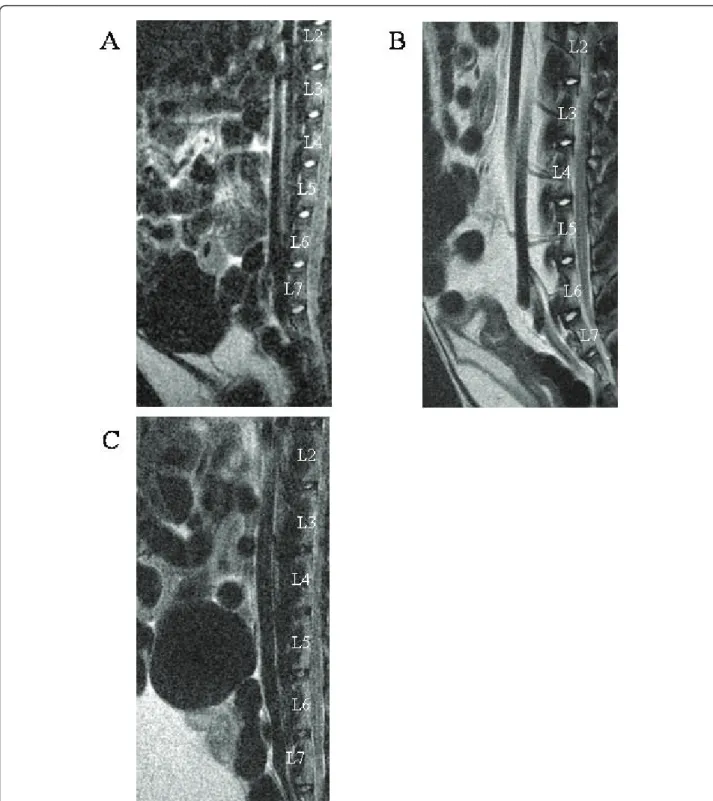
In order to characterize the aging process of IVD, the T2ws intensity of lumbar spine from one, six and 30-month-old rabbits was assessed. Representative MRI pic-ture of one-month-old IVD on Figure 1A indicates a grade I according to the Pfirrmann’grading to IVD from L2 to L6 levels. The lowest lumbar IVD (L6-L7) appears non-homogeneous with a dark transverse band. A grade II was assigned to this disc. The six-month-old rabbits’ (Figure 1B) NP from L2 to L7 level showed a
nonhomogeneous T2ws intensity with the presence of a dark transverse band and were assigned as grade II. Six-month-old disc height was not altered as compared to that of one-month old disc but the distinction between NP and AF became less discernable. For the 30-month-old rabbits (Figure 1C), all the IVD at lumbar level exhibited a non-homogeneous T2 signal intensity (gray). The IVD height was slightly decreased, and no distinc-tion between NP and AF could be observed. These were scored as grade III. In summary, MRI data indicate that rabbits exhibit scalable IVD changes evolving as early as one-month old.
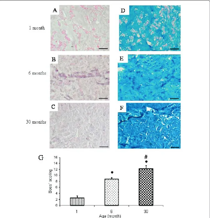
To further address whether the age-dependent MRI changes of rabbit IVD may correlate with some tissue alteration, we then performed histological stainings. Analysis of histological staining revealed a dramatic decrease in cell density as a function of age (Figure 2A, B, C). In addition, mucous degeneration and granular changes, shown by the presence of large decoloured and heterogeneous areas in granulated regions, were mark-edly increased (Figure 2D, E, F). Finally, the formation of tears and clefts shown by the presence of small thin defects was also affected by aging. To quantitatively assess the histological damage observed as a function of age, we next used a modified Boos’ scoring [13] (Figure 2G). Our results show a significant increase in the mod-ified Boos’ scoring from one to 30 months old. This scoring was 5.5-times higher in the 30-month-old rab-bits compared to one-month old rabrab-bits (p < 0.05).
Alteration of transcript expression patterns during IVD aging
To address whether the structural age-related changes identified by MRI and histology may be related to altera-tion of cell phenotype, we analysed the expression levels of
Table 1 real-time PCR investigation
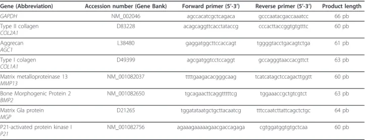
Gene (Abbreviation) Accession number (Gene Bank) Forward primer (5’-3’) Reverse primer (5’-3’) Product length
GAPDH NM_002046 agccacatcgctcagaca gcccaatacgaccaaatcc 66 pb
Type II collagen COL2A1 D83228 acagcaggttcacctataccg cccacttaccggtgtgtttc 60 pb Aggrecan AGC1 L38480 gaggatggcttccaccagt tggggtacctgacagtctga 61 pb Type I colagen COL1A1 D49399 agcgatggtcctccaggt gccagggtaaccacgttct 63 pb Matrix metalloproteinase 13 MMP13 NM_001082037 ttttgaagacacgggcaag tcatcatagctccagacttggtt 60 pb Bone Morphogenic Protein 2
BMP2
NM_001082650 tgcagaacttcaggtttttcg tggaaaccgctgtcgtct 63 pb Matrix Gla protein
MGP
D21265 tggatataatgctgcttacaatcg tttccaatcttattcagctctgc 64 pb P21-activated protein kinase I
P21
NM_001082756 agaaagaaaaagaacgaccagaga cgtggatggtgtgctcaa 60 pb
transcript coding for several IVD associated genes in freshly isolated NP cells from increasing ages. We first focused our attention on transcript coding for ECM genes (type II collagen COL2A1, type I collagen COL1A1 and aggrecan AGC1). Our data indicated that COL2A1 mRNA
levels decreased significantly with aging in NP cells (Figure 3A). The COL2A1 mRNA level was 100-fold lower in the 30-month-old rabbit compared to that of a one-month-old rabbit (p < 0.05). In parallel, a significant 2.5-fone-month-old decrease in AGC1 mRNA levels was also noted between
Figure 1 MRI images of the lumbar spine of rabbits with increasing ages. T2-weighted midsagittal images of (A) one-, (B) six- and (C) 30-month-old rabbits. Representative MRI images are shown.
Clouet et al. BMC Musculoskeletal Disorders 2011, 12:147 http://www.biomedcentral.com/1471-2474/12/147
one and 30-month-old rabbits (p < 0.05) (Figure 3B). In contrast to the decrease in COL2A1 and AGC1 mRNA levels, the COL1A1 mRNA level increased significantly with aging in NP cells (Figure 3C) with a 2.5-fold increase in COL1A1 mRNA at the age of 30-months compared to one-month. Taken together, these data indicate that the
expression of chondrogenic markers (COL2A1 and AGC1) decreases with age whereas that of the dedifferen-tiation marker (COL1A1) increases as early as thirty months.
Since it has been reported that the expression of matrix metalloprotease 13 (MMP13), bone morphogenetic
Figure 2 Histological characterization of theNucleus pulposus from IVD of rabbits with increasing ages. Histological sections of IVD from (A,D) one-, (B,E) six-, (C,F) 30-month-old rabbits were stained with (A, B, C) Haematoxilin Phloxin Safran and (D, E, F) alcian blue. Bar: 50μm. Representative images are shown. (G) A Modified Boos’s scoring was used to assess quantitatively the age-dependent tissue changes. *: p < 0.05 as compared to 1-month old rabbits; #: p < 0.05 as compared to 6-month old rabbits.
protein-2 (BMP2) and the matrix Gla protein (MGP) were affected during the course of osteoarthritis [14] and IVD aging [15], we were interested in deciphering whether these genes may also be affected by aging in rabbit NP. Whilst a significant decrease in MMP-13 mRNA levels was found between one and six-month-old NP, our data
indicate that MMP13 mRNA levels increased significantly between 6 and 30-month old rabbit (2.8-fold higher) (p < 0.05) (Figure 3D). In addition, BMP2 mRNA levels was found to be significantly up-regulated between one and 30-month-old rabbits with a 2.5 fold increase (p < 0.05) (Figure 3E). MGP mRNA levels were significantly
Figure 3 Phenotypic markers inNucleus pulposus cells. Analysis of the expression levels of transcripts coding for the phenotypic markers (A) type II collagen, (B) Aggrecan, (C) type I collagen, (D) MMP-13, (E) BMP-2, (F) MGP and (G) p21 in Nucleus pulposus (NP) cells from IVD of rabbits with increasing ages. Messenger RNAs were purified from freshly isolated NP cells and analyzed by real-time PCR as described in the materials and methods section. Results are reported as fold change in gene expression related to one month old rabbits. *: p < 0.05 as compared to one-month-old rabbits; #: p < 0.05 as compared to six-month old rabbits.
Clouet et al. BMC Musculoskeletal Disorders 2011, 12:147 http://www.biomedcentral.com/1471-2474/12/147
increased as a function of age with a significant seven-fold increase between one-and 30-month-old rabbits (Figure 3F).
Finally, since IVD aging has recently been associated with cellular senescence [16], we investigated whether the level of p21 mRNA may vary as a function of age (Figure 3G). Interestingly, our transcript analysis revealed that p21 mRNA level was significantly up-regu-lated (2 fold increase) between six month and 30-month-old rabbits.
Viewed together, our data strongly suggest that the age-dependent tissue changes evidenced by MRI and histology were associated with alteration of the pheno-type of NP cells.
Discussion
The current therapeutic strategies for patients with lower back pain remain symptomatic and are mainly dedicated to relieving painful symptoms. Our current understanding of the physiopathology of IVD degenera-tion allows us to consider regenerative medicine as a promising strategy. Indeed, recent reviews have consid-ered the imbalance between anabolism and catabolism as a pivotal factor of the IVD degeneration process. This imbalance between anabolism and catabolism contri-butes to the disorganization of the ECM. Accordingly, increasing attention has been paid to the regeneration of functional tissue based on the restoration of the ECM integrity by cell therapy and/or tissue engineering [3,17]. Nevertheless, before these promising biotherapies may enter the therapeutic arsenal, some preclinical tests in adapted animal models that closely mimic the physiolo-gical aging and degeneration process of human IVD should be performed. In this context, the present study aims at characterizing IVD aging and degeneration in the rabbit to propose this model as a suitable tool to i) gain new insights in the complex mechanisms of IVD degeneration and ii) to perform preclinical experiments dedicated to the evaluation of the safety and efficacy of IVD tissue engineering strategies.
In humans, MRI is the gold standard for the clinical investigation of IVD integrity. This technique allows the definition of IVD based on the tissue hydration shown by the intensity of the T2ws in the NP. The pictures of rabbit lumbar spines showed an age-dependent decrease in the T2ws and the appearance of a dark transverse band. This data is quite similar to that observed in humans during the course of IVD degeneration [18]. The decrease in T2ws intensity in human IVD after 20 years of age is well-acknowledged and reflects the decrease in water and proteoglycan contents during aging [19]. The dark transverse band is considered to be a normal structure in persons of 30 years or older [18]. Histological studies have thus suggested that this dark
transverse band may likely to be due to the accumula-tion of fibrous tissue in the central zone of NP [20]. To precisely assess disc degeneration on routine T2ws in human, Pfirrmann et al. have developed a reliable grad-ing system [11]. Interestgrad-ingly, the use of Pfirrmann’s sys-tem to grade IVD degeneration in rabbit reveals the same profile of T2ws variation as a function of age, thereby confirming the reliability of the different periods of life in rabbits previously described by Murakami et al. [21]: grade I for childhood (one-month old in rabbits is similar to a child in humans), grade II for adolescent rabbit (six months old in rabbits is similar to an adoles-cent in humans) and grade III-IV in adult age (30 months old in rabbits is similar to an adult in humans). All these MRI data suggest that the age-related changes that occurs in rabbit IVD share consistent similarities with those traditionally observed during the course of human IVD aging.
To further address whether the age-related changes observed by MRI may correlate with some tissue altera-tion, we then performed histological stainings. We were interested in addressing whether the modified Boos’ scoring, originally developed to histologically assess IVD changes in humans, could be transposed to rabbit IVD aging. Accordingly, the main features of human IVD degeneration were found in rabbit IVD with mucous degeneration, granular changes as well as cracks and tears being formed as a function of age. Interestingly, these histological changes perfectly corroborate the changes observed by MRI (loss of T2ws intensity and formation of a dark band). Taken together, our MRI and histological data show some similarities between the process of IVD aging in rabbits and humans thereby strongly suggesting that rabbits could be considered as a valuable spontaneous model of age-dependent IVD changes. In addition to being a reliable model of IVD aging, this spontaneous model may provide a closer representation of the pathophysiology of IVD aging in humans compared to the degeneration induced by nee-dle aspiration or chemonucleolysis [9]. Despite our data strongly suggesting that the rabbit is a relevant model of IVD aging, some limitations should, however, be consid-ered. Contrarily to humans, rabbits have notochordal cells in their NP at least up to 6 months of age [22]. In addition, in small quadrupeds and despite a relative dis-crepancy in disc diameter, the spine is likely to be loaded with smaller forces as compared to humans [23,24]. Whether both of these limits affect the rele-vance of the rabbit model is not yet known.
To our knowledge, only two studies have partially described IVD aging in rabbits [25,26]. In these previous studies, rabbit IVD aging was evaluated longitudinally between the age of 6 and 42 months. In this previous study, rabbit IVD aging is longitudinally evaluated
between the age of six and 42 months. On the contrary to our study, no result is available for the period ranging from one to six months, which could be an essential period for the onset of IVD aging. In fact, it is well known that the NP of less than twelve-month-old rab-bits contain some remnant cells originating from the embryonic notochord [22]. Whether the age-dependent loss of notochordal cells, most likely by apoptosis, is intimately related to IVD aging is not yet fully deci-phered but has been proposed as a possible initiating mechanism for IVD degeneration [27].
NP is the structure in which IVD degeneration is initiated by dehydration followed by an alteration of the structural organization of the tissue [13]. Therefore, to strengthen our MRI and histological data, we finally sought to decipher whether the above described age-dependent tissue changes may correlate with alteration in the NP cell phenotype. To address this issue, we focused our attention on the expression levels of mole-cules that were previously shown to be modulated dur-ing the onset of IVD degeneration or osteoarthritis (COL1A1, COL2A1, AGC1, BMP2, MMP13, MGP and P21) [14,16,28]. Since it remains difficult to quantita-tively evaluate the level of the corresponding proteins, particularly in the rabbit, we embarked on the analysis of the corresponding transcript levels by real-time RT-PCR. Our data describing an age-dependent increase in COL1A1 expression with a concomitant decrease in COL2A1 and AGC1 expression suggest that NP cells experience a process of dedifferentiation. Interestingly, this dedifferenciation process has been well described in cultured articular chondrocytes [29] and in osteoar-thritic joints [30]. Our data also suggests that during the process of IVD aging, the molecules with a longer half-life (collagen type II) exhibit an early decrease in the corresponding transcript levels. Conversely, the mole-cules with a shorter half-life (aggrecan) have a transcript expression levels that starts to decline later on. Whether this differential regulation of ECM molecules may be of importance for the maintenance of IVD integrity deserves further attention.
Among the various genes modulated in osteoarthritic chondrocytes [14], MMP-13 is probably the most trust-worthy. We were therefore interested to find a signifi-cant modulation of these genes during IVD degeneration. MMP-13 is known to degrade collagens and glycosaminoglycans [31]. The increase in MMP13 could therefore be a major contributor of IVD degenera-tion [16] as it has been extensively reported in cartilage degradation during OA [14].
BMP-2, a growth factor that stimulates the production of ECM components in IVD [21] and articular cartilage [32], was also shown to be stimulated as a function of age in rabbit IVD. One can assume that the increase in
BMP2 expression during IVD aging is related to the existence of reparative mechanisms that could contri-bute to slowing down ECM degradation. In line with this suggestion, MGP, one of the most potent inhibitors of calcification in mammals [33], was also found to be up-regulated as a function of age [15] thereby support-ing our hypothesis of the existence of such compensa-tory mechanisms. Of note, similar compensacompensa-tory mechanisms have been described in osteoarthritic carti-lage, which draws attention to the physiopathological vicinity of IVD degeneration and osteoarthritis. Finally, the age-dependent increase in p21, a cycline dependant kinase inhibitor that in addition to being involved in cel-lular senescence [16] has also been reported to inhibit type II collagen expression in articular chondrocytes [34], further highlights the similarities between OA and IVD degeneration.
Conclusions
This study describes the consistency of the rabbit as a model of age-dependent IVD changes. Our data also highlight some similarities between the physiopathologi-cal processes involved in the onset of IVD aging and those described for osteoarthritis. Finally, our study makes the rabbit a valuable tool (i) to gain new insights into the complex molecular mechanisms that govern IVD aging and (ii) to test the preclinical efficacy of tis-sue engineering strategy that may offer the possibility of regenerating damaged IVD.
Acknowledgements
This study was supported by grants from Société Française de
Rhumatologie, Fondation de l’Avenir pour la Recherche Médicale Appliquée (ET8-491 and ET9-541), Agence Nationale de la Recherche AAP TeCSAN (chondrograft project), Agence de la Biomédecine, Institut National de la Santé et de la Recherche Médicale, région des Pays de la Loire (BIOREGOSproject), and the University Hospital of Nantes. The authors gratefully acknowledge the technical assistance of Pierre Weiss, Maïthe Gatius-Perré, Pierre Monmousseau, Patrice Roy, Stéphane Madec, Dominique Rouleau and Christian Raphael, as well as Joanna Ashton-Chess for the critical reading of the manuscript.
Author details
1INSERM (Institut National de la Santé et de la Recherche Médicale) U791,
LIOAD, Group Skeletal Tissue Engineering and Physiopathology (STEP), University of Nantes, Nantes F-44042, France.2PresUNAM, University of
Nantes, Nantes, France.3Department of Pharmacy, University Hospital of
Nantes, Nantes F-44093, France.4Department of Rheumatology, University Hospital of Nantes, Nantes F-44093, France.5Department of experimental
Surgery, CRIP-VC, National Veterinary School (ONIRIS), Nantes F-44307, France.6Department of Imagery, CRIP-VC, National Veterinary School
(ONIRIS), Nantes F-44307, France.7INRA UMR703, National Veterinary School (ONIRIS), Nantes F-44307, France.8Graftys SAS, Aix en Provence, France.
Authors’ contributions
JC and MPV have conceived and coordinated the study. They participated in MRI analysis, Boos’scoring and RT-PCR analysis. They also performed the statistical analysis and drafted the manuscript. MM performed the PCR analysis. JL performed the histological stainings. BHF, OG participated in the design of the study, particularly in animal management. MF performed MRI imaging and participated in analysis. YC participated in histological analysis. Clouet et al. BMC Musculoskeletal Disorders 2011, 12:147
http://www.biomedcentral.com/1471-2474/12/147
GG and YM have helped in the writing of the manuscript. JG and CV helped to the conception of the study, and participated in its design and have helped in the writing of the manuscript. All authors read and approved the final manuscript.
Competing interests
The authors declare that they have no competing interests.
Received: 29 March 2011 Accepted: 4 July 2011 Published: 4 July 2011 References
1. Sive JI, Baird P, Jeziorsk M, et al: Expression of chondrocyte markers by cells of normal and degenerate intervertebral discs. Mol Pathol 2002, 55(2):91-7.
2. Clouet J, Grimandi G, Pot-Vaucel M, et al: Identification of phenotypic discriminating markers for intervertebral disc cells and articular chondrocytes. Rheumatology 2009, 48(11):1447-50.
3. Clouet J, Vinatier C, Merceron C, et al: The intervertebral disc: from pathophysiology to tissue engineering. Joint Bone Spine 2009, 76(6):614-8. 4. Hadjipavlou AG, Tzermiadianos MN, Bogduk N, Zindrick MR: The
pathophysiology of disc degeneration: a critical review. J Bone Joint Surg Br 2008, 90(10):1261-70.
5. Raj PP: Intervertebral disc: anatomy-physiology-pathophysiology-treatment. Pain Pract 2008, 8(1):18-44.
6. Ruan D, He Q, Ding Y, et al: Intervertebral disc transplantation in the treatment of degenerative spine disease: a preliminary study. Lancet 2007, 369(9566):993-9.
7. Alini M, Li W, Markovic P, et al: The potential and limitations of a cell-seeded collagen/hyaluronan scaffold to engineer an intervertebral disc-like matrix. Spine 2003, 28(5):446-54, 453.
8. Roughley P, Hoemann C, DesRosiers E, et al: The potential of chitosan-based gels containing intervertebral disc cells for nucleus pulposus supplementation. Biomaterials 2006, 27(3):388-96.
9. Alini M, Eisenstein SM, Ito K, et al: Are animal models useful for studying human disc disorders/degeneration? Eur Spine J 2008, 17(1):2-19. 10. Lotz JC: Animal models of intervertebral disc degeneration: lessons
learned. Spine 2004, 29(23):2742-50.
11. Pfirrmann CW, Metzdorf A, Zanetti M, Hodler J, Boos N: Magnetic resonance classification of lumbar intervertebral disc degeneration. Spine 2001, 26(17):1873-8.
12. Vinatier C, Gauthier O, Masson M, et al: Nasal chondrocytes and fibrin sealant for cartilage tissue engineering. J Biomed Mater Res A 2009, 89(1):176-85.
13. Boos N, Weissbach S, Rohrbach H, et al: Classification of age-related changes in lumbar intervertebral discs: 2002 Volvo Award in basic science. Spine 2002, 27(23):2631-44.
14. Goldring MB, Otero M, Tsuchimochi K, Ijiri K, Li Y: Defining the roles of inflammatory and anabolic cytokines in cartilage metabolism. Ann Rheum Dis 2008, 67(Suppl 3):iii75-82.
15. Lee CR, Sakai D, Nakai T, et al: A phenotypic comparison of intervertebral disc and articular cartilage cells in the rat. Eur Spine J 2007,
16(12):2174-85.
16. Le Maitre CL, Freemont AJ, Hoyland JA: Accelerated cellular senescence in degenerate intervertebral discs: a possible role in the pathogenesis of intervertebral disc degeneration. Arthritis Res Ther 2007, 9(3):R45. 17. Kalson NS, Richardson S, Hoyland JA: Strategies for regeneration of the
intervertebral disc. Regen Med 2008, 3(5):717-29.
18. Aguila LA, Piraino DW, Modic MT, et al: The intranuclear cleft of the intervertebral disk: magnetic resonance imaging. Radiology 1985, 155(1):155-8.
19. Sether LA, Yu S, Haughton VM, Fischer ME: Intervertebral disk: normal age-related changes in MR signal intensity. Radiology 1990, 177(2):385-8. 20. Yu SW, Haughton VM, Ho PS, et al: Progressive and regressive changes in
the nucleus pulposus. Part II. The adult. Radiology 1988, 169(1):93-7. 21. Murakami H, Yoon ST, Attallah-Wasif ES, et al: The expression of anabolic
cytokines in intervertebral discs in age-related degeneration. Spine 2006, 31(16):1770-4.
22. Hunter CJ, Matyas JR, Duncan NA: The notochordal cell in the nucleus pulposus: a review in the context of tissue engineering. Tissue Eng 2003, 9(4):667-77.
23. Smit TH: The use of a quadruped as an in vivo model for the study of the spine - biomechanical considerations. Eur Spine J 2002, 11(2):137-44. 24. Wilke HJ, Rohlmann A, Neller S, et al: ISSLS prize winner: A novel
approach to determine trunk muscle forces during flexion and extension: a comparison of data from an in vitro experiment and in vivo measurements. Spine 2003, 28(23):2585-93.
25. Sowa G, Vadala G, Studer R, et al: Characterization of intervertebral disc aging: longitudinal analysis of a rabbit model by magnetic resonance imaging, histology, and gene expression. Spine 2008, 33(17):1821-8. 26. Murakami H, Yoon TS, Attallah-Wasif ES, et al: Quantitative differences in
intervertebral disc-matrix composition with age-related degeneration. Med Biol Eng Comput 2010, 48(5):469-74.
27. Hunter CJ, Matyas JR, Duncan NA: The functional significance of cell clusters in the notochordal nucleus pulposus: survival and signaling in the canine intervertebral disc. Spine 2004, 29(10):1099-104.
28. Canfield AE, Hadfield KD, Rock CF, Wylie EC, Wilkinson FL: HtrA1: a novel regulator of physiological and pathological matrix mineralization? Biochem Soc Trans 2007, 35(Pt 4):669-71.
29. Schnabel M, Marlovits S, Eckhoff G, et al: Dedifferentiation-associated changes in morphology and gene expression in primary human articular chondrocytes in cell culture. Osteoarthritis Cartilage 2002, 10(1):62-70. 30. Clouet J, Vinatier C, Merceron C, et al: From osteoarthritis treatments to
future regenerative therapies for cartilage. Drug Discov Today 2009, 14(19-20):913-25.
31. Roberts S, Caterson B, Menage J, et al: Matrix metalloproteinases and aggrecanase: their role in disorders of the human intervertebral disc. Spine 2000, 25(23):3005-13.
32. Cheng C, Conte E, Pleshko-Camacho N, Hidaka C: Differences in matrix accumulation and hypertrophy in superficial and deep zone
chondrocytes are controlled by bone morphogenetic protein. Matrix Biol 2007, 26(7):541-53.
33. Luo G, Ducy P, McKee MD, et al: Spontaneous calcification of arteries and cartilage in mice lacking matrix GLA protein. Nature 1997,
386(6620):78-81.
34. Sironen RK, Karjalainen HM, Elo MA, et al: cDNA array reveals mechanosensitive genes in chondrocytic cells under hydrostatic pressure. Biochim Biophys Acta 2002, 1591(1-3):45-54.
Pre-publication history
The pre-publication history for this paper can be accessed here: http://www.biomedcentral.com/1471-2474/12/147/prepub
doi:10.1186/1471-2474-12-147
Cite this article as: Clouet et al.: Characterization of the age-dependent intervertebral disc changes in rabbit by correlation between MRI, histology and gene expression. BMC Musculoskeletal Disorders 2011 12:147.
Submit your next manuscript to BioMed Central and take full advantage of:
• Convenient online submission
• Thorough peer review
• No space constraints or color figure charges
• Immediate publication on acceptance
• Inclusion in PubMed, CAS, Scopus and Google Scholar
• Research which is freely available for redistribution
Submit your manuscript at www.biomedcentral.com/submit