HAL Id: hal-02152327
https://hal.archives-ouvertes.fr/hal-02152327
Submitted on 11 Jun 2019HAL is a multi-disciplinary open access archive for the deposit and dissemination of sci-entific research documents, whether they are pub-lished or not. The documents may come from teaching and research institutions in France or abroad, or from public or private research centers.
L’archive ouverte pluridisciplinaire HAL, est destinée au dépôt et à la diffusion de documents scientifiques de niveau recherche, publiés ou non, émanant des établissements d’enseignement et de recherche français ou étrangers, des laboratoires publics ou privés.
Endothelial Dysfunction in Wistar Rats
Joseph Iharinjaka Randriamboavonjy, Sandrine Heurtebise, Pierre Pacaud,
Gervaise Loirand, Angela Tesse
To cite this version:
Joseph Iharinjaka Randriamboavonjy, Sandrine Heurtebise, Pierre Pacaud, Gervaise Loirand, Angela Tesse. Moringa oleifera Seeds Improve Aging-Related Endothelial Dysfunction in Wistar Rats. Ox-idative Medicine and Cellular Longevity, Hindawi, 2019, 2019, pp.2567198. �10.1155/2019/2567198�. �hal-02152327�
Research Article
Moringa oleifera Seeds Improve Aging-Related Endothelial
Dysfunction in Wistar Rats
Joseph Iharinjaka Randriamboavonjy
,
1,2Sandrine Heurtebise,
1Pierre Pacaud,
1Gervaise Loirand
,
1,2and Angela Tesse
11L’institut du thorax, INSERM, CNRS, UNIV Nantes, Nantes, France 2L’institut du thorax, CHU Nantes, Nantes, France
Correspondence should be addressed to Angela Tesse; [email protected] Received 28 December 2018; Accepted 14 February 2019; Published 13 May 2019 Guest Editor: Albino Carrizzo
Copyright © 2019 Joseph Iharinjaka Randriamboavonjy et al. This is an open access article distributed under the Creative Commons Attribution License, which permits unrestricted use, distribution, and reproduction in any medium, provided the original work is properly cited.
Vascular aging is characterized by functional and structural changes of the vessel wall, including endothelial dysfunction, with decreased endothelial NO⋅bioavailability and elevated vasoconstrictor and inflammatory mediator production, vascular rigidity, and tone impairment. Moringa oleifera (MOI) is a little tree, and different parts of which are used in traditional medicine in tropical Africa, America, and Asia for therapeutic applications in several disorders including cardiovascular disease. The present study is aimed at assessing the effect of MOI on aging-associated alteration of the endothelial function in Wistar rats. Middle-aged Wistar rats (46-week-old males) have been fed with food containing or not 750 mg/kg/day of MOI seed powder for 4 weeks. A group of young Wistar rats (16-week-old) was used as control. Measurement of isometric contraction, western blot analysis, and immunostaining has then been performed in the aortas and mesenteric arteries to assess the endothelium function. MOI treatment improved carbachol-induced relaxation in both aortas and mesenteric arteries of middle-aged rats. In the aortas, this was associated with an increased Akt signalling and endothelial NO synthase activation and a downregulation of arginase-1. In the mesenteric arteries, the improvement of the endothelial-dependent relaxation was related to an EDHF-dependent mechanism. These results suggest a vascular protective effect of MOI seeds against the vascular dysfunction that develops during aging through different mechanisms in conductance and resistance arteries.
1. Introduction
Vascular aging corresponds to functional and structural changes of the arterial wall characterized by endothelial dys-function and vascular rigidity [1, 2]. A primary mechanism responsible for aging-induced endothelial dysfunction is the decrease in the bioavailability and the production of nitric
oxide (NO⋅), mainly resulting from the increased oxidative
stress. NADPH-derived superoxide anions (O2-) interact
with NO to form peroxynitrite (ONOO-), which induces
damaged protein accumulation and vascular inflammation
and remodelling [3]. Peroxynitrite oxidizes the endothelial NO synthase (eNOS) cofactor BH4, thereby inactivating
eNOS and reducing NO⋅ production [4, 5]. Moreover, BH4
reduction can result in eNOS uncoupling, leading to the
generation of ROS rather than NO⋅ and thus perpetuating
endothelial dysfunction [5]. Increased arginase expression and activity are also involved in endothelial dysfunction in aged vessels [6, 7]. Since arginase and eNOS compete for their common L-arginine (L-Arg) substrate, a rise in arginase
activity or expression limits endothelial NO⋅ production in
the vasculature [6–8]. Furthermore, the increased methyl-ation of L-Arg and the resulting production of dimethyl L-Arg, observed in aged rats and humans, also contribute to the inhibition of eNOS activity [9].
In addition to decreased NO⋅ production and
bioavail-ability, aging-associated endothelial dysfunction in resistance arteries also comprised oxidative stress-dependent impair-ment of prostacyclin and EDHF production that contributes to the defective endothelium-mediated vasorelaxation [5, 10].
Hindawi
Oxidative Medicine and Cellular Longevity Volume 2019, Article ID 2567198, 9 pages https://doi.org/10.1155/2019/2567198
Several studies suggested that polyphenols and other natural compounds contained in bioactive extracts and foods can protect against vascular aging through their
anti-inflammatory and antioxidant properties as well as for their
ability to improve eNOS activity and NO⋅ bioavailability
[11–13]. Moringa oleifera (MOI) is a little tree used in
Malagasy traditional medicine to treat several pathological
states such as hypertension and inflammation [14, 15].
MOI seed oil showed free radical scavenging activity due to
molecules such as flavonoids known to have antioxidant
properties [16]. We previously described the cardiovascular protective effect of the oral administration of MOI seeds against cardiac complications induced by high blood pressure
(left ventricle hypertrophy andfibrosis), vascular
inflamma-tion, and oxidative stress in spontaneously hypertensive rats
(SHR), thus showing the anti-inflammatory and antioxidant
action of a diet containing MOI seeds [17, 18].
The aim of the present work was to investigate the
potential beneficial effects of MOI seeds administrated
during 4 weeks against established aging-related vascular dysfunction in middle-aged Wistar rats (MAWR) by analys-ing endothelial function in conductance (aorta) and resistance (mesenteric) arteries.
2. Methods
2.1. Animals. Male Wistar rats (46 weeks old, middle-aged) were divided into two groups: a group receiving normal food (MAWR) and a group fed with food containing MOI seed powder (750 mg/day) mixed with standard pellet diet (MOI MAWR) for 4 weeks. This dose is within the range of concentrations classically used in experimental rodent models [18, 19]. A control group of young rats (16 weeks old, YWR), receiving normal food, was also used to check that middle-aged rats did indeed have arterial dysfunction. All groups received water ad libitum. At the end of the exper-imental protocol, all the rats were euthanized. The thoracic aorta and mesenteric arteries were then collected for vascular reactivity, western blot, and immunohistological analyses. All experiments were conducted in agreement with our Ethical Committee Guide for the Care and Use of Laboratory Animals (authorisation number 00909.01).
2.2. Arterial Reactivity. The aortas andfirst branches of
supe-rior mesenteric arteries were collected in physiological saline
solution (in mM; 130 NaCl, 5.6 KCl, 1 MgCl2, 2 CaCl2, 11
glucose, and 10 Tris, pH 7.4 with HCl), cleaned, and cut in 2 mm long rings. Arterial rings were then mounted on multi-channel isometric myograph, bathed in Krebs-Henseleit
solution at 37°C bubbled with 95% O2-5% CO2, and
connected to a force transducer (Pioden Controls Ltd., Canterbury, UK, for aortic rings; Danish Myo Technology, Aarhus, Denmark, for mesenteric artery rings). After equili-bration, the contractile response to KCl (60 mM) was mea-sured. Endothelial function was tested by measuring the relaxing response to cumulative doses of carbachol (CCh,
1 nM-10μM, Sigma-Aldrich) of rings precontracted by
phenylephrine (PhE, 1μM, Sigma-Aldrich) in the absence
and presence of L-NG-nitroarginine methyl ester (L-NAME,
100μM, Sigma-Aldrich) alone or in association with the
cyclooxygenase (COX) inhibitor, indomethacin (10μM,
Sigma-Aldrich). Digital data were recorded by a MacLab/4e recorder and analysed using a LabChart v7 software (AD Instruments, Paris, France).
2.3. Staining and Confocal Microscopy Imaging. Frozen
sections of the aortas (7μm thick) were fixed with cold
100% methanol and incubated for 2 h at room temperature
in blocking buffer (5% of albumin in PBS). Tissue sections
were then incubated overnight (4°C) with a mouse
monoclo-nal antibody against the phosphorylated-(Ser 1179)-eNOS (1 : 50, Santa Cruz Biotechnology) or a rabbit polyclonal anti-body against phosphorylated-(Ser473)-Akt (p-Akt) protein (1/500, Cell Signaling). Three washes were followed by incubation (1 h, at room temperature, in the dark) with the
secondary mouse or rabbit fluorescent Alexa
fluor-647-conjugated antibody (1 : 500, Molecular Probes). A Nikon A1-RS inverted laser scanning confocal microscope was used for the optical sectioning of the tissue. Digital image recording was performed using the NIS element software. Images were analysed and processed by Fiji software.
2.4. Western Blot Analysis. The aortas were homogenized and
lysed. Proteins (50μg) were separated in precoated
SDS-PAGE 4-15% (MiniPROTEAN® TGX™, Bio-Rad), trans-ferred to nitrocellulose membrane, and then incubated (2 h at room temperature) in blocking buffer (5% nonfat dry milk
in PBS). The membranes were probed overnight at 4°C with
primary rabbit polyclonal antibody directed to arginase-1 (1/200, Santa Cruz Biotechnology). After 3 washes, immuno-reactive bands were revealed with a secondary peroxidase-conjugated anti-rabbit IgG (1 : 5000, Beckman Coulter), detected by enhanced chemiluminescence system (ECL Plus,
Amersham Biosciences), and quantified by densitometry. A
polyclonal anti-α-tubulin antibody (1 : 5000, Sigma-Aldrich)
was used to check protein gel loading and to normalize protein expression. The analysis of the blots was performed with Image Lab™ software v4.1 (Bio-Rad).
2.5. Data Analysis. The endothelial relaxation in response to cumulative doses of CCh was expressed as a percentage of the amplitude of PhE-induced precontraction. For the statistical analysis of these data, we used a two-way analysis of variance for repeated measures with subsequent Bonferroni post hoc test. To calculate the area under the curve (AUC) of the concentration-dependent relaxation in response to CCh, we used the GraphPad Prism 5.02 software. A one-way ANOVA with subsequent Bonferroni post hoc test or the ANOVA on ranks was performed for the AUC of relaxation and protein expression data. All statistical analysis were realized with
SigmaStat 3.5 software. All values are presented asmean ±
SD of n repetitive measurements, with n representing the
number of rat samples. ∗p < 0 05 was considered to be
statistically significant.
3. Results
3.1. MOI Administration Restores CCh-Induced Relaxation
treatment on endothelial-dependent relaxation of aorta rings from MAWR by measuring the CCh-dependent relaxation in the three groups of rats and calculating the AUC in the absence and in the presence of L-NAME (Figure 1). As expected, CCh-induced endothelium-dependent relaxation was reduced in the aortas of MAWR compared to YWR aortas (Figures 1(a) and 1(c)). MOI seed administration almost completely restored CCh-induced relaxation in the aortas of MAWR that returned similar to that observed in YWR (Figures 1(b) and 1(c)). L-NAME completely abolished CCh-induced relaxation in the aortas from YWR, MAWR, and MOI MAWR (Figures 1(b) and 1(c) and Supplementary Figure 1) indicating that the relaxation response to CCh in
the aorta is completely dependent of NO⋅ in the three
groups of rats. These data suggest that the improvement of the endothelial function induced by MOI treatment in the aortas could result from the correction of aging-induced
NO⋅signalling defect.
3.2. MOI Administration Improves eNOS Signalling in the
Aortas. To confirm the effect of MOI treatment on NO⋅
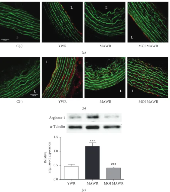
signalling, we directly assessed eNOS activity by immuno-staining on cross sections of the aortas of active phosphory-lated-(Ser1179)-eNOS and active phosphorylated-(Ser473)-Akt, one of the main kinases phosphorylating eNOS at
Ser1179 [20] (Figures 2(a) and 2(b)). Both endothelial phos-phorylated-(Ser1179)-eNOS and phosphorylated-(Ser473)-Akt staining observed in YWR were lost in MAWR but were restored in MOI MAWR (Figures 2(a) and 2(b)) suggesting a role of Akt-induced eNOS activation in the beneficial effect of MOI on endothelial function in MAWR. We next assessed the expression of arginase-1 that might also
contribute to the reduced production of NO⋅with aging [21]
(Figure 2(c)). Arginase-1 expression, increased in MAWR aortas compared to YWR, was decreased to a level similar to YWR in MOI MAWR (Figure 2(c)). These data are
consis-tent with a beneficial effect of MOI on eNOS activity in the
aortas of MAWR.
3.3. MOI Administration Improves Endothelial Function in AWR Mesenteric Arteries. We also assessed the effect of MOI on the endothelial function of resistance arteries from MAWR. Both the dose-dependent relaxation curve to CCh and the AUC show that the relaxation induced by CCh was decreased in MAWR mesenteric arteries compared to YWR vessels (Figures 3(a) and 3(c)). MOI seeds improved the endothelial relaxation in the mesenteric arteries of MAWR compared to nontreated MAWR (Figures 3(b) and 3(c)). In YWR, L-NAME induced only a rightward shift of the concentration-relaxation curve to CCh without change of
−10 −9 −8 −7 −6 −5 0 20 40 60 80 100 YWR MAWR ⁎⁎⁎ ⁎⁎⁎ ⁎⁎ Log (CCh) M Relaxa tio n (%) (a) −10 −9 −8 −7 −6 −5 0 20 40 60 80 100 MAWR
MAWR+L‑NAME MOI MAWR MOI MAWR+L‑NAME ### ### ### ### $$ $$$ $$$ ⁎⁎⁎ ⁎⁎⁎ ⁎⁎⁎ Log (CCh) M Relaxa tio n (%) (b) ### $$$ $$$ $$$ ⁎⁎⁎
MAWR MOI MAWR YWR − + − + − + 0 50 100 150 200 250 L-NAME A.U .C. (c)
Figure 1: (a) Carbachol- (CCh-) induced relaxation in the aortas from young rats (YWR: 16 weeks old) and control middle-aged rats (MAWR, 50 weeks old;∗∗p < 0 01,∗∗∗p < 0 001, YWR versus MAWR). (b) Relaxation curves to CCh in the aortas from MAWR and MOI-treated middle-aged rats (MOI MAWR) in the absence and in the presence of L-NAME (100μM) (∗∗∗p < 0 001, MOI MAWR versus MAWR;###p < 0 001, MAWR versus MAWR+L-NAME;$$p < 0 01,$$$p < 0 001, MOI MAWR versus MOI MAWR+L-NAME). (c) Histogram showing the area under the curve (AUC) of CCh-induced relaxation without and with L-NAME in YWR, MAWR, and MOI MAWR (∗∗∗p < 0 001, MAWR versus YWR;###p < 0 001 MOI MAWR versus MAWR;$$$p < 0 001 without L-NAME versus with L-NAME in the same group. Results are expressed inmean ± SD; n = 5).
3 Oxidative Medicine and Cellular Longevity
the maximal relaxation, indicating that NO⋅ did not play a major role in CCh-induced relaxation of YWR mesenteric arteries (Supplementary Figure 2). Accordingly, the AUC
was only decreased by ~25% by L-NAME in YWR
mesenteric arteries (Figure 3(c)). In contrast, L-NAME strongly reduced the CCh-induced relaxation in MAWR as
illustrated by the ~80% reduction of the maximal
CCh-induced relaxation (Figure 3(b)) and the~70% reduction of
the AUC observed in the presence of L-NAME (Figure 3(c)).
This suggests that aging increased the NO⋅ component of
CCh-induced relaxation in the mesenteric arteries. MOI increased the relaxation response in MAWR mesenteric arteries, and L-NAME reduced only partially the CCh-induced relaxation in MOI MAWR, suggesting that the beneficial effect of MOI on the endothelial function was not
mainly mediated by NO⋅ signalling but involved other
mechanism(s) (Figures 3(b) and 3(c)).
3.4. MOI Administration Enhances EDHF-Dependent
Relaxation in MAWR Mesenteric Arteries. We hypothesized that MOI improved endothelium-dependent relaxation in the mesenteric arteries of MAWR by increasing the contri-bution of EDHF. We therefore directly measured the EDHF-dependent component of the CCh-induced relaxation in the presence of both L-NAME and INDO to block both
NO⋅- and COX-deriving relaxing factors, respectively
(Figure 4). The EDHF-mediated relaxation of the mesenteric
arteries in response to CCh was significantly decreased in
MAWR compared to YWR, indicating that in mesenteric arteries CCh-induced relaxation was essentially dependent
L
L L
L
YWR
C(-) MAWR MOI MAWR
(a)
L L
L L
YWR
C(-) MAWR MOI MAWR
(b)
Arginase-1 𝛼‑Tubulin
YWR MAWR MOI MAWR
0.0 0.5 1.0 1.5 ### ⁎⁎⁎ Relative arginase-1 expression (c)
Figure 2: (a) Immunohistochemical representative red staining of phosphorylated-(Ser 1179)-eNOS or phosphorylated-(Ser 473)-Akt (b) in the aortas from YWR, control MAWR, and MOI MAWR. Greenfluorescence corresponds to elastin autofluorescence. L = lumen of the vessel. C(-) is a negative control without the primary antibody. Scalebar = 100 μm. (c) Representative western blot and corresponding densitometric analysis of arginase-1 expression normalized toα-tubulin in the aortas of YWR, MAWR, and MOI MAWR (mean ± SD, n = 3;∗∗∗p < 0 001 MAWR versus YWR;###p < 0 001 MOI MAWR versus MAWR).
−10 −9 −8 −7 −6 −5 0 20 40 60 80 100 ∗ ∗∗∗ ∗∗∗ MAWR YWR Log (CCh) M Relaxation (%) (a) −10 −9 −8 −7 −6 −5 0 20 40 60 80 100 ⁎ ⁎ MAWR
MAWR+L-NAME MOI MAWRMOI MAWR+LNAME
### ## ## Log (CCh) M Relaxation (%) (b) − + − + − + 0 100 200 300 ⁎⁎⁎ ### $ $$ $ + L-NAME A.U.C.
YWR MAWR MOI MAWR
(c)
Figure 3: (a) Carbachol- (CCh-) induced relaxation in the mesenteric arteries of YWR and control MAWR (∗p < 0 05,∗∗∗p < 0 001, MAWR versus YWR). (b) Relaxation curves to CCh in the mesenteric arteries from MAWR and MOI MAWR in the absence and in the presence of L-NAME (100μM) (∗p < 0 05, MAWR+L-NAME versus MAWR;##p < 0 01,###p < 0 001, MOI MAWR+L-NAME versus MOI MAWR). (c) Histograms showing the area under the curve (AUC) without and with L-NAME for YWR, MAWR, and MOI MAWR (∗∗∗p < 0 001, MAWR versus YWR;###p < 0 001, MOI MAWR versus MAWR;$p < 0 05,$$p < 0 01, without L-NAME versus with L-NAME in the same group;+p < 0 05 MOI MAWR+L-NAME versus MAWR+L-NAME). Results were expressed in mean ± SD of n = 5 rats.
−10 −9 −8 −7 −6 −5 0 20 40 60 80 100 MAWR+L‑NAME+INDO MOI MAWR+L‑NAME+INDO YWR+L‑NAME+INDO ## ## ∗ ∗ ∗∗∗ ∗∗∗ Log (CCh) M Relaxa tio n (%) ∗ ∗ (a)
YWR MAWR MOI MAWR
0 50 100 150 200 250 ⁎⁎⁎ ## A.U .C. (b)
Figure 4: (a) EDHF-dependent relaxation to carbachol (CCh) in the mesenteric arteries from YWR, MAWR, and MOI MAWR obtained in the presence of L-NAME and indomethacin (INDO), L-NAME+INDO (∗p < 0 05,∗∗∗p < 0 001, versus YWR+L-NAME+INDO). (b) Histograms showing the area under the curve (AUC) of CCh-dependent relaxation of YWR, MAWR, and MOI MAWR mesenteric artery in the presence of L-NAME and INDO (L-NAME+INDO) (∗∗∗p < 0 001, MAWR+L-NAME+INDO versus YWR+L-NAME+INDO; ##p < 0 01, MOI MAWR+L-NAME+INDO versus MAWR+L-NAME+INDO; results are expressed in mean ± SD with n = 5 per group).
5 Oxidative Medicine and Cellular Longevity
on EDHF in YWR and that aging strongly reduced this EDHF-mediated relaxation (Figures 4(a) and 4(b)). MOI not totally but strongly restored EDHF-mediated relaxation in MAWR (Figures 4(a) and 4(b)). These data thus indicate that the altered endothelium-dependent relaxation response to CCh in MAWR mesenteric arteries resulted from a loss of its EDHF-mediated component which was partially corrected by MOI.
4. Discussion
Our study provides evidence for the beneficial arterial effects of MOI seed administration in MAWR with an established aging-induced endothelial dysfunction in both the aorta and mesenteric arteries. Diet containing MOI seeds improves the endothelial function in MAWR by boosting eNOS activ-ity in the aorta and enhancing EDHF-dependent relaxation
in the mesenteric arteries. These data suggest a different effect
of MOI seeds on the endothelial aging process in conduc-tance and resisconduc-tance arteries.
Vascular aging consists of a set of changes in the mechanical and structural properties of the vascular wall including a decrease in endothelium-dependent relaxation
by reducing the bioavailability of NO⋅ and/or EDHF
depending on the arterial bed [4, 10, 22]. As previously
described [23, 24], we confirm that the
endothelium-dependent CCh-induced relaxation in the aortas was
exclusively mediated by NO⋅ both in YWR and MAWR.
Aging was associated with a reduction of
endothelium-dependent NO⋅ signalling attested by the loss of the
CCh-induced relaxation and the decrease of phosphoryla-tion of eNOS and its upstream kinase Akt, in agreement with the role of Akt/eNOS signalling impairment in aging-induced endothelial dysfunction [20, 25]. MOI admin-istration corrected the Akt/eNOS defect and restored
NO⋅-mediated endothelium-dependent relaxation in MAWR.
NO signalling has been identified as a target of the beneficial
action of polyphenol-rich diets on endothelial function and cardiovascular protection [26, 27]. Indeed, similar improve-ments of endothelial function by activating the Akt/eNOS pathway were previously demonstrated in a diet
supple-mented with other natural substances such as (R)-α-lipoic
acid contained in several vegetal foods and other polyphenols such as delphinine and resveratrol contained in red wine [25, 28, 29]. Polyphenolic compounds contained in MOI seeds could thus be responsible for or participate in the
beneficial effects of MOI seeds on Akt/eNOS signalling
in the endothelium of MAWR [30, 31]. Indeed, MOI seeds contain glucosinolates (glucomoringin) and isothiocyanates known for their ability to improve endothelial-dependent relaxation in rat aortas [32].
In addition to the stimulation of Akt/eNOS signalling, we observed that MOI reduced arginase-1 expression which was upregulated in MAWR. This MOI-induced reduction of arginase-1 expression can therefore provide
an additional mechanism by which MOI improves NO⋅
signalling, by increasing the amount of the eNOS substrate
L-Arg required for NO⋅ biosynthesis. Targeting arginase is
considered as an emergent strategy to elevate NO⋅ level in
disorders involving endothelial dysfunction and thus as a potential target for the treatment of cardiovascular disease [33]. Several plant-derived substances, especially polyphe-nols, have been shown to inhibit arginase activity [34]. In particular, MOI leaf extracts inhibited arginase activity
in a dose-dependent manner [35]. This effect has been
ascribed to the polyphenols (gallic acid, catechin, chloro-genic acid, ellagic acid, epicatechin, rutin, quercitrin, iso-quercitrin, quercetin, and kaempferol) identified in the extract. Here, we show that MOI seeds downregulate arginase-1 protein expression, suggesting that plant-derived polyphenols can reduce the deleterious effect of
arginase-1 on endothelial NO⋅signalling by inhibiting both
its activity and its expression.
In the mesenteric arteries, MOI restored the EDHF-mediated CCh-induced relaxation that was decreased in MAWR. Although not demonstrated in this study, hydrogen
peroxide (H2O2), known as the main hyperpolarizing factor
in the mesenteric arteries, could be involved in the effect of MOI [10, 36]. Other mechanisms such as the upregulation
of Ca2+-activated small (SKCa), intermediate (IKCa), and
large (BKCa) conductance potassium channels, Na+/K+
ATPase pump, and the myoendothelial gap junctions can
be also involved in the NO-independent effect of MOI in
the mesenteric arteries [37, 38]. Indeed, the improvement of aging-related endothelial dysfunction, particularly its EDHF component, produced by red wine polyphenolic com-pounds has been ascribed, at least in part, to the
normaliza-tion of SKCa and IKCa expression, reduced in MAWR
mesenteric arteries [10]. Similar to MOI, the beneficial effect of dietary supplementation with red wine polyphenolic com-pounds on endothelial dysfunction results from the potentia-lization of the EDHF-dependent relaxation in resistance
arteries while it was due to the increase in NO⋅bioavailability
in conductance arteries [39]. Thus, the endothelial effect of
dietary MOI on the EDHF pathway is probably also due to the polyphenolic compounds contained in MOI seeds, in agreement with the polyphenolic compound-related antioxi-dant and anti-inflammatory effects of a diet containing MOI seeds that we previously described [18]. Complementary experiments to measure plasma biomarkers of inflammation, oxidative stress, and endothelial dysfunction such as resistin, ICAM-1, VCAM-1, and E-selectin could be useful to refine the mechanism of action of MOI.
The beneficial effect of MOI on endothelium function has
been observed in male MAWR. However, it has been described that male and female rodents are not equally prone to age-induced endothelial dysfunction [40]. It thus would be interesting to assess whether MOI seeds would also be able to improve endothelial function in female rats.
5. Conclusion
This study shows that dietary supplementation with MOI
corrects aging-induced endothelial dysfunction. The bene
fi-cial effects of MOI seeds result from the upregulation of
NO⋅and EDHF signalling in the aorta and mesenteric artery,
respectively. They are likely due to the polyphenolic compounds contained in MOI seeds. Our data suggest the
interest of dietary supplementation with MOI seeds in middle-aged or elderly people to limit aging-related endo-thelial dysfunction and prevent the development of cardio-vascular disease. This could be particularly relevant in MOI-producing countries that may have limited access to pharmacological treatments and also in Western countries, as a nondrug mean for healthy aging.
Abbreviations
MAWR: Middle-aged Wistar rats
CCh: Carbachol
COX: Cyclooxygenase
EDHF: Endothelium-derived hyperpolarizing factor
eNOS: Endothelial NO synthase
INDO: Indomethacin
iNOS: Inducible NO synthase
L-Arg: L-arginine
L-NAME: L-NG-nitroarginine methyl ester
MOI: Moringa oleifera
MOI MAWR: Middle-aged Wistar rats treated with Mor-inga oleifera seeds
NO: Nitric oxide
PHE: Phenylephrine
RAS: Renin/angiotensin system
ROS: Reactive oxygen species
RWPs: Red wine polyphenols
SOD: Superoxide dismutase
AUC: Area under the curve
YWR: Young Wistar rats.
Data Availability
The data of the vascular relaxation, the area under the curves of relaxation, and the values of protein expression (arginase-1) will be available on request asking directly the corresponding author writing a mail at [email protected].
Disclosure
Preliminary results of the manuscript have been pre-sented as an abstract in Archives of Cardiovascular Diseases Supplements [41].
Conflicts of Interest
The authors declare that there is no conflict of interest
regarding the publication of this article.
Authors
’ Contributions
Gervaise Loirand and Angela Tesse contributed equally to this work.
Acknowledgments
We thank Philippe Hulin and Steven Nedellec and the platform MicroPicell (SFR Francois Bonamy, Nantes) for tech-nical assistance in confocal microscopy. We also value the
support provided by the animal facility unit of the Univer-sity of Nantes. This work was supported by a grant from the INSERM and Région Pays de la Loire (PROVASC project).
Supplementary Materials
Supplementary Figure 1: carbachol- (CCh-) induced relaxa-tion in the aortas from young rats (YWR: 16 weeks old) in
the absence and in the presence of L-NAME (100μM)
(∗∗∗p < 0 001, +L-NAME versus without L-NAME. Results
are expressed inmean ± SD with n = 5 per group).
Supple-mentary Figure 2: carbachol- (CCh-) induced relaxation in the mesenteric arteries from young rats (YWR: 16 weeks old) in the absence and in the presence of L-NAME (100μM) (∗∗p < 0 01, +L-NAME versus without L-NAME;
results are expressed in mean ± SD with n = 5 per group).
(Supplementary Materials)
References
[1] M. El Assar, J. Angulo, S. Vallejo, C. Peiro, C. F. Sànchez-Ferrer, and L. Rodriguez-Manas,“Mechanisms involved in the aging-induced vascular dysfunction,” Frontiers in Physiology, vol. 3, 2012.
[2] M. D. Herrera, C. Mingorance, R. Rodriguez-Rodriguez, and M. Alvarez de Sotomayor,“Endothelial dysfunction and aging: an update,” Ageing Research Reviews, vol. 9, no. 2, pp. 142–152, 2010.
[3] B. van der Loo, R. Labugger, J. N. Skepper et al.,“Enhanced peroxynitrite formation is associated with vascular aging,” The Journal of Experimental Medicine, vol. 192, no. 12, pp. 1731–1744, 2000.
[4] Y. Higashi, Y. Kihara, and K. Noma,“Endothelial dysfunction and hypertension in aging,” Hypertension Research, vol. 35, no. 11, pp. 1039–1047, 2012.
[5] U. Forstermann and T. Munzel, “Endothelial nitric oxide synthase in vascular disease,” Circulation, vol. 113, no. 13, pp. 1708–1714, 2006.
[6] C. Zhu, Y. Yu, J. P. Montani, X. F. Ming, and Z. Yang, “Argi-nase-I enhances vascular endothelial inflammation and senes-cence through eNOS-uncoupling,” BMC Research Notes, vol. 10, no. 1, article 82, 2017.
[7] L. Santhanam, D. W. Christianson, D. Nyhan, and D. E. Berkowitz,“Arginase and vascular aging,” Journal of Applied Physiology, vol. 105, no. 5, pp. 1632–1642, 2008.
[8] Z. Yang and X. F. Ming,“Arginase: the emerging therapeutic target for vascular oxidative stress and inflammation,” Fron-tiers in Immunology, vol. 4, 2013.
[9] D. Tsikas, A. Bollenbach, E. Hanff, and A. A. Kayacelebi, “Asymmetric dimethylarginine (ADMA), symmetric dimethy-larginine (SDMA) and homoarginine (hArg): the ADMA, SDMA and hArg paradoxes,” Cardiovascular Diabetology, vol. 17, no. 1, 2018.
[10] D. A. Long, M. A. Newaz, S. S. Prabhakar et al.,“Loss of nitric oxide and endothelial-derived hyperpolarizing factor-mediated responses in aging,” Kidney International, vol. 68, no. 5, pp. 2154–2163, 2005.
[11] N. Idris Khodja, T. Chataigneau, C. Auger, and V. B. Schini-Kerth, “Grape-derived polyphenols improve aging-related endothelial dysfunction in rat mesenteric artery: role of
7 Oxidative Medicine and Cellular Longevity
oxidative stress and the angiotensin system,” PLoS One, vol. 7, no. 2, article e32039, 2012.
[12] B. Monsalve, A. Concha-Meyer, I. Palomo, and E. Fuentes, “Mechanisms of endothelial protection by natural bioactive compounds from fruit and vegetables,” Anais da Academia Brasileira de Ciências, vol. 89, 1 Supplement, pp. 615–633, 2017.
[13] R. G. Feresin, J. Huang, D. S. Klarich et al.,“Blackberry, rasp-berry and black rasprasp-berry polyphenol extracts attenuate angio-tensin II-induced senescence in vascular smooth muscle cells,” Food & Function, vol. 7, no. 10, pp. 4175–4187, 2016. [14] P. Siddhuraju and K. Becker, “Antioxidant properties of
various solvent extracts of total phenolic constituents from three different agroclimatic origins of drumstick tree (Moringa oleifera Lam.) leaves,” Journal of Agricultural and Food Chemistry, vol. 51, no. 8, pp. 2144–2155, 2003.
[15] J. W. Fahey, “Moringa oleifera: a review of the medical evidence for its nutritional, therapeutic and prophylactic prop-erties,” Trees for life Journal, vol. 1, 2005.
[16] H. A. Ogbunugafor, F. U. Eneh, A. N. Ozumba et al., “Physico-chemical and antioxidant properties in Moringa oleifera seed oil,” Pakistan Journal of Nutrition, vol. 10, no. 5, pp. 409– 414, 2011.
[17] J. I. Randriamboavonjy, G. Loirand, N. Vaillant et al.,“Cardiac protective effects of Moringa oleifera seeds in spontaneous hypertensive rats,” American Journal of Hypertension, vol. 29, no. 7, pp. 873–881, 2016.
[18] J. I. Randriamboavonjy, M. Rio, P. Pacaud, G. Loirand, and A. Tesse,“Moringa oleifera seeds attenuate vascular oxidative and nitrosative stresses in spontaneously hypertensive rats,” Oxidative Medicine and Cellular Longevity, vol. 2017, Article ID 4129459, 10 pages, 2017.
[19] S. J. Stohs and M. J. Hartman,“Review of the safety and effi-cacy of Moringa oleifera,” Phytotherapy Research, vol. 29, no. 6, pp. 796–804, 2015.
[20] M. B. Harris, H. Ju, V. J. Venema et al.,“Reciprocal phosphor-ylation and regulation of endothelial nitric-oxide synthase in response to bradykinin stimulation,” Journal of Biological Chemistry, vol. 276, no. 19, pp. 16587–16591, 2001.
[21] Z. S. Katusic,“Mechanisms of endothelial dysfunction induced by aging: role of arginase I,” Circulation Research, vol. 101, no. 7, pp. 640-641, 2007.
[22] R. L. Matz, M. A. de Sotomayor, C. Schott, J. C. Stoclet, and R. Andriantsitohaina, “Vascular bed heterogeneity in age-related endothelial dysfunction with respect to NO and eicosanoids,” British Journal of Pharmacology, vol. 131, no. 2, pp. 303–311, 2000.
[23] F. T. Ruschitzka, R. H. Wenger, T. Stallmach et al., “Nitric oxide prevents cardiovascular disease and determines survival in polyglobulic mice overexpressing erythropoietin,” Proceed-ings of the National Academy of Sciences, vol. 97, no. 21, pp. 11609–11613, 2000.
[24] R. Das, G. M. Kravtsov, H. J. Ballard, and C. Y. Kwan, “L-NAME inhibits Mg(2+)-induced rat aortic relaxation in the absence of endothelium,” British Journal of Pharmacol-ogy, vol. 128, no. 2, pp. 493–499, 1999.
[25] A. R. Smith and T. M. Hagen, “Vascular endothelial dys-function in aging: loss of Akt-dependent endothelial nitric oxide synthase phosphorylation and partial restoration by (R)-α-lipoic acid,” Biochemical Society Transactions, vol. 31, no. 6, pp. 1447–1449, 2003.
[26] M. Forte, V. Conti, A. Damato et al.,“Targeting nitric oxide with natural derived compounds as a therapeutic strategy in vascular diseases,” Oxidative Medicine and Cellular Longevity, vol. 2016, Article ID 7364138, 20 pages, 2016.
[27] M. H. Oak, C. Auger, E. Belcastro, S. H. Park, H. H. Lee, and V. B. Schini-Kerth,“Potential mechanisms underlying cardiovascular protection by polyphenols: role of the endo-thelium,” Free Radical Biology & Medicine, vol. 122, pp. 161–170, 2018.
[28] M. Chalopin, A. Tesse, M. C. Martínez, D. Rognan, J. F. Arnal, and R. Andriantsitohaina,“Estrogen receptor alpha as a key target of red wine polyphenols action on the endothelium,” PLoS One, vol. 5, no. 1, article e8554, 2010.
[29] C. M. Klinge, N. S. Wickramasinghe, M. M. Ivanova, and S. M. Dougherty, “Resveratrol stimulates nitric oxide pro-duction by increasing estrogen receptor alpha-Src-caveolin-1 interaction and phosphorylation in human umbilical vein endothelial cells,” The FASEB Journal, vol. 22, no. 7, pp. 2185–2197, 2008.
[30] A. Leone, A. Spada, A. Battezzati, A. Schiraldi, J. Aristil, and S. Bertoli,“Moringa oleifera seeds and oil: characteristics and uses for human health,” International Journal of Molecular Sciences, vol. 17, no. 12, p. 2141, 2016.
[31] M. Premi and H. K. Sharma,“Effect of extraction conditions on the bioactive compounds from Moringa oleifera (PKM 1) seeds and their identification using LC-MS,” Journal of Food Measurement and Characterization, vol. 11, no. 1, pp. 213– 225, 2017.
[32] A. T. Dinkova-Kostova and R. V. Kostov,“Glucosinolates and isothiocyanates in health and disease,” Trends in Molecular Medicine, vol. 18, no. 6, pp. 337–347, 2012.
[33] J. Pernow and C. Jung,“Arginase as a potential target in the treatment of cardiovascular disease: reversal of arginine steal?,” Cardiovascular Research, vol. 98, no. 3, pp. 334–343, 2013.
[34] B. R. Minozzo, D. Fernandes, and F. L. Beltrame, “Phenolic compounds as arginase inhibitors: new insights regarding endothelial dysfunction treatment,” Planta Medica, vol. 84, no. 5, pp. 277–295, 2018.
[35] G. Oboh, A. O. Ademiluyi, A. O. Ademosun et al.,“Phenolic extract from Moringa oleifera leaves inhibits key enzymes linked to erectile dysfunction and oxidative stress in rats’ penile tissues,” Biochemistry Research International, vol. 2015, Article ID 175950, 8 pages, 2015.
[36] C. R. Triggle, S. M. Samuel, S. Ravishankar, I. Marei, G. Arunachalam, and H. Ding,“The endothelium: influencing vascular smooth muscle in many ways,” Canadian Journal of Physiology and Pharmacology, vol. 90, no. 6, pp. 713–738, 2012.
[37] H. A. Coleman, M. Tare, and H. C. Parkington,“Endothelium potassium channels, endothelium-dependent hyperpolariza-tion and the regulahyperpolariza-tion of vascular tone in health and in dis-ease,” Australian Physiological and Pharmacological Society, vol. 34, pp. 55–64, 2004.
[38] G. Edwards, M. Félétou, and A. H. Weston, “Endothelium-derived hyperpolarising factors and associated pathways: a synopsis,” Pflügers Archiv, vol. 459, no. 6, pp. 863–879, 2010.
[39] A. Agouni, A. H. Lagrue-Lak-Hal, H. A. Mostefai et al.,“Red wine polyphenols prevent metabolic and cardiovascular alter-ations associated with obesity in Zucker fatty rats (Fa/Fa),” PLoS One, vol. 4, no. 5, article e5557, 2009.
[40] Y. Takenouchi, T. Kobayashi, T. Matsumoto, and K. Kamata, “Gender differences in age-related endothelial function in the murine aorta,” Atherosclerosis, vol. 206, no. 2, pp. 397–404, 2009.
[41] J. I. Randriamboavonjy, G. Loirand, S. Heurtebise, P. Pacaud, and A. Tesse,“0515 : Moringa oleifera seeds improve vascular function in aged Wistar rats,” Archives of Cardiovascular Diseases Supplements, vol. 8, no. 3, p. 218, 2016.
9 Oxidative Medicine and Cellular Longevity
Stem Cells
International
Hindawi www.hindawi.com Volume 2018 Hindawi www.hindawi.com Volume 2018 INFLAMMATIONEndocrinology
International Journal ofHindawi www.hindawi.com Volume 2018 Hindawi www.hindawi.com Volume 2018
Disease Markers
Hindawi www.hindawi.com Volume 2018 BioMed Research InternationalOncology
Journal of Hindawi www.hindawi.com Volume 2013 Hindawi www.hindawi.com Volume 2018 Oxidative Medicine and Cellular Longevity Hindawiwww.hindawi.com Volume 2018
PPAR Research
Hindawi Publishing Corporation
http://www.hindawi.com Volume 2013 Hindawi www.hindawi.com
The Scientific
World Journal
Volume 2018 Immunology Research Hindawi www.hindawi.com Volume 2018 Journal ofObesity
Journal of Hindawi www.hindawi.com Volume 2018 Hindawi www.hindawi.com Volume 2018 Computational and Mathematical Methods in Medicine Hindawi www.hindawi.com Volume 2018Behavioural
Neurology
Ophthalmology
Journal of Hindawi www.hindawi.com Volume 2018Diabetes Research
Journal ofHindawi
www.hindawi.com Volume 2018
Hindawi
www.hindawi.com Volume 2018
Research and Treatment
AIDS
Hindawi
www.hindawi.com Volume 2018
Gastroenterology Research and Practice
Hindawi www.hindawi.com Volume 2018