HAL Id: hal-02658841
https://hal.inrae.fr/hal-02658841
Submitted on 30 May 2020
HAL is a multi-disciplinary open access
archive for the deposit and dissemination of
sci-entific research documents, whether they are
pub-lished or not. The documents may come from
teaching and research institutions in France or
abroad, or from public or private research centers.
L’archive ouverte pluridisciplinaire HAL, est
destinée au dépôt et à la diffusion de documents
scientifiques de niveau recherche, publiés ou non,
émanant des établissements d’enseignement et de
recherche français ou étrangers, des laboratoires
publics ou privés.
myostatin-null mice reveal PI3K and apoptotic pathways
as myostatin targets
Ilham Chelh, Bruno Meunier, Brigitte Picard, Mark James Reecy, Catherine
Chevalier, Jean-François Hocquette, Isabelle Cassar-Malek
To cite this version:
Ilham Chelh, Bruno Meunier, Brigitte Picard, Mark James Reecy, Catherine Chevalier, et al..
Molec-ular profiles of quadriceps muscle in myostatin-null mice reveal PI3K and apoptotic pathways as
myo-statin targets. BMC Genomics, BioMed Central, 2009, 10 (196), 13 p. �10.1186/1471-2164-10-196�.
�hal-02658841�
Open Access
Research article
Molecular profiles of Quadriceps muscle in myostatin-null mice
reveal PI3K and apoptotic pathways as myostatin targets
Ilham Chelh
1, Bruno Meunier
1, Brigitte Picard
1, Mark James Reecy
2,
Catherine Chevalier
3, Jean-François Hocquette
1and Isabelle Cassar-Malek*
1Address: 1INRA, UR1213, Unité de Recherches sur les Herbivores, Equipe Croissance et Métabolisme du Muscle, Theix, F-63122 Saint-Genès-Champanelle, France, 2Iowa State University, Animal Science Dept, 2255 Kildee Hall, Ames, IA 50011-3150, USA and 3Plateforme Puces à ADN, Ouest Génopole, U915, Institut du Thorax, Faculté de Médecine 1, rue Gaston Veil, 44035 Nantes cédex, France
Email: Ilham Chelh - ilham.chelh@clermont.inra.fr; Bruno Meunier - bruno.meunier@clermont.inra.fr; Brigitte Picard - Brigitte.Picard@clermont.inra.fr; Mark James Reecy - jreecy@iastate.edu;
Catherine Chevalier - catherine.chevalier@nantes.inserm.fr; Jean-François Hocquette - Jean-Francois.Hocquette@clermont.inra.fr; Isabelle Cassar-Malek* - isabelle.cassar-malek@clermont.inra.fr
* Corresponding author
Abstract
Background: Myostatin (MSTN), a member of the TGF-β superfamily, has been identified as a
negative regulator of skeletal muscle mass. Inactivating mutations in the MSTN gene are responsible for the development of a hypermuscular phenotype. In this study, we performed transcriptomic and proteomic analyses to detect altered expression/abundance of genes and proteins. These differentially expressed genes and proteins may represent new molecular targets of MSTN and could be involved in the regulation of skeletal muscle mass.
Results: Transcriptomic analysis of the Quadriceps muscles of 5-week-old MSTN-null mice (n = 4)
and their controls (n = 4) was carried out using microarray (human and murine oligonucleotide sequences) of 6,473 genes expressed in muscle. Proteomic profiles were analysed using two-dimensional gel electrophoresis coupled with mass spectrometry. Comparison of the transcriptomic profiles revealed 192 up- and 245 down- regulated genes. Genes involved in the PI3K pathway, insulin/IGF pathway, carbohydrate metabolism and apoptosis regulation were up-regulated. Genes belonging to canonical Wnt, calcium signalling pathways and cytokine-receptor cytokine interaction were down-regulated. Comparison of the protein profiles revealed 20 up- and 18 down-regulated proteins spots. Knockout of the MSTN gene was associated with up-regulation of proteins involved in glycolytic shift of the muscles and down-regulation of proteins involved in oxidative energy metabolism. In addition, an increased abundance of survival/anti-apoptotic factors were observed.
Conclusion: All together, these results showed a differential expression of genes and proteins
related to the muscle energy metabolism and cell survival/anti-apoptotic pathway (e.g. DJ-1, PINK1, 14-3-3ε protein, TCTP/GSK-3β). They revealed the PI3K and apoptotic pathways as MSTN targets and are in favour of a role of MSTN as a modulator of cell survival in vivo.
Published: 27 April 2009
BMC Genomics 2009, 10:196 doi:10.1186/1471-2164-10-196
Received: 18 November 2008 Accepted: 27 April 2009
This article is available from: http://www.biomedcentral.com/1471-2164/10/196 © 2009 Chelh et al; licensee BioMed Central Ltd.
This is an Open Access article distributed under the terms of the Creative Commons Attribution License (http://creativecommons.org/licenses/by/2.0), which permits unrestricted use, distribution, and reproduction in any medium, provided the original work is properly cited.
Background
Myostatin (MSTN), a member of the TGF-β superfamily, has emerged as a key factor of muscle development and maintenance of muscle mass homeostasis [1]. This has strengthened the idea that MSTN is a potential target for novel therapeutic strategies to improve the disease symp-toms with potential long-term benefits in a variety of mus-cular dystrophies [2], or for improving the management of muscular growth in livestock animals [3].
Inactivating mutations of the MSTN gene are responsible for the development of a hypermuscular phenotype in cat-tle [4,5], dogs [6], mice [7] and humans [8]. The hyper-muscular phenotype associated with constitutive MSTN loss-of-function results from both hyperplasia (increased number of fibers) and hypertrophy (increased size of indi-vidual fibers). An hypermuscular phenotype of the same magnitude as that observed for constitutive knockout has also been observed in mice with a conditional MSTN knockout (postnatal inactivation of MSTN) generated by a Cre-Lox system [9]. This demonstrated that MSTN mod-ulates the muscle mass throughout development. The ability of MSTN to control myoblast cell proliferation and differentiation has been demonstrated. Examination of the molecular action of MSTN has revealed an inhibitory influence on proliferation through the control of cell cycle progression [10,11]. MSTN also inhibits myoblast differ-entiation [12] partially through a decreased expression of Myogenic Regulatory Factors (reviewed by [13]). Myo-genin and p21CKI have been identified as the major phys-iological targets of endogenous MSTN in murine cells [14]. MSTN has also been shown to negatively regulate satellite cell activation and self-renewal [15] and this action may involve a regulation of Pax7 [16]. Further-more, we recently demonstrated that MSTN can regulate satellite cell proliferation via regulation of WNT4 [17].
Recent data established that MSTN induces muscle atro-phy. In mice, muscle disuse-induced atrophy generated by hindlimb unloading is associated with a reversible increase in MSTN mRNA abundance [18]. MSTN has been implicated in muscle wasting in various diseases (HIV, cachexia, cancer, Duchenne's muscular dystrophy) and ageing [19]. Transgenic mice that overexpress MSTN selec-tively in skeletal muscle have lower muscle mass [20]. In adult rats, ectopic MSTN expression induces atrophy of skeletal muscle as shown by a significant decrease in mus-cle mass, fiber cross-sectional area and protein content [21]. This is associated with decreased expression of genes structural proteins (myosin heavy chain IIb, troponin I and desmin) and myogenic transcription factors. Moreo-ver, inhibition of MSTN rescues the muscular atrophy of caveolin-3-deficient mice [22] and protects against muscle atrophy due to glucocorticoid treatment [23]. If some of
the mechanisms by which MSTN contributes to atrophy have been clarified, e.g. FOXO1 activation and subse-quent activation of ubiquitin proteolytic system [24], the relative contribution of MSTN to the regulation of the bal-ance between atrophic and hypertrophic processes in muscle remains to be further elucidated. Some questions remain to be answered such as the possibility that MSTN might inhibit muscle hypertrophy rather than induce atrophy [24,25].
We have previously analysed the metabolic profile of dou-ble-muscled cattle [26] and more recently the molecular profiles in muscle in the context of MSTN loss-of-function [27,28] and hypothesised that these should help in the understanding of molecular mechanisms by which MSTN regulates muscle mass homeostasis. The main objective of the present study was to identify MSTN targets involved in the regulation of muscle mass. For this, we examined the transcriptional and proteomic profiles in the Quadriceps muscle of MSTN-null mice and their control littermates. The originality of this work was to combine two genomic approaches, namely transcriptomics and proteomics, pro-viding complementary data.
Results
Transcriptomic profiles in myostatin-null mice muscle To search for novel candidate MSTN targets, we used oli-gonucleotide microarrays (human and murine oligonu-cleotide sequences) of 6,473 genes expressed in muscle. We examined mRNA expression in individual samples from 4 constitutive MSTN-null and 4 control littermates. A reference sample allowing hybridization with detection of 57 to 68% reliable spots was used to compare the pat-terns of muscle gene expression between groups of mice. The hybridization results showed that, among 6,473 genes represented on the chip, an average of 2,496 (39%) gave valid expression values. Significance analysis of microarray (SAM) was conducted to detect statistically sig-nificant expression changes. In total, SAM analysis revealed the differential expression of 192 up- and 245 down- regulated genes (FDR <5%, see additional files 1 and 2).
Bioinformatic analysis indicated that genes belonging to the Phosphatidyl Inositol 3-kinase (PI3K) pathway, insu-lin/IGF pathway protein kinase β signalling, carbohydrate metabolism, cell differentiation were up-regulated (Table 1). Conversely, genes belonging to the canonical Wnt sig-nalling pathway, calcium sigsig-nalling pathway and cytokine-cytokine receptor interaction were down-regu-lated (Table 1). Twenty-three of the differentially expressed genes encoded for transcription factors of which 8 were up-regulated (e.g. foxa3) and 15 were down-regu-lated (e.g. Lef1).
Expression of upstream components of the PI3K survival pathway (Akt, GSK-3β) were surveyed. The steady-state level of Akt mRNA (Table 1) and phosphorylated protein (Table 2) were increased in the muscles of MSTN-null mice. Differential expression of the GSK-3β was con-firmed by qPCR and western-blot (Table 2) analyses. In addition, the ratio of serine-9 phosphorylated GSK-3β/ GSK-3β ration was increased, which illustrates GSK-3β activation in MSTN-null versus wild-type skeletal muscle (Table 2).
Proteomic profiles in myostatin-null mice muscle
We have used two-dimensional polyacrylamide gel elec-trophoresis, high-throughput image analysis and candi-date picking, coupled with mass spectrophotometry to investigate the proteome profile of MSTN-null mice vs
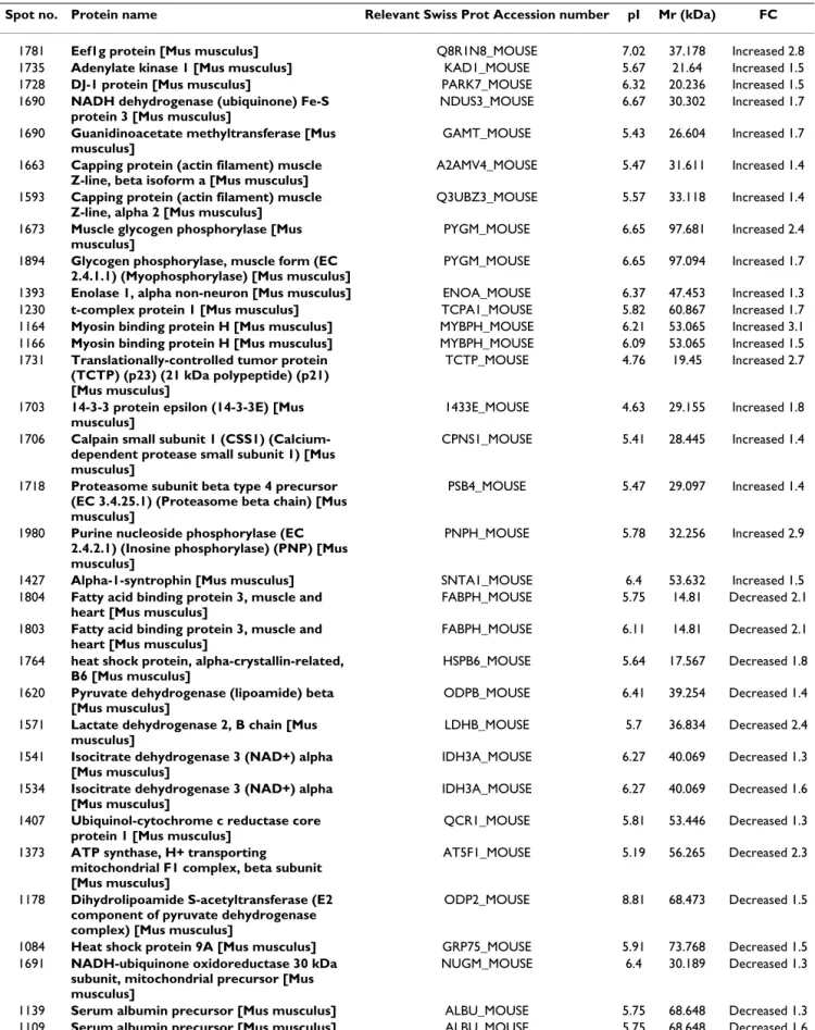
control littermates. Comparison of the protein profiles using SAM (FDR <5%) revealed the differential abun-dance of 38 protein spots (20 up-regulated and 18 down-regulated) in MSTN-null mice vs wild-type controls. Mass spectrometry analyses allowed the identification of the candidate spots (Table 3). Differential expression was confirmed for some selected proteins by western-blot analysis (Table 2).
Bioinformatic analysis indicated that most of the differen-tial proteins were related to muscle energy metabolism. Enzymes of the citrate cycle (e.g. pyruvate dehydrogenase lipoamide, dihydrolipoamide S-acetyl transferase E2, iso-citate dehydrogenase, Succinate dehydrogenase) and the respiratory chain (e.g. ATP synthase F1 beta, ATP synthase D chain, ubiquinol cytochrome c reductase core protein 1,
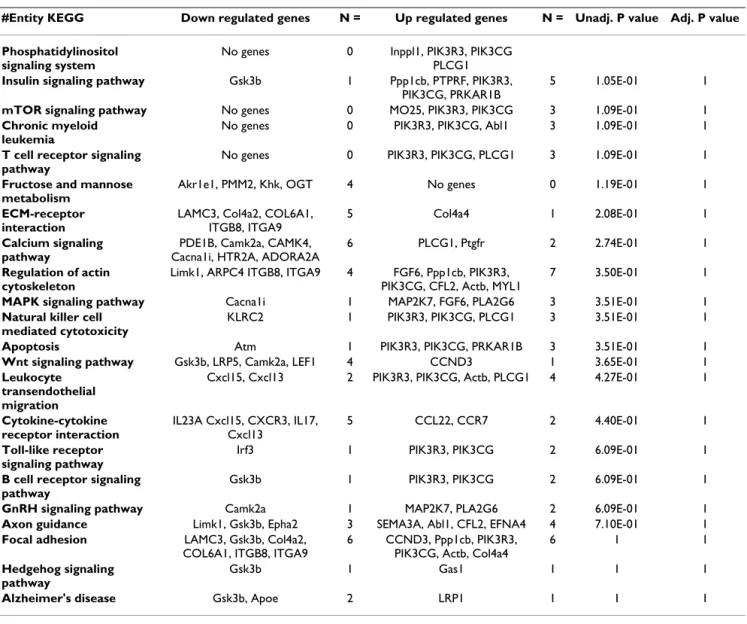
Table 1: Distribution of differential genes in myostatin-null mice vs control mice according to KEGG pathways.
#Entity KEGG Down regulated genes N = Up regulated genes N = Unadj. P value Adj. P value
Phosphatidylinositol signaling system
No genes 0 Inppl1, PIK3R3, PIK3CG PLCG1
Insulin signaling pathway Gsk3b 1 Ppp1cb, PTPRF, PIK3R3,
PIK3CG, PRKAR1B
5 1.05E-01 1
mTOR signaling pathway No genes 0 MO25, PIK3R3, PIK3CG 3 1.09E-01 1
Chronic myeloid leukemia
No genes 0 PIK3R3, PIK3CG, Abl1 3 1.09E-01 1
T cell receptor signaling pathway
No genes 0 PIK3R3, PIK3CG, PLCG1 3 1.09E-01 1
Fructose and mannose metabolism
Akr1e1, PMM2, Khk, OGT 4 No genes 0 1.19E-01 1
ECM-receptor interaction
LAMC3, Col4a2, COL6A1, ITGB8, ITGA9
5 Col4a4 1 2.08E-01 1
Calcium signaling pathway
PDE1B, Camk2a, CAMK4, Cacna1i, HTR2A, ADORA2A
6 PLCG1, Ptgfr 2 2.74E-01 1
Regulation of actin cytoskeleton
Limk1, ARPC4 ITGB8, ITGA9 4 FGF6, Ppp1cb, PIK3R3, PIK3CG, CFL2, Actb, MYL1
7 3.50E-01 1
MAPK signaling pathway Cacna1i 1 MAP2K7, FGF6, PLA2G6 3 3.51E-01 1
Natural killer cell mediated cytotoxicity
KLRC2 1 PIK3R3, PIK3CG, PLCG1 3 3.51E-01 1
Apoptosis Atm 1 PIK3R3, PIK3CG, PRKAR1B 3 3.51E-01 1
Wnt signaling pathway Gsk3b, LRP5, Camk2a, LEF1 4 CCND3 1 3.65E-01 1
Leukocyte transendothelial migration
Cxcl15, Cxcl13 2 PIK3R3, PIK3CG, Actb, PLCG1 4 4.27E-01 1
Cytokine-cytokine receptor interaction IL23A Cxcl15, CXCR3, IL17, Cxcl13 5 CCL22, CCR7 2 4.40E-01 1 Toll-like receptor signaling pathway
Irf3 1 PIK3R3, PIK3CG 2 6.09E-01 1
B cell receptor signaling pathway
Gsk3b 1 PIK3R3, PIK3CG 2 6.09E-01 1
GnRH signaling pathway Camk2a 1 MAP2K7, PLA2G6 2 6.09E-01 1
Axon guidance Limk1, Gsk3b, Epha2 3 SEMA3A, Abl1, CFL2, EFNA4 4 7.10E-01 1
Focal adhesion LAMC3, Gsk3b, Col4a2,
COL6A1, ITGB8, ITGA9
6 CCND3, Ppp1cb, PIK3R3, PIK3CG, Actb, Col4a4
6 1 1
Hedgehog signaling pathway
Gsk3b 1 Gas1 1 1 1
Alzheimer's disease Gsk3b, Apoe 2 LRP1 1 1 1
N: Number of genes involved in each biological process; Unadj. P value: Unadjusted P value, P value from Fisher's exact test without adjusting for multiple comparisons; Adj. P value: adjusted P value, after correction for multiple testing.
NADH-ubiquinone oxidoreductase), and the heart-fatty acid binding protein (H-FABP) were down-regulated (Table 2; Table 3). Conversely two fragments of Glycogen phosphorylase, and enolase 1 (an enzyme of the glycoly-sis/gluconeogenesis pathway) were up-regulated (Table 3). Abundance levels of Guanidinoacetate methyltrans-ferase, which catalyzes the last step of creatine biosynthe-sis, purine nucleoside phosphorylase, which catalyzes the reversible cleavage of inosine and guanosine to their respective bases hypoxanthine and guanine, and the ade-nylate kinase 1, which is an important enzyme in cellular adenine nucleotide homeostasis, were also increased. Myosin-binding protein H, which is a protein involved in the contractile apparatus, was revealed to be up-regulated in MSTN-null mice vs wild-type controls and this was con-firmed by western-blot analysis (Table 2). Fast glycolytic Myosin Heavy chain isoforms (MyHCII) were also more abundant in MSTN-null mice vs wild-type controls in western-blot experiments (Table 2). α1-Syntrophin was found to be up-regulated in MSTN-null mice (Table 2; Table 3). All together, these data are representative of greater fast glycolytic metabolism in the muscle of MSTN-null mice.
A differential abundance of survival/mortality factors and some chaperones (Table 3) was also detected with the down regulation of Hsp alpha crystallin-related B6, Hsp
9A (mortalin), and the up-regulation of Tcp1, TCTP, DJ-1, and 14-3-3E. The differential abundance levels were con-firmed by western-blot analyses for mortalin, DJ-1, TCTP and 14-3-3E (Table 2 and Figure 1). All together these data indicate that myostatin inactivation was associated with up-regulation of survival/anti-apoptotic factors, in favour to increased cell survival.
Discussion
In this study, we have looked for novel myostatin targets and mechanisms involved in the control of muscle home-ostasis. For this, we have used a functional genomics strat-egy, which combined both transcriptomic and proteomic profiling of MSTN-null vs wild-type control skeletal mus-cle. We hypothesized that examination of molecular pro-files in the muscles of young growing mice in the context of MSTN loss-of-function would allow the identification of myostatin downstream targets. However any changes in gene and protein expression may also reflect conse-quences of loss of MSTN function. Anyway, this approach should give information on direct or indirect MSTN tar-gets by revealing altered physiological pathways. A similar percentage of genes (17.5%) and proteins (15.3%) were declared to be differentially expressed with both tech-niques as indicated by the ratio of the number of differen-tial genes and proteins to the total number of genes spotted on chips and proteins detected in 2-DE gels in the
Table 2: Differential expression of myostatin targets genes confirmed by qPCR and by western-blot experiments in MSTN-null mice vs their control littermates.
Proteomics Transcriptomics qPCR Western-Blot
H-FABP -2.1 -9.1 MYBPH 3.1 1.4 MyHCII 4.9 Syntrophin A 1.5 2.8 p-Akt 2.2 GSK-3β -1.9 -2.4 GSK-3β(ser9)/GSK-3β 1.7 t PI3KR3 1.1 1.8 DJ-1 1.5 1.7 2.2 PINK1 1.9 3.4 PTEN -1.6 t -1.6 Mortalin -1.5 -1.4 -2.1 TCTP 2.7 1.6 14-3-3E 1.8 1.4 1.7 mfgf6 1.9
Results are expressed as Fold Changes and all the differences have been declared significant (P < 0.05) except for some tendencies (t: P < 0.10). H-FABP: Heart-Fatty acid binding protein H; MyBP-H: Myosin binding protein H; MyHCII: fast myosin heavy chain isoform; pAkt: phosphorylated Akt; GSK-3β: Glycogen synthase kinase 3β; GSK-3β (ser9): Glycogen synthase kinase 3β (phosphorylated at serine 9); PI3KR3: Phosphatidylinositol 3-kinase regulatory subunit; DJ-1: cancer- and Parkinson's disease-associated protein (park7); PINK1: PTEN-induced putative kinase 1; PTEN: tumour-suppressor phosphatase with tensin homology. TCTP: Translationally controlled tumor protein; 14-3-3E: 14-3-3ε protein; mfgf6: murine Fibroblast growth factor 6.
Table 3: Differentially expressed muscle proteins in myostatin-null mice vs control mice.
Spot no. Protein name Relevant Swiss Prot Accession number pI Mr (kDa) FC
1781 Eef1g protein [Mus musculus] Q8R1N8_MOUSE 7.02 37.178 Increased 2.8
1735 Adenylate kinase 1 [Mus musculus] KAD1_MOUSE 5.67 21.64 Increased 1.5
1728 DJ-1 protein [Mus musculus] PARK7_MOUSE 6.32 20.236 Increased 1.5
1690 NADH dehydrogenase (ubiquinone) Fe-S protein 3 [Mus musculus]
NDUS3_MOUSE 6.67 30.302 Increased 1.7 1690 Guanidinoacetate methyltransferase [Mus
musculus]
GAMT_MOUSE 5.43 26.604 Increased 1.7 1663 Capping protein (actin filament) muscle
Z-line, beta isoform a [Mus musculus]
A2AMV4_MOUSE 5.47 31.611 Increased 1.4 1593 Capping protein (actin filament) muscle
Z-line, alpha 2 [Mus musculus]
Q3UBZ3_MOUSE 5.57 33.118 Increased 1.4 1673 Muscle glycogen phosphorylase [Mus
musculus]
PYGM_MOUSE 6.65 97.681 Increased 2.4 1894 Glycogen phosphorylase, muscle form (EC
2.4.1.1) (Myophosphorylase) [Mus musculus]
PYGM_MOUSE 6.65 97.094 Increased 1.7
1393 Enolase 1, alpha non-neuron [Mus musculus] ENOA_MOUSE 6.37 47.453 Increased 1.3
1230 t-complex protein 1 [Mus musculus] TCPA1_MOUSE 5.82 60.867 Increased 1.7
1164 Myosin binding protein H [Mus musculus] MYBPH_MOUSE 6.21 53.065 Increased 3.1
1166 Myosin binding protein H [Mus musculus] MYBPH_MOUSE 6.09 53.065 Increased 1.5
1731 Translationally-controlled tumor protein (TCTP) (p23) (21 kDa polypeptide) (p21) [Mus musculus]
TCTP_MOUSE 4.76 19.45 Increased 2.7
1703 14-3-3 protein epsilon (14-3-3E) [Mus musculus]
1433E_MOUSE 4.63 29.155 Increased 1.8 1706 Calpain small subunit 1 (CSS1)
(Calcium-dependent protease small subunit 1) [Mus musculus]
CPNS1_MOUSE 5.41 28.445 Increased 1.4
1718 Proteasome subunit beta type 4 precursor (EC 3.4.25.1) (Proteasome beta chain) [Mus musculus]
PSB4_MOUSE 5.47 29.097 Increased 1.4
1980 Purine nucleoside phosphorylase (EC 2.4.2.1) (Inosine phosphorylase) (PNP) [Mus musculus]
PNPH_MOUSE 5.78 32.256 Increased 2.9
1427 Alpha-1-syntrophin [Mus musculus] SNTA1_MOUSE 6.4 53.632 Increased 1.5
1804 Fatty acid binding protein 3, muscle and heart [Mus musculus]
FABPH_MOUSE 5.75 14.81 Decreased 2.1 1803 Fatty acid binding protein 3, muscle and
heart [Mus musculus]
FABPH_MOUSE 6.11 14.81 Decreased 2.1 1764 heat shock protein, alpha-crystallin-related,
B6 [Mus musculus]
HSPB6_MOUSE 5.64 17.567 Decreased 1.8 1620 Pyruvate dehydrogenase (lipoamide) beta
[Mus musculus]
ODPB_MOUSE 6.41 39.254 Decreased 1.4 1571 Lactate dehydrogenase 2, B chain [Mus
musculus]
LDHB_MOUSE 5.7 36.834 Decreased 2.4 1541 Isocitrate dehydrogenase 3 (NAD+) alpha
[Mus musculus]
IDH3A_MOUSE 6.27 40.069 Decreased 1.3 1534 Isocitrate dehydrogenase 3 (NAD+) alpha
[Mus musculus]
IDH3A_MOUSE 6.27 40.069 Decreased 1.6 1407 Ubiquinol-cytochrome c reductase core
protein 1 [Mus musculus]
QCR1_MOUSE 5.81 53.446 Decreased 1.3 1373 ATP synthase, H+ transporting
mitochondrial F1 complex, beta subunit [Mus musculus]
AT5F1_MOUSE 5.19 56.265 Decreased 2.3
1178 Dihydrolipoamide S-acetyltransferase (E2 component of pyruvate dehydrogenase complex) [Mus musculus]
ODP2_MOUSE 8.81 68.473 Decreased 1.5
1084 Heat shock protein 9A [Mus musculus] GRP75_MOUSE 5.91 73.768 Decreased 1.5
1691 NADH-ubiquinone oxidoreductase 30 kDa subunit, mitochondrial precursor [Mus musculus]
NUGM_MOUSE 6.4 30.189 Decreased 1.3
1139 Serum albumin precursor [Mus musculus] ALBU_MOUSE 5.75 68.648 Decreased 1.3
range of pH 4–7, respectively. Transcriptomics and pro-teomics provided complementary data and led us to ana-lyse both gene expression by qPCR and protein abundance by western blotting for some candidate targets.
As expected, the expression of several genes and abun-dance of proteins involved in muscle energy metabolism were altered. Specifically we found an up-regulation of those involved in glycolytic metabolism and a down-reg-ulation of mitochondrial proteins. Similar to previously published results [27-29], this provided additional evi-dence of a glycolytic shift within skeletal muscle in absence of functional myostatin. Additionally, expression level of α1-syntrophin, a predominant syntrophin iso-form in skeletal and cardiac muscle, significantly increased in MSTN-null mice. This result is in agreement with the work of Vandebrouck and co-workers [30], which indicates that α1-syntrophin might play a potential signalling role in the regulation of calcium influx in mus-cle cells and thereby regulate musmus-cle activity.
Increased potential for PI3K pathway
Our study revealed that upstream components of the PI3K/Akt/GSK-3β, a signalling network that promotes cell survival and proliferation, were targets of myostatin inac-tivation. The core of the PI3K signalling pathway begins with PI3K activation and subsequent generation of the second messenger lipid phosphatidyl (3,4,5) triphos-phate, which recruits and activates PI-dependent kinase. The latter phosphorylates and activates protein kinase B/ Akt [31]. GSK-3β is a substrate for phosphorylated Akt and is inactivated upon phosphorylation. We detected the differential expression of mRNAs that encode for PIK3R3, PIK3CG, PLCG1, and GSK-3β. Increases in the abundance of phophorylated-Akt and in the activation ratio of GSK-3β (phosphorylated GSK-GSK-3β at serine 9/GSK-GSK-3β) were also detected (Table 2). This finding is original since the PI3K pathway is mainly regulated through post-translational modification namely phosphorylation. Thus, our results indicate that myostatin inactivation resulted in increased potential for PI3K/Akt activation and decreased potential for GSK-3β activity both at the transcriptional and post-translational levels.
Interestingly, we also detected the differential abundance of factors regulating the PI3K pathway e.g. DJ-1, PTEN and PINK1. DJ-1, a cancer- and Parkinson's disease-associated protein also called park7, protects cells from toxic stresses and is implicated in mitochondria and oxidative stress-related survival pathways (reviewed in [32]). It associates with chaperonnes including mortalin. DJ-1 has also been reported to be a novel regulator of the PI3K-PTEN path-way [33]. The tumour-suppressor phosphatase with tensin homology (PTEN) is the most important negative regulator of the cell-survival signalling initiated by PI3K (Figure 1). DJ-1 interacts with PTEN-induced putative kinase 1 (PINK1), which encodes a Ser/Thr kinase with a mitochondrial-targeting signal, and collaborate to protect cells against oxidative stress induced cell death [34]. Inter-estingly, our data show up-regulation of DJ-1 and PINK1 and down-regulation of PTEN both at the transcriptional and protein levels and this could account for activation of the PI3K pathway in MSTN-null mice.
Abundance of candidate proteins to be myostatin targets as revealed by western-blotting in the muscles of control and MSTN-null mice
Figure 1
Abundance of candidate proteins to be myostatin targets as revealed by western-blotting in the mus-cles of control and MSTN-null mice. A-D: control mice;
E-H: MSTN-null mice. pAkt: phosphorylated akt; GSK-3β: Glycogen synthase kinase 3β; GSK-3β (ser9): Glycogen syn-thase kinase 3β (phosphorylated at serine 9); DJ-1: cancer- and Parkinson's disease-associated protein (park7); PINK1: PTEN-induced putative kinase 1; PTEN: tumour-suppressor phosphatase with tensin homology. TCTP: Translationally controlled tumor protein; 14-3-3E: 14-3-3ε protein. Actin was used as a loading control protein.
GSK-3ȕ (total) A B C D E F G H Actin DJ-1 PTEN p-Akt PINK1 TCTP A B C D E F G H Actin 14-3-3E GSK-3ȕ (ser9) GSK-3ȕ (total) GSK-3ȕ (total) A B C D E F G H Actin DJ-1 PTEN p-Akt PINK1 TCTP A B C D E F G H Actin Actin DJ-1 DJ-1 PTEN PTEN p-Akt p-Akt PINK1 PINK1 TCTP TCTP A B C D E F G H Actin 14-3-3E GSK-3ȕ (ser9)
1739 ATP synthase D chain, mitochondrial [Mus musculus]
ATP5H_MOUSE 5.52 18.607 Decreased 1.3 1152 Succinate dehydrogenase [ubiquinone]
flavoprotein subunit, mitochondrial precursor [Mus musculus]
DHSA_MOUSE 7.06 72.539 Decreased 1.7
1400 Ubiquinol-cytochrome-c reductase complex core protein I, mitochondrial precursor [Mus musculus]
UQCR1_MOUSE 5.75 52.735 Decreased 1.5
1608 Troponin T, fast skeletal muscle (TnTf) (Fast skeletal muscle troponin T) (fTnT) [Mus musculus]
TNNT3_MOUSE 5.26 32.09 Decreased 1.6
Accession number: reference of each protein in Swiss-Prot database; pI: isoelectric point; Mr (kDa): molecular weight; FC: Fold-Change
The PI3K/Akt pathway plays a pivotal role in the promo-tion of cell proliferapromo-tion and inhibipromo-tion of cell death. It plays an important role in at least two signal transductory systems, namely, the Wnt/wingless and PI3K pathways, which influence proliferation and cell survival, respec-tively. Our data agree well with data that demonstrate a correlation between activation of PI3K pathway and increase in cell size [35] and the activation of Akt, which stimulates muscle hypertrophy and antagonizes the loss of muscle protein [36]. Akt signalling through GSK-3β, mTOR (a PI3K-related kinase) and Foxo1 was shown to be associated with skeletal muscle hypertrophy and atrophy processes in humans [37]. Recently, Amirouche and work-ers have shown that MSTN negatively regulates Akt/mTOR signalling pathway [38]. It was also shown that MSTN activates GSK-3β and decreases cyclin D1 by inhibiting the PI3K/Akt pathway and this has been proposed to be involved in the progression of muscle-disuse atrophy [39]. Lastly, the PI3K/Akt pathway is the major intracellu-lar pathway activated by IGF-I stimulation during myo-genesis [40]. A primary effect of GSK-3β inhibition in muscle is to promote myogenic differentiation and it has been proposed that IGF-mediated hypertrophy involves the negative regulation of GSK-3β activity [41].
Altered expression/abundance of factors involved in cell survival/apoptosis
Our data revealed changes in the expression of genes and proteins related to cell survival/apoptosis pathways. Actu-ally, we detected the down-regulation of some factors (e.g. alpha crystallin-related B6, heat shock protein 9A) and up-regulation of others (e.g. Dad1, survivin, TCTP, 14-3-3E) that may be a signature of increased cell survival and anti-apoptotic processes. Indeed, alpha crystallin-related B6, a heat shock protein 20, is thought to protect cells from apoptosis [42]. Heat shock protein 9A (Mortalin, Grp75), a member of the heat shock protein 70 family, plays a central role in mitochondrial import, energy gen-eration and chaperoning of mis-folded proteins (for review [43]). This factor interacts with the tumor suppres-sor protein p53 and inactivates its transcriptional activa-tion and apoptotic funcactiva-tions [44]. Up-regulaactiva-tion of mortalin contributes significantly to tumorigenesis [45]. Dad1 (defender against apoptotic death) has been reported to be a programmed cell death suppressor neces-sary for cell survival in a mammalian cell line [46] and the loss of Dad1 protein triggers apoptosis [47]. Survivin (Birc5) is a member of the inhibitor of apoptosis (IAP) family that prevents apoptotic cell death in various cell types including hematopoietic, endothelial, embryonic and neuronal cells [48]. Translationally controlled tumor protein (TCTP) is involved in the calcium binding and microtubule stabilization [49] and has been implicated in cell growth, cell cycle progression, malignant transforma-tion and in protectransforma-tion of cells against various stress
condi-tions and apoptosis [50]. 3-3E is a member of the 14-3-3 family of phosphoserine-binding adapter molecules that mediate a general survival-promoting function through interaction with target proteins, by enhancing survival signalling while suppressing activity of pro-apoptotic proteins (reviewed by [51]). They are involved in the regulation of apoptosis through multiple interac-tions with proteins of the core mitochondrial machinery, pro-apoptotic transcription factors and their upstream sig-nalling pathways. One prominent mechanism for the sup-pression of apoptosis is through 14-3-3-mediated sequestration of pro-apoptotic proteins (e.g. BAD, Bcl-2-antagonist of cell death; [52]). Furthermore, 14-3-3 pro-teins regulate the Akt/mTOR pathway via interaction with PRAS40, a mTOR binding partner [53]. In our study, 14-3-3E showed greater expression in the hypertrophied muscles of MSTN-null mice. Interestingly, some of the dif-ferential proteins detected in our study (e.g. DJ-1, T-com-plex protein 1, proteasome B chain) were reported to belong to the 14-3-3 interacting "phosphoproteome" [54].
Interestingly, in a previous study conducted in the context of MSTN loss of function in cattle [28], we have already found an up-regulation of genes encoding proteins involved in apoptosis (e.g DDX, PDCD6) and a down-reg-ulation of anti-apoptotic genes belonging to the Bcl2 fam-ily. Supporting the hypothesis of decreased apoptosis in the muscles of MSTN-null mice, we found that the bioac-tive fragment of the initiator Caspase 8 was decreased in these mice compared to their littermates (data not shown). The functional significance of proteins related to cell survival/apoptosis to muscle hypertrophy remains to be further studied. However, the results of the present study suggest that MSTN may behave as an arbiter of the survival decision in the muscles. In MSTN-null mice, increased abundance of anti-apoptotic factors probably induces a by-pass of apoptosis and consequently increased cell survival, a process leading to hypertrophy (Figure 1). The involvement of MSTN in apoptosis remains scarce and controversial. There are conflicting results regarding MSTN and apoptosis. It is not clear whether this factor protects myoblasts from apoptosis [14,55] or promotes the apoptotic process [39,56]. This discrepancy may be explained by the different experimen-tal conditions in which MSTN effect was examined in vitro (e.g. proliferation vs differentiation phase, myogenic cell lines vs primary myoblast cultures). Although skeletal muscle is considered to be rather resistant to apoptosis partly due to high concentration of endogenous inhibi-tors (reviewed in [57]), there is evidence that apoptotic events occur in skeletal muscle under physiological (exer-cise) and pathological conditions (e.g. muscle disuse-induced atrophy, denervation, ageing, dystrophinopa-thies) and may contribute to atrophy and loss of fibers
[58-61]. "Myonuclear apoptosis" is a predominant form of apoptosis by which a loss of myonuclei occurs without cell death during muscle atrophy [62,63]. This process may have major consequence since (1) the number of myonuclei is a critical determinant for protein synthesis capacity (2) apoptosis within muscle may alter the myo-nuclear to cytosolic ratio (protein/DNA). Thus, modula-tion of myonuclear number or myonuclear domain size or both is a mechanism by which MSTN may contribute to muscle homeostasis. Inhibition of apoptosis in the context of MSTN loss-of-function could lead to increased myonucler survival and consequently to increased protein biosynthesis. Thus, this could contribute to the hyper-trophic pathway, especially in growing mice. Work is progress to assay apoptotic process in muscles according to MSTN activity.
Conclusion
In conclusion, integration of transcriptomic and teomic data indicate that MSTN inactivation could pro-mote survival and resistance to apoptosis in muscle through the PI3K/Akt/GSK-3β survival pathway and through a mechanism that involves regulation of pro- and anti-apoptotic factors. A combination of increased poten-tial for PI3K activation and decreased myonuclear apopto-sis could be crucial to muscle hypertrophy in MSTN-null mice (Figure 2).
Methods
Muscle samples
The MSTN-null mice have a constitutive invalidation of myostatin gene [7]. These mice were bred at the Iowa State University, were slaughtered at 5 weeks postnatally, were dissected and their muscle masses were weighted. At this age the mice were nearly adults and this developmental stage was chosen in order to dissociate the influence of MSTN on hypertrophy from that of myofiber hyperplasia. Muscle samples were taken from Quadriceps muscle (mixed slow oxidative) immediately after slaughter, fro-zen in liquid nitrogen and kept at -80°C until protein extraction was performed. The Quadriceps muscle was cho-sen for the genomic experiments owing to its weight and marked hypertrophy (+87%, data not shown).
Microarray experiments
Transcriptomic analysis was performed with a microarray of 6,473 genes expressed in muscle ("Myochips" available from OUEST-genopole®, France) as described in [28].
These Myochips consisted in 6,473 oligonucleotides from human (76%) and murine (24%) origin and 919 con-trols. Microarray experiments were performed according to recently proposed standards (MIAME consortium). Data were incorporated into the BASE database and the NCBI Gene Expression Omnibus (GEO) http://
www.ncbi.nlm.nih.gov/projects/geo/ and are accessible through GEO Series GSE5561 and GSE5456.
Total RNA was extracted from muscle tissue samples with TRIZOL® reagent (Life Technologies) according to the
manufacturer's recommendation. The RNA was then puri-fied and treated with DNAse using the RNeasy® Mini kit
(Qiagen, France). RNA integrity was checked using Lab Chip Agilent technology, as previously described [28]. Each individual sample was compared to a reference pool consisting of skeletal muscle transcripts isolated from dif-ferent muscles of four mice lines (Balb, C57bl6, B129) of different age and gender, and from cultured murine myo-genic cells (C2C12).
Labeling was performed using a "Pronto Plus Direct Sys-tems" kit (Corning-Promega) according to the supplier's guidelines. Five μg of total RNA was reverse transcribed into cDNA in the presence of cyanine (Cy3-dCTP/Cy5-dCTP, Amersham). cDNA purification was performed according to the manufacturer's instructions, and concen-tration and frequency of incorporation were determined by spectrophotometry. Three chips were hybridized per sample comparison. After washing, the chips were scanned on an Affymetrix 428™ Array Scanner. Images were analyzed using GenePix Pro V6 software (Axon instrument, Inc). Raw intensity data were normalized using the MADSCAN program (http://cardios erve.nantes.inserm.fr/mad/madscan/, 15). In order to identify differentially expressed genes, the Cy3/Cy5 ratios were statistically analyzed using Significance Analysis of Microarrays (SAM) method (FDR <5%; [64]). With the available sample size (4 animals per group) and 3 repeated measurements per sample and taking account a test power (1-β) set at 80% [65,66] and the biological var-iability (Standard Deviation SD = 0.083), the minimum difference was 11% corresponding to a threshold fold-change (FC) of 1.11. Thus we only retained appropriately powered genes.
Proteomics
Preparation of muscle samples for 2-DE
Sample preparation was performed according to Bouley et al. [67]. Briefly, frozen muscle tissue (50–100 mg) was homogenized in a lysis buffer containing 8.3 M urea, 2 M thiourea, 1% DTT, 2% CHAPS and 2% IPG buffer pH 3– 10 with a polytron and centrifuged at 10,000 g for 30 min. The supernatant was harvested and protein concentration for each sample was determined as described in the Plu-sOne 2-DE Quant kit (Amersham Biosciences).
Protein quantification was determined using the 2-DE Quant kit (Amersham Uppsala, Sweden), with bovine serum albumin as a standard. Average amount of protein was 9.6 μg. μl-1 ± 1.48 μg.μl-1.
2-DE separation
2-DE separation was performed according to the method of Bouley et al. [67]. Briefly, for the first dimension (IEF), proteins were solubilized in a rehydration solution (8 M urea, 1 M thiourea, 0.28% DTT, 2% CHAPS, 2% IPG buffer pH 4–7 and 0.01% CBBR-250). IPG strips, 18 cm, covering a pH range of 4–7 were rehydrated overnight in 330 ml of this protein solution under low viscosity paraf-fin oil (sample loading by rehydration). IPG strips con-taining 700 μg of proteins were subjected to IEF (73.5 kVh) in a Multiphor II gel apparatus at 20.5°C. After com-pletion of the IEF, proteins on the strip were equilibrated for 15 min in a buffer containing 6 M urea, 1% DTT, 30% glycerol, 50 mM Tris base, 2% sodium dodecyl sulphate (SDS), and DTT and then for an additional 15 min in the same solution except that DTT was replaced by 5% iodoa-cetamide and 0.002% bromophenol blue was added. The
IPG strips were then transferred onto 11% T, 2.6% C sep-arating polyacrylamide gels, and proteins were separated in the second dimension using a Hoefer DALTsix system (Hoefer, Scientific Instruments, San Francisco, CA, USA). 2-DE gels were stained using a colloidal CBB G-250 pro-cedure [67] and digitized on a calibrated scanner (ImageS-canner, Amersham).
Image and Data analysis
ImageMaster 2D Platinum software (Amersham Bio-sciences) was used to match and quantify protein spots on 2-DE gels. Parameters used for spot detection were mini-mal area = 70 pixels, smooth factor = 2.0, and saliency = 20. An artificial reference gel was created combining all the spots present in the different gels. The reference gel was then used for matching of corresponding protein spots between gels. The volume of individual spot was normalized (% volume) according to the mean volume of 249 protein spots which were strictly common to all gels. Comparisons between groups were performed using SAM statistical test [68]. Differences were declared significant for FDR <5%. Taking into account the biological variabil-ity which is 21%, the power analysis [66] allowed retain-ing proteins which present a minimum difference of 41% corresponding to a threshold FC of 1.41.
Identification of spots by Mass Spectrometry
All differential protein spots were excised from the poly-acrylamide gel and placed in a Multiscreen solvinert 96-well filtration system plate (Millipore Corp., Bedford, MA). These proteins of interest were digested proteolyti-cally in the plate with 3 volumes of trypsin solution (10 ng/μL; V5111, Promega, Madison, WI) at 37°C for 5 h and extracted with 8–12 μL of ACN. The resulting peptide fragments were mixed with an equal volume of CHCA sat-urated solution in 0.1% TFA, 50% aqueous ACN.
Peptide mass fingerprintings (PMF) of tryptic peptides were identified in a positive reflectron mode using a voy-ager DE Pro MALDI-TOF (Perspective Biosystem, Farm-inghan, MA, USA). External calibration was performed with a standard peptide solution (Proteomix, LaserBio Labs, Sophia-Antipolis, France). Internal calibration was performed using peptides resulting from auto-digestion of porcine trypsin, with protonated masses of 842.509, 1045.564, and 2211.104 Da. Peptides mass fingerprints were compared with mammalian databases [NCBI http:// www.ncbi.nlm.nih.gov/Database/ and SWISS-PROT http://www.expasy.org/sprot/] using MASCOT and Pro-Found software [http://www.matrixscience.com and http://prowl.rockefeller.edu, respectively (June 2007 and October 2007)].
The search criteria used were 1 missing trypsin cleavage site, partial methionine oxidation, partial
carbamid-In MSTN-null mice, regulators of the PI3K pathway and cell survival/apoptosis likely contribute to muscle hypertrophy Figure 2
In MSTN-null mice, regulators of the PI3K pathway and cell survival/apoptosis likely contribute to muscle hypertrophy. PI3K signalling pathway: PI3K, Phosphatidyl
Inositol 3 Kinase; Akt, protein kinase B; GSK-3β, Glycogen synthase kinase 3β; eIF2B, eukaryotic protein synthesis initia-tion factor 2B; PTEN, tumour-suppressor phosphatase with tensin homology (the most important negative regulator of the cell survival signalling initiated by PI3K); DJ-1, a cancer- and Parkinson's disease-associated protein (a novel regulator of the PI3K-PTEN pathway). Survival/Anti-apoptotic factors: PINK1, PTEN-induced putative kinase; Dad1, defender against apoptotic death; TCTP, Translationally controlled tumour protein; 14-3-3E, 14-3-3 protein epsilon; Birc5, sur-vivin. N DJ-1 Increased myonuclear survival Activation of the PI3K pathway GSK-3ȕ downstream survival signalling pathway
Hypertrophy
Decreased apoptosis
P PTEN
N DJ-1
N Survival anti-apoptotic factors (e.g. PINK1, dad1, TCTP,
14-3-3E, Birc5) P Protein synthesis via eIF2B activation PI3K AKT GSK-3ȕ N DJ-1 Increased myonuclear survival Activation of the PI3K pathway GSK-3ȕ downstream survival signalling pathway
Hypertrophy
Decreased apoptosis
P PTEN
N DJ-1
N Survival anti-apoptotic factors (e.g. PINK1, dad1, TCTP,
14-3-3E, Birc5) P Protein synthesis via eIF2B activation PI3K AKT GSK-3ȕ N DJ-1 Increased myonuclear survival Activation of the PI3K pathway GSK-3ȕ downstream survival signalling pathway
Hypertrophy
Decreased apoptosis
P PTEN
N DJ-1
N Survival anti-apoptotic factors (e.g. PINK1, dad1, TCTP,
14-3-3E, Birc5) P Protein synthesis via eIF2B activation PI3K AKT GSK-3ȕ PI3K AKT GSK-3ȕ
omethylation of cysteine and mass deviation lower than 30 ppm. We required at least 5 matched peptides per pro-tein for identification and used the ProFound and MAS-COT probabilistic score and the accuracy of the experimental-to-theoretical isoelectric point (pI) and molecular weight (Mr).
Bioinformatic tools
To understand biological function or molecular mecha-nism of the main molecular targets of MSTN, we used a set of bioinformatics web tools such as Genomatix, DAVID and PANTHER for gene identification and String for inter-actions networks between proteins. The data mining allowed to visualize KEGG (Kyoto Encyclopedia of Genes and Genomes) pathway and to classify gene and protein by their function.
Validation of Proteins and genes expression Western blotting
Western-blotting was used to validate the main differen-tially expressed proteins. These proteins were separated by gel electrophoresis using SDS-PAGE for 2 h. After migra-tion, the proteins were transferred onto PVDF transfer membrane Millipore (Dutscher Dominique SAS, France). Membranes were then blocked with 5% non-fat milk in T-TBS 1× buffer containing (blocking solution) and incu-bated under gentle agitation all the night at room temper-ature in the presence of the a primary antibodies: DJ-1, 1:2000 dilution of purified rabbit polyclonal anti-DJ-1 antibody (sc 32874, Santa Cruz); MyBP, 1:4000 dilution of purified mouse monoclonal anti-MyBP antibody (H0000 4608-MO1, ABNOVA); TCTP, 1:2000 dilution of purified rabbit polyclonal anti-HRF antibody (sc 30124, Santa Cruz); 14-3-3ε, 1:4000 dilution of purified goat pol-yclonal anti-14-3-3ε protein antibody (sc 1615, Santa Cruz); α-1 syntrophin, 1:500 dilution of purified rabbit polyclonal anti-SNTA1 antibody (sc- 50460, Santa Cruz); H-FABP, 1:2000 dilution of purified rabbit polyclonal anti-H-FABP, was a gift from Dr. Jacques Veerkamp and
Dr. Herman Van Moerkerk (Dept. Biochemistry, Nijmegen, Netherlands) [63]; mortalin, 1:250 dilution of purified mouse monoclonal anti-GRP75 antibody (MAB3584, Clone 419612, R&D systems); PINK1, 1:2000 dilution of purified rabbit polyclonal PINK1 anti-body (sc- 33796, Santa Cruz); GSK-3β, 1:2000 dilution of purified rabbit polyclonal anti-GSK-3β antibody (sc-9166, Santa Cruz); GSK-3β (ser9), 1:4000 dilution of purified goat polyclonal anti-p-GSK-3β (Ser9) antibody (sc- 11757, Santa Cruz); MyHC, 1:2000 dilution of puri-fied mouse monoclonal anti-MyHC antibody [S5 8H2, lot: 9013-1, obtained under "Noé" project who associates our team and Biocytex company [69] and now this anti-body is commercialized by Agro-Bio society]; PTEN, 1:2000 dilution of purified rabbit polyclonal anti-PTEN antibody (sc- 9145, Santa Cruz); p-Akt, 1:2000 dilution of purified rabbit polyclonal p-Akt 1/2/3 (Ser473) anti-body (sc- 33437-R, Santa Cruz); Actin, 1:20000 dilution of purified mouse monoclonal anti-actin antibody (IgG2a, Clone AC40, Sigma Chemical Company, St Louis, MO, USA), which are able to bind to its specific protein. The blots were extensively washed with Tris buffer and incubated under gentle agitation with the secondary anti-bodies (anti-mouse, anti-goat or anti-rabbit IgG com-bined with conjugated with horseradish peroxidase 1: 20000 dilution) for 1 h. The activity of the enzyme was revealed by Enhanced Chemiluminescence kit (Amer-sham).
The power analysis [66] allowed retaining proteins which present a minimum difference of 45% corresponding to a threshold FC of 1.45.
Real-Time Polymerase Chain Reaction Analysis
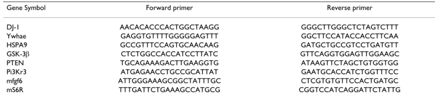
Seven genes shown to be differentially expressed were selected from the results obtained by microarray analysis and by proteomic analysis. The quantitative RT-PCR (qPCR) was realized using SYBR Green I dye. PCRs were performed using gene-specific primers pairs (Table 4). The
Table 4: Primer sequences used in quantitative real-time PCR.
Gene Symbol Forward primer Reverse primer
DJ-1 AACACACCCACTGGCTAAGG GGGCTTGGGCTCTAGTCTTT
Ywhae GAGGTGTTTTGGGGGAGTTT GGCTTCCATACCACCTTCAA
HSPA9 GCCGTTTCCAGTGCAACAAG GATGCTGCCGTCCTGATGTT
GSK-3β CTCTGGCCACCATCCTTATC GTTCAGGTGGAGTTGGAAGC
PTEN TGCAGAAAGACTTGAAGGTG ATAAGTTCTAGCTGTGGTGG
Pi3Kr3 ATGAGAACCTGCCGCATTAT GAATGCACCATCTGGTTTCC
mfgf6 ATTGGGAAAGCGGCTATTTGC CTCGTGTGTTCCACTGATGC
mS6R TTTGATTCTGAAAGCCATGCG CGGTCCATCAGGATTCTATTG
All primer sequences were designed with the Primer3 software.
DJ-1: cancer- and Parkinson's disease-associated protein (park7); 14-3-3E: 14-3-3ε protein; HSPA9: mortalin, GSK-3β: Glycogen synthase kinase 3β; PI3KR3: Phosphatidylinositol 3-kinase regulatory subunit; PTEN: tumour-suppressor phosphatase with tensin homology; mfgf6: murine Fibroblast growth factor 6; mS6R: mouse ribosomal protein S6.
absolute quantity of each target gene was determined by using serially diluted standards to generate a standard curve. The concentration in pg/μmol of each gene was determined by using a linear relationship between amounts of standards cDNA and their Ct (Cycle thresh-old). Real-Time PCR experiment was controlled using the ribosomal protein S6 as reference gene. The power analy-sis [66] allowed retaining genes which present a mini-mum difference of 54% corresponding to a threshold FC of 1.54.
Western-blot and RT-PCR data analysis
The difference of proteins and genes levels between myostatin-null mice and their controls was analyzed by Student t-test. Results are expressed as the mean ± stand-ard error of mean (SEM). A difference between the two groups was considered significant when P < 0.05.
Abbreviations
MSTN: myostatin; SAM: Significance Analysis of Microar-rays; DNA: Desoxyribo Nucleic Acid; FC: Fold Change; FDR: False Discovery Rate; PI3K: the Phosphatidyl Inosi-tol 3-kinase.
Authors' contributions
IC and ICM carried out the experiments. ICM conceived the experimental design in collaboration with BP and JFH who provided useful advice for data analysis and interpre-tation. IC and BM were actively involved in data analyses and interpretation. JR bred the experimental animals and provided muscle samples. CC provided the array and skilled advice for microarray analyses. IC and ICM wrote the manuscript. All authors read the manuscript, signifi-cantly contributed either to the presentation, the interpre-tation or the discussion of the results, and were highly involved in writing the manuscript.
Additional material
Acknowledgements
The authors thank members of the U533 Inserm group's (Faculté de Méde-cine de Nantes France) for providing the Myochips and bioinformatics tools and for skilled technical advice; G Gentès, N Dunoyer and C Barboiron for excellent technical assistance in molecular biology, C Chambon (Plateforme de Theix "Exploration du Métabolisme: des gènes aux métabolites") for protein identification. This work was supported by a grant from the French Muscular Dystrophy Association (AFM).
References
1. Guttridge : Signaling pathways weigh in on decisions to make
or break skeletal muscle. Curr Opin Clin Nutr Metab Care 2004, 7(4):443-450.
2. Tsuchida K: Activins, Myostatin and Related TGF-beta Family
Members as Novel Therapeutic Targets for Endocrine, Met-abolic and Immune Disorders. Curr Drug Targets Immune Endocr Metabol Disord 2004, 4:157-166.
3. Sillence MN: Technologies for the control of fat and lean
dep-osition in livestock. Vet J 2004, 167(3):242-257.
4. Holmes JH, Ashmore CR: A histochemical study of
develop-ment of muscle fiber type and size in normal and "double muscled" cattle. Growth 1972, 36:351-372.
5. Grobet L, Martin LJ, Poncelet D, Pirottin D, Brouwers B, Riquet J, Schoeberlein A, Dunner S, Menissier F, Massabanda J, et al.: A
dele-tion in the bovine myostatin gene causes the double-muscled phenotype in cattle. Nat Genet 1997, 17:71-74.
6. Mosher DS, Quignon P, Bustamante CD, Sutter NB, Mellersh CS, Parker HG, Ostrander EA: A Mutation in the Myostatin Gene
Increases Muscle Mass and Enhances Racing Performance in Heterozygote Dogs. PLoS Genetics 2007, 3(5):79.
7. McPherron AC, Lawler AM, Lee S-J: Regulation of skeletal muscle
mass in mice by a new TGF-beta superfamily member. Nature 1997, 387(6628):83-90.
8. Schuelke M, Wagner KR, Stolz LE, Hubner C, Riebel T, Komen W, Braun T, Tobin JF, Lee S-J: Myostatin Mutation Associated with
Gross Muscle Hypertrophy in a Child. N Engl J Med 2004, 350(26):2682-2688.
9. Grobet L, Pirottin D, Farnir F, Poncelet D, Royo LJ, Brouwers B, Christians E, Desmecht D, Coignoul F, Kahn R, et al.: Modulating
skeletal muscle mass by postnatal, muscle-specific inactiva-tion of the myostatin gene. Genesis 2003, 35(4):227-238.
10. Thomas M, Langley B, Berry C, Sharma M, Kirk S, Bass J, Kambadur R: Myostatin, a Negative Regulator of Muscle Growth,
Func-tions by Inhibiting Myoblast Proliferation. J Biol Chem 2000, 275(51):40235-40243.
11. Taylor WE, Bhasin S, Artaza J, Byhower F, Azam M, Willard DH Jr, Kull FC Jr, Gonzalez-Cadavid N: Myostatin inhibits cell
prolifera-tion and protein synthesis in C2C12 muscle cells. Am J Physiol Endocrinol Metab 2001, 280(2):E221-228.
12. Rios R, Carneiro I, Arce VM, Devesa J: Myostatin is an inhibitor of
myogenic differentiation. Am J Physiol Cell Physiol 2002, 282(5):C993-999.
13. Joulia-Ekaza D, Cabello G: Myostatin regulation of muscle
devel-opment: Molecular basis, natural mutations, physiological aspects. Exp Cell Res 2006, 312:2401-2414.
14. Joulia D, Bernardi H, Garandel V, Rabenoelina F, Vernus B, Cabello G:
Mechanisms involved in the inhibition of myoblast prolifera-tion and differentiaprolifera-tion by myostatin. Experimental Cell Research
2003, 286(2):263-275.
15. McCroskery S, Thomas M, Maxwell L, Sharma M, Kambadur R:
Myostatin negatively regulates satellite cell activation and self-renewal. J Cell Biol 2003, 162(6):1135-1147.
16. McFarland : The role of myostatin in chicken (Gallus
domesti-cus) myogenic satellite cell proliferation and differentiation. Gen Comp Endocrinol 2007, 151:351-357.
17. Steelman CA, Recknor JC, Nettleton D, Reecy JM: Transcriptional
profiling of myostatin-knockout mice implicates Wnt signal-ing in postnatal skeletal muscle growth and hypertrophy. Faseb J 2006, 20(1):580-582.
18. Carlson CJ, Booth FW, Gordon SE: Skeletal muscle myostatin
mRNA expression is fiber-type specific and increases during hindlimb unloading. Am J Physiol 1999, 277(2 pt 2):R601-606.
Additional file 1
Up-regulated gene list with fold change (FC). The data provided here
represent the statistical analysis (SAM) of up-regulated genes in the mus-cles of MSTN-null mice.
Click here for file
[http://www.biomedcentral.com/content/supplementary/1471-2164-10-196-S1.doc]
Additional file 2
Down-regulated gene list with fold change (FC). The data provided
here represent the statistical analysis (SAM) of down-regulated genes in the muscles of MSTN-null mice.
Click here for file
[http://www.biomedcentral.com/content/supplementary/1471-2164-10-196-S2.doc]
19. Solomon AM, Bouloux PMG: Modifying muscle mass – the
endo-crine perspective. J Endocrinol 2006, 191(2):349-360.
20. Reisz-Porszasz S, Bhasin S, Artaza JN, Shen R, Sinha-Hikim I, Hogue A, Fielder TJ, Gonzalez-Cadavid NF: Lower skeletal muscle mass
in male transgenic mice with muscle-specific overexpression of myostatin. Am J Physiol Endocrinol Metab 2003, 285(4):E876-888.
21. Durieux A-C, Amirouche A, Banzet S, Koulmann N, Bonnefoy R, Pas-deloup M, Mouret C, Bigard X, Peinnequin A, Freyssenet D: Ectopic
expression of myostatin induces atrophy of adult skeletal muscle by decreasing muscle gene expression. Endocrinology
2007, 148(7):3140-3147.
22. Ohsawa Y, Hagiwara H, Nakatani M, Yasue A, Moriyama K, Murakami T, Tsuchida K, Noji S, Sunada Y: Muscular atrophy of
caveolin-3-deficient mice is rescued by myostatin inhibition. J Clin Invest
2006, 116(11):2924-2934.
23. Gilson H, Schakman O, Combaret L, Lause P, Grobet L, Attaix D, Ketelslegers JM, Thissen JP: Myostatin Gene Deletion Prevents
Glucocorticoid-Induced Muscle Atrophy. Endocrinology 2007, 148(1):452-460.
24. McFarlane C, Plummer E, Thomas M, Hennebry A, Ashby M, Ling N, Smith H, Sharma M, Kambadur R: Myostatin induces cachexia by
activating the ubiquitin proteolytic system through an NF-kappaB-independent, FoxO1-dependent mechanism. J Cell Physiol 2006, 209:501-514.
25. McMahon CD, Popovic L, Oldham JM, Jeanplong F, Smith HK, Kam-badur R, Sharma M, Maxwell L, Bass JJ: Myostatin-deficient mice
lose more skeletal muscle mass than wild-type controls dur-ing hindlimb suspension. Am J Physiol Endocrinol Metab 2003, 285(1):E82-87.
26. Hocquette J-F, Bas P, Bauchart D, Vermorel M, Geay Y: Fat
parti-tioning and biochemical characteristics of fatty tissues in relation to plasma metabolites and hormones in normal and double-muscled young growing bulls. Comp Biochem Physiol A Molec Integr Physiol 1999, 122(1):127-138.
27. Bouley J, Meunier B, Chambon C, De Smet S, Hocquette JF, Picard B:
Proteomic analysis of bovine skeletal muscle hypertrophy. Proteomics 2005, 5(2):490-500.
28. Cassar-Malek I, Passelaigue F, Bernard C, Leger J, Hocquette J-F:
Tar-get genes of myostatin loss-of-function in muscles of late bovine fetuses. BMC Genomics 2007, 8(1):63.
29. Steelman CA, Recknor JC, Nettleton D, Reecy JM: Transcriptional
profiling of myostatin-knockout mice implicates Wnt signal-ing in postnatal skeletal muscle growth and hypertrophy. FASEB J 2006, 20(3):580-582.
30. Vandebrouck A, Sabourin J, Rivet J, Balghi H, Sebille S, Kitzis A, Ray-mond G, Cognard C, Bourmeyster N, Constantin B: Regulation of
capacitative calcium entries by {alpha}1-syntrophin: associa-tion of TRPC1 with dystrophin complex and the PDZ domain of {alpha}1-syntrophin. FASEB J 2007, 21(2):608-617.
31. Chan TO, Rittenhouse SE, Tsichlis PN: Akt/PKB and other D3
phosphoinositide-regulated kinases: Kinase Activation by Phosphoinositide-Dependent Phosphorylation. Annu Rev Bio-chem 1999, 68(1):965-1014.
32. Lev N, Roncevich D, Ickowicz D, Melamed E, Offen D: Role of DJ-1
in Parkinson's disease. J Mol Neurosci 2006, 29(3):215-225.
33. Kim RH, Peters M, Jang Y, Shi W, Pintilie M, Fletcher GC, DeLuca C, Liepa J, Zhou L, Snow B, et al.: DJ-1, a novel regulator of the
tumor suppressor PTEN. Cancer Cell 2005, 7(3):263-273.
34. Tang B, Xiong H, Sun P, Zhang Y, Wang D, Hu Z, Zhu Z, Ma H, Pan Q, Xia J-h, et al.: Association of PINK1 and DJ-1 confers digenic
inheritance of early-onset Parkinson's disease. Hum Mol Genet
2006, 15(11):1816-1825.
35. Cully M, You H, Levine AJ, Mak TW: Beyond PTEN mutations:
the PI3K pathway as an integrator of multiple inputs during tumorigenesis. Nat Rev Cancer 2006, 6(3):184-192.
36. Frost RA, Lang CH: Protein kinase B/Akt: a nexus of growth
factor and cytokine signaling in determining muscle mass. J Appl Physiol 2007, 103(1):378-387.
37. Leger B, Cartoni R, Praz M, Lamon S, Deriaz O, Crettenand A, Gobe-let C, Rohmer P, Konzelmann M, Luthi F, et al.: Akt signalling
through GSK-3{beta}, mTOR and Foxo1 is involved in human skeletal muscle hypertrophy and atrophy. J Physiol
2006, 576(3):923-933.
38. Amirouche A, Durieux A-C, Banzet S, Koulmann N, Bonnefoy R, Mouret C, Bigard X, Peinnequin A, Freyssenet D: Down-Regulation
of Akt/Mammalian Target of Rapamycin Signaling Pathway
in Response to Myostatin Overexpression in Skeletal Muscle. Endocrinology 2009, 150(1):286-294.
39. Yang W, Zhang Y, Li Y, Wu Z, Zhu D: Myostatin Induces Cyclin
D1 Degradation to Cause Cell Cycle Arrest through a Phos-phatidylinositol 3-Kinase/AKT/GSK-3beta Pathway and Is Antagonized by Insulin-like Growth Factor 1. J Biol Chem 2007, 282(6):3799-3808.
40. Stitt TN, Drujan D, Clarke BA, Panaro F, Timofeyva Y, Kline WO, Gonzalez M, Yancopoulos GD, Glass DJ: The IGF-1/PI3K/Akt
pathway prevents expression of muscle atrophy-induced ubiquitin ligases by inhibiting FOXO transcription factors. Mol Cell 2004, 14:395-403.
41. Velden JLJ van der, Langen RCJ, Kelders MCJM, Wouters EFM, Jans-sen-Heininger YMW, Schols AMWJ: Inhibition of glycogen
syn-thase kinase-3{beta} activity is sufficient to stimulate myogenic differentiation. Am J Physiol Cell Physiol 2006, 290(2):C453-462.
42. Fan G-C, Chu G, Mitton B, Song Q, Yuan Q, Kranias EG: Small
Heat-Shock Protein Hsp20 Phosphorylation Inhibits {beta}-Agonist-Induced Cardiac Apoptosis. Circ Res 2004, 94(11):1474-1482.
43. Kaul SC, Deocaris CC, Wadhwa R: Three faces of mortalin: A
housekeeper, guardian and killer. Exp Geront 2007, 42(4):263-274.
44. Wadhwa KTaSCK: An Hsp70 family chaperone, mortalin/
mthsp70/PBP74/Grp75: what, when, and where? Cell Stress Chaperones 2002, 7(3):309-316.
45. Wadhwa R, Takano S, Kaur K, Deocaris CC, Pereira-Smith OM, Red-del RR, Kaul SC: Upregulation of mortalin/mthsp70/Grp75
contributes to human carcinogenesis. Int J Cancer 2006, 118(12):2973-2980.
46. Sugimoto A, Hozak RR, Nakashima T, Nishimoto T, Rothman JH:
dad-1, an endogenous programmed cell death suppressor in Caenorhabditis elegans and vertebrates. EMBO J 1995, 14(18):4434-4441.
47. Nakashima T, Sekiguchi T, Kuraoka A, Fukushima K, Shibata Y, Komi-yama S, Nishimoto T: Molecular cloning of a human cDNA
encoding a novel protein, DAD1, whose defect causes apop-totic cell death in hamster BHK21 cells. Mol Cell Biol 1993, 13(10):6367-6374.
48. Gurbuxani S, Xu Y, Keerthivasan G, Wickrema A, Crispino JD:
Dif-ferential requirements for survivin in hematopoietic cell development. Proc Natl Acad Sci USA 2005, 102(32):11480-11485.
49. Yarm FR: Plk Phosphorylation Regulates the
Microtubule-Sta-bilizing Protein TCTP. Mol Cell Biol 2002, 22(17):6209-6221.
50. Bommer U-A, Thiele B-J: The translationally controlled tumour
protein (TCTP). Int J Biochem Cell Biol 2004, 36(3):379-385.
51. Porter GW, Khuri FR, Fu H: Dynamic 14-3-3/client protein
interactions integrate survival and apoptotic pathways. Sem Cancer Biol 2006, 16(3):193-202.
52. Datta SR, Katsov A, Hu L, Petros A, Fesik SW, Yaffe MB, Greenberg ME: 14-3-3 Proteins and Survival Kinases Cooperate to
Inac-tivate BAD by BH3 Domain Phosphorylation. Mol Cell 2000, 6(1):41-51.
53. Haar EV, Lee S-i, Bandhakavi S, Griffin TJ, Kim D-H: Insulin
signal-ling to mTOR mediated by the Akt/PKB substrate PRAS40. Nat Cell Biol 2007, 9(3):316-323.
54. Pozuelo Rubio M, Geraghty KM, Wong BHC, Wood NT, Campbell DG, Morrice N, Mackintosh C: 14-3-3-affinity purification of
over 200 human phosphoproteins reveals new links to regu-lation of cellular metabolism, proliferation and trafficking. Biochem J 2004, 379(2):395-408.
55. Ríos R, Carneiro I, Arce VM, Devesa J: Myostatin Regulates Cell
Survival during C2C12 Myogenesis. Biochem Biophys Res Com-mun 2001, 280(2):561-566.
56. Yang W, Zhang Y, Ma G, Zhao X, Chen Y, Zhu D: Identification of
gene expression modifications in myostatin-stimulated myoblasts. Biochem Biophys Res Commun 2005, 326(3):660-666.
57. Adhihetty PJ, Hood DA: Mechanisms of Apoptosis in Skeletal
Muscle. Basic Appl Myol 2003, 13(4):171-179.
58. Koçtürk S, Kayatekin B, Resmi H, Açýkgöz O, Kaynak C, Özer E: The
apoptotic response to strenuous exercise of the gastrocne-mius and soleus muscle fibers in rats. Eur J Appl Physiol 2008, 102(5):515-524.
59. Spencer MJ, Walsh C, Dorshkind KA, Rodriguez EM, Tidball JG:
per-Publish with BioMed Central and every scientist can read your work free of charge "BioMed Central will be the most significant development for disseminating the results of biomedical researc h in our lifetime."
Sir Paul Nurse, Cancer Research UK Your research papers will be:
available free of charge to the entire biomedical community peer reviewed and published immediately upon acceptance cited in PubMed and archived on PubMed Central yours — you keep the copyright
Submit your manuscript here:
http://www.biomedcentral.com/info/publishing_adv.asp
BioMedcentral
forin-mediated cytotoxicity. J Clin Invest 1997, 99(11):2745-2751.
60. Dupont-Versteegden EE: Apoptosis in muscle atrophy:
Rele-vance to sarcopenia. Exp Geront 2005, 40(6):473-481.
61. Busquets S, Deans C, Figueras M, Moore-Carrasco R, López-Soriano FJ, Fearon KCH, Argilés JM: Apoptosis is present in skeletal
mus-cle of cachectic gastro-intestinal cancer patients. Clin Nutr
2007, 26(5):614-618.
62. Allen DL, Roy RR, Edgerton VR: Myonuclear domains in muscle
adaptation and disease. Muscle Nerve 1999, 22(10):1350-1360.
63. Primeau A, Adhihetty PJ, Hood DA: Apoptosis in heart and
skel-etal muscle. Can J Appl Physiol 2002, 27(4):349-395.
64. Tusher VG, Tibshirani R, Chu G: Significance analysis of
micro-arrays applied to the ionizing radiation response. Proc Natl Acad Sci 2001, 98:5116-5120.
65. Mera R, Thompson H, Prasad C: How to calculate sample size
for an experiment: A case-based description. Nutr Neurosci
1998, 1:87-91.
66. Lenth RV: Statistical power calculations. J Anim Sci 2007,
85(13_suppl):E24-29.
67. Bouley J, Chambon C, Picard B: Mapping of bovine skeletal
mus-cle proteins using two-dimensional gel electrophoresis and mass spectrometry. Proteomics 2004, 4(6):1811-1824.
68. Meunier B, Bouley J, Piec I, Bernard C, Picard B, Hocquette J-F: Data
analysis methods for detection of differential protein expres-sion in two-dimenexpres-sional gel electrophoresis. Anal Biochem
2005, 340(2):226-230.
69. Picard B, Duris M-P, Jurie C: Caractérisation des chaînes lourdes
de myosine dans le muscle de bovin. INRA Prod Anim 1998, 11(2):150-152.