REGULAR ARTICLES
A 3-year long study of
Staphylococcus aureus isolates
from subclinical mastitis in three Azawak zebu herds
at the Sahelian experimental farm of Toukounous, Niger
Abdoulkarim Ibrahim Issa1,2&Jean-Noël Duprez1&Rianatou Bada-Alambedji3&Mamane Djika4&Jacques Georges Mainil1&Marjorie Bardiau1,5
Received: 20 May 2015 / Accepted: 5 November 2015 / Published online: 19 November 2015 # Springer Science+Business Media Dordrecht 2015
Abstract Staphylococcus (S.) aureus is one of the most im-portant pathogens causing bovine mastitis. The aim of the present work was to follow in three herds and during the 3 years the clonality of S. aureus isolated from California Mastitis Test (CMT)-positive cows at the experimental station of Toukounous (Niger) by (i) comparing their pulsed field gel electrophoresis (PFGE) fingerprints, (ii) identifying their virulotypes by PCR amplification and (iii) assessing the pro-duction of capsule and the formation of biofilm. The 88 S. aureus isolates belonged to 14 different pulsotypes, 3 of them being predominant: A (30 %), D (27 %), B (15 %). A and B pulsotypes had the highest profile similarity coefficient (94 %), while others had similarity coefficients under 60 %. Seventy-five S. aureus isolates were further studied for their virulotypes, capsular antigens and biofilm production. Most surface factor-, leukocidin- and haemolysin-, but not the
enterotoxin-encoding genes were detected in the majority (>75 %) of the isolates and were evenly distributed between the A, B and D pulsotype isolates. The majority of the 72 S. aureus positive with the cap5H or cap8H PCR produced the CP5 (82 %) or the CP8 (88 %) capsular antigen, respec-tively. Biofilm production by the 57 icaA-positive isolates was strong for 8 isolates, moderate for 31 isolates but weak for 18 isolates, implying that the icaA gene may not be expressed in vitro by one third of the positive isolates. Similar to other studies, those results confirm that a restricted number of S. aureus clones circulate within the three herds at Toukounous and that their specific virulence-associated prop-erties must still be further studied.
Keywords Azawak zebu . Mastitis . Staphylococcus aureus . Pulsed field gel electrophoresis . Virulotyping . PCR
Introduction
Livestock farming is the second most important economic activity after crop production in the Sahel region. The specific objective of the Sahelian experimental station of Toukounous (SEST) located 220 km northeast of Niamey in Niger is the selection and the diffusion of the Azawak zebu breed with improved production levels, especially dairy production. Azawak zebu breed is indeed amongst the best dairy cattle in West Africa, with a mean daily production of 7 to 8 l in good standard management practice (Issa et al.2014).
Still, like all dairy cattle worldwide, Azawak zebu cows suffer from mammary gland infections that impair the level of milk production. Unfortunately, publications on mastitis in Niger are scarce and surveys are highly needed to improve the knowledge on their aetiology and epidemiology (Issa et al.
2013,2014). During such a first survey at the SEST in 2009 * Jacques Georges Mainil
1
Department of Infectious Diseases (Bacteriology), Faculty of Veterinary Medicine and Institute for Fundamental and Applied Research in Animal Health (FARAH), University of Liège, Campus du Sart Tilman (B43a), Quartier Vallée II, Avenue de Cureghem 6, 4000 Liège, Belgium
2 Department of Diagnosis, Epidemiological Investigations and
Applied Research, Laboratoire Central d’Elevage (LABOCEL), BP 485, Niamey, Niger
3
Department of Immunology and Infectious Pathology (Microbiology), Interstate School of Veterinary Science and Medicine, BP 5077, Dakar, Senegal
4
National School of Public Health, BP 290, Niamey, Niger
5 Present address: Environment & Public Health Research Group,
School of Environment & Technology, University of Brighton, Cockcroft Building, Lewes Road, Brighton BN2 4GJ, UK
(Issa et al.2013), 55 bacterial isolates from 104 cows positive at the California Mastitis Test (CMT) were identified, despite absence of evidence of clinical mastitis, with half of them (51 %) belonging to the genus Staphylococcus (S.) and 42 % to the species Staphylococcus aureus.
S. aureus is indeed one of the most important contagious mastitis-causing bacterial pathogens worldwide and is responsible for clinical and subclinical as well as chron-ic and acute mastitis (Radostits et al. 2010; Zadoks et al. 2011; Keefe 2012). S. aureus produces several putative virulence factors contributing to its pathogenic-ity: protein adhesins, named BMicrobial Surface Compo-nents Recognizing Adhesive Matrix Molecules^ (or MSCRAMMs) interacting with host extracellular com-ponents; polysaccharide intercellular adhesins involved in the production of biofilm; other surface antigens in-tervening in the evasion of the innate and acquired im-mune responses (capsules, protein A) and secreted toxins causing cellular and tissue damages, such as haemolysins, leukocidins and enterotoxins (Foster and Höök 1998; Dinges et al. 2000; Srinivasan et al. 2006; Nishifuji et al. 2008; Ote et al. 2011; Gogoi-Tiwari et al. 2015).
Besides the virulotyping, a standard discriminatory tech-nique is the pulsed field gel electrophoresis (PFGE), or pulsotyping (Hallin et al.2007). The comparison of the PFGE profiles or pulsotypes of S. aureus isolates is very effective and highly discriminative to characterizing the genetic diver-sity of S. aureus during outbreaks of mastitis in herds and to identifying the contamination source and transmission route (Zadoks et al.2011).
The purpose of this work was to repeat and complete during the 3 years (from 2010 to 2012) the 2009 survey by isolating S. aureus from CMT-positive Azawak zebu cows at the SEST and by comparing their pulsotypes, virulotypes and ability to produce a capsule and to form a biofilm.
Materials and methods
Study design andS. aureus identification
At the SEST, the cows are divided into three herds according to their age and level of milk production:Bprimiparous^ cows, Bnon-elite^ cows (<1400 kg milk/lactation) and Belite^ cows (>1400 kg milk/lactation). Due to scarcity of resources, the milkers apply no hygienic or antisepsis measures during the milking process that is performed manually twice daily (Issa et al.2014).
Between 2010 and 2012, the Californian Mastitis Test (CMT) was performed on all lactating cows of the herds: a quarter was considered positive when the score was (++), (+++) or (++++); negative when the score was (−) and doubt-ful when the score was (+). The CMT-positive cows were sampled for bacteriological analysis and milk samples were stored at−20 °C in 10 % (v/v) glycerol until further use (Issa et al. 2013). The procedure was repeated three times on the same cows at 2-week intervals in 2010 (Table1).
Isolated colonies were pre-identified at the Laboratory of Bacteriology of the National Public Health School of Niamey. A loopful of each milk sample was inoculated onto Columbia sheep blood agar (BioMérieux). Staphylococci were suspected on the basis of colony shape, Gram staining, bacte-rial cell shape and catalase production and were subsequently frozen at−20 °C in glycerol.
Each year, all suspect isolates were air shipped to Belgium in polystyrene boxes (import permits no. CONT/IEC/FRT/ 589554 in 2010, CONT/IEC/FRT/813231 in 2011 and CONT/IEC/FRT/1016090 in 2012). As soon as they arrived at the Laboratory of Bacteriology of the Veterinary Faculty of the University of Liège, the bacteria were grown on Columbia sheep blood agar, to check purity (BioMérieux) and on selec-tive Mannitol salt phenol red agar (Merck) to check growth and mannitol fermentation. After confirmation of the initial results obtained in Niamey, mannitol-fermenting colonies Table 1 Number of CMT-positive cows and of S. aureus identified per year and per herd
Year No. of CMT-positive cows/no. of cows tested No. of S. aureus-positive cows/no. of cows sampled Primiparous cows Non-elite cows Elite cows Primiparous cows Non-elite cows Elite cows
2010-t0 19/39 20/40 25/65 1a/19 5/20 3/25 2010-t+2w ND ND ND 7/19 5/20 7/25 2010-t+4w ND ND ND 7/19 4/20 6/25 2010-t+6w ND ND ND 6/19 6a/20 9/25 2011 ND 24/48 19/44 ND 4/24 4/19 2012 ND 19/29 32/51 ND 4/19 8/32
2010-t0=first of four sampling times in 2010; 2010-t+2w=second sampling 2 weeks later; etc. ND not done
a
were picked up and tested by the BD BBL® coagulase test with rabbit plasma (Becton Dickinson).
The final identification was performed on mannitol-fermenting coagulase-positive colonies using API-STAPH® (BioMérieux). After confirmation of their identity, S. aureus isolates were frozen at−80 °C in glycerol until further typing. Pulsotyping
All S. aureus isolates were compared by PFGE at once at the end of the study according to the procedure described by Bardiau et al. (2014). In brief, the whole genome DNA was extracted from lysostaphin pre-treated bacterial cells embed-ded in 1.8 % certified low-melt agarose (Bio-Rad). After en-zymatic digestion with SmaI restriction enzyme (Sigma-Al-drich), the DNA fragments were separated in a 1 % pulsed field-certified agarose (Bio-Rad) gel using a CHEF MAPPER (Bio-Rad) at 6.0 V/cm for 21 h, with pulsed times ranging from 5 to 60 s, an angle of 120° and a linear ramp factor. The dendrogram was prepared by the Unweighted Pair Group Method using arithmetic average Algorithm (UPGMA) using Biogene Software (Vilber Lourmat) as described by Bardiau et al. (2013).
Virulotyping
Each year, the S. aureus isolates were tested by PCR as de-scribed by Ote et al. (2011) for the presence of genes coding for MSCRAMMs (clfA, clfB, fnbA, cna, ebpS, sdrC), biofilm formation (icaA), capsular antigens (cap5H, cap8H), protein A (spa), haemolysins (hla, hlb, hld, hlgAC), leukocidins (lukD, lukM, lukF-PV, lukS-PV) and enterotoxins (sea, seb, sec4, sed, see, seg, seh, sei, sej).
Capsular serotyping
The existence of 11 capsule serotypes is reported with two of them, CP5 and CP8, being most frequent in mastitis-associated S. aureus (O’Riordan and Lee 2004). Capsular serotyping was performed on all isolates at once by ELISA assays with specific monoclonal and polyclonal antibodies detecting the CP5 and CP8 antigens (Bardiau et al.2014). Optical density (OD) values were compared to those obtained with S. aureus reference strains, namely the human CP5-positive strain Lowenstein (ATCC 49521) and the human CP8-positive strain Wright (ATCC 49525).
Biofilm production
Biofilm production was assayed for all isolates at once in microtitre plates using safranin staining (Bardiau et al.
2014). The quantitative classification of biofilm production was based on OD values at 490 nm using a microplate reader
(Bio-Rad). Isolates are classified into non-producers and weak, moderate or strong producers.
Results
S. aureus identification and pulsotyping
From a total of 158 CMT-positive cows that were sampled, 88 S. aureus were identified: 68 in 2010, 8 in 2011 and 12 in 2012. Thirty-seven S. aureus were isolated from cows of the elite herd, 29 from cows of the non-elite herd and 22 from cows of the primiparous herd (Table1).
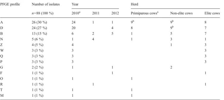
The 88 S. aureus isolates exhibited 14 different PFGE fin-gerprints arbitrarily designated by a capital letter (Table2). A and B pulsotypes had the highest profile similarity coefficient (94 %), while the other pulsotypes had similarity coefficients ranging between ca. 10 and 60 % (Fig. 1). A, D and B pulsotypes were the most frequent: 26 isolates (30 %), 24 isolates (27 %) and 13 isolates (15 %), respectively (Table 2). The remaining 11 pulsotypes were identified in one to five isolates each.
A and B pulsotypes were identified during the 3 years and D pulsotype in 2010 and 2012, while most other pulsotypes were restricted to the year 2010 (Table2). A and D pulsotype isolates were equally distributed in the three herds whereas B pulsotype isolates were almost restricted to the non-elite and elite herds (12/13 isolates), as were the other 6 pulsotypes identified in more than one isolate (Table2). In 2010, 12 of the 14 pulsotypes were identified during at least one of the four sampling events: 7 in the primiparous, 5 in the non-elite and 7 in the elite herds. But only A pulsotype was identified in the three herds at the four sampling events.
Twenty-one cows were S. aureus positive more than once between 2010 and 2012 (Table3). S. aureus isolated during two consecutive years belonged to different pulsotypes; nev-ertheless, in 2010, the same pulsotype was identified at least twice in eight of the 17 cows with more than one positive sample (Table 3). Assuming those isolates had high clonal relationships, only the first ones were further studied, making the total of isolates tested for their virulotypes, capsular anti-gens and biofilm production to 75.
PCR virulotyping
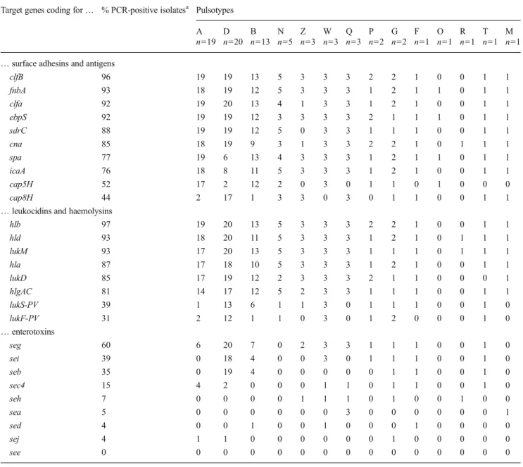
Apart from the cap5H and cap8H genes, all surface factor-encoding genes were detected in the majority (>75 %) of the 75 S. aureus (Table 4) and three of them were not equally distributed between the A, B and D pulsotype isolates: the cna gene (85 % of the isolates) was detected in only two thirds of the B pulsotype isolates and the spa and icaA genes (77 and 76 % of the isolates, respectively) in 30 to 40 % of the D pulsotype isolates, respectively. The cap5H gene (52 % of
the isolates) was highly associated with A and B pulsotypes and the cap8H gene (44 % of the isolates) with D pulsotype. Three isolates (4 %) tested negative with both cap5H and cap8H PCR.
The lukD, lukM, hla, hlb, hld and hlgAC toxin-encoding genes were also detected in the majority of the 75 S. aureus (>80 %) and, with the exception of the hlgAC gene, were equally distributed within the A, B and D pulsotype isolates (Table4). Conversely, the lukF-PV and lukS-PV genes were present in only 30 to 40 % of the isolates, respectively, and were more frequently identified in D, than in A and B pulsotype isolates.
Although 51 S. aureus (68 %) tested positive for at least one enterotoxin-encoding gene, only the seg gene was detect-ed in a majority of the 75 isolates (60 %). The seb and sei genes were identified in 35 to 40 % of the isolates, respective-ly, but the other genes were detected in none to 15 % of the
isolates (Table 4). The seg, sei and seb enteroxin-encoding genes were all highly associated with D pulsotype (>90 %), compared to A and B pulsotypes (0 to 50 %) (Table4). Capsular antigen ELISA and biofilm formation
Of the 39 isolates genotyped as cap5H, 32 (82 %) were CP5 ELISA positive, and of the 33 isolates genotyped as cap8H, 29 (88 %) were CP8 ELISA positive (Table5). Irrespective of the identity of the capsular antigen the highest correlation was with D pulsotype isolates (100 %), followed by B (85 %) and A (68 %) pulsotype isolates.
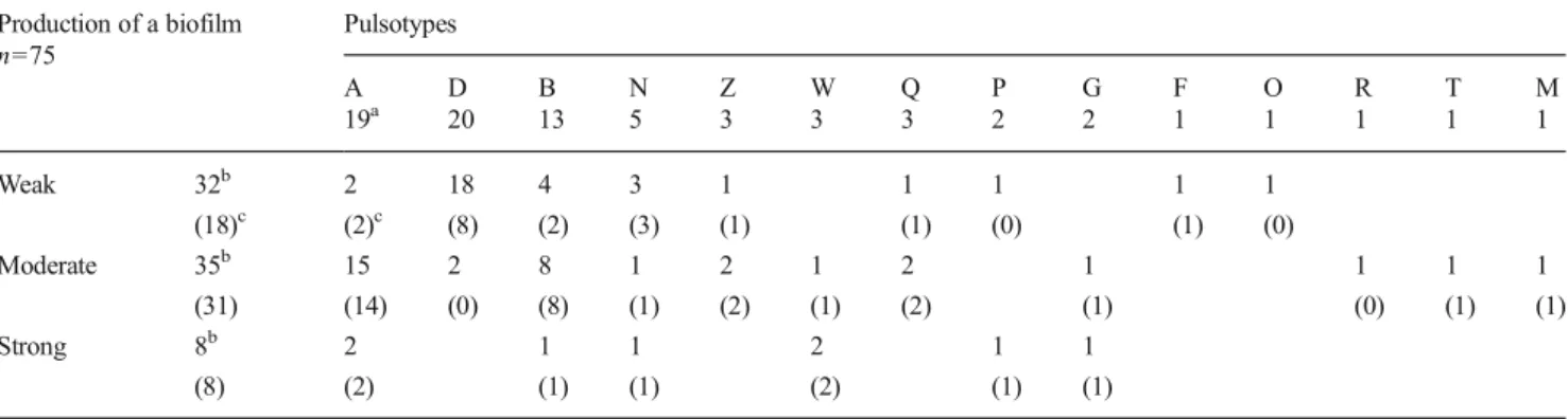
All 75 S. aureus isolates produced biofilm to some extent: 32 (43 %), 35 (47 %) and 8 (11 %) isolates were weak, mod-erate and strong biofilm producers, respectively (Table6). All 8 strong and 89 % of the moderate producer isolates (31/35) harboured the icaA gene while 56 % of the weak producer Fig. 1 Dendrogram of the
S. aureus isolates derived from the PFGE profiles. The pulsotype C isolate was identified in 2009 (Issa et al.2013), but not between 2010 and 2012
Table 2 Number of pulsotypes per year and per herd
PFGE profile Number of isolates Year Herd
n=88 (100 %) 2010a 2011 2012 Primiparous cowsa Non-elite cows Elite cows
A 26 (30 %) 24 1 1 9b 9b 8 D 24 (27 %) 20 4 8 9b 7 B 13 (15 %) 6 2 5 1 5 7 N 5 (6 %) 1 4 1 3 1 Z 4 (5 %) 4 1 3 W 3 (3 %) 3 3 Q 3 (3 %) 3 3 P 3 (3 %) 3 3 G 2 (2 %) 1 1 2 F 1 (1 %) 1 1 O 1 (1 %) 1 1 R 1 (1 %) 1 1 T 1 (1 %) 1 1 M 1 (1 %) 1 1
aPrimiparous cows were sampled only in 2010 b
isolates (18/32) also tested positive. The majority of A and B pulsotype isolates not only harboured the icaA gene (95 and 85 %, respectively) but also were moderate to strong biofilm producers (89 and 69 %, respectively). In contrast, only 10 % of D pulsotype isolates were moderate and none were strong biofilm producers though 40 % tested positive for the icaA gene (Table6).
Discussion
Though the 88 S. aureus isolated between 2010 and 2012 be-long to 14 different pulsotypes, 3 of them (A, B, D) group together 72 % and 5 of them (A, B, D, N, Z), 83 % of the isolates, confirming, like in other studies, that a restricted num-ber of S. aureus clones are present within a herd or a farm (Hata et al.2006; Aires-de-Sousa et al. 2007; Rabello et al.2007; Anderson et al.2012). Nevertheless, apart from the A and B pulsotypes (>90 % similarity), the other pulsotypes are not closely related and represent distinct lineages (Fig.1), empha-sizing different contamination origins and/or ways. Although
some influence of the differences in the sampling methods and in the bacteriological analysis cannot be ruled out, this diversity and distribution of pulsotypes may indeed be explained by different management practices at the SEST. The absence of hand and udder washing and disinfection during the milking process certainly represents the most important problem, but other practices, such as the occasional exchange of the milkers between the herds, the mixing of all pregnant cows in the same maternity building and the introduction of recently purchased cows, also represent opportunities for new strains to be intro-duced and transferred between cows (Middleton et al.2002; Sommerhauser et al.2003; De Vliegher et al.2012).
The majority of pulsotypes are identified during the four sampling events in 2010, but only A and B pulsotypes are present in the three herds from 2010 to 2012, the latter being isolated only once from the primiparous cows (Table 2). Al-ready in 2009, A, B and D pulsotypes had been identified, as had C pulsotype (Issa et al.2013) which is absent in this study, and B pulsotype was restricted to the non-elite and elite herds (Table2). Time (over years) diversity and distribution of the pulsotypes is also illustrated by the results obtained for the 21 cows followed with more than one positive milk samples dur-ing this survey (Table3), though in 2010, isolates belonging to the same pulsotype were identified more than once from eight of these cows. To avoid redundancy, only the first isolate was kept for further typing, making the total of isolates tested to 75. Even if pulsotypes can evolve as consequences of point mu-tations at the height of the restriction sites and of genetic rear-rangement such as deletion, insertion and/or inversion of DNA fragments, the predominance of a limited number of pulsotypes suggests that those isolates have a greater capacity to infect the mammary gland, to persist over time within the mammary gland and/or to spread between cows (Rabello et al.2007) and, there-fore, privileged association(s) with specific virulence factors.
However, according to the PCR results, the virulotypes do not greatly differ between pulsotypes (Table 4). The MSCRAMM-, capsule-, haemolysin-, leukocidin (except lukS-PV and lukF-PV)-, and one enterotoxin (seg)-encoding genes are present in a majority of isolates, while the other genes are detected in ca. less than one third of them, similarly to other published results. The most noticeable difference is the much higher prevalence of the lukS-PV (39 %) and lukF-PV (31 %) genes coding for the Panton-Valentine leukocidin (PVL) com-pared to the results of other studies in Europe and Africa (Rainard et al.2003; Fueyo et al.2005; Karahan et al.2009; Ikawaty et al.2010; Kadja et al.2010; Ote et al.2011; Issa et al.
2013). Since the PVL toxin is strongly associated with human S. aureus isolates causing skin and soft-tissue infections (Shallcross et al.2013), the lukS-PV- and/or lukF-PV-positive isolates in Toukounous might therefore be of human rather than of cattle origin, transferred during the manual milking process. In addition, different genes are unevenly distributed be-tween the most frequent three pulsotypes (Table4): the spa, Table 3 Pulsotypes distribution of S. aureus isolates from 21 cows with
>1 positive sample over the 3 years
Herds Cow 2010 2010 2010 2010 2011 2012 t0* t+2w t+4w t+6w Elite 56/11 Q W A 60/11 Q F 61/11 Z Z R D 63/11 A D Q N B 65/11 P P B 66/11 A A B 68/11 D A B 70/11 D A 71/11 Z D B 76/11 W D Non-elite 79/11 A A A A B G 86/11 D D A-Da N D 94/11 B G N D 96/11 A N B 98/11 D D Primiparous 35/11 D A nd nd 38/11 D D nd nd 40/11 A-Aa M A A nd nd 43/11 A D nd nd 50/11 A D nd nd 52/11 D O nd nd
Cows with more than one isolate in 2010 belonging to the same pulsotype are in italics
nd not done
a
Table 4 Relation between the positive PCR reactions of the 75 S. aureus isolates and their pulsotypes Target genes coding for… % PCR-positive isolatesa Pulsotypes
A D B N Z W Q P G F O R T M
n=19 n=20 n=13 n=5 n=3 n=3 n=3 n=2 n=2 n=1 n=1 n=1 n=1 n=1 … surface adhesins and antigens
clfB 96 19 19 13 5 3 3 3 2 2 1 0 0 1 1 fnbA 93 18 19 12 5 3 3 3 1 2 1 1 0 1 1 clfa 92 19 20 13 4 1 3 3 1 2 1 0 0 1 1 ebpS 92 19 19 12 3 3 3 3 2 1 1 1 0 1 1 sdrC 88 19 19 12 5 0 3 3 1 1 1 0 0 1 1 cna 85 18 19 9 3 1 3 3 2 2 1 0 1 1 1 spa 77 19 6 13 4 3 3 3 1 2 1 1 0 1 1 icaA 76 18 8 11 5 3 3 3 1 2 1 0 0 1 1 cap5H 52 17 2 12 2 0 3 0 1 1 0 1 0 0 0 cap8H 44 2 17 1 3 3 0 3 0 1 1 0 0 1 1
… leukocidins and haemolysins
hlb 97 19 20 13 5 3 3 3 2 2 1 0 0 1 1 hld 93 18 20 11 5 3 3 3 1 2 1 0 1 1 1 lukM 93 17 20 13 5 3 3 3 1 1 1 0 1 1 1 hla 87 17 18 10 5 3 3 3 1 2 1 0 0 1 1 lukD 85 17 19 12 2 3 3 3 2 1 1 0 0 0 1 hlgAC 81 14 17 12 5 2 3 3 1 1 1 0 0 1 1 lukS-PV 39 1 13 6 1 1 3 0 1 1 1 0 0 1 0 lukF-PV 31 2 12 1 1 0 3 0 1 2 0 0 0 1 0 … enterotoxins seg 60 6 20 7 0 2 3 3 1 1 1 0 0 1 0 sei 39 0 18 4 0 0 3 0 1 1 1 0 0 1 0 seb 35 0 19 4 0 0 0 0 0 1 1 0 0 1 0 sec4 15 4 2 0 0 0 1 1 0 1 1 0 0 1 0 seh 7 0 0 0 0 1 1 1 0 1 0 0 1 0 0 sea 5 0 0 0 0 0 0 3 0 0 0 0 0 0 1 sed 4 0 0 1 0 0 1 0 0 0 1 0 0 0 0 sej 4 1 1 0 0 0 0 0 0 1 0 0 0 0 0 see 0 0 0 0 0 0 0 0 0 0 0 0 0 0 0 a
Total % of the 75 S. aureus that tested positive with the PCR for that gene
Table 5 Comparison of the results of the PCR for the cap5H and cap8H genes and of the ELISA for the production of CP5 and CP8 capsular polysaccharides ELISA-positive isolates n=75 Pulsotypes A D B N Z W Q P G F O R T M 19a 20 13 5 3 3 3 2 2 1 1 1 1 1 CP5 antigen 32b 13 2 10 1 0 3 0 1 1 0 1 0 0 0 (39)c (17)c (2) (12) (2) (0) (3) (0) (1) (1) (0) (1) (0) (0) (0) CP8 antigen 29b 0 17 1 3 3 0 3 0 0 1 0 0 0 1 (33)c (2) (17) (1) (3) (3) (0) (3) (0) (1) (1) (0) (0) (1) (1)
aNumber of isolates belonging to the pulsotype (Table2) b
Number of isolates producing the CP5 or CP8 capsular antigens
c
icaA and cap5H genes are detected more frequently in A and B pulsotype isolates; the cap8H, lukS-PV, lukF-PV, seg, sei and seb genes, in D pulsotype isolates. Still, to our knowledge, none of these results can actually explain, from the one hand, the higher prevalence of these three pulsotypes, or, from the other hand, the difference of their isolation rates between years and herds. Indeed if some of the hundreds of the spa gene variants and if the seg and sei genes are more frequently pres-ent in S. aureus associated with persistpres-ent infections and sub-clinical mastitis (Haveri et al.2007; Mitra et al. 2013; Spa Server Database 2015), no pulsotype association has been described to our knowledge.
As already reported, the vast majority of the 75 S. aureus (96 %) test positive for the cap5H or cap8H capsule-encoding genes and are not at random distributed between the different pulsotypes (Ikawaty et al.2010), but the reason for this is unknown. The prevalence of isolates actually expressing CP5 and CP8 (85 %) is high and similar for either antigen (Table5) in contrast to other studies (Camussone et al.2012; Bardiau et al.2014). Several parameters can indeed influence the expression of CP5 and CP8 capsular antigens, including the S. aureus growth conditions, the duration of the culture and, of course, mutations in the encoding genes (Poutrel et al.
1995; Herbert et al.2001; Cocchiaro et al.2006).
Finally, the frequency of the icaA gene in S. aureus isolated from bovine mastitis can be highly different between studies: 15 % (Darwish and Asfour2013), 58 % (Chavhan et al.2012), 86 % (Ote et al.2011), 98 % (Castelani et al.2015) vs. 76 % in the present study. Biofilm production by the 57 icaA-positive isolates was strong for 8 isolates (belonging to different pulsotypes), moderate for 31 isolates (including most A and B pulsotype isolates) but weak for 18 isolates (including all 8 D pulsotype isolates) (Table6), emphasizing not only that the icaA gene may not be expressed by one third of the positive isolates in this in vitro assay but also that this lack of expres-sion could be related to the pulsotype.
In conclusion, S. aureus isolates collected between 2010 and 2012 can be divided into 14 pulsotypes of which three are pre-dominant. The virulotyping identified some differences between them but none could explain the differences observed in their distribution between herds and years, nor their actual association with only subclinical mastitis. Additional long-term surveys fo-cusing on the isolation and typing of S. aureus from the hands of milkers and from non-milk sites (dairy cow teat skin, teat canals, skin lesion and environment of the cows and of the milkers) will help to better understand the epidemiology and the virulence of the different pulsotypes of S. aureus-associated mastitis at Toukounous. The results of such surveys will also help to set up effective education programmes for milkers and farmers about the application of hygienic and antiseptic procedures throughout the dairy production chain (Castelani et al. 2013), not only at the SEST but also in urban and peri-urban cattle farms in Niger (Bada-Alambedji et al.2005; Harouna et al.2009).
Acknowledgments Abdoulkarim Issa Ibrahim is a PhD student at the University of Liège (Belgium) and is supported financially by the Bel-gium Technical Cooperation (CTB). We are grateful to the staff of the SEST and especially to Dr. Chanono Mogueza, the manager. We also thank Dr. Jon Caplin of the School of Environment & Technology, Uni-versity of Brighton, England, UK, and Dr. Damien Thiry of the Faculty of Veterinary Medicine and of FARAH, University of Liège, Belgium, for critical reading and editorial reviewing of the manuscript.
Conflict of interest The authors declare that they have no competing interests.
References
Aires-de-Sousa, M., Parente, C.E.S., Vieira-da-Motta, O., Bonna, I.C.F., Silva, D.A. and Lencastre, H., 2007. Characterization of Staphylococcus aureus isolates from buffalo, bovine, ovine, and caprine milk samples collected in Rio de Janeiro State, Brazil, Applied and Environmental Microbiology, 73, 3845–3849. Table 6 Comparison of the pulsotypes with the results of the biofilm production and of the PCR for the icaA gene
Production of a biofilm n=75 Pulsotypes A D B N Z W Q P G F O R T M 19a 20 13 5 3 3 3 2 2 1 1 1 1 1 Weak 32b 2 18 4 3 1 1 1 1 1 (18)c (2)c (8) (2) (3) (1) (1) (0) (1) (0) Moderate 35b 15 2 8 1 2 1 2 1 1 1 1 (31) (14) (0) (8) (1) (2) (1) (2) (1) (0) (1) (1) Strong 8b 2 1 1 2 1 1 (8) (2) (1) (1) (2) (1) (1) a
Number of isolates belonging to the pulsotype (Table2)
b
Number of isolates producing weak, moderate or high levels of a biofilm
Anderson, K.L., Lyman, R., Moury, K., Ray, D., Watson, D.W. and Correa, M.T., 2012. Molecular epidemiology of Staphylococcus aureus mastitis in dairy heifers, Journal of Dairy Science, 95, 4921–4930.
Bada-Alambedji, R., Kane, Y., Issa Ibrahim, A., Vias, F.G. and Akakpo, A.J., 2005. Bactéries associées aux mammites subcliniques dans les élevages bovins laitiers urbains et périurbains de Niamey (Niger), Revue Africaine de Santé et de Productions Animales, 3, 119–124. Bardiau M., Yamazaki K., Duprez J.N., Taminiau B., Mainil J. and Ote I., 2013. Genotypic and phenotypic characterisation of methicillin-resistant Staphylococcus aureus (MRSA) isolated from milk of bo-vine mastitis, Letters in Applied Microbiology, 57, 181–186. Bardiau, M., Detilleux, J., Farnir, F., Mainil, J.G. and Ote, I., 2014.
Associations between properties linked with persistence in a collec-tion of Staphylococcus aureus isolates from bovine mastitis, Veterinary Microbiology, 169, 74–79.
Camussone, C., Rejf, P., Pujato, N., Schwab, A., Marcipar, I. and Calvinho, L., 2012. Genotypic and phenotypic detection of capsular polysaccharides in Staphylococcus aureus isolated from bovine intramammary infections in Argentina, Brazilian Journal of Microbiology, 43, 1010–1014.
Castelani, L., Santos, A.F., Dos Santos, M.M., Zafalon, L.F., Pozzi, C.R. and Arcaro, J.R., 2013. Molecular typing of mastitis-causing Staphylococcus aureus isolated from heifers and cows, International Journal of Molecular Sciences, 14, 4326–4333. Castelani, L., Pilon, L.E., Martins, T., Pozzi, C.R. and Arcaro, J.R., 2015.
Investigation of biofilm production and icaA and icaD genes in Staphylococcus aureus isolated from heifers and cows with mastitis, Animal Science Journal, 86, 340–344.
Chavhan, S.K., Kalorey, D.R., Nagdive, A.A., Purohit, H.J., Barbuddhe, S.B. and Kurkure, N.V., 2012. Molecular characterization of inter-cellular adhesion gene in Staphylococcus aureus isolated from bo-vine mastitic milk, Tropical Animal Health and Production, 44, 247–252.
Cocchiaro, J.L., Gomez, M.I., Risley, A., Solinga, R., Sordelli, D.O. and Lee, J.C., 2006. Molecular characterization of the capsule locus from non-typeable Staphylococcus aureus, Molecular Microbiology, 59, 948–960.
Darwish, S.F. and Asfour, H.A., 2013. Investigation of biofilm forming ability in staphylococci causing bovine mastitis using phenotypic and genotypic assays, The Scientific World Journal, http:// dx.doi.org/10.1155/2013/378492(9 pages).
De Vliegher, S., Fox, L.K., Piepers, S., McDougall, S. and Barkema, H.W., 2012. Mastitis in dairy heifers: nature of the disease, potential impact, prevention and control, Journal of Dairy Science, 95, 1025– 1040.
Dinges, M.M., Orwi, P.M. and Schlievert, P.M., 2000. Exotoxins of Staphylococcus aureus, Clinical Microbiological Reviews, 13, 16– 34.
Foster, T.J. and Höök, M., 1998. Surface protein adhesins of Staphylococcus aureus, Trends in Microbiology, 6, 484–488. Fueyo, J.M., Mendoza, M.C., Rodicio, M.R., Muniz, J., Alvarez, M.A.
and Martin, M.C., 2005. Cytotoxin and pyrogenic toxin superantigen profiles of Staphylococcus aureus associated with sub-clinical mastitis in dairy cows and relationships with macrorestriction genomic profiles, Journal of Clinical Microbiology, 43, 1278–1284.
Gogoi-Tiwari, J., Waryah, C.B., Eto, K.Y., Tau, M., Wells, K., Costantino, P., Tiwari, H.K., Isloor, S., Hegde, N. and Mukkur, T., 2015. Relative distribution of virulence-associated factors among Australian bovine Staphylococcus aureus isolates: Potential rele-vance to development of an effective bovine mastitis vaccine, Virulence, 6, 419–423.
Hallin, M., Deplano, A., Denis, O., De Mendonca, R., De Ryck, R. and Struelens, M.J., 2007. Validation of pulsed-field gel electrophoresis and spa typing for long-term, nationwide epidemiological
surveillance studies of Staphylococcus aureus infections, Journal of Clinical Microbiology, 45, 127–133.
Harouna, A., Zecchini, M., Locatelli, C., Scaccabarozzi, L., Cattaneo, C., Amadou, A., Bronzo, V., Marichatou, H., Boettcher, P.J., Zanoni, M.G., Alborali, L. and Moroni, P., 2009. Milk hygiene and udder health in the periurban area of Hamdallaye, Niger, Tropical Animal Health and Production, 41, 705–710.
Hata, E., Katsuda, K., Kobayashi, H., Ogawa, T., Endo, T. and Eguchi, M., 2006. Characteristics and epidemiologic genotyping of Staphylococcus aureus isolates from bovine mastitis milk in Hokkaido, Japan, The Journal of Veterinary Medical Science, 68, 165–170.
Haveri, M., Roslöf, A., Rantala, L. and Pyörälä, S., 2007. Virulence genes of bovine Staphylococcus aureus from persistent and non-persistent intramammary infections with different clinical characteristics, Journal of Applied Microbiology, 103, 993–1000.
Herbert, S., Newell, S.W., Lee, C., Wieland, K.P., Dassy, B., Fournier, J.M., Wolz, C. and Doring, G., 2001. Regulation of Staphylococcus aureus type 5 and type 8 capsular polysaccharides by CO2, Journal
of Bacteriology, 183, 4609–4613.
Ikawaty, R., Brouwer, E.C., Van Duijkeren, E., Mevius, D., Verhoef, J. and Fluit, A.C., 2010. Virulence factors of genotyped bovine mas-titis Staphylococcus aureus isolates in The Netherlands, International Journal of Dairy Science, 5, 60–70.
Issa, I.A., Bada-Alambedji R., Duprez, J.-N., Djika, M., Moula, N., Ote, I., Bardiau, M. and Mainil, J.G., 2013. Bacterial mastitis in the Azawak zebu breed at the Sahelian experimental station in Toukounous (Niger): Identification and typing of Staphylococcus aureus, International Research Journal of Microbiology, 4, 168– 178.
Issa, I.A., Bada-Alambedji, R. and Mainil, J.G., 2014. Le Zébu Azawak dans l’élevage bovin au Sahel, Revue Africaine de Santé et de Productions Animales, 12, 71–77.
Kadja, M., Kane, Y., Tchassou, K., Kaboret, Y., Mainil, J. and Taminiau, B., 2010. Typing of Staphylococcus aureus strains isolated from milk cows with subclinical mastitis in Dakar, Senegal, Bulletin of Animal Health and Production in Africa, 58, 195–205.
Karahan, M., Acik, M.N. and Cetinkaya, B., 2009. Investigation of toxin genes by polymerase chain reaction in Staphylococcus aureus strains isolated from bovine mastitis in Turkey, Foodborne Pathogens and Disease, 6, 1029–1035.
Keefe, G., 2012. Update on control of Staphylococcus aureus and Streptococcus agalactiae for management of mastitis. Veterinary Clinics of North America: Food Animal Practice, 28, 203–216. Middleton, J., Fox, L., Gay, J., Tyler, J. and Besser, T., 2002. Use of
pulsed-field gel electrophoresis for detecting differences in Staphylococcus aureus strain populations between dairy herds with different cattle importation practices, Epidemiology and Infection, 129, 387–395.
Mitra, S.D., Velu, D., Bhuvana, M., Krithiga, N., Banerjee, A., Shome, R., Rahman, H., Ghosh, S.K. and Shome, B.R., 2013. Staphylococcus aureus spa type t267, clonal ancestor of bovine subclinical mastitis in India, Journal of Applied Microbiology, 114, 1604–1615.
Nishifuji, K., Sugai, M. and Amagai, M., 2008. Staphylococcal exfolia-tive toxins: "molecular scissors" of bacteria that attack the cutaneous defense barrier in mammals, Journal of Dermatological Science, 49, 21–31.
O'Riordan, K. and Lee, J.C., 2004. Staphylococcus aureus capsular poly-saccharides, Clinical Microbiological Reviews, 17, 218–234. Ote, I., Taminiau, B., Duprez, J-N., Dizier, I. and Mainil, J.G., 2011.
Genotypic characterization by polymerase chain reaction of Staphylococcus aureus isolates associated with bovine mastitis, Veterinary Microbiology, 153, 285–292.
Poutrel, B., Gilbert, F.B. and Lebrun, M., 1995. Effects of culture condi-tions on production of type 5 capsular polysaccharide by human and
bovine Staphylococcus aureus strains, Clinical and Diagnostic Laboratory Immunology, 2, 166–171.
Rabello, R.F., Moreira, B.M., Lopes, R.M.M., Teixeira, L.M., Riley, L.W. and Castro, A.C.D., 2007. Multilocus sequence typing of Staphylococcus aureus isolates recovered from cows with mastitis in Brazilian dairy herds, Journal of Medical Microbiology 56, 1505– 1511.
Radostits, O.R., Gay, C.C., Hinchcliff, K.W. and Constable, P.D., 2010. Veterinary medicine: A textbook of the diseases of cattle, horses, sheep, pigs, and goats, (Saunders Elsevier: London), 697–702. Rainard, P., Corrales, J.C., Barrio, M.B., Cochard, T. and Poutrel, B.,
2003. Leucotoxic activities of Staphylococcus aureus strains isolat-ed from cows, ewes, and goats with mastitis: importance of LukM/ LukF'-PV leukotoxin, Clinical and Diagnostic Laboratory Immunology, 10, 272–277.
Shallcross, L.J., Fragaszy, E., Johnson, A.M. and Hayward, A.C., 2013. The role of the Panton-Valentine leucocidin toxin in staphylococcal
disease: a systematic review and meta-analysis. The Lancet Infectious Diseases, 13, 43–54.
Sommerhauser, J., Kloppert, B., Wolter, W., Zschock, M., Sobiraj, A. and Failing, K., 2003. The epidemiology of Staphylococcus aureus in-fections from subclinical mastitis in dairy cows during a control programme, Veterinary Microbiology, 96, 91–102.
Spa Server Database. Ridom GmbH.http://spa.ridom.de/index.shtml. Accessed 24 Febuary 2015.
Srinivasan, V., Sawant, A.A., Gillespie, B.E., Headrick, S.J., Ceasaris, L. and Oliver, S.P., 2006. Prevalence of enterotoxin and toxic shock syndrome toxin genes in Staphylococcus aureus isolated from milk of cows with mastitis, Foodborne Pathogens and Disease, 3, 274– 283.
Zadoks, R.N., Middleton, J.R., McDougall, S., Katholm, J. and Schukken, Y.H., 2011. Molecular epidemiology of mastitis patho-gens of dairy cattle and comparative relevance to humans, Journal of Mammary Gland Biology and Neoplasia, 16, 357–372.