Université de Montréal
Neuraminidases as triggers of atherosclerosis
Par Viktorija Smutova
Département de biochimie Faculté de médecine
Thèse présentée à la Faculté des Études Supérieures en vue de l’obtention du grade de Philosophiae Doctoral (Ph.D.)
en Biochimie May, 2017
Dedicated to
Summary
Atherosclerosis is a chronic vascular disease characterized by lipid retention and inflammation in the vessel wall. Atherosclerosis of the coronary arteries (Coronary Artery Disease or CAD) is the leading current cause of death worldwide. The disease is triggered by the uptake by resident macrophages of low-density lipoproteins (LDL) forming arterial fatty streaks and eventually atheromatous plaques. Our recent data suggest that enzymes of the neuraminidase (sialidase) family, present on the surface of hematopoietic cells and arterial endothelium, also contribute to atherosclerosis. These enzymes are involved in removal of sialic acids (Sia) from glycan chains of glycoproteins and glycolipids, modulating molecular and cellular recognition as well as cellular signaling pathways. Previously human neuraminidases (Neu1, Neu2, Neu3, and Neu4) were not considered to be clinical targets for prevention or treatment of atherosclerosis, but we found that desialylation of a major LDL glycoprotein – apolipoprotein B 100 (ApoB) in human LDL by Neu1 or Neu3 increases LDL uptake by cultured human macrophages and leads to accumulation of LDL in the aortic wall of live mice. We further showed that in a murine model of atherosclerosis, Apolipoprotein E (ApoE) knockout mice, genetic deficiency of Neu1 and Neu3 significantly delayed the formation of fatty streaks in the aortic root. We suggest that neuraminidase inhibition-based therapies merit clinical evaluation for treating or preventing atherosclerosis and coronary artery disease.
Key words: atherosclerosis, coronary artery disease, neuraminidase, sialic acid, low-density lipoproteins, apolipoprotein B 100.
Résumé
L'athérosclérose est une maladie vasculaire chronique qui se caractérise par une accumulation de lipides et une inflammation dans la paroi artérielle. L'athérosclérose est un facteur de risque majeur de la maladie artérielle coronarienne qui représente actuellement la principale cause de mortalité dans le monde. La maladie se déclenche lorsque les macrophages résidents captent les lipoprotéines de basse densité (LDL) formant ainsi la strie lipidique et éventuellement une plaque d'athérome. Nos récentes données suggèrent que les enzymes de la famille des neuraminidases (sialidases), présentes à la surface des cellules hématopoïétiques et de l'endothélium vasculaire, contribuent au développement de l'athérosclérose. Les neuraminidases catalysent la suppression des résidus d’acides sialiques (Sia) présents dans les chaînes glycanes des glycoprotéines et des glycolipides, modulant ainsi les événements de reconnaissance moléculaire et cellulaire ainsi que les voies de signalisation. Jusqu'alors les neuraminidases humaines (Neu1, Neu2, Neu3, et Neu4) n'ont pas été considérées comme des cibles thérapeutiques potentielles pour la prévention ou le traitement de l'athérosclérose. Nous avons démontré que la desialylation par Neu1 et Neu3 de l'apolipoprotéine B 100 (ApoB), glycoprotéine majeure contenue dans les LDL humains, augmente la captation des LDL par les monocytes humains en culture ainsi que l'accumulation des LDL dans la paroi artérielle de souris sauvages. De plus, dans un modèle murin d'athérosclérose, les souris déficientes en apolipoprotéine E (ApoE KO), la déficience génétique en Neu1 et Neu3 diminue significativement la formation de la strie lipidique au niveau du sinus aortique. L'ensemble de ces résultats suggèrent que des thérapies basées sur l'inhibition des neuraminidases méritent une évaluation
clinique pour la prévention et le traitement de l'athérosclérose et des maladies artérielles coronariennes.
Mots clés : athérosclérose, maladie artérielle coronariennes, neuraminidases, acide sialique, lipoprotéines de basse densité, apolipoprotéine B 100.
Table of Contents
Summary ... ii
Résumé ... iii
List of figures ... vii
List of tables ... viii
Abbreviations ... ix
Acknowledgements ... xiii
Chapter 1. General introduction ...1
1.1 Structure of arterial wall ...2
1.2 Atherosclerosis overview ...3
1.3 Risk factors of atherosclerosis ...6
1.4 Inflammation in atherosclerosis ...7
1.5 Recognized mechanisms for triggering atherogenesis ...9
1.6 Treatment and prevention of atherosclerosis ...11
1.7 Mouse models of atherosclerosis ...13
1.7.1 ApoE-/- mouse model of atherosclerosis ... 14
1.7.2 Ldlr-/- mouse model of atherosclerosis ... 16
1.8 Lipids and lipoproteins ...16
1.9 Lipoprotein classification ...17
1.10 Structure and biochemical composition of LDL ...19
1.11 Structure and function of apolipoproteins ...19
1.12 ApoE and ApoB - lipoproteins most associated with atherosclerosis...20
1.12.1 Apolipoprotein E ... 20
1.12.2 Apolipoprotein B ... 21
1.13 Lipoprotein metabolism ...22
1.13.1 Exogenous (dietary) pathway – chylomicron processing ... 22
1.13.2 Endogenous pathway – VLDL synthesis and processing. Conversion of VLDL to LDL ... 24
1.13.3 Reverse cholesterol transport – role of HDL ... 24
1.14 LDL receptor pathway...25
1.15 LDL and atherosclerosis ...26
1.16 Foam cell formation ...29
1.17 Sialic acids ...30
1.18 Neuraminidases ...32
1.19 Mouse models of neuraminidase deficiency ...37
1.20 Hypothesis and objectives ...39
Chapter 2. Structural basis for substrate specificity of mammalian neuraminidases (Published paper) 41 2.1 Abstract ...43
2.3.1 Synthesis of BODIPY-labeled sialyloligosaccharides ... 48
2.3.2 Neuraminidases... 48
2.3.3 Neuraminidase activity assay ... 49
2.3.4 Animals. ... 50
2.3.5 Neuraminidase assay in mouse brain tissues. ... 51
2.3.6 Molecular modeling... 52
2.4 Results ...54
2.4.1 Substrate specificity of neuraminidases ... 54
2.4.2 Molecular modeling of substrate binding in the active sites of Neu2 and Neu3. ... 59
2.4.3 Neuraminidase activity in the mouse brain tissues... 62
2.5 Discussion ...65
2.6 Supporting Information ...69
Chapter 3. Neuraminidases 1 and 3 as triggers for atherosclerosis (Paper in preparation) ....71
3.1 Abstract ...72
3.2 Introduction ...73
3.3 Materials and methods ...75
3.3.1 Isolation of LDL and LPDS... 75
3.3.2 LDL modification and labeling ... 76
3.3.3 Production and purification of human neuraminidases 1-4 ... 76
3.3.4 Lectin blotting ... 77
3.3.5 Analysis of LDL uptake in cultures of human monocyte-derived macrophages ... 78
3.3.6 Analysis of LDL incorporation in the wall of aortic root ... 79
3.3.7 Analysis of atherosclerotic lesions in ApoE-/- mice deficient in neuraminidases 1,3 or 4 .. 80
3.3.8 Statistical analysis ... 81
3.4 Results ...82
3.4.1 ApoB in human LDL can be desialylated by mammalian neuraminidases 3 and 4. ... 82
3.4.2 Desialylation of LDL increases their uptake by cultured human monocyte-derived macrophages. ... 86
3.4.3 Desialylation of LDL does not change the rate of their uptake by HepG2 cells. ... 88
3.4.4 The uptake of desialylated LDL can be inhibited by oxidized LDL ... 90
3.4.5 Desialylation of LDL increases its incorporation into the mouse aortic root wall after systemic injection. ... 92
3.4.6 Early stage of atherosclerosis is delayed in gene-targeted neuraminidase 1 and neuraminidase 3 deficient mice. ... 94
3.5 Discussion ...98
Chapter 4. General Discussion and Future directions ... 102
4.1 Overview... 103
4.2 Analysis and future directions ... 104
4.3 Clinical implications and perspectives ... 108
4.4 Conclusions ... 109
List of figures
Chapter 1.
Figure 1. Structure of the arterial wall. ...2
Figure 2. Initiation of atherosclerosis: formation of the fatty streak. ...4
Figure 3. Initiation and progression of atherosclerosis. ...5
Figure 4. Timeline of lesion formation in the ApoE-/- mice. ...15
Figure 5. A schematic diagram representing the structure of lipoproteins. ...17
Figure 6. Relative size of plasma lipoproteins according to their density. ...18
Figure 7. Major normal lipoprotein metabolic pathways. ...23
Figure 8. Sequential steps in the LDL receptor pathway. ...26
Figure 9. Lp(a) Structure. ...27
Figure 10. Chemical structure of the most common human sialic acid, N-acetyl neuraminic acid ...30
Figure 11. Proposed roles of Neu1 in regulation of cell signaling. ...36
Chapter 2. Figure 1. Chemical structures of the fluorescent substrates used in this study. ...55
Figure 2. Substrate specificity of mammalian neuraminidases. ...58
Figure 3. Modeling of S1 and S5 binding to the Neu2 active site. ...61
Figure 4. Modeling of S1 binding to Neu3 active site. ...62
Figure 5. Neuraminidase activity in mouse brain tissues. ...64
Figure S1. Relative expression of neuraminidase mRNA in mouse brain tissues...69
Figure S2. 4MU-NANA neuraminidase activity in mouse brain tissues. ...70
Chapter 3. Figure 1. Neuraminidases 3 and 4 remove sialic acids from the glycan chains of ApoB molecule. ...83
Figure 2. Desialylation of LDL increases their uptake by human monocyte-derived macrophages. ...87
Figure 3. Desialylation of LDL does not affect their uptake by cultured hepatocytes. ...89
Figure 4. Oxidized LDL only partially block the uptake of desialylated LDL by macrophages ...91
Figure 5. Desialylation increases incorporation of LDL into the mouse aortic wall. ...93
Figure 6. Reduced size of fatty streaks in the aortic root of female ApoE-/- mice deficient in Neu1. ...95
Figure 7. Lipid analysis in female mouse plasma. ...97
List of tables
Chapter 1.
Table 1. General properties of four mammalian neuraminidases. ...34 Chapter 2.
Table 1. Kinetic data from substrate studies with recombinant neuraminidases. ...57 Table 2. Predicted protein-ligand contacts in Neu2 and Neu3 ...59 Chapter 3.
Abbreviations
4-Mu-NANA 4-Methylumbelliferyl-N-acetyl-α-D-neuraminic acid ABCA1 ATP-binding cassette transporter A1
ABCG1 ATP-binding cassette transporter G1 ACAT Acyl-CoA cholesterol acyltransferase AKT Protein kinase B
Alexa Fluorescent dye ApoB Apolipoprotein B ApoC Apolipoprotein C ApoE Apolipoprotein E Arg Arginine Asn Asparagine ATP Adenosine-5’-triphosphate BODIPY Boron-dipyrromethene BSA Bovine serum albumin CAD Coronary artery disease CathA Cathepsin A
CD Cluster of differentiation
CETP Cholesterol ester transfer protein
CM Chylomicron
COS 7 Fibroblast-like cell line derived from monkey kidney tissue DANA 2-deoxy-2,3-dehydro-N-acetylneuraminic acid
DAPI 4, 6-diamidino-2-phenylindole desLDL Desialylated low-density lipoprotein DiI 3,3′-dioctadecylindocarbocyanine DMEM Dulbecco’s modified eagle medium DMSO Dimethyl sulfoxide
ECL Enhanced chemiluminescence substrate EDTA Ethylenediamine tetraacetic acid
ER Endoplasmic reticulum FBS Fetal bovine serum FcγR Fc-gamma receptors FFA Free fatty acid
FPLC Fast protein liquid chromatography
Fuc Fucose Gal Galactose GalNAc N-Acetylgalactosamine Glc Glucose GlcNAc N-acetyl-D-glucosamine GM Monosialotetrahexosyl ganglioside GST glutathione S-transferase fusion proteins HDL High density lipoprotein
HEK293 Human embryonic kidney cells 293 HepG2 Human liver cancer cell line
HDL High density lipoprotein
His Histidine
HMG-CoA 3-hydroxy-3-methylglutaryl-coenzyme A HRP Horseradish peroxidase
HTGL Hepatic triglyceride lipase
ICAM-1 Intercellular adhesion molecule-1 IDL Intermediate-density lipoprotein
IL Interleukin
INF- γ Interferon-γ
KI knockin
KM Michaelis constant
KO knockout
LCAT Lecithin-cholesterol acyltransferase LDL Low-density lipoprotein
LDLR Low-density lipoprotein receptor LOX-1 Lectin-like oxLDL receptor
Lp(a) lipoprotein(a)
LPDS Lipoprotein deficient serum LPL Lipoprotein lipase
LRP Low-density lipoprotein receptor-related protein MAL Maackia amurensis leukoagglutinin
MBP Maltose-binding protein
MHC II Major histocompatibility complex-2 MCP-1 Monocyte chemoattractant protein-1 M-CSF Macrophage-colony stimulating factor mRNA Messenger RNA
MS/MS Tandem mass spectrometry
MTTP Microsomal triglyceride-transfer protein Neu1 Neuraminidase 1
Neu2 Neuraminidase 2 Neu3 Neuraminidase 3 Neu4 Neuraminidase 4
Neu5Ac N-Acetylneuraminic acid
NF-κB Nuclear factor kappa-light-chain-enhancer of activated B cells nLDL Native low-density lipoproteins
NPT Isothermal–isobaric ensemble
OCT Optimum cutting temperature compound oxLDL Oxidized LDL
PAGE Polyacrylamide gel electrophoresis PBS Phosphate-buffered saline
PCR Polymerase chain reaction
PCSK9 Proprotein convertase subtilisin/kexin type 9 PME Particle mesh Ewald
PMN Polymorphonuclear leukocyte
PNA Peanut (Arachis hypogaea) agglutinin
Rho Kinase belonging to the AGC family of serine-threonine kinases RMPI Roswell Park Memorial Institute medium
RNA Ribonucleic acid
S Substrate
SDS Sodium dodecyl sulphate Sia Sialic acid
SMC Smooth muscle cell SNA Sambucus nigra agglutinin SR-A Scavenger receptor A
SR-B1 Scavenger receptor class B member 1 SREBP Sterol regulatory element-binding protein TBS Tris buffered saline
TG Triglyceride (triacylglycerol)
THP-1 Tamm-Horsfall protein 1 (human acute monocytic leukemia cell line) TLR Toll-like receptor
TNF Tumor necrosis factor
VCAM-1 Vascular cell adhesion molecule-1 VLDL Very low density lipoprotein
Vmax Maximal velocity of enzymatic hydrolysis
Acknowledgements
First and above all, I want to express my deepest gratitude to my supervisor, Dr. Alexey Pshezhetsky for giving me the opportunity to pursue this study. His profound knowledge, excellent insight and genuine enthusiasm for advancing science have been the driving force of this work. I will be forever grateful for all his assistance throughout my PhD, for the constructive guidance and for everything that I learned from him during these years. Through this experience, I have acquired invaluable lessons in the scientific world as well as everyday life.
I am grateful to Dr. Christopher W. Cairo and his group at the University of Alberta for their collaboration and important contribution to the original publication of this dissertation. That was a pleasure to work with such a professional and friendly team.
I wish to thank Dr. Alexander Orekhov for catalyzing my PhD journey and introducing me the world of atherosclerosis; Dr. Nicolai Bovin and his team for synthesizing the substrates for neuraminidases; Dr. Muriel Laffargue and her group for their guidance and help in analyzing atherosclerotic lesions; Dr. Émile Levy and his team, especially Carole Garofalo, who taught me how to work with the lipoproteins and always gave time to answer all my questions. I am grateful to Dr. Anne Monique Nuyt and her group for their kind help in plasma analysis.
I want to express my sincere thanks to my advisory committee members, Dr. Éric Thorin, Dr. Nikolaus Heveker and Dr. Michel Bouvier for their time and exceptional expertise. Many thanks for helpful discussions regarding my research, their advice and unwavering support.
I greatly appreciate Dr. Mila Ashmarina and our talented undergraduate student Rachel Héon-Roberts for their helpfulness during the revision process of my thesis.
Thanks to all the technical and other supportive staff in the CHU Sainte-Justine Research Center and the University of Montreal. There were so many people directly and indirectly supporting this project as well as helping me bring it to fruition and I am grateful to all of them.
A big thank you to all the members of our laboratory. Especially, thanks to Dr. Xuefang Pan and Dr. Anne Fougerat for their invaluable contributions to this project, for their support and guidance. It has been a delight to work with Anne and to share out-of-laboratory time on various occasions. I am grateful for our friendship.
I would like to thank all my dear friends for their interest in my work and enormous support.
I owe my deepest thanks to my dear parents. I am blessed to have a wonderful family. Thank you, mama and papa, and my dear brother Kirill, for your love, your unwavering belief in me, endless patience, encouragement and for your constant support for all my endeavors throughout my life. I deeply appreciate my parents-in-law for their care, belief, and compassion. Finally, I want to express my heartfelt gratitude to my spouse, Guillaume, for giving me the greatest love, support, motivation and understanding; for his encouragement and assistance; and, for our lovely son, Maxime. I dedicate this thesis to my men.
1.1 Structure of arterial wall
The arterial wall is composed of three morphologically distinct layers that surround the luminal cavity (Figure 1). The innermost layer – the tunica intima in its turn has three layers: the endothelium, the intima and the basement membrane (Lusis, 2000). An endothelium monolayer acts as a physical and functional barrier from the blood stream; besides endothelial cells play a regulatory role in the arterial biology participating in blood pressure regulation, leukocyte trafficking and vascular tone regulation via the production of vasoactive mediators (Sudano et al., 2006). The outer membrane of the tunica intima known as the elastica interna separates it from the next layer – the tunica media. The tunica media consists of concentric layers of vascular smooth muscle cells. The next layer, the elastica externa separates the tunica media from the outermost layer of the arterial wall, the tunica adventitia, which consist of connective tissue with sporadic fibroblasts, progenitor cells and muscle cells (Lusis, 2000).
Figure 1. Structure of the arterial wall.
1.2 Atherosclerosis overview
The term atherosclerosis has a Greek origin, and means thickening of the intimal layer of arteria and accumulation of fat (athere meaning gruel, and skleros meaning hardness) (Mallika, Goswami, & Rajappa, 2007). Atherosclerosis is a progressive inflammatory disease characterized by accumulation of lipids and fibrous elements in arteries of all sizes with the most dramatic effects in the aorta, coronary and cerebral arteries (Figure 2, 3). Atherosclerosis leads to coronary artery disease (CAD), one of the leading causes of mortality in western society (Libby, 2002). According to a World Health Organization report published in 2016 as a part of “Global Hearts” initiative cardiovascular disease is responsible for over 17.5 million deaths annually (~31% of all deaths) and this value is projected to increase to 23.6 million annual deaths by 2030 (World Health Organization, 2017). The disease is initiated by infiltration of the subendothelial space of the artery wall by cholesterol-carrying low-density lipoprotein (LDL) particles from the circulation (Lusis, 2000). LDL particles become modified and recognized by residential macrophages, which leads to uncontrolled accumulation of cholesterol in these cells. The appearance of lipid loaded macrophages – foam cells – is the main characteristics of the early stage of atherosclerosis (X. H. Yu, Fu, Zhang, Yin, & Tang, 2013). This is a complex process, that involves a large number of growth factors, cytokines, regulatory molecules and different cell types (Koenen & Weber, 2010). Activation of endothelial cells of the arterial wall leads to secretion of proinflammatory cytokines and chemokines and increased expression of the adhesion molecules on their surface (Steinberg, 1997). It results in recruitment and attachment of circulating monocytes that migrate into the
arterial wall and become macrophages and eventually, foam cells. Further, foam cells combine to form arterial fatty streaks.
Figure 2. Initiation of atherosclerosis: formation of the fatty streak.
The fatty streak phase of atherosclerosis begins with dysfunctional endothelial cells and the retention of LDL in the subendothelial space. Retained LDL become modified (oxidation, glycation, enzymatic processing) and promote activation of endothelial cells. Activation of endothelial cells results in monocytes migration into the intimal space. The monocytes differentiate into macrophages and express receptors that mediate the internalization of modified LDL to become foam cells. Inflammatory signaling pathways are activated in macrophage foam cells leading to more cell recruitment and LDL modification. Modified from Linton et al. (Linton et al., 2000).
In humans, fatty streak lesions can be found in the aorta already during the first decade of life. In general, they do not have any clinical significance, but may become precursors of more advanced lesions. By the third decade of life they can occupy as much as one third of the aorta surface (Stary et al., 1995). These fatty deposits called atheromatous plaques appear in the inner layers of arteries in certain areas, that correspond to the regions of branching or high vessel curvature (Davies, 2000). These areas have specific hemodynamic features and are typically associated with low shear stress and oscillatory or turbulent flow (Zarins et al., 1983). These factors promote the appearance of a proinflammatory endothelial cell phenotype, that leads to
production of leukocytes adhesion molecules, increased NADPH oxidase activity and other events contributing to chronic inflammation (Cybulsky & Jongstra-Bilen, 2010; Libby, Ridker, & Maseri, 2002).
Figure 3. Initiation and progression of atherosclerosis.
a, Atherogenic LDL enter the intima. Unrestricted uptake of atherogenic lipoproteins by macrophages leads to the
generation of foam cells. The accumulation of foam cells results in the formation of fatty streaks. b, SMC secrete large amounts of collagen. c, Death of foam cells causes release of cellular debris and crystalline cholesterol. SMC form a fibrous cap beneath the endothelium that separates plaque from the blood stream. The plaque can rupture or the endothelium can erode, resulting in the formation of a thrombus. The thrombus can block the artery, which causes an acute coronary syndrome or myocardial infarction (heart attack). d, If the plaque does not rupture and the lesion continues to grow, the lesion can encroach on the lumen and result in clinically obstructive disease (Rader & Daugherty, 2008).
At some point healthy macrophages become unable to keep up with necrosis and apoptosis of foam cells and this stage corresponds to the development of the necrotic lipid core (Tabas, 2010). The deposition of cholesterol crystals in the arterial wall and its underlying smooth muscle cell (SMC) leads to the formation of atherosclerotic plaques. The SMC proliferate and migrate into advanced lesions. SMC also produce cytokines and growth factors (IL-1, TNF), which cause migration of SMC into the luminal side of the arterial wall (Steinbrecher, Parthasarathy, Leake, Witztum, & Steinberg, 1984). Proteinases secreted by activated leukocytes damage the extracellular matrix and at the same time pro-inflammatory cytokines downregulate synthesis of
new collagen. Collagen covers the surface of the plaque, forming a fibrous cap that protects the necrotic core and stabilizes the lesion. (Hopkins, 2013). The growing lipid core thins the fibrous cap and makes it susceptible to rupture. Once the plaque ruptures, platelets activated by thrombin initiate thrombus formation. A thrombus can result in complete blockage of blood flow causing a heart attack or stroke (Libby, Ridker, Hansson, & Leducq Transatlantic Network on, 2009; Ross, 1993).
1.3 Risk factors of atherosclerosis
The exact causes of atherogenesis are still unclear, but atherosclerotic lesions can occur throughout life. The earliest type of lesions, the fatty streaks are common in young children and even in infants (Napoli et al., 1997). They are qualified as strictly inflammatory lesions because they consist only of macrophages and T-lymphocytes (Stary et al., 1994). Although all risk factors of atherogenesis are still unknown, certain conditions and habits have been shown to increase the chance of developing the disease. First of all, there is a strong correlation between total serum cholesterol and increased risk of atherosclerosis (Law, Wald, & Thompson, 1994). Also, a number of non-lipoprotein risk factors have been identified. They include age, sex, genetic predisposition, tobacco smoking, hypertension, type II diabetes, obesity and the lack of physical activity (Tegos, Kalodiki, Sabetai, & Nicolaides, 2001). It has been shown that women have a lower risk than men, because of the antiatherogenic effects of estrogen hormones during the premenopausal period (Kalin & Zumoff, 1990).
The association of defects in lipid metabolism with the risk of CAD has been examined for decades. Major studied parameters were the total blood cholesterol, triglycerides and
lipoprotein cholesterols including low density lipoprotein (LDL) cholesterol and high density lipoprotein (HDL) cholesterol (Grundy, 1995). These studies identified a conventional rule for the atherosclerosis risk: the less LDL, the better (Carmena, Duriez, & Fruchart, 2004), which is opposite for HDL – the more the better (Superko et al., 2012).
1.4 Inflammation in atherosclerosis
Inflammation is recognized as a central pathogenic process in atherosclerosis (Libby et al., 2009). However, it is not a single inflammatory cascade that is involved in atherogenesis, instead, it involves all branches of the immune system including innate immunity, adaptive immunity and humoral immunity.
The first steps of the inflammatory process in atherosclerosis belong to the innate immunity – its components such as monocytes, dendritic cells, mast cells and platelets are retaliated since the beginning of the pathogenic process (Libby et al., 2009). Activated endothelial cells of the arterial intima attract circulating monocytes via leukocyte adhesion molecules, such as E-selectin, P-selectin, intercellular adhesion molecule-1 (ICAM-1) and vascular cell adhesion molecule-1 (VCAM-1) (Rocha & Libby, 2009). It has been shown that endothelial cells express VCAM-1 in response to modified LDL (Nakashima, Raines, Plump, Breslow, & Ross, 1998). Also, chemokines induce migration of the monocytes to the intima. The chemokines best known to be associated with atherosclerosis are monocyte chemoattractant protein-1 (MCP-1) and its receptor, CC-receptor-2, CC-chemokine ligand 5 and its receptor, CC-receptor 5 as well as CX3-chemokine receptor-1 (Boring, Gosling, Cleary, & Charo, 1998). The activation of macrophages in the intima leads to upregulation of scavenger and toll-like receptors (TLR) that recognize a large
spectrum of molecules, including modified LDL and apoptotic cell fragments (Janeway & Medzhitov, 2002). Stimulation of macrophages through TLR and scavenger receptors leads to activation of the proinflammatory transcription factor – nuclear factor κ-B (NF-κB) and subsequent expression of various cytokines (Vatten & Kvinnsland, 1990). With the appearance of lipid-rich foam cells, macrophages release growth factors and cytokines, which induce further inflammation. They also secrete proteases that degrade collagen and stimulate apoptosis of smooth muscle cells (Hansson, 2005).
The adaptive immune response is involved in atherogenesis due to the requirement of T-lymphocytes, since they are as essential to atherosclerotic plaque as monocytes (Mallat, Taleb, Ait-Oufella, & Tedgui, 2009). CD4+ T-cells or helper T-cells are the major lymphocyte subclass in the atheromatous plaque. Th1 cells – the subtype of helper T-cells, are known to induce production of proinflammatory cytokines, such as interferon-γ (INF- γ) and tumor necrosis factor-α (TNF-factor-α). In its turn, expression of these cytokines upregulates production of many other inflammatory and cytotoxic factors in macrophages and vascular cells, that leads to further development of atherosclerosis (Hansson, 2005). 30% of T-cells in human atheromatous plaques are CD8+ T-cells, which are capable of destroying other cells via cell-cell contact (Libby et al., 2009). Activation of these cells in mice leads to destruction of smooth muscle cells and macrophages in arteries, which accelerates atherosclerosis (Ludewig et al., 2000). Lipid antigens activate natural killer T-cells that are also present in early atherosclerotic lesions (Tupin et al., 2004). In contrast, B-cells responsible for humoral immunity have anti-atherogenic effects (Caligiuri, Nicoletti, Poirier, & Hansson, 2002).
1.5 Recognized mechanisms for triggering atherogenesis
Since clinical observations showed that development of atherosclerosis has a non-random pattern, multiple studies have been focused on the investigation of the mechanism triggering this disease (Ross, 1993; Schwartz, Valente, Sprague, Kelley, & Nerem, 1991; Stary et al., 1994). Various hypotheses have been proposed to explain monocyte recruitment and lipid loading of the differentiated macrophages. The three most recognized (elaborated) hypotheses of atherogenesis are: “response to injury”, “response to retention” and “oxidative modification”. Response to injury -
This hypothesis proposes that atherogenesis is initiated by injury and denudation of the endothelial cell monolayer of the intima, leading to monocyte and platelet recruitment to the site of the injury and induction of the inflammatory response. However, the fact that an intact endothelium can also give rise to the progression of atherosclerosis weakens the idea of injury being the initiating factor of the disease and prompts alternative hypotheses for the initiation of atherosclerosis (Ross, 1999).
Response to retention -
According to this hypothesis the central pathogenic process in atherogenesis is subendothelial LDL retention (Nievelstein, Fogelman, Mottino, & Frank, 1991). The retention of lipoprotein involves the association of major LDL apolipoprotein, ApoB with proteoglycans in the arterial wall (Camejo, Fager, Rosengren, Hurt-Camejo, & Bondjers, 1993; Yla-Herttuala et al., 1987). This theory has been supported by evidence that transgenic mice expressing mutant ApoB incapable of binding to proteoglycans have a greatly reduced atherogenic potential (Flood et al.,
2002; Skalen et al., 2002; Veniant et al., 1997). Another argument that supports the “response to retention” hypothesis is the presence of lipolytic and lysosomal enzymes in the extracellular matrix, since binding of LDL to endothelial cells is strongly dependent on the activity of lipoprotein lipase (the enzyme that hydrolyzes lipoprotein triglycerides into free fatty acids) (Williams, Petrie, Brocia, & Swenson, 1991). Lysosomal enzymes including cathepsin D and lysosomal acid lipase (Hakala et al., 2003) also induce aggregation of LDL and proteoglycans and avidly facilitate foam cell formation in the arterial wall (Ismail, Alavi, & Moore, 1994).
Oxidative modification -
Oxidative modification is the most well-known hypothesis of atherogenesis mechanism. It focuses on the concept that LDLs themselves are not atherogenic until they become modified. LDL trapped in the sub-endothelial region can undergo oxidation mediated by the endothelial cells, macrophages and smooth muscle cells (Stocker & Keaney, 2004). Oxidized LDL (oxLDL) becomes then recognized by macrophages through the scavenger receptor pathway resulting in uncontrolled uptake of LDL and formation of foam cells (Steinberg, Parthasarathy, Carew, Khoo, & Witztum, 1989). Importantly, antioxidant supplements such as vitamins E and C alone as well as potent non-vitamin antioxidant (succinobucol) provided no benefit to CAD patients in secondary prevention (Steinberg & Witztum, 2002; Tardif, Gregoire, et al., 2008; Tardif, McMurray, et al., 2008; Thomson, Puntmann, & Kaski, 2007). Besides, multiple studies reviewed below have shown that there are many other modifications of LDL that can be responsible for inducing their accumulation in macrophages. Thus, the theory of oxidative modification can be considered as popular, but not central.
1.6 Treatment and prevention of atherosclerosis
The primary prevention of atherogenesis is based on promoting a healthy lifestyle including normal body weight, increased physical activity, smoking cessation and proper nutrition habits. However, for millions of people at high risk of atherosclerosis complications, lifestyle changes are not enough to prevent the disease. Current clinical treatments of atherosclerosis in general are based on the drugs that lower plasma cholesterol and blood pressure (Chhatriwalla et al., 2009; Ishii et al., 2013; Roy, 2014), yet it does not help to completely eliminate mortality associated with atherosclerosis.
Two thirds of the total cholesterol are synthesized in the body, the rest coming from food. Statins, the most clinically and financially successful drugs for the treatment of atherosclerosis, target the cholesterol de novo synthesis pathway by inhibiting the 3-hydroxy-3-methylglutaryl-coenzyme A (HMG-CoA) reductase (Babelova, Sedding, & Brandes, 2013; Ray, Cannon, & Braunwald, 2007). At the same time, all statins raise the level of HDL, which is considered as an antiatherogenic effect (Barter, Brandrup-Wognsen, Palmer, & Nicholls, 2010). Inhibitors of the HMG-CoA reductase also have immunomodulatory effects due to inhibition of interferon gamma (IFN-γ) induced major histocompatibility complex-2 (MHC II)-mediated T-cells activation and expression of monocyte adhesion molecules (Danesh et al., 2003). Statins also induce Akt and Rho/Rho-kinase systems that inhibit migration of smooth muscle cells and mobilize endothelial progenitor cells (Q. Zhou & Liao, 2009). Through this mechanism inhibitors of the HMG-CoA reductase reduce plaque progression even without lowering LDL level. However, the high doses of statins needed for a sufficient lowering LDL-cholesterol were found to have a greater risk of
side effects (Grundy, 2002). In particular, it has been shown that statins inhibit the synthesis of vitamin K2 and accelerate artery calcification (Saremi, Bahn, Reaven, & Investigators, 2012). Another study showed that statins decrease the concentration of mitochondria in muscle tissues (Larsen et al., 2013) and lead to other adverse effects, including stimulation of atherosclerosis, heart failure, carcinogenicity, central and peripheral nervous disorders and hepatic injury (de Lorgeril & Salen, 2014; Okuyama et al., 2015).
Blocking absorption of cholesterol in the intestine becomes another clinical approach to reduce LDL and total circulating cholesterol. Ezetimible is a first agent which is known to inhibit absorption of cholesterol from the lumen of the intestine (Meng, 2002). The complex of caveolin-1 and annexin-2 is the target of ezetimible in regulating intestinal cholesterol transport (Smart, De Rose, & Farber, 2004). This medication in combination with low doses of statins was as effective in lowering LDL-cholesterol as high doses of statins (Kerzner et al., 2003).
Some compounds such as fibric acid also improve the lipid profile, however they do not show significant results in reducing cardiovascular risk in patients (Zoungas & Patel, 2010). Another potential target for reducing atherogenesis is the prevention of leukocyte adhesion and migration through endothelium (Zeiher, Fisslthaler, Schray-Utz, & Busse, 1995). Several preclinical studies have demonstrated that vitamin E (α-tocopherol), the major lipid-soluble antioxidant of LDL, prevents the progression of atherosclerosis. However, the results of clinical trials are still controversial (Cherubini et al., 2005).
The inhibitors of proprotein convertase subtilisin/kexin type 9 (PCSK9) represent a novel class of anti-cholesterol drugs. Once the LDLR and LDL particle complex undergoes endocytosis, PCSK9 binds to LDLR, marking the receptor for lysosomal degradation (Abifadel et al., 2003;
Verbeek, Stoekenbroek, & Hovingh, 2015). The increased activity of PCSK9 has been associated with high LDL cholesterol and premature CAD (Steinberg & Witztum, 2009). Recently, monoclonal antibody therapies to inhibit PCSK9 were suggested as a potential drug to treat atherosclerosis. In preclinical experiments the antibody showed significant reduction of LDL cholesterol encouraging further development (Schmidli, 2016). A monoclonal antibody to PCSK9, Bococizumab, was being developed by Pfizer, however the company withdrew the drug from development in November 2016, explaining that it was "not likely to provide value to patients, physicians or shareholders" (Danehy, 2016). Other PCSK9 inhibitors, alirocumab (Praluent; Sanofi/ Regeneron, Bridgewater, NJ, USA) and evolocumab (Repatha; Amgen, Thousand Oaks, CA, UDA), showed significant effectiveness in lowering LDL cholesterol in patients with familial hypercholesterolemia in monotherapy or taken together with statins (AlHajri, AlHadhrami, AlMheiri, AlMutawa, & AlHashimi, 2017; Colletti, Derosa, & Cicero, 2016; Lepor & Kereiakes, 2015), however their long-term efficacy in preventing atherosclerosis has yet to be evaluated.
1.7 Mouse models of atherosclerosis
Wild type mice are resistant to atherosclerosis due to a favorable lipoprotein profile (Meir & Leitersdorf, 2004): most of the cholesterol in mouse circulation is carried by anti-atherogenic HDL (Jawien, Nastalek, & Korbut, 2004). However, being fed a diet containing high fat and/or high cholesterol C57bl/6J mice develop small atherosclerotic lesions in the aortic root (Paigen, Morrow, Brandon, Mitchell, & Holmes, 1985). The standard laboratory diet for mice contains approximately 6% of fat by weight and a minor amount of cholesterol. Several formulations of diets based on elevated levels of fat and cholesterol were developed to induce or accelerate
atherosclerosis in mice (Getz & Reardon, 2006). The most commonly used atherogenic diet is a so-called Western type diet that is usually comprised of 21% fat and 0.15% cholesterol by weight (Plump et al., 1992).
Genetic inactivation of proteins involved in LDL and VLDL clearance in the C57bl/6J mice resulted in reliable murine models of experimental atherosclerosis (Daugherty, 2002). The two most used are ApoE-/- and Ldlr-/- mice. In the case of ApoE-/- mice atherosclerosis develops already on the normal diet, but it can be accelerated if mice are fed the Western diet. Inducing atherosclerosis in Ldlr-/- mice requires a high fat diet. These mice develop atherosclerosis plaques
mainly in the areas of large arteries where blood flow is non-laminar. First atherosclerotic plaques in both models appear in the aortic sinus, the dilation of the aorta that is represented by a three-leaflet valve structure and separates the left heart ventricle from the ascending aorta. The next regions where lesions appear are the aortic arch, brachiocephalic artery and descending aorta (Reardon & Getz, 2001).
1.7.1 ApoE-/- mouse model of atherosclerosis
The important role in chylomicron and VLDL metabolism belongs to apolipoprotein E (ApoE) that is synthesized by the liver cells and is instrumental for the clearance of chylomicrons and VLDL remnant lipoprotein particles via LDLR and LRP (Meir & Leitersdorf, 2004) (discussed in details further in the chapter 1.12.1). Mice deficient in ApoE have a lipid profile similar to that of humans with the majority of cholesterol present as chylomicrons, VLDL, and LDL rather than HDL. They also have a high total plasma cholesterol as compared to that of wild type mice (~10-15 mM vs. ~2 mM) even when they are maintained on a regular low fat, low cholesterol diet (Plump et
al., 1992; S. H. Zhang, Reddick, Piedrahita, & Maeda, 1992). ApoE-/- mice fed on a regular diet
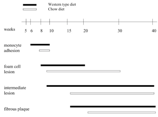
develop spontaneous atherosclerotic lesions resembling human plaques at a relatively young age (3-5 month) (Nakashima, Plump, Raines, Breslow, & Ross, 1994). Early lesions such as adhesion of monocytes to the aortic wall in the aortic sinus occur at approximately 2 months of age, progressing to formation of foam cells-rich fatty streaks by 3 months. The intermediate lesions can be observed by 15 weeks, fibrous plaques by 20 weeks, calcification and wall thinning by 32 weeks of age (Meir & Leitersdorf, 2004). As in humans, atherogenesis in ApoE-/- mice can be
further accelerated by keeping them on high fat diet (Figure 4) (Jawien et al., 2004). For all these reasons ApoE-/- mice are considered to be the most reliable mouse model of atherosclerosis and
are widely used in atherosclerosis studies.
Figure 4. Timeline of lesion formation in the ApoE-/- mice.
Diagram illustrates delayed lesion formation in chow-fed mice as compared with mice fed the Western diet (Jawien et al., 2004)
1.7.2 Ldlr-/- mouse model of atherosclerosis
Originally Ldlr-/- mice were described as a model of human familial hypercholesterolemia
(Fazio & Linton, 2001), but further studies established this line as a model of diet-induced atherosclerosis (Ishibashi, Goldstein, Brown, Herz, & Burns, 1994). On a normal chow diet Ldlr
-/-mice have a 2-3-fold elevated plasma cholesterol level and begin to develop early atherosclerotic lesions around 6-7 months of age, but advanced lesions are normally not observed even further in life. However when Ldlr-/- mice are fed the Western diet (Daugherty, 2002) this leads to
appearance of early atherosclerotic lesions already after 4-6 weeks of feeding, whereas intermediate lesions are observed between 12 and 16 weeks of feeding. More advanced lesions appear after prolonged feeding, between 16 and 20 weeks (Whitman, 2004).
1.8 Lipids and lipoproteins
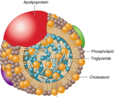
Body lipids can be classified as the exogenous lipids that are resynthesized in the gut from dietary fats, and endogenous lipids, that are synthesized in the liver. Lipoproteins play a key role in the absorption and transport of dietary lipids from the small intestine, in the transport of endogenous lipids synthesized in the liver to peripheral tissues, as well as in the transport of lipids from peripheral tissues to the liver and intestine (reverse lipid transport). Another major function of lipoproteins is the transport of toxic hydrophobic and amphipathic compounds from the areas of invasion and infection (Feingold & Grunfeld, 2012). Lipoproteins are spherical macromolecular complexes composed of a hydrophilic coat of phospholipids, free cholesterol and specific amphipathic proteins, apoproteins and a hydrophobic core of non-polar lipids such as cholesterol esters and triglycerides (Hoofnagle & Heinecke, 2009) (Figure 5).
Figure 5. A schematic diagram representing the structure of lipoproteins.
Copyright © 2005 by Elsevier Inc.
1.9 Lipoprotein classification
The lipid-protein complexes vary in their size, composition and content of lipid and protein. Traditionally plasma lipoproteins are classified by density defined as the ratio of lipid to protein affecting their flotation during density gradient ultracentrifugation. Because lipids occupy a greater molecular volume then proteins and are therefore less dense, lipoproteins become progressively denser and smaller in size as the ratio of lipid to protein decreases (Lewis, 1973). Lipoproteins are divided into five major classes based on the relative contents of lipids and proteins: chylomicrons (CM), very low-density lipoproteins (VLDL), intermediate-density lipoproteins (IDL), low-density lipoproteins (LDL) and high-density lipoproteins (HDL) (Figure 6) (Hegele, 2009).
Figure 6. Relative size of plasma lipoproteins according to their density.
Copyright © 2005 by Elsevier Inc.
Besides density, lipoproteins are also classified according to their net charge. All lipoproteins exhibit a negative net charge, determined by both the apoprotein and lipid constituents (Sparks & Phillips, 1992). Based on the charge measured by their electrophoretic mobility lipoproteins can be separated into α, pre β, β and broad β lipoprotein classes. The electrophoretic mobility of a lipoprotein is mainly dependent upon its protein content: those with higher protein content will move faster in the agarose gel towards the anode and those with low protein content will have lower mobility. Thus, HDL are α, VLDL are pre β, LDL are β and IDL are broad beta lipoproteins (Lewis, 1973; Noble, 1968). In addition, lipoproteins can be classified on the basis of their immunological characteristics, particle size and by conferred principal apoproteins (Morita, 2016).
1.10 Structure and biochemical composition of LDL
LDL is a spheroidal particle with an average diameter of 22-28 nm and a density of 1.019-1.063 g/ml. Its’ hydrophobic central core contains approximately 1600 molecules of cholesteryl esters and 170 molecules of triglycerides. The core is surrounded by a phospholipid-rich shell, which consists of 600 molecules of free cholesterol, approximately 700 phospholipid molecules and a small amount of lyso-phospholipids, gangliosides, sphingomyelin, phosphatidylethanolamine, phosphatidylserine and phosphatidylinositol (Esterbauer, Gebicki, Puhl, & Jurgens, 1992). Each LDL particle contains one molecule of apolipoprotein B 100 (ApoB) attached to the phospholipid-rich surface, that plays a crucial role in the interaction of the lipoprotein particle with the hepatocyte LDL receptor (LDLR), but not with other members of the LDLR family (Schumaker, Phillips, & Chatterton, 1994) (Esser, Limbird, Brown, Goldstein, & Russell, 1988).
1.11 Structure and function of apolipoproteins
Apolipoproteins are essential for lipoprotein structural assembly, secretion and function. Apolipoproteins have several major roles: they guide formation of the lipoproteins and support their structure; they act as ligands for lipoprotein receptors; and they serve as activators or inhibitors of enzymes involved in the metabolism of lipoproteins(Hoofnagle & Heinecke, 2009). Plasma apolipoproteins can be classified in two groups: nonexchangeable apolipoproteins such as ApoB 100, or ApoB48 and exchangeable apolipoproteins such as ApoE, I, II, ApoA-IV, ApoC-I, ApoC-II, or ApoC-III (Segrest, Jones, De Loof, & Dashti, 2001). Each lipoprotein class
has a characteristic apolipoprotein composition. One or more apolipoproteins (proteins or polypeptides) are present in each lipoprotein. The major apolipoproteins of HDL (α-lipoprotein) are designated as A class. The main apolipoproteins of LDL (β-lipoprotein) belong to the B class, represented by ApoB. They are also found in VLDL. Chylomicrons contain a truncated from of ApoB (B48) synthesized in the intestine, while a full-size ApoB is synthesized in the liver. ApoE is found in VLDL, HDL, chylomicrons and chylomicrons remnants (Stocker & Keaney, 2004).
1.12 ApoE and ApoB - lipoproteins most associated with atherosclerosis
1.12.1 Apolipoprotein E
ApoE, is a 34-kDa (299 amino acids) polymorphic multifunctional plasma apoprotein with a single O-linked glycan chain (Wernette-Hammond et al., 1989). In human plasma approximately 20% of all ApoE molecules are sialylated and a sialylation has been linked to their clearance rate (Millar, 2001).
ApoE is a ligand for the low-density lipoprotein receptor (LDLR) and LDLR-related protein (LRP). The majority of plasma ApoE is produced by the liver and macrophages but the protein is also synthesized by other organs such as the brain, kidneys and spleen (Boisvert, Spangenberg, & Curtiss, 1995). As a ligand for the receptor-mediated clearance of lipoproteins, ApoE is an important modulator of atherosclerosis. ApoE promotes the internalization of TG-rich lipoprotein remnants by hepatocytes, so the lack of it leads to lipoprotein accumulation (Schaefer et al., 1986). ApoE knockout mice have high levels of plasma cholesterol and spontaneously develop atherosclerosis (S. H. Zhang et al., 1992). Besides, in the atherosclerotic lesions ApoE directly
modifies the macrophage-mediated immune response that contributes to atherosclerosis (Curtiss & Boisvert, 2000; Perrey et al., 2001).
1.12.2 Apolipoprotein B
Apolipoprotein B (ApoB) is the main structural surface protein present in all beta-lipoproteins (chylomicrons, VLDL, IDL and LDL) in stoichiometry of one molecule of ApoB per lipoprotein. ApoB has two isoforms: ApoB100, a 4536-amino acid-secretory glycoprotein (550kDa) and ApoB48, a 2152-amino acid protein corresponding to the N-terminal 48% sequence of ApoB100 (Segrest et al., 2001). ApoB48 is produced in the proximal small intestine by tissue-specific mRNA splicing that generates a truncated form of ApoB. ApoB48 directs the formation of chylomicrons which main function is absorption and transport of lipids absorbed by the intestine. A small amount of APOB mRNA however escapes editing, resulting in a low level of ApoB100 present in the intestine (Kane, 1983). The full-length protein is constitutively synthesized in the liver by hepatocytes, where it assembles in VLDL. After VLDL are excreted and undergo lipolysis they become IDL and LDL (Kane, 1983).
Human ApoB100, one of the largest known monomeric proteins, is highly hydrophobic (Shelness & Sellers, 2001). Its folding requires assistance of chaperone proteins, post-translational lipidation, formation of disulfide bonds, and glycosylation. The fraction of ApoB100 that remains misfolded post-transnationally is eliminated through autophagy (Macri & Adeli, 1997). There are 8 disulfide bonds in ApoB100, 7 of them are found within the N-terminal 21% of its sequence (Yang et al., 1990). The amino acid sequence of ApoB100 contains 19 potential N-glycosylation sites, 17 of these are occupied by high-mannose and complex forms of
oligosaccharides (Taniguchi, Ishikawa, Tsunemitsu, & Fukuzaki, 1989) (Triplett & Fisher, 1978). Most of the ApoB100 N-glycans contain terminal sialic acid residues present in the α2-6 conformation. Six of the mature ApoB100 N-glycans are located close to the LDLR-binding region (Yang et al., 1986), however they do not play a significant role in LDL endocytosis (Shireman & Fisher, 1979). The potential function of ApoB100 glycans in post-hepatic secretion is still unknown.
1.13 Lipoprotein metabolism
Lipoprotein metabolism involves an exogenous pathway, endogenous pathway and reverse cholesterol transport.
1.13.1 Exogenous (dietary) pathway – chylomicron processing
Hydrolyzed dietary fats enter intestinal cells (enterocytes) via fatty acid transporters (Figure 7). Reconstituted TG are packaged with esterified cholesterol and ApoB48 into chylomicrons by the microsomal TG-transfer protein (MTTP) through a vesicular pathway (Hegele, 2009). In circulation, the nascent chylomicrons acquire ApoC and ApoE from plasma HDL in exchange for phospholipids. The acquisition of ApoC-II from HDL is essential to activate lipoprotein lipase (LPL) (Huff, Pollex, & Hegele, 2006). CM bind to membrane-bound LPL located on adipose and muscle tissues where the TG are hydrolyzed into free fatty acid (FFA). FFA can be further taken up by the liver, adipose tissue or by muscles. In the adipose cells FFA are re-processed into triacylglycerols and stored, whereas in the muscle, FFA are oxidized to provide energy (Morita, 2016). As the tissues absorb the fatty acids, CM progressively diminish in size and
become chylomicron remnants, where ApoE is exposed on the particle surface and the released ApoC is returned to plasma HDL. The ApoE mediate uptake of chylomicron remnants by the liver via the LDLR and LRP. The ApoC proteins are continuously recycled between chylomicrons and HDL (Morita, 2016).
Figure 7. Major normal lipoprotein metabolic pathways.
ABCA1, ATP binding cassette transporter 1; CE, cholesterol ester; CETP, cholesteryl ester transfer protein; FFA, free fatty acid; HTGL, hepatic triglyceride lipase; IDL, intermediate-density lipoprotein; LCAT, lecithin: cholesterol acyltransferase; LDL-R, LDL receptor; Lp(a), lipoprotein(a); LPL, lipoprotein lipase; LRP, LDL-R–related protein; SR B1, scavenger receptor B1; TG, triglyceride (Kwan, Kronenberg, Beddhu, & Cheung, 2007).
1.13.2 Endogenous pathway – VLDL synthesis and processing. Conversion of VLDL to LDL
In hepatocytes TG assemble with cholesterol and ApoB into VLDL that are secreted via exocytosis (Figure 7). Like chylomicrons VLDL acquire ApoC and ApoE from HDL. VLDL are processed by the membrane-bound LPL of adipose and muscle tissues. LPL hydrolyses TG into FFA (Kwan et al., 2007) that are transported to the adipose cells for further synthesis and storage of TG or to the muscles to be oxidized and produce energy. After hydrolysis of the TG by LPL, VLDL release ApoC and phospholipids to HDL. VLDL considerably reduced in size forms IDL. IDL can be taken up by the liver or they can be further hydrolyzed by LPL, eventually loosing ApoE to form LDL (Morita, 2016). LDL are endocytosed by peripheral cells and hepatocytes through LDLR specifically recognizing ApoB. Approximately 75% of all LDL are absorbed by the liver. The remaining LDL are removed by other cells via LRP and other scavenger receptors (Brown & Goldstein, 1983).
1.13.3 Reverse cholesterol transport – role of HDL
HDL via ApoA-I and ApoA-IV mediate reverse cholesterol transport by interacting with ATP-binding cassette A1 (ABCA1) and ATP-binding cassette G1 (ABCG1) transporters on non-hepatic cells (Figure 7) (Morita, 2016). HDL remove excess cholesterol from peripheral cells and carry it to the liver, for conversion into bile salts. Precursors of HDL particles having a disk-shaped structures are secreted by the liver and intestine (Kwan et al., 2007). They acquire spherical shape while they absorb cholesterol from cell membranes and TG from other lipoproteins. The major apolipoprotein of HDL, ApoA-I activates lecithin-cholesterol acyltransferase (LCAT) that catalyzes the transfer of long-chain fatty acids from phospholipids to cholesterol forming cholesterol esters
to be further incorporated into HDL (Morita, 2016). After remodeling by the cholesterol ester transfer protein (CETP), that mediates exchange of cholesterol esters between lipoproteins, and by endothelial lipase, larger in size HDL particles enter hepatocytes via the scavenger receptor class B type I (SRB1) expressed in the liver and steroidogenic tissues (Acton et al., 1996). When HDL grow in size they also acquire multiple copies of ApoE, so they can further be taken up by the liver via LDLR (Bruce, Chouinard, & Tall, 1998).
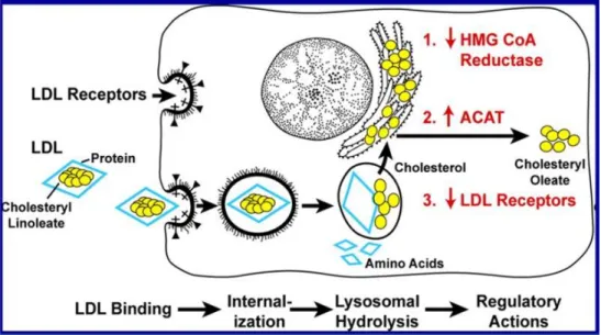
1.14 LDL receptor pathway
The LDL receptor, LDLR is a 160 kDa glycoprotein present in all mammalian cell types (T. Yamamoto et al., 1984) that has affinity for two apolipoproteins, ApoB and ApoE (Willnow, 1997). LDLR plays a major role in cellular cholesterol homeostasis and also participates in a variety of regulatory modulations (Figure 8). LDL catabolism is primarily mediated through the hepatic LDLR. Following the internalization LDL degrade in the lysosomes, releasing LDL cholesterol. This suppresses expression of HMG-CoA (3-hydroxy-3-methylglutaryl coenzyme A) reductase, the key enzyme in the biosynthesis of cholesterol, and induces expression of LDLR. Thus, cholesterol content regulates the level of LDLR. Proprotein convertase subtilisin/kexin type 9 (PCSK9) diverts LDLR into the endosomal-lysosomal pathway for degradation (Seidah et al., 2003). Besides, LDL can suppress LDLR expression through the inhibition of the SREBP (sterol regulatory element-binding protein) pathway (Brown & Goldstein, 1999). A high level of cellular cholesterol activates the cholesterol-esterifying enzyme, cholesterol acyl-transferase (ACAT) so the excess of cholesterol can be stored as cholesteryl ester droplets in the cytoplasm (Brown, Dana, &
Goldstein, 1975). All these actions allow cells to regulate the amount of LDLR and level of cholesterol for their metabolic needs without causing cholesterol over accumulation.
Figure 8. Sequential steps in the LDL receptor pathway.
HMG-CoA, 3-hydroxy-3-methylglutaryl coenzyme A reductase; ACAT, cholesterol acyl-transferase. Modified from Brown and Goldstein (Brown & Goldstein, 1979).
1.15 LDL and atherosclerosis
A high level of cholesterol in LDL particles is a well-known risk factor for the development and progression of atherosclerosis (Bentzon, Otsuka, Virmani, & Falk, 2014; Keys, 1997; Martin, Hulley, Browner, Kuller, & Wentworth, 1986). However, 46% of first cardiovascular events occur in people with LDL levels at the normal range (Packard & Libby, 2008). Atherogenicity of LDL can be caused not only by the quantity, but also by certain properties of LDL, such as a small size, high density (Griffin, 1999) or their modification (Ahotupa, Suomela, Vuorimaa, & Vasankari, 2010). LDL are defined as lipoprotein particles with a density between 1.019 and 1.063 g/ml, however their composition is extremely heterogeneous and includes particles with different sizes, structures and chemical compositions (Atkinson, Deckelbaum, Small, & Shipley, 1977;
Fisher, 1972; Shen, Krauss, Lindgren, & Forte, 1981). Specifically, predominance of small dense LDL (d=1.044-1.060 g/ml) was associated with increased risk of CAD (Krauss, 1995).
Another plasma lipoprotein(a), Lp(a), that represents a subclass of LDL has been also associated with development of CAD (G. T. Jones et al., 2007). Lp(a) has as a protein moiety ApoB linked covalently to a single molecule of Apo(a) a specific multi-kringle protein of the plasminogen family (Figure 9) (Scanu, Lawn, & Berg, 1991). Lp(a) is unique to humans, apes, monkeys and hedgehog (Sabarinath & Appukuttan, 2015). The Apo(a) molecule is heavily glycosylated (glycans contribute to 28% of Apo(a) molecular mass (Fless, ZumMallen, & Scanu, 1986)) and linked to the apoprotein ApoB by a disulphide bond. It has been shown that Lp(a) is the dominant lipoprotein in human atherosclerotic plaques and its level increases with the plaque progression (Pepin, O'Neil, & Hoff, 1991; van Dijk et al., 2012).
Figure 9. Lp(a) Structure.
Lp(a) consists of a LDL lipid core with ApoB attached by a disulfide bond to Apo(a). Apo(a) contains a variable number of kringle domains (KIV-1–10) that have a high homology to K4 of plasminogen, one KV kringle similar to K5 of plasminogen, and a proteolytic-like domain. KIV-2 is present in 2–50 copies, imparting extreme heterogeneity to Lp(a) (Hoover-Plow & Huang, 2013).
Biochemical modifications of ApoB contribute to LDL atherogenicity. There are many modifications that can occur with ApoB, such as: partial enzymatic hydrolysis (Bhakdi et al., 1995), chlorination or nitration (Heinecke, 1999), free radical oxidation of the polypeptide (Bruce et al., 1998), and modifications of ApoB glycan chains including their desialylation (Tertov et al., 1993). In macrophages, oxidized LDL (oxLDL) are taken up with the help of scavenger receptors such as CD36, SR-A1 and lectin-like oxLDL receptor-1 (LOX-1) (Gimbrone & Garcia-Cardena, 2013). Oxidation or acetylation of LDL in vitro converts them into ligands of scavenger receptors, but whether oxidation is the primary LDL modification in vivo is still unknown.
Another modification of LDL that may lead to atherosclerosis is desialylation (Orekhov, Tertov, & Mukhin, 1991). Fractions of ApoB-containing LDL with reduced content of sialic acids (Sia) were found in human blood (Bartlett & Stanley, 1998). Sia content of LDL in patients with atherosclerosis is lower than in healthy subjects (Ruelland, Gallou, Legras, Paillard, & Cloarec, 1993; Tertov et al., 1993). Desialylated LDL (desLDL) are rapidly taken up and accumulated by peripheral blood monocytes and by smooth muscle cells isolated from human arterial intima (Bartlett, Grewal, De Angelis, Myers, & Stanley, 2000).
Combined treatment with trypsin, cholesterol esterase, and neuraminidase transforms LDL into particles with a lipid structure, biological properties and composition similar to that of lipids extracted from atherosclerotic lesions (Bhakdi et al., 1995). Another study reported that enzymatic desialylation of LDL caused physical and chemical modifications in the structure of ApoB and resulted in accumulation of neutral lipids and cholesteryl esters in human aortic intimal cells (Tertov et al., 1992). DesLDL have exposed Gal residues (Taniguchi et al., 1989) which makes them a high affinity ligand for lectin receptors such as the asialoglycoprotein receptor (Lee et al.,
1983). Importantly, lipopolysaccharide-stimulated macrophages expressing increased amounts of Gal/GalNAc-specific lectin on their surface (Grewal, Bartlett, Burgess, Packer, & Stanley, 1996) showed high uptake rates of desLDL. Desialylated LDL can also bind scavenger receptors, and cellular surface proteoglycans (Camejo, Lopez, Lopez, & Quinones, 1985). DesLDL are smaller in size, higher in density, have lower negative charge and reduced affinity to the LDL receptor (Orekhov et al., 1992). Besides, desialylation of LDL can lead to their aggregation, which is a key factor for intracellular lipid accumulation (Musliner, McVicker, Iosefa, & Krauss, 1987).
1.16 Foam cell formation
Circulating monocytes differentiate into macrophages after migration into the intima. In macrophages, a range of genes related to lipid metabolism, such as LDLR, PCSK9, APOB play critical roles in maintaining normal cholesterol homeostasis (Kjolby, Nielsen, & Petersen, 2015; Lamon-Fava, 2013; Nozue et al., 2016).
Atherogenic modifications of LDL prevent their recognition by (or reduce their affinity for) the low-density lipoprotein receptor (LDLR) (Tabas, 2002). As a result, modified LDL become recognized by another group of receptors – scavenger receptors – that are expressed on the cell surface of macrophages. Scavenger receptors identified in macrophages include: scavenger receptor AI/II (SR-AI/II), scavenger receptors cluster of differentiation 36 (CD36), LOX-I, SR-BI (Levitan, Volkov, & Subbaiah, 2010). Scavenger receptor type A (SR-A) was shown to be specific for the recognition and the uptake of acetylated and oxidized LDL (Kodama et al., 1990). Cytokines and modified LDL themselves induce expression of scavenger receptors. It is also increased during differentiation of monocytes to macrophages (Ye et al., 2009). Unlike the LDLR,
scavenger receptors do not undergo negative feedback regulation in response to intracellular cholesterol accumulation (Moore & Freeman, 2006). LDL, recognized via scavenger receptors are endocytosed and lysosomes loaded with LDL accumulated in the cytoplasm, resulting in the formation of the foam cells (Morita, 2016). Some lipid-loaded macrophages can leave the arterial wall, thus taking away the lipids from the artery. Otherwise, they contribute to the consequential growth of atheroma (X. H. Yu et al., 2013).
1.17 Sialic acids
Sialic acids (Sia) are negatively charged N-or O-acyl derivatives of a 9-carbon sugar called neuraminic acid (5-amino- 3, 5-dideoxy-2 nonulosonic acid) (Figure 10) (X. Chen & Varki, 2010). Sia are common terminal sugar components of the oligosaccharide chains of glycoproteins and glycolipids.
Figure 10. Chemical structure of the most common human sialic acid, N-acetyl neuraminic acid
Sia are involved in a surprising variety of biological processes, including conformational stabilization of molecules, regulation of cell surface charge, cell recognition, cell differentiation, interaction, migration, adhesion, immune response and metastasis (X. Chen & Varki, 2010; Drake, Balbis, Wu, Bergeron, & Posner, 2000). The majority of soluble secreted and lysosomal proteins contain Sia as part of their glycan chains, and this modification extends their half-life. Sia are also
present on the surface of erythrocytes and platelets and the level of sialylation determines the life span of these cells in circulation (Aminoff, Bell, Fulton, & Ibgebrigtsen, 1976). It is thought that sialylation provides for the negative surface charge of erythrocytes which is important for electrostatic repulsion between the cells (Chien, 1986). Members of the Siglecs (sialic acid binding immunoglobulin-like lectins) superfamily mediate intracellular interactions which contribute to the scavenging function of macrophages, pathogen uptake and antigen presentation (Kahn, 2000). Glycosynapses enriched in sialylated glycoproteins and glycolipids mediate cell signaling and participate in cell adhesion, motility and growth (Brunzell et al., 1976). Cancer cells have long been recognized to have a significant over-expression of Sia on the cell surface (Bevan et al., 2000). Lipid- and protein-bound Sia are elevated in plasma from cancer patients (Contreres, Faure, Baquiran, Bergeron, & Posner, 1998) and linked with acute phase condition and chronic disease (C. Yu et al., 2002). The majority of Sia in the blood are bound to proteins and glycolipids. In particular Sia associated with lipoproteins are attached to the carbohydrate chains of apolipoproteins (Taniguchi et al., 1989) and ganglioside molecules (mainly GM3 ganglioside) present in the lipid core. The content of Sia decreases from VLDL to LDL and further to HDL (Harada, Carvalho, Passarelli, & Quintao, 1998). Glycosylation plays an important role in the biology of LDL as all LDL particles contain sialylated oligosaccharides (Millar, 2001; Orekhov et al., 1991). Every ApoB molecule in human plasma LDL contains approximately 12-14 Sia residues (Millar, 2001). Apolipoproteins CII and CIII, which are found to VLDL and are responsible for the interaction of VLDL with lipoprotein lipase are also heavily sialylated with functional consequences (Mauger, Couture, Bergeron, & Lamarche, 2006). Sialic acid is also largely present in the glycan chains of the mature LDLR (Cummings et al., 1983) and is responsible
for the net negative charge of this protein (Goldstein, Brown, Anderson, Russell, & Schneider, 1985). Importantly sialylation of LDLR plays an important role in its interaction with LDL, evidenced by the fact that treatment of fibroblasts and endothelial cells with bacterial neuraminidase had significantly altered LDL binding, internalization and degradation by these cells(Sprague, Moser, Edwards, & Schwartz, 1988). PCSK9 also binds to the highly glycosylated ligand-binding region of LDLR, thus adding further potential implications for sialylation in LDLR regulation and degradation (T. Yamamoto, Lu, & Ryan, 2011). Several studies showed that a high level of free sialic acid in human plasma is associated with CAD and atherosclerosis (Knuiman, Watts, & Divitini, 2004; Wakabayashi, Sakamoto, Yoshimoto, & Kakishita, 1994). Patients with atherosclerosis contain LDL in their blood with a 2.5-5 fold lower sialic acid content compared to that of healthy subjects, at the same time there was no difference in protein and lipid content between the two groups of patients (Orekhov et al., 1991). The sialylation of ApoB has been directly implicated into the risk of atherosclerosis (Mel'nichenko et al., 2005; Sobenin, Tertov, Orekhov, & Smirnov, 1991).
1.18 Neuraminidases
Neuraminidases (sialidases) are enzymes that catalyze removal of terminal Sia residues from glycoproteins, oligosaccharides, and sialylated glycolipids. The mammalian genomes contain four genes, which encode members of the neuraminidase family (NEU1-NEU4 in humans). These enzymes have different, yet overlapping tissue expression, intracellular localization and substrate specificity (Milner et al., 1997; Monti et al., 2000; Monti, Preti, Nesti, Ballabio, & Borsani, 1999; Seyrantepe et al., 2004) (Table 1). NEU1 is ubiquitously expressed with