Original Article
Activation of the calcium-sensing receptor before renal
ischemia/reperfusion exacerbates kidney injury
Laurent Weekers1, Pascal de Tullio2, Christophe Bovy1, Laurence Poma3, Raphaël Marée4,
Catherine Bonvoisin1, Jean-Olivier Defraigne3, Jean-Marie Krzesinski1,3, François Jouret1,3
1Division of Nephrology, University of Liège Hospital (ULg CHU), Liège, Belgium; 2Centre for Interdisciplinary
Re-search on Medicines (CIRM), University of Liège, Liège, Belgium; 3Groupe Interdisciplinaire de Génoprotéomique
Appliquée (GIGA), Cardiovascular Sciences, University of Liège, Liège, Belgium; 4Groupe Interdisciplinaire de
Gé-noprotéomique Appliquée (GIGA), Bioinformatics and Systems and Modeling, University of Liège, Liège, Belgium
Received October 14, 2014; Accepted November 25, 2014; Epub January 19, 2015; Published January 30, 2015 Abstract: Activation of the calcium-sensing receptor (CaSR) by ischemia/reperfusion (I/R) favours apoptosis in car-diomyocytes, hepatocytes and neurons. Its role in renal I/R is unknown. We investigated the impact of pharmacologi-cal preactivation of the CaSR on kidney structure and function in a murine model of bilateral renal 30-min ischemia and 48-hour reperfusion, and in a 6-year cohort of kidney transplant recipients (KTR). C57BL/6J mice were admin-istered daily with CaSR agonist, R-568, or with vehicle for 48 hours. Evaluation of serum urea and creatinine levels, renal histology and urine metabolome by nuclear magnetic resonance showed that R-568 was not nephrotoxic per
se. Following I/R, serum urea and creatinine levels increased higher in R-568-treated animals than in controls.
Jablonski’s score was significantly greater in R-568-treated kidneys, which showed a higher rate of cell prolifera-tion and apoptosis in comparison to controls. Next, we retrospectively identified 36 patients (10.7% of our cohort) who were treated by CaSR agonist, cinacalcet, at the time of kidney transplantation (KTx). After matching these to 61 KTR upon type of donor, cold ischemic time, residual diuresis, and donor age, we observed that delayed graft function, i.e. need for dialysis in the first week after KTx, occurred in 42 and 23% of cinacalcet-treated and control groups, respectively (p≤0.05). These data suggest that pharmacological preactivation of the CaSR before renal I/R exacerbates kidney injury.
Keywords: Ca2+-sensing receptor, kidney, ischemia/reperfusion, cinacalcet, transplantation, delayed graft function
Introduction
The Ca2+-sensing receptor (CaSR) belongs to
family C of the G-protein-coupled receptor (GPCR) superfamily, and is ubiquitously exp- ressed [1, 2]. Its principal physiological ligand is the ion Ca2+. By sensing the variations of
extracellular Ca2+ ([Ca2+]
e) concentration, the
CaSR plays a critical role in Ca2+ homeostasis.
It notably controls parathormone (PTH) secre-tion by the parathyroid glands and modulates Ca2+ fluxes in kidneys, intestine and bones
[3-5]. The importance of the CaSR in Ca2+
homeostasis is supported by the chronic hyper- or hypocalcemia observed in patients harbor-ing loss- or gain-of-function mutations, respec-tively, in the CASR gene [6]. In addition, the EC50 value for Ca2+ binding to the CaSR can be
significantly modified by several physiological
parameters, including ionic strength, extracel-lular pH, L-aromatic amino acids and poly-amines, or by drugs, like the calcimimetic com-pounds cinacalcet and R-568 [4]. Hence, cinacalcet is used in clinical routine to treat patients with primary hyperparathyroidism or with secondary hyperparathyroidism caused by chronic kidney disease (CKD), including individ-uals registered on kidney transplantation (KTx) waiting list [7, 8].
The CaSR has been identified in numerous tis-sues and cells that are not directly involved in Ca2+ homeostasis, in which its role remains
unclear [9, 10]. In epithelia, the CaSR particu-larly regulates cell proliferation, survival and Ca2+-induced polarization and differentiation
[11-14]. Moreover, the CaSR has been implicat-ed ex vivo and in vivo in the
ischemia/reperfu-sion (I/R) cascade in cardiomyocytes [15, 16] and neurons [17], as well as in hepatocytes [18]. The occurrence of I/R typically involves the reduction or interruption of organ perfusion with a subsequent reflow. Such I/R event in- duces significant cell metabolism modifications [19], including increased expression of the CaSR, intracellular Ca2+ overload and activation
of the mitochondrial and mitogen-activated protein kinase apoptotic pathways [15-18]. Transient I/R is the primary cause of acute kidney injury (AKI), a common situation curren- tly defined as a rapid fall of glomerular filtration rate (GFR) and/or a decline in urine output [20, 21]. One of the paradigms of renal I/R is KTx. Indeed, graft procurement and storage neces-sarily require the temporary interruption of renal blood flow, with subsequent reperfusion after transplantation. Such unavoidable I/R events contribute to delay graft recovery post KTx [22]. The role of the CaSR in kidney I/R has not been studied thus far. In order to transla-tionally investigate the impact of CaSR activa-tion before renal I/R on kidney structure and function, we first took advantage of a conven-tional mouse model of bilateral clamping of renal pedicles. Next, we retrospectively studied a prospective cohort of kidney transplant recip-ients (KTR) to test whether the use of cinacal-cet (Sensipar®/Mimpara®) at the time of KTx
influences early graft recovery. Materials and methods Acute renal ischemia in mice
All animal protocols were approved by the Ethics Committee for Animal Care and Use at the University of Liege School of Medicine (pro-tocol number #1335). Ten-week-old male C57BL/6 mice weighing ~20 g were treated with i.p. injections of CaSR agonist, R-568 (AMGEN), at 250 µg/d [23] or with a similar vo- lume of DMSO for 2 days before surgery. Animals were then anesthetized with pentobar-bital (60 mg/kg, Ceva®) by i.p. injection and,
using aseptic techniques, subjected to a lapa-rotomy with bilateral renal pedicle clamping (“ischemia”) for 30 min. Ischemia was con-firmed by color change observed in kidneys fol-lowing clamping. Supportive fluids were given throughout the operative period, and hypother-mia was prevented by use of an isothermal heating pad and warming lights. Following
sur-gery, animals were kept in light- and tempera-ture-controlled conditions for 48 hours, with a twice-daily clinical evaluation of scar and gen-eral health status. At 24 h post surgery, mice were placed in metabolic cages for 24 hours with ad libitum access to food and drinking water. Urine was collected on 2%-Na+ azide
solution (Sigma®) with one drop of mineral oil
(Sigma®) to prevent bacterial proliferation and
evaporation, respectively. Urine metabolome was analyzed using 1H-NMR (see infra). Blood
was obtained by vena cava puncture at the time of sacrifice. Serum levels of Ca2+, urea and
creatinine were measured on a COBAS 6000 C501 device (Roche-Hitachi®). At 48h
post-sur-gery, the animals were once again anesthetized and subjected to a laparotomy. Kidneys were collected, fixed in 4% paraformaldehyde (Boe- hringer Ingelheim, Heidelberg, Germany) in 0.1 mol/L phosphate buffer, pH 7.4, prior to embed-ding in paraffin, and further subjected to histo-logical analyses [24].
Immunostaining
Six-µm sections were incubated for 30 minutes with 0.3% hydrogen peroxide to block endoge-nous peroxidase. Antigen retrieval was per-formed by incubating sections in 0.01 mol/L citrate buffer, pH 6.1, for 11 minutes, in an autoclave heated at 121°C, before cooling down for 20 min, and rinsing. Following incuba-tion with 10% normal serum for 30 minutes, sections were incubated for 45 minutes with the primary antibodies diluted in phosphate-buffered saline (PBS) containing 2% bovine serum albumin (BSA). After washing, sections were successively incubated with biotinylated secondary anti-immunoglobulin G antibodies (Dako®), avidin-biotin peroxidase (Merck®), and
3,3-diaminobenzidine (DAB, Dako®). The
speci-ficity of immunostaining was tested by incuba-tion (i) in absence of primary antiserum, or (ii) with control IgG (Vector Lab®). The use of
ApopTag kit S7101 followed the manufacturer’s recommendations (Millipore®). Following
immu-nostaining, kidney sections were scanned using the NanoZoomer 2.0 HT (Hamamatsu®).
The regions of interest were first distinguished (Supplementary Figure 1). The cortex is located between the capsula and the cortico-medullary junction (CMJ). CMJ is distinguishable from the cortex upon the absence of glomeruli, the pres-ence of large venous spaces, and the shape of S3 straight PT. CMJ was further isolated from
the medulla according to the longitudinal vs. transversal orientation of the tubules. Next, immuno-reactive cells were systematically identified in each kidney section and counted using both direct visual method and www.cyto-mine.be software (Supplementary Figure 1) [25]. Following a hybrid human-computer approach, tens of positive and negative cell examples were manually annotated. Then, a tree-based machine learning algorithm was used to build a positive pixel segmentation model further combined with connected-com-ponent analysis and filtering operations based on size and circularity criteria. Automatic posi-tive cell detections were proofread by experts. The number of positive cells and area statistics were then computed for each region of interest for each image. In addition to immunostaining experiments, routine colorations, i.e. Hema- toxylin-Eosine (HE) and periodic acid-Shiff, were classically performed. Sections were viewed under a Leica DM 1000 LED coupled to a Leica MC170 digital camera (Leica®). The severity of
I/R-associated AKI was graded according to Jablonski’s score [26] by a pathologist special-ized in renal pathology, unaware of animal groups.
1H-NMR metabolomics
Urine samples (150 μl) were supplemented with 450 μl of deuterated phosphate buffer (DPB, pH 7.4), 100 μl of a 5 mM solution of maleic acid and 10 μl of a 10 mg/ml trimethyl-silyl-3-propionic acid-d4 D2O (TMSP) solution.
1H-NMR spectra were acquired using a 1D
NOESY sequence with presaturation on a Bruker Avance spectrometer operating at 500.13 MHz for proton and equipped with a TCI
entire range of the spectra and the δ scale was calibrated to 0 ppm using the internal standard TMSP. For statistical analysis, optimized 1H-
NMR spectra were automatically baseline cor-rected and reduced to ASCII files using AMIX software (version 3.9; Bruker). The spectral intensities were normalized to the creatinine signal at 3.05 ppm and reduced to integrated regions of equal width (0.04 ppm) corre-sponding to the 0.5-10.00 ppm region. Beca- use of the residual water signal, the region between 4.5 and 5.5 ppm was removed before further analysis. The matrices obtained were used for the statistical analysis. The output data were then submitted to a “principal com-ponent analysis” (PCA) using AMIX software. Cohort of patients
All KTR from 2007 to 2012 at University of Liège Hospital (ULg CHU) were prospectively included in a database. Patients actively treat-ed with cinacalcet on the day of KTx were retro-spectively identified from this database and matched 1:2 with controls on (i) type of donor (living (LD), deceased after brain or circulatory death (DCD)); (ii) cold ischemic time (CIT) ± 1 hour; (iii) residual diuresis (± 500 mL); and (iv) donor age (± 5 years). Delayed graft function (DGF) was defined as the need for RRT in the first postoperative week post KTx. Baseline characteristics of both groups are summarized in Table 3.
Results
Administration of CaSR agonist, R-568, is not nephrotoxic in mouse
Male 10-week-old C57BL/6 mice were daily administered with CaSR agonist, R-568, or with Table 1. Biological parameters before and after renal
ischemia/re-perfusion in DMSO- and R-568-treated mice
Creatinine (mg/dl) BUN (g/L) Ca2+ (mmol/L)
Before I/R
DMSO-treated mice 1.02 ± 0.08 0.63 ± 0.09 2.66 ± 0.21 R-568-treated mice 1.13 ± 0.28 0.81 ± 0.16 2.37 ± 0.27
p (unpaired Student t-test) ns ns p<0.05
Following I/R
DMSO-treated mice 0.73 ± 0.13 0.78 ± 0.21 R-568-treated mice 5.74 ± 1.13 4.54 ± 0.82
p (unpaired Student t-test) p<0.05 p<0.05
I/R, ischemia/reperfusion; BUN, blood urea nitrogen; ns, not significant n=8 mice in each group.
cryoprobe at 298 K. The Noesypresat experiment us- ed a RD-90°-T1-90°-Tm -90°-acquire sequence with a relaxation delay of 4 s, a mix-ing time (Tm) of 10 ms and a fixed T1 delay of 4 µs. Water suppression pulse was placed during the relaxation delay (RD). The number of transient is 128 (64K data points) and a number of 4 dummy scans is chosen. Acquisition time is fixed to 3.2769001 s. Phase and baseline corrections were performed manually over the
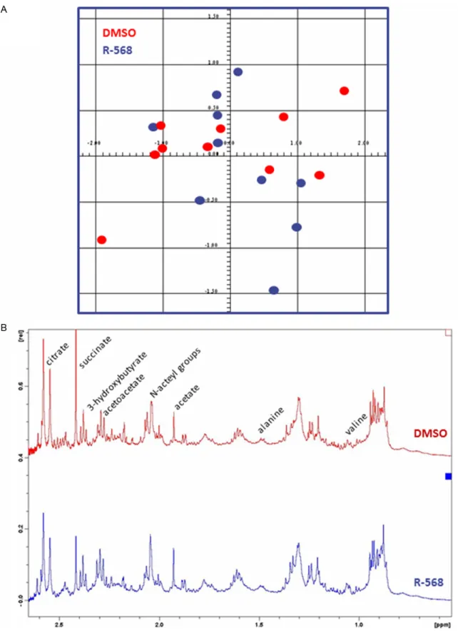
Figure 1. 1H nuclear magnetic resonance (1H-NMR) metabolomics analysis on urine samples from DMSO-treated
and R-568-treated mice. A. Representative score plot of principal component analysis (PCA, AMIX software) of urinary metabolomes of DMSO-treated (red dots) and R-568-treated (blue dots) mice. No significant difference is observed between urine metabolite profiles of both groups. B. Representative 1H-NMR spectrum of urine
metabo-lome of a DMSO-treated (upper panel) and an R-568-treated (lower panel) mice. The relative abundance of urinary acetoacetate, alanine, 3-hydroxybutyrate and valine, is similar in samples from either group.
an equivalent volume of dimethyl-sulfoxide (DMSO) by intraperitoneal (i.p.) injections for 48 hours. Urine was collected in metabolic cages for the last 24 hours, whereas blood and kidney samples were obtained after anesthesia and laparotomy. As previously described [23], R-568 administration was associated with a significant decrease of serum Ca2+ levels in
comparison to DMSO treatment (Table 1). Conversely, no significant difference in serum creatinine and urea levels was observed between R-568-treated versus non-treated ani-mals (Table 1). In order to further assess renal safety of R-568 administration in mouse, we performed 1H nuclear magnetic resonance (1
H-NMR) metabolomics analysis on urine from R-568-treated and control mice. 1H-NMR
pro-files were undistinguishable between groups (Figure 1A). Furthermore, the relative abun-dance of urinary metabolites previously associ-ated with proximal tubule (PT) toxicity [27], including glucose, acetoacetate, alanine, 3-hy- droxybutyrate and valine, was similar in sam-ples from either group (Figure 1B). Finally, com-parative histological examination of kidneys from R-568-treated and DMSO-treated mice was undistinguishable in terms of glomerular or tubular injury, cell proliferation and apoptosis (Table 2). Altogether, these functional and structural data support that i.p. administration of R-568 in mice does not cause significant nephrotoxicity per se.
Administration of CaSR agonist, R-568, before renal I/R exacerbates acute kidney injury in mice
Male 10-week-old C57BL/6 mice were daily administered with CaSR agonist, R-568, or with DMSO by i.p. injections for 48 hours before
sur-Histological examination of kidneys from DMSO-pretreated mice revealed acute tubular necrosis (ATN), with tubulorrhexis of epithelial cells and cylinders of cellular debris in renal tubules located in the cortex and at the cortico-medullary junction (Figure 2). Jablonski’s score of ATN severity reached 1 ± 1 in DMSO-treated animals (Table 2). Furthermore, numeric quan-tification of cells positive for proliferative cell nuclear antigen (PCNA) and terminal deoxynu-cleotidyl transferase nick end labeling (TUNEL) in kidneys from DMSO-treated mice showed a higher rate of cell proliferation and apoptosis after I/R in comparison to baseline conditions (Table 2). In animals treated by CaSR agonist, R-568, before renal I/R, Jablonski’s score of ATN severity was significantly higher (3 ± 1, p<0.05) in comparison to control group (Figure 2 and Table 2). Moreover, the number of prolif-erating cells and apoptotic cells was significant-ly increased in the cortex and at the cortico-medullary junction of R-568-treated versus DMSO-treated kidneys (Figure 2 and Table 2). These observations, including functional and histological parameters, suggest that i.p. administration of CaSR agonist, R-568, before renal I/R worsens the extent of ATN in mouse. Treatment with CaSR agonist, cinacalcet, at the time of kidney transplantation is associ-ated with delayed graft function
At the time of KTx, I/R is an unavoidable event, which has been associated with delayed graft recovery [28]. By definition, delayed graft func-tion (DGF) corresponds to the need for renal replacement therapy (RRT) in the first postop-erative week [22]. Cinacalcet is an analog of R-568 compound, with an improved bioavail-ability and metabolic profile [29]. Since cinacal-Table 2. Histological parameters before and after renal
ischemia/reper-fusion in DMSO- and R-568-treated mice
Jablons-ki score PCNA-positive cells/mm² ApopTag-positive cells/mm² cortex c-m junction cortex c-m junction Before I/R
DMSO-treated mice 0 16 ± 6 5 ± 3 2 ± 1 2 ± 1
R-568-treated mice 0 13 ± 6 7 ± 4 3 ± 2 3 ± 2
unpaired Student t-test ns ns ns ns ns
Following I/R
DMSO-treated mice 1 ± 1 66 ± 12 23 ± 10 8 ± 3 14 ± 8 R-568-treated mice 3 ± 1 117 ± 31 78 ± 33 12 ± 5 76 ± 19
unpaired Student t-test p<0.05 p<0.05 p<0.05 p<0.05 p<0.05
gical clamping of bilater-al renbilater-al pedicles for 30 minutes. After unclamp-ing, mice were kept alive for 48 hours with ad libi-tum access to food and drinking water. Analysis of serum samples ob- tained by vena cava puncture at the time of sacrifice showed a sig-nificantly worse AKI in R-568-treated mice than in controls, as reflected by serum creatinine and urea levels (Table 1).
cet is used in CKD patients with secondary hyperparathyroidism [7], individuals registered on KTx waiting list are prone to be actively treat-ed by cinacalcet at the time of KTx. Such a par-ticular clinical setting provides a unique trans-lational situation of CaSR activation before renal I/R. In our prospective 6-year database of 337 KTR, we retrospectively identified 36 (10.7%) patients who were treated by cinacal-cet at the time of KTx. These patients were matched 1:2 to controls according to
well-established factors of DGF, i.e. (i) type of donor, (ii) cold ischemic time (CIT), (iii) residual diure-sis, and (iv) donor age. Control group included 61 patients. Characteristics of patients and donors were compared between groups using Student’s t-test or Chi-2 as appropriate, as summarized in Table 3. The occurrence of DGF was significantly superior in cinacalcet-treated patients than in controls (42% versus 23%, p=0.05). These retrospective observations in a single-centre cohort of KTR suggest that CaSR
Figure 2. Histological examination of kidneys from DMSO-pretreated and R-568-pretreated mice following ischemia/ reperfusion. Representative kidney sections following I/R injury in mice pretreated with DMSO (A, C, E) or R-568 (B, D, F). Periodic acid-Shiff coloration (A, B), immunostaining against Proliferative Cell Nuclear Antigen (PCNA) (C, D), or terminal deoxynucleotidyl transferase nick end labeling (TUNEL, ApopTag®) (E, F) were performed. Pictures are
taken at the cortico-medullary junctions. Arrowheads in panels (A, B) indicate tubulorrhexis-associated intratubular debris. Scale bar represents 200 µm and 50 µm in panels (A-F) and insets, respectively.
activation at the time of KTx is more often asso-ciated with a need for RRT in the first postop-erative week.
Discussion
Kidney I/R is the leading cause of AKI, which accounts for up to 20% of admissions to intensive care units [30]. Furthermore, I/R cascade at the time of KTx is responsible for early graft dysfunction and enhanced graft immunogenicity, which impairs both short- and long-term graft survival [28]. Treatment of AKI remains largely supportive, including fluid main-tenance, vasoactive drugs, cytoprotective ther-apy and extrarenal epuration [30]. Therefore, advances in deciphering the pathophysiology of renal I/R injury are urgently required to develop both preventive and curative approaches of AKI [19, 31]. In our mouse model of renal I/R, we observed that administration of the CaSR ago-nist, R-568, before I/R was associated with a significantly worsened AKI, at both functional and architectural levels. Furthermore, the num-ber of apoptotic cells in kidney parenchyma was significantly greater in R-568-treated ani-mals in comparison to controls. As previously described, apoptosis particularly occurred in S3 PT segments, which are located at the corti-co-medullary junction and are highly vulnerable to ischemia [32, 33]. These findings concur with recent observations in various ex vivo and in vivo models of I/R [15-18]. In cardiomyo-cytes, the CaSR is involved in I/R-induced apoptosis through the mitochondrial pathway
[16]. Exposure to the CaSR agonist, GdCl3, before I/R causes a significant increase in phosphorylated protein kinase C δ (PKCδ) translocation to the mitochondria, with en- hanced release of cytochrome c (Cyt-c) and marked reduction of mitochondrial potential. Similarly, incubation of Buffalo rat liver cells with GdCl3 at the time of simulated I/R further induced CaSR expression, increased [Ca2+]
i,
and favored apoptosis through both mitochon-drial and mitogen-activated protein kinase pathways [18]. In vivo, forebrain I/R by tran-sient bilateral occlusion of carotid arteries in C57/BL6 mice increases CaSR expression and promotes cell death [17]. Altogether, these observations in distinct experimental models of I/R suggest a deleterious role of the CaSR in I/R-associated cellular cascades.
The CaSR has been ubiquitously identified [3, 4]. In the kidney, its distribution remains debat-ed [34]. In rats, CaSR mRNA is detectdebat-ed along essentially the entire nephron, including glom-eruli, S1-S2 convoluted and S3 straight PT, thick ascending limbs of Henlé’s loop (TAL), dis-tal convoluted tubules (DCT), and collecting ducts (CD) [35]. Similarly, immunohistochemis-try using CaSR-specific antisera documented the localization of the CaSR protein in PT, TAL, DCT and CD, with a variable polarity upon renal cell types [36]. Still, contradictory experimenta-tions in rats, mice and humans only found the CaSR in the TAL [37, 38]. In mice, the i.p. admin-istration of the CaSR agonist, R-568, induces hypocalcaemia [23], as we did observe in our Table 3. Clinical features of cinacalcet-treated and control patients at the time of kidney transplanta-tion
Cinacalcet (n=36) Controls (n=61) p
Recipient Age at Tx (years) 50.2 ± 10.3 49 ± 13.5 0.92
Sex ratio (% female) 47 41 0.55
Dialysis vintage (years) 3.7 ± 2.1 3.3 ± 3.8 0.57
Resting diuresis (ml) 430 ± 655 444 ± 541 0.91
Multi-organ Tx (%) 5.6 1.7 0.28
Donor Age (years) 46.8 ± 11.4 47 ± 11.4 0.93
Sex ratio (% female) 42 46 0.67
LD (%) 2.8 1.6 0.70
DCD (%) 30.6 21.3 0.31
Transplantation CIT (min) 779 ± 297 825 ± 255 0.43
HLA mismatches A 0.8 ± 0.5 0.9 ± 0.5 0.75
B 1.2 ± 0.7 1 ± 0.5 0.08
DR 0.8 ± 0.4 0.8 ± 0.3 0.99
Tx, transplantation; LD, living donor; DCD, donation after cardiac death; CIT, cold ischemic time; HLA, human leukocyte anti-gen.
studies. By contrast, the compound R-568 did not cause significant nephrotoxicity, as evi-denced by biological and histological assess-ment of renal function and by 1H-NMR
metabo-lomics analysis. 1H-NMR is currently used to
assess drug-induced kidney toxicity [27]. More specifically, our findings using 1H-NMR
metabo-lomics show that R-568 administration in mouse does not significantly modify the urinary metabolome previously linked to S3 PT toxicity. In humans, no deleterious impact on kidney function has been reported thus far after cina-calcet treatment (www.amgen.com). In KTR, Spanish multicenter observational retrospec-tive studies reported that cinacalcet therapy initiated 20 months after KTx induced a signifi-cant and sustained decrease of both serum Ca2+ and PTH levels, but did not affect renal
function during a 3-year follow-up [39]. Therefore, CaSR activation by the compound R-568 should not be regarded as nephrotoxic per se, but might make renal tubular cells more prone to apoptosis in case of I/R injury. The mechanism by which CaSR activation at the time of renal I/R enhances AKI severity is unknown. It might involve either a direct impact on tubular cells, with a rise of [Ca2+]
i and
stimu-lation of mitochondria-related apoptotic path-ways, and/or an indirect effect on the blood perfusion of kidney parenchyma. Indeed, intra-venous infusion of the calcimimetic, R-568, induces hypotensive effects in both normoten-sive and spontaneously hypertennormoten-sive rats [40, 41]. The cause of blood pressure drop following R-568 administration remains debated, with in vitro arguments for both CaSR-dependent and CaSR-independent production of nitric oxide (NO) [42].
KTx currently represents the best treatment for end-stage renal disease. Data from the Euro- Transplant Network (www.eurotransplant.org) show graft survival rates for primary kidney transplants of 84%, 78%, and 68% after 1, 3, and 5 years, respectively. Acute vascular rejec-tion and chronic allograft nephropathy remain the most relevant risk factors for renal graft dysfunction [43]. In addition to immunogenicity, organ preservation and I/R participate to kid-ney injury. DGF is a form of ATN resulting in post-transplantation oliguria, increased allo- graft immunogenicity and risk of acute rejec-tion episodes, and decreased long-term surviv-al. Factors related to the donor and prerenal, renal, or postrenal transplant factors related to the recipient can contribute to this condition
[44]. The reported frequency of DGF greatly var-ies worldwide, from 2% to 50%, upon the ambi-guity in its definition [22]. Preclinical and clini-cal studies have particularly demonstrated that both ischaemia and reinstitution of blood flow in ischemically damaged kidneys after hypo-thermic preservation play a pivotal part in the development of DGF [28]. Therefore, efforts to limit I/R injury may help minimize its deleteri-ous impact on early graft recovery. Our retro-spective analysis of a single-centre cohort of KTR suggests that cinacalcet treatment at the time of KTx is more often associated with a need for RRT in the first postoperative week. Cinacalet is an agonist of the CaSR, which is routinely prescribed to treat secondary hyper-parathyroidism in CKD patients [7]. Mineral and bone disorders (MBD) are thought to play a part in extraskeletal calcification and dimin-ished vascular compliance observed in CKD patients, thereby contributing to their increased cardiovascular risk [45]. Cinacalcet treatment may attenuate the progression of vascular and cardiac-valve calcification in hemodialysis pa- tients, although its beneficial impact on the risk of death or major cardiovascular events re- mains unproven [46, 47].
Our present translational study has several lim-itations. Our mouse model is restricted to bilat-eral clamping of renal vascular pedicles, with-out actual KTx, which does not allow us to take into account immune donor/recipient interac-tions. In addition, mice were only administered with the CaSR agonist, R-568, for 48 h before I/R, whereas KTR had been chronically treated with cinacalcet before KTx. Finally, the retro-spective pattern of the analysis of our single-center cohort limits its interpretation. Selection bias was reduced by matching 1:2 cinacalcet-treated and control individuals according to well-established factors of DGF. As a whole, our data suggest that CaSR activation at the time of renal I/R exacerbates AKI. The regulation of CaSR activity might thus serve as a novel phar-macological target to prevent and treat such a common condition.
Acknowledgements
The authors cordially thank the surgeons (M. Meurisse, C. Coimbra Marques, A. De Roover, E. Hamoir, P. Honoré, L. Kohnen, N. Meurisse and J-P Squifflet), the physicians (S. Grosch and P. Xhignesse), the technician (J-P Cheramy-Bien), and the members of the local transplant coordination center (M-H. Delbouille, M-H.
Hans, J. Mornard) for their personal and profes-sional commitment to kidney transplantation at the University of Liège Academic Hospital in Liège, Belgium. They also thank Eric Brevers (in E. Cavalier’s lab) for the measurements of serum creatinine, urea and Ca2+ levels, as well
as all members of the GIGA Bioinformatics Platform for providing access to computing servers and to the Cytomine software (http:// www.cytomine.be/). This work is supported by the Fonds National de la Recherche Scientifique (FNRS, Research Credit), University of Liège (Fonds Spéciaux à la Recherche) and from Fonds Leon Frédéricq (ACiiRT grant).
Disclosure of conflict of interest
The R-568 compound was provided by AMGEN Company (Thousand Oaks, CA) under the agr- eements MMFA 2012578383 and RPA 2012- 578387.
Address correspondence to: Dr. François Jouret, Division of Nephrology (Tower 1 - 6th floor), University of Liège Hospital (ULg CHU), Avenue de l’Hôpital, 1 (Building B35), B-4000 Liège, Belgium. Tel: +32.4. 366.25.40; Fax: +32.4.366.72.05; E-mail: francois. jouret@chu.ulg.ac.be
References
[1] Brown EM, Gamba G, Riccardi D, Lombardi M, Butters R, Kifor O, Sun A, Hediger MA, Lytton J and Hebert SC. Cloning and characterization of an extracellular Ca(2+)-sensing receptor from bovine parathyroid. Nature 1993; 366: 575-580.
[2] Brauner-Osborne H, Wellendorph P and Jen-sen AA. Structure, pharmacology and thera-peutic prospects of family C G-protein coupled receptors. Curr Drug Targets 2007; 8: 169-184.
[3] Chakravarti B, Chattopadhyay N and Brown EM. Signaling through the extracellular calci-um-sensing receptor (CaSR). Adv Exp Med Biol 2012; 740: 103-142.
[4] Geibel JP. The calcium-sensing receptor. J Nephrol 2010; 23 Suppl 16: S130-135. [5] Garg MK. The intestinal calcistat. Indian J
En-docrinol Metab 2013; 17: S25-s28.
[6] Hannan FM and Thakker RV. Calcium-sensing receptor (CaSR) mutations and disorders of calcium, electrolyte and water metabolism. Best Pract Res Clin Endocrinol Metab 2013; 27: 359-371.
[7] Torres PU. Cinacalcet HCl: a novel treatment for secondary hyperparathyroidism caused by
chronic kidney disease. J Ren Nutr 2006; 16: 253-258.
[8] Verheyen N, Pilz S, Eller K, Kienreich K, Fah-rleitner-Pammer A, Pieske B, Ritz E and Tomas-chitz A. Cinacalcet hydrochloride for the treat-ment of hyperparathyroidism. Expert Opin Pharmacother 2013; 14: 793-806.
[9] Magno AL, Ward BK and Ratajczak T. The calci-um-sensing receptor: a molecular perspective. Endocr Rev 2011; 32: 3-30.
[10] Riccardi D and Kemp PJ. The calcium-sensing receptor beyond extracellular calcium homeo-stasis: conception, development, adult physiol-ogy, and disease. Annu Rev Physiol 2012; 74: 271-297.
[11] Tu CL, Chang W, Xie Z and Bikle DD. Inactiva-tion of the calcium sensing receptor inhibits E-cadherin-mediated cell-cell adhesion and calcium-induced differentiation in human epi-dermal keratinocytes. J Biol Chem 2008; 283: 3519-3528.
[12] Gong Y, Renigunta V, Himmerkus N, Zhang J, Renigunta A, Bleich M and Hou J. Claudin-14 regulates renal Ca(+)(+) transport in response to CaSR signalling via a novel microRNA path-way. EMBO J 2012; 31: 1999-2012.
[13] Jouret F, Wu J, Hull M, Rajendran V, Mayr B, Schofl C, Geibel J and Caplan MJ. Activation of the Ca2+-sensing receptor induces deposition of tight junction components to the epithelial cell plasma membrane. J Cell Sci 2013; 126: 5132-5142.
[14] Gama L, Baxendale-Cox LM and Breitwieser GE. Ca2+-sensing receptors in intestinal epi-thelium. Am J Physiol 1997; 273: C1168-1175. [15] Jiang CM, Han LP, Li HZ, Qu YB, Zhang ZR,
Wang R, Xu CQ and Li WM. Calcium-sensing receptors induce apoptosis in cultured neona-tal rat ventricular cardiomyocytes during simu-lated ischemia/reperfusion. Cell Biol Int 2008; 32: 792-800.
[16] Zheng H, Liu J, Liu C, Lu F, Zhao Y, Jin Z, Ren H, Leng X, Jia J, Hu G, Dong S, Zhong X, Li H, Yang B, Xu C and Zhang W. Calcium-sensing recep-tor activating phosphorylation of PKCdelta translocation on mitochondria to induce car-diomyocyte apoptosis during ischemia/reper-fusion. Mol Cell Biochem 2011; 358: 335-343. [17] Kim JY, Kim N, Yenari MA and Chang W. Mild
Hypothermia Suppresses Calcium-Sensing Re-ceptor (CaSR) Induction Following Forebrain Ischemia While Increasing GABA-B Receptor 1 (GABA-B-R1) Expression. Transl Stroke Res 2011; 2: 195-201.
[18] Xing WJ, Kong FJ, Li GW, Qiao K, Zhang WH, Zhang L, Bai SZ, Xi YH, Li HX, Tian Y, Ren H, Wu LY, Wang R and Xu CQ. Calcium-sensing recep-tors induce apoptosis during simulated isch-aemia-reperfusion in Buffalo rat liver cells. Clin Exp Pharmacol Physiol 2011; 38: 605-612.
[19] Erpicum P, Detry O, Weekers L, Bonvoisin C, Lechanteur C, Briquet A, Beguin Y, Krzesinski JM and Jouret F. Mesenchymal stromal cell therapy in conditions of renal ischaemia/re-perfusion. Nephrol Dial Transplant 2014. [20] Khwaja A. KDIGO Clinical Practice Guidelines
for Acute Kidney Injury. Nephron Clin Pract 2012; 120: 179-184.
[21] Schrier RW, Wang W, Poole B and Mitra A. Acute renal failure: definitions, diagnosis, pathogenesis, and therapy. J Clin Invest 2004; 114: 5-14.
[22] Mallon DH, Summers DM, Bradley JA and Pet-tigrew GJ. Defining delayed graft function after renal transplantation: simplest is best. Trans-plantation 2013; 96: 885-889.
[23] Lavi-Moshayoff V, Silver J and Naveh-Many T. Human PTH gene regulation in vivo using transgenic mice. Am J Physiol Renal Physiol 2009; 297: F713-719.
[24] Gailly P, Jouret F, Martin D, Debaix H, Parreira KS, Nishita T, Blanchard A, Antignac C, Willnow TE, Courtoy PJ, Scheinman SJ, Christensen EI and Devuyst O. A novel renal carbonic anhy-drase type III plays a role in proximal tubule dysfunction. Kidney Int 2008; 74: 52-61. [25] Maree R, Stevens B, Rollus L, Rocks N, Lopez
XM, Salmon I, Cataldo D and Wehenkel L. A rich internet application for remote visualiza-tion and collaborative annotavisualiza-tion of digital slides in histology and cytology. Diagn Pathol 2013; 8: S26.
[26] Jablonski P, Howden BO, Rae DA, Birrell CS, Marshall VC and Tange J. An experimental model for assessment of renal recovery from warm ischemia. Transplantation 1983; 35: 198-204.
[27] Boudonck KJ, Rose DJ, Karoly ED, Lee DP, Law-ton KA and Lapinskas PJ. Metabolomics for early detection of drug-induced kidney injury: review of the current status. Bioanalysis 2009; 1: 1645-1663.
[28] Cooper JE and Wiseman AC. Acute kidney inju-ry in kidney transplantation. Curr Opin Nephrol Hypertens 2013; 22: 698-703.
[29] Nemeth EF, Heaton WH, Miller M, Fox J, Bal-andrin MF, Van Wagenen BC, Colloton M, Kar-bon W, Scherrer J, Shatzen E, Rishton G, Scully S, Qi M, Harris R, Lacey D and Martin D. Phar-macodynamics of the type II calcimimetic com-pound cinacalcet HCl. J Pharmacol Exp Ther 2004; 308: 627-635.
[30] Thakar CV. Perioperative acute kidney injury. Adv Chronic Kidney Dis 2013; 20: 67-75. [31] Wever KE, Menting TP, Rovers M, van der Vliet
JA, Rongen GA, Masereeuw R, Ritskes-Hoitinga M, Hooijmans CR and Warle M. Ischemic pre-conditioning in the animal kidney, a systematic
review and meta-analysis. PLoS One 2012; 7: e32296.
[32] Chien CT, Lee PH, Chen CF, Ma MC, Lai MK and Hsu SM. De novo demonstration and co-local-ization of free-radical production and apopto-sis formation in rat kidney subjected to isch-emia/reperfusion. J Am Soc Nephrol 2001; 12: 973-982.
[33] Bagnasco S, Good D, Balaban R and Burg M. Lactate production in isolated segments of the rat nephron. Am J Physiol 1985; 248: F522-526.
[34] Brown EM and MacLeod RJ. Extracellular cal-cium sensing and extracellular calcal-cium signal-ing. Physiol Rev 2001; 81: 239-297.
[35] Riccardi D, Lee WS, Lee K, Segre GV, Brown EM and Hebert SC. Localization of the extra-cellular Ca(2+)-sensing receptor and PTH/ PTHrP receptor in rat kidney. Am J Physiol 1996; 271: F951-956.
[36] Riccardi D, Hall AE, Chattopadhyay N, Xu JZ, Brown EM and Hebert SC. Localization of the extracellular Ca2+/polyvalent cation-sensing protein in rat kidney. Am J Physiol 1998; 274: F611-622.
[37] Yang T, Hassan S, Huang YG, Smart AM, Briggs JP and Schnermann JB. Expression of PTHrP, PTH/PTHrP receptor, and Ca(2+)-sensing re-ceptor mRNAs along the rat nephron. Am J Physiol 1997; 272: F751-758.
[38] Loupy A, Ramakrishnan SK, Wootla B, Cham-brey R, de la Faille R, Bourgeois S, Bruneval P, Mandet C, Christensen EI, Faure H, Cheval L, Laghmani K, Collet C, Eladari D, Dodd RH, Ruat M and Houillier P. PTH-independent regulation of blood calcium concentration by the calcium-sensing receptor. J Clin Invest 2012; 122: 3355-3367.
[39] Torregrosa JV, Morales E, Diaz JM, Crespo J, Bravo J, Gomez G, Gentil MA, Rodriguez Benot A, Garcia MR, Jimenez VL, Gutierrez Dalmau A, Jimeno L, Saez MJ, Romero R and Gomez Alamillo C. Cinacalcet for hypercalcaemic sec-ondary hyperparathyroidism after renal trans-plantation: a multicentre, retrospective, 3-year study. Nephrology (Carlton) 2014; 19: 84-93. [40] Rybczynska A, Boblewski K, Lehmann A,
Or-lewska C and Foks H. Pharmacological activity of calcimimetic NPS R-568 administered intra-venously in rats: dose dependency. Pharmacol Rep 2006; 58: 533-539.
[41] Rybczynska A, Lehmann A, Jurska-Jasko A, Bo-blewski K, Orlewska C, Foks H and Drewnows-ka K. Hypertensive effect of calcilytic NPS 2143 administration in rats. J Endocrinol 2006; 191: 189-195.
[42] Bonomini M, Giardinelli A, Morabito C, Di Sil-vestre S, Di Cesare M, Di Pietro N, Sirolli V, For-moso G, Amoroso L, Mariggio MA and Pandolfi
A. Calcimimetic R-568 and its enantiomer S-568 increase nitric oxide release in human endothelial cells. PLoS One 2012; 7: e30682. [43] Bon D, Chatauret N, Giraud S, Thuillier R,
Fa-vreau F and Hauet T. New strategies to opti-mize kidney recovery and preservation in transplantation. Nat Rev Nephrol 2012; 8: 339-347.
[44] Perico N, Cattaneo D, Sayegh MH and Remuzzi G. Delayed graft function in kidney transplan-tation. Lancet 2004; 364: 1814-1827. [45] London GM, Marchais SJ, Guerin AP and
Me-tivier F. Arteriosclerosis, vascular calcifications and cardiovascular disease in uremia. Curr Opin Nephrol Hypertens 2005; 14: 525-531.
[46] Raggi P, Chertow GM, Torres PU, Csiky B, Naso A, Nossuli K, Moustafa M, Goodman WG, Lo-pez N, Downey G, Dehmel B and Floege J. The ADVANCE study: a randomized study to evalu-ate the effects of cinacalcet plus low-dose vita-min D on vascular calcification in patients on hemodialysis. Nephrol Dial Transplant 2011; 26: 1327-1339.
[47] Chertow GM, Block GA, Correa-Rotter R, Drueke TB, Floege J, Goodman WG, Herzog CA, Kubo Y, London GM, Mahaffey KW, Mix TC, Moe SM, Trotman ML, Wheeler DC and Parfrey PS. Effect of cinacalcet on cardiovascular dis-ease in patients undergoing dialysis. N Engl J Med 2012; 367: 2482-2494.
Supplementary Figure 1. Hybrid human-computer approach to count immunoreactive cells on kidney slides using the Cytomine software. A. Immunostaining is performed using antibodies directed against Proliferative Cell Nuclear Antigen (PCNA). Whole kidney sections are scanned using the NanoZoomer 2.0 HT (Hamamatsu®). B. Examples
of PCNA-positive cells are manually encircled. C. Regions of interests, i.e. cortex (red zone) and cortico-medullary junction (purple zone), are manually delineated. Then a tree-based machine learning algorithm is used to build a positive pixel segmentation model further combined with connected-component analysis and filtering operations based on size and circularity criteria.