Copyright© 1989, American Society for Microbiology
Varicella-Zoster Virus Infection of Adult
Rat
Sensory
Neurons
In
Vitro
M.-P. MERVILLE-LOUIS,1 C. SADZOT-DELVAUX,1 P. DELREE,2 J. PIETTE,. G. MOONEN.3 AND B. RENTIER1
Sector ofVirology, Departmetnt of Mi(crobiology,1B23Sacrt Tilmflan1, Laborator-ies
of
Cytology-Histology2
antidPhysiology-Physiopathologv,
University
ofLicge,
4000
Likge,
Belgiium
Received 29 November 1988/Accepted 8March 1989
Wereportherean in vitro model of neuronal infection by varicella-zoster virus (VZV). Such a model has been achieved by using dissociated adult rat dorsal root ganglia cells infected by cocultivation with VZV-infected MRC5 cells or with cell-free virus. Indirect VZV immunolabeling, in situ hybridization, and neuron-specific immunolabeling demonstrated that VZV infection occurred selectively in neurons. VZV-specificimmunolabeling detectedafewneurons1or2 dayspostinfection butnotlater.Genome detection using cloned VZV DNA probes revealedahybridization signalprimarily with RNA. WithinIto6 dayspostinfection,
a progressive increase of VZV-specific hybridization was observed in up to 50% of the neurons. RNAs corresponding toimmediate-early, early, and late genes werefound, and transcripts of immediate-earlygene 63were particularly abundant.
Varicella-zoster virus (VZV) is a human herpesvirus
which causes a primary infection in childhood, becomes
latent, presumably in dorsal root ganglia (DRG), and is reactivated many years later to produce shingles in adults (14). VZV has been localized by genomic hybridization
techniques in human ganglia which have not been recently exposed to VZV (12, 15). VZV-specific proteins have been detected in explanted human ganglia and may be related to
genes62and 63.Moreover,insituhybridization revealed the
presence ofgene 63 RNA in the same ganglia, suggesting
that immediate-early (IE) genes62 and63 expressed during
latencymay playarole inestablishing and maintaining VZV
latency in humans (21).
Knowledge concerning the molecular aspects of VZV infection is rudimentary incomparison with that relatingto
otherhuman herpesviruses. One majorreason is that, even
though VZVis ahighly productivevirus in skincellsduring clinical infection, it remains predominantly cell associated throughout its replication cycle in vitro. Molecular cloning and physical mapping of the VZV genome (9, 20) and its
completeDNA sequence (7) haveprovided important
infor-mation on the size of the genome and gene organization. There are several polypeptide homologs between herpes
simplex virus type 1 (HSV-1) and VZV, and from these, three putative IE genes (ORF4, ORF62, and ORF63) have been identified (7, 11). Inproductive systems, transcription mapping of theVZV genomerevealed 58 uniquetranscripts
ranginginsize from0.8to6.5kilobases (kb)andencodedby
sequences spanning the entire genome (16).
The first study of VZV-neuron interaction reported the
acute infection of human fetal DRG cells which resulted in the development of virus-specific cytopathic effect, viral antigen expression, and production of virus particles (22).
Since theperipheral nervoussystemis adult when the initial infectionoccursin childhood and becausevirus reactivation
happensinadults, leadingtoshingles,wehave chosen adult
rat ganglia neurons in dissociated cultures to study VZV infection and gene expression in nerve cells.
Two strainsof VZVwereusedinthesestudies; they were Correspondingauthor.
isolated by sterile aspiration ofvesicular fluids of patients during varicella infection. Restrictionanalysis of the purified DNAs shows the typical pattern of VZV strain Ellen. Infected MRC5 (human embryonic lung) cells and cell-free virus wereused as inocula. Cell-free viruswas obtained by
ultrasonication of infected MRC5 cellsinPSGC medium(5% sucrose, 0.1% sodium glutamate, 10%/ fetal calf serum in
phosphate-buffered saline
[PBS])
andclarified by centrifuga-tion at 5,000 x g (10 min, 4° C) (13). Stocks were stored at-800C.
Neuronal cultureswere prepared by usingDRG dissected from 3-to6-month-old Wistarratsessentiallyasdescribed in
prior characterization of the DRG cultures (P. Delree, P. Leprince,J. Schoenen, and G. Moonen, J. Neurosci. Res., in press). Notice however that the last purification step of the procedure described by Delree et al. (in press) was omitted in the present study so that mixed neuronal and nonneuronal cellpopulationswereobtained.Seeding density was 800 to 1,200 neurons per cover slip, and cultures contained 10% neurons.
At 24 h after being seeded in culture, adult rat dorsal ganglianeurons wereinfected with VZVby overlayingcover
slips with atrypsinized suspension ofan equivalent surface areaof VZV-infected MRC5 cellsorwith cell-freevirusata
multiplicity ofinfection of 0.1. After 18h ofincubation, the inoculum was removed and washed with culture medium, and cells were maintained at 37° C in Dulbecco modified essential medium-fetal calf serum (5%). After various incu-bation times (1 to 10days), thecover slips were washed in PBS and fixed inacetoneforimmunostainingor in
paraform-aldehyde (4%)for in situ hybridization.
VZV DNAwaspurifiedfromnucleocapsids (20),andVZV DNA fragments generated by digestion with BaimHI or EcoRI were cloned into pBR322 and pUC19, respectively, and amplified in Escerichia (coli HB101. Cloned DNA fragments were labeled in vitro by nick translation (18) or random priming (10) with biotin 11-dUTP (Bethesda Re-search Laboratories, Inc.) or with [35SJdCTP (Amersham
Corp.). The incorporation of biotin was checked by dotting the probes on nitrocellulose, exposing them to an
avidin-3155
by on November 25, 2009
jvi.asm.org
peroxidase complex, and revealing them with
H102
(0.1%)-3,3' diaminobenzidine-tetrachlorhydrate (0.5 mg/ml).Infected and control cells used for in situ hybridization studies(2, 4)weretreated in 0.2 NHCI(10
min)-0.1%
TritonX-100 (2 min) followed by 15
min
at37° C
in 20 mM Tris hydrochloride (pH7.4) containing 1 p.gofproteinase Kper ml. Samples treated or untreated with RNase were given a15-min postfixation in 4% paraformaldehyde in PBS and washed in PBS for 5 min. The hybridization mixture
con-tained 50% (vol/vol) deionized formamide, 10% (wt/vol) dextran sulfate, 250,ug of sonicated carrier DNA (herring sperm)perml,2x SSC(1x is 0.15 MNaCl, 0.015 M sodium
citrate), and 4,ugofdenatured probe DNAperml.
We have combinedinsituhybridization and immunolabel-ingtodetectVZVnucleic acids and specificneuronproteins
in the same cells. Infected cells were fixed with 0.5%
formaldehyde (10 min at room temperature) followed by a
5-min incubation in 70% ethanol. The neurons were labeled
with an antineurofilament monoclonal antibody that was
revealedwithperoxidase-labeled rabbit anti-mouse immuno-globulin G (IgG) amplified with aperoxidase rabbit
antiper-oxidase complex. After being stainedwith DAB, the same
cellswereimmediately treated for in situhybridization with
35S-labeled probes. After proteolytic treatment(see above), acetylated cellswere postfixed in paraformaldehyde (4% in
PBS), dehydrated in graded ethanol solutions, and prehy-bridized at 37° C for 3 h. Then, 106 cpm of the 35S-labeled
probewas added to the solution, andhybridization was for
18 h at 37° C. Extensive washing was carriedout at
65° C
in 0.2x SSC.The structure of the large sensory neurons in infected
cultures appeared to be ratherwell preserved, and infected cultures survivedforatleast10days.Staining of the neurons
with an antineurofilament monoclonal antibody
demon-strated the presence of neurofilaments in cell bodies and processesof the neuronal cells (Fig. la). Nodifferenceswere
observedbetween infected and noninfected controlneurons,
and the number of cells remained stable, indicating that the cells survived the infection. During the 1 to 10days ofthe adultratDRG cultivation, the virus wasnot released in the culture medium,asverifiedbymediuminoculationtoMRC5 cell culture.No cytopathic effectwasdetected, and noviral
antigens appearedin MRC5cellsevenafter8days inculture
with mediumconditioned for various periods(upto 7days)
by infected neuronal cells. In addition, VZV antigens were
notdetected in the DRG culture medium by enzyme immu-noassay. The viruswasthus maintained in DRG cultures as
cell-associated virus.
The presence of cell-associated VZVantigens was
exam-ined by indirect immunofluorescenceof thesurfaces of living
cells withahumanserumcontainingahigh titer ofanti-VZV
antibodies revealedwith afluorescein-conjugated
monoclo-nal anti-human IgG. Within 1to 2days after infection, only
a few neurons (1%) expressed VZV antigens (Fig. lb). At later. times in the infection process, antigen expression
decreased and disappeared totally by5 days afterinfection.
Insituhybridizationexperimentsweredone first withprobes
spanning the complete genome and labeled with biotin.
Control experiments were done with uninfected or
VZV-infected MRC5 cells (Fig. lc). Neither a vector plasmid
(Ml3mpl9) nor a human cytomegalovirus probe, both
la-beled with biotin, could hybridize to these VZV-infected
cells. Hybridizationof HSV-infected MRC5 cells with VZV
probeswasnegative. Examination of infected adultratDRG
culturesrevealedastronghybridizationbetween the labeled
probe and VZV nucleic acid sequences. This hybridization
was restricted to neurons; nonneuronal cell (fibroblasts and Schwann cells) turned out to be free of viral nucleic acids (Fig.ld). In most neurons, the labeling was mainly observed over the cytoplasmic area, including processes. However, hybridization reactions could also be observed over some neuronal
nuclei,
although with various intensity levels. Treatment of the neuron cultures with ribonuclease reduced significantly the hybridization signal, indicating that hybrid-ization of the VZV probe was primarily with VZV mRNA. This finding suggests that the VZV DNA copy number in the neuronal nuclei is low and that active VZV replication does not take place in these cultured rat neurons.Within 1 to 6 days after VZV infection of the adult rat DRG neuron cultures, a progressive increase of VZV-spe-cific hybridization was observed. As early as 1 day after infection, 20% of the neurons were infected by VZV. This percentage increased regularly and reached 50% after 6 days (Fig. 2). However, by 8 to 10 days after infection, the number of infected neurons remained stable, and beyond that time, we observed enlargement and aggregation of some neurons. In order to select a preferential infection window, the DRG cultures were infected at various times
(1
to 9 days) after dissociation and seeding in culture. These cultures were studied by in situ hybridization 3 days after infection. The proportion of positive neurons exhibiting a hybridiza-tion signal remained the same whatever the delay between neuronal seeding and infection by coculture with MRC5 cells. In addition, the same results in terms of efficiency of infection were obtained when the DRG cultures were in-fected at the time of seeding, and we can conclude that the percentage of infected neurons remained identical regardless of the time in culture before VZV infection.Although it is easy to distinguish neurons from nonneu-ronal cells on the sole basis of morphological criteria, we have tried to detect simultaneously the presence of VZV nucleic acids and neuron-specific proteins in the same cells. This double labeling could be done after modifying the conditions of cell fixation and hybridization procedures to preserve the reactivity of nucleic acids and antigens. As expected, detection of neurofilaments had to be done first, since antigens are destroyed by hybridization procedures (3). When
35S-labeled
VZV probes were used, the cells exhibiting a hybridization signal were also labeled by an-tineurofilament antibodies (Fig. 3). These results demon-strated conclusively that VZV infection of DRG cultures occurred selectively in neurons.The results obtained by immunolabeling and cytohybrid-ization indicated that VZV infection in neurons by coculti-vation was nonproductive. To segregate between an abortive and a persistent infection, we have analyzed VZV gene transcription in infected neurons and compared it with VZV or HSV-1 gene transcription in human latent neuron infec-tion (5, 21). The VZV-specific transcripts in infected neurons were analyzed by in situ hybridization with probes specific for VZV genes that were representative of the temporal gene classes (IE, early, and late). The 4.0-kb subfragment ob-tained after successive restrictions of the VZV
EcoRI
A fragment withSspI
(7.0 kb) and TthIII-I was used to detect RNA from the regions encoding IE gene 62. A 0.9-kb probe corresponding to theEcoRI
A-SspI
subfragment (4.0 kb) restricted byHpaI
andBamHI
was used to detect RNA from IE gene 63. TheEcoRI
C-KpnI
(3.4-kb) subfragment and the BamHIH-PstI
(2.6-kb) subfragment were used to detect RNA from the regions encoding IE gene 4 and early gene 36 (thymidine kinase), respectively. The EcoRIB-Hindll
(2.6-kb) probe was used to detect specificallygpII
RNA (a lateby on November 25, 2009
jvi.asm.org
a
.11 Ib
.ic
d
I. t. ... /2.d p,:I' I t.:. .41 I *0 4. 4.S * 'I Dea
, w4,-.:
to
_i,f
*' Its j1#1
.,
(.
I.k% FE ~6 A''~~~~~~~~~l
S
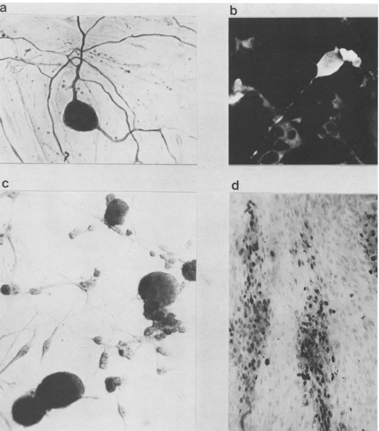
FIG. 1. Indirect immunolabeling and in situ hybridization analysis of adultrat DRG neuronsafterVZVinfection(2days postinfection).
(a)Primarymonoclonalantineurofilament followedbyaperoxidase-conjugated rabbit anti-mouse IgG antibody. (b) Primaryhumanserumrich in anti-VZV antibodies followed by a fluorescein-conjugated monoclonal anti-human IgG antibody. Magnification, x410. (c) In situ
hybridization of infected MRC5 cells probed with VZV DNA BtamnHI fragments labeled with biotin. Magnification, x220. (d) In situ
hybridization (biotin probes)of infected adultrat DRGneuronsat2days postinfection. Magnification, x640.The VZV DNAprobe was an
equimolarmixture of recombinant VZV DNAs spanning the VZVgenome, BanHI A, B. C. E, H, 1, J, Kfragments. Afterbeing washed,
biotin probes wererevealed withgoatanti-biotin antibody and peroxidase-labeled rabbit anti-goat IgG.
_ ..._ON, .# by on November 25, 2009 jvi.asm.org Downloaded from
50T 45t 40 35 labeled neurons (%)
a
30 +-25 -20 15 10- 5-0 0 1 2 3 4 5 6 7 8 daysp.l.FIG. 2. Percentageofinfectedneuronsobtained after 1to8days postinfection (p.i.). This percentage was determined after in situ hybridization by counting neurons with a positive or a negative hybridization signal.
gene). None
of
theseprobes
exhibitedahybridization signal
withuninfected
control DRG neurons(Fig.
4a).
Positivehybridization
signals
occurred withprobes
that corre-sponded toregions
encoding
IE genes4,
62,
and63, early
gene 36
(thymidine
kinase),
and late gene 31(gpII),
indicat-ing
that these genes are transcribed in infected neurons.Hybridization
with thefragment
corresponding
toIE gene 63 wasnoticeably stronger than that with the otherprobes
(Fig.
4b
and
c).Similarly,
neurons infected with cell-free virus showedpositive hybridization
signals,
with individualprobes corresponding
toregions
encoding
IE genes4,
62,
and 63
and late
gene 31. These results fitparticularly
well,f,.;
*
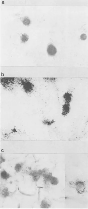
FIG.3.Siulanou detectio of VZ RN and neurFiaen
proteins in infected DRG cultures. The cells were fixed at 3 days postinfection, and double labeling was done with antineurofilament antibody revealed with a peroxidase-labeled second IgG. In situ hybridization was done on the same cells with
BalmHl
probes labeled with35S. Magnification, x560.b
.
INE~
i
C
FIG. 4. Detection of VZV RNA in infected neurons by in situ hybridization with 35S-labeled probes. (a) Control uninfectedcells probed withafragment thatcorrespondedtoIE gene 63. Infected cells(3 dayspostinfection)wereprobedwithafragment correspond-ing to lE gene 63 (b) and IE gene 62 (c). Original magnification, x470. For the detection of35S-labeled probes, cells were dehy-drated in graded ethanol, dipped in Ilford K2 autoradiographic emulsion, exposed for 2to5daysat4° C,anddevelopedinKodak D19. 0 .:~ "
4 &I,
t..iv..
p-,N
f. i. ., '4 "*,-;.4i by on November 25, 2009 jvi.asm.org Downloaded fromwith those obtained when the infection was propagated by cocultivation with MRC5 cells. In addition, a similar tran-scription pattern was recorded by using cell-free virus infec-tion and cocultivation, confirming that these genes are expressed in the neuronal cells.
Several relevant conclusions can be drawn from the data presented. A specific VZV infection of adult rat DRG neurons has been obtained by cocultivation with infected MRC5 cells or by cell-free virus. As shown by immunoflu-orescence,in situ hybridization, and simultaneous detection ofVZV and neuronal protein, neurons are the only cell type which is infected in vitro, but VZV-specific antigens are not expressed in neurons. Viral nucleic acids are detected in infected neurons, and the hybridization signals increase with thetime postinfection. At 6 days, almost half of the neurons are infected by VZV. In addition, no infectious virus and no antigens are released from the infected neurons, nor can they be recovered in the culture medium. These findings indicate that neuron-specific infection of adult rat DRG is persistent because (i) the viral genome is present, (ii) the complete infectious virus is not produced, and (iii) the number of neurons is maintained stable up to 10 days postinfection, showing that the infection process is not lytic. It remains now to determine the nature of the chemical, physical, or biological treatments which may be used to trigger virus replication.
The percentage of infected neurons visualized by in situ hybridization remained the same whatever the time of DRG culture before infection with VZV. The same efficiency of neuron infection at any time of culture suggests that infec-tion is not mediated by a molecular event related to acute neuronal damage or regeneration. It would be interesting to know whether neuronal infection is random or whether there is selectivity in terms of the neuronal subpopulation (8), considering for instance the neurotransmitter phenotype (J. Schoenen, P. Delrde, P. Leprince, and G. Moonen, J. Neurosci. Res., in press).
The analysis of VZV transcripts by in situ hybridization has shown the presence of RNA transcripts from regions corresponding to the three temporal gene classes (IE, early, and late), suggesting that transcription is not blocked in the
IE
or early stage of infection. Moreover, hybridization signals were stronger when neurons were probed with a fragment corresponding toIE gene 63, indicating an active transcription of this gene in infected neurons. Further exper-iments are required to fully characterize the complete VZV genomic transcription in infected rat DRG cultures either by in situ hybridization or by Northern (RNA) hybridization. Like VZV, HSV-1 establishes latent infection in neurons of sensory ganglia and expressesIE genes which encode infect-ed-cell polypeptides (ICP) (17, 19). HSV ICP4 protein has been detected in latently infected ganglia and shown to be a regulatory protein in vivo and in vitro (1). ICPO RNA has also been detected in neuronal nuclei of latently infected murine (19) and human ganglia from cadavers (5). These transcripts overlapped and had a polarity opposite to that of mRNA of the ICPO gene and could be referred to as an antisense transcript. The authors suggest that these tran-scripts may play a role in maintaining the latency of HSV. The situation is quite different for VZV, because no homolog of this HSV-1 ICPO gene has yet been described in its genome. DNA sequence analysis (7) and recent reports (11) have identified VZV genes similar to HSVIE genes. VZV gene 62 encodes a protein of 175 kilodaltons which is the functional analog of HSV-1 ICP4. VZV gene 63 displays a sequence homology to HSV-1ICP22 (30 kilodaltons). VZVproteins of similar size have been detected in explanted humanganglia (21) andmaybe relatedto VZV genes 62and 63.
Moreover,
insitu hybridization revealed the presenceof gene63 RNA in thesameganglia.
These results suggestthat IEgenes62and 63expressedduring latency mayplayarole in establishing and maintaining VZV latency in humans.In a recent report (6), Croen et al. studied the cellular localization and viral transcription pattern of acute and latent VZV infection of human sensory nerve ganglia by in situ hybridization. The authors claimed that latent VZV infection involves nonneuronal cells and that multiple but not all VZV genes are transcribed. Transcripts have been detectedwith unidirectional probes from BamHI-E (ORF4), BamHI-J
(ORF63),
andEcoRI-B
(ORF29,
ORF30, andORF31).
During varicella
disease,
both neuronal and non-neuronal cells are infected and all regions of the VZV genome appear to be expressed.InVZVinfectionofrat sensory neurons, wehavedetected transcripts of IE, early, and late genes but more abundant transcripts of IE gene63, suggesting thatthe latter mayplay a role in repressing a productive infection in rat sensory neurons.
We thank S. Straus and M. Levine for critical reading of the manuscript and M. Lion-Jassogne for technical assistance.
M.-P.M.-L. is a senior research assistant from IRSIA (Brussels, Belgium), C.S.D. and J.P. are, respectively, research assistant and senior research associate from the National Fund for Scientific Research, Brussels, Belgium. This work was supported by research grants from NFSR, FMSR, and IRSIA, by the Belgian National Lottery, and by the Queen Elizabeth Medical Foundation of Bel-gium.
LITERATURE CITED
1. Beard, P., S. Farber, K. W. Wilcox, and L. I. Pizer. 1986. Herpes simplex virusimmediate early infected-cell polypeptide 4 binds to DNA and promotes transcription. Proc. Natl. Acad. Sci. USA 83:4016-4020.
2. Brahic, M., and A. T. Haase. 1978. Detection of viral sequences of lowreiteration by in situhybridization. Proc.Natl. Acad.Sci. USA 75:6125-6129.
3. Brahic, M., A. T. Haase, and E. Cash. 1984.Simultaneousinsitu detection of viral RNA and antigens. Proc. Natl. Acad. Sci. USA 81:5445-5448.
4. Brigati, D. J., D. Myerson, J. J. Leary, B. Spaholz, S. Z. Travis, C. K. Y. Fong, G. D. Hsiung, and D. C. Ward. 1983. Detection of viral genomes in cultured cells and paraffin embedded tissue sections using biotin-labeled hybridization probes. Virology 126:32-50.
5. Croen, K. D., J. M. Ostrove, L. J. Dragovic, J. E. Smialek, and S. E. Straus. 1987. Latent herpes simplex virus in human trigeminal ganglia. N. Engl. J. Med. 317:1427-1432.
6. Croen, K. D., J. M. Ostrove, L. J. Dragovic, and S. E. Straus. 1988. Patterns of gene expression and sites of latency in human nerve ganglia are different for varicella-zoster and herpes sim-plex viruses. Proc. NatI. Acad. Sci. USA 85:9773-9777. 7. Davison, A. J., and J. E. Scott. 1986. The complete DNA
sequence ofvaricella-zoster virus.J. Gen. Virol. 67:1759-1816. 8. Dood, J., D. Solter, and T. M. Jessel. 1984. Monoclonal anti-bodies against carbohydrate differentiation antigens identify subsets of primary sensory neurons. Nature (London) 311:
469-472.
9. Ecker, J. R., and R. W. Hyman. 1982. Varicella-zoster virus exists as two isomers. Proc. NatI. Acad. Sci. USA 79:156-160. 10. Feinberg, A. P., and B. Vogelstein. 1983. Technique for radio-labeling DNA restriction endonuclease fragment forhigh spe-cificactivity. Anal. Biochem. 132:6-13.
11. Felser, J. M., P. R. Kinchington, G. Inchaupse, S. E.Straus,and J. M. Ostrove. 1988. Cell lines containing varicella-zoster virus open reading frame 62 and expressing the "IE"175 protein complement ICP4 mutants of herpes simplex virus type 1. J.
by on November 25, 2009
jvi.asm.org
Virol.62:2076-2082.
12. Gilden, D. H., A. Vafai, Y. Shtram, Y. Becker, M. Devlin, and M.Wellish.1983.Varicella-zoster virusDNAinhumansensory
ganglia. Nature (London) 306:478-480.
13. Grose,C., D. M. Perrotta, P. A. Brunell, and G. C.Smith.1979. Cell-free varicella-zoster virus in cultured human melanoma cells. J. Gen. Virol. 43:15-27.
14. Hope-Simpson,E. R. 1965.Thenatureofherpes-zoster: a long
termstudy anda newhypothesis. Proc. R. Soc. Med. 58:9-20.
15. Hyman,R. W., J. R. Ecker, and R. B. Tenser. 1983.
Varicella-zostervirus inhuman trigeminal ganglia. Lancet ii:814-816. 16. Ostrove,J. M., W. Reinhold, C. M. Fan,S. Horn,J. Hay, and
S.E.Straus. 1985. Transcription mapping of the varicella-zoster virusgenome. J. Virol.56:600-606.
17. Puga, A.,and A. L. Notkins. 1987. Continued expression ofa
poly(A)+ transcript of herpes simplex virustype1in trigeminal ganglia of latently infected mice. J. Virol. 61:1700-1703. 18. Rigby, P., M. Dieckemann, C. Rhodes, and P. Berg. 1977.
Labeling of deoxyribonucleic acid to high specific activity in
'itloby nick translationwith DNApolymerase 1. J. Mol.Biol.
113:237-251.
19. Stevens,J.G.,E. K.Wagner, G. B. Devi-Rao, M. L.Cook, and
L. T. Feldman. 1987. RNAcomplementarytoaherpes virusa
genemRNA is prominent inlatently infected neurons.Science
235:1056-1059.
20. Straus, S. E., H. S. Aulack, W.T. Ruyechan, J. Hay, T.A.
Casey, G.F. VandeWoude, J. Owens, and H. A. Smith. 1981. Stucture of varicella-zoster virus DNA. J. Virol. 40:516-525. 21. Vafai, A., R. S. Murray, M. Wellish, M. Devlin, and D.H.
Gilden. 1988. Expression of varicella-zoster virus in normal human trigeminal ganglia. Proc. Natl. Acad. Sci. USA 85:
2362-2366.
22. Wigdalh, B., B. Lan Rong, and E. Kinney-Thomas. 1986. Vari-cella-zostervirus infectionof humansensory neurons.Virology
152:384-399.
by on November 25, 2009
jvi.asm.org