HAL Id: tel-02138957
https://tel.archives-ouvertes.fr/tel-02138957
Submitted on 24 May 2019HAL is a multi-disciplinary open access archive for the deposit and dissemination of sci-entific research documents, whether they are pub-lished or not. The documents may come from teaching and research institutions in France or abroad, or from public or private research centers.
L’archive ouverte pluridisciplinaire HAL, est destinée au dépôt et à la diffusion de documents scientifiques de niveau recherche, publiés ou non, émanant des établissements d’enseignement et de recherche français ou étrangers, des laboratoires publics ou privés.
System for Chronic Monitoring of Biological Tissue
Composition
Achraf Lamlih
To cite this version:
Achraf Lamlih. Design of an Integrated Bioimpedance Measurement System for Chronic Monitoring of Biological Tissue Composition. Optics / Photonic. Université Montpellier, 2018. English. �NNT : 2018MONTS070�. �tel-02138957�
DE L’UNIVERSITE DE MONTPELLIER
DE L’UNIVERSITE DE MONTPELLIER
En Systèmes Automatiques et Microélectroniques (SYAM) École doctorale : Information, Structures, Systèmes
Unité de recherche : LIRMM
Conception d’un système intégré de mesure de
bioimpédance pour le suivi long terme de la composition
des tissus biologiques
Conception d’un système intégré de mesure de
bioimpédance pour le suivi long terme de la composition
des tissus biologiques
Présentée par Achraf LAMLIH.
Le 26-11-2018
Sous la direction de Serge Bernard
Devant le jury composé de
BRAGOS Ramon, Associate Professor , UPC Barcelona Rapporteur
ROMAIN Olivier, Professeur, Université de Cergy - ETIS Président du jury/Rapporteur YUFERA GARCIA Alberto, Professor, Universidad de Sevilla - IMSE-CNM Examinateur
BERNARD Serge, Directeur de Recherche, LIRMM - CNRS Directeur KERZERHO Vincent, Chargé de Recherche, LIRMM - CNRS Co-encadrant ROUYER Tristan, Chercheur, MARBEC - IFREMER Co-encadrant RENOVELL Michel, Directeur de Recherche, CNRS-LIRMM Invité SOULIER Fabien, Maître de Conférences, LIRMM - UM Invité BONHOMMEAU Sylvain, Chercheur, IFREMER - DOI Invité
Doctoral Thesis
Design of an Integrated
Bioimpedance Measurement
System for Chronic Monitoring of
Biological Tissue Composition
Author:
Achraf LAMLIH
Supervisors:
Serge Bernard
Vincent kerzérho
Fabien Soulier
Tristan Rouyer
Sylvain Bonhommeau
A thesis submitted in fulfillment of the requirements
for the degree of Doctor of Philosophy
in the
SYAM - I2S
LIRMM - IFREMER
“The most exciting phrase to hear in science, the one that heralds the most discoveries, is not ‘Eureka!’ but ‘That’s funny. . . ’ ”
Acknowledgements
I would like to take this opportunity to thank all those who have contributed to the achievement of this work. In particular I would like to express my gratitude to the following persons.
Special mention goes to my enthusiastic supervisor, Serge Bernard. My PhD has been an amazing experience and I thank Serge wholeheartedly, not only for his tremendous academic support and thought-provoking ideas but also for giv-ing me so many wonderful opportunities. Similar, profound gratitude goes to Vincent Kerzérho and Fabien Soulier, who have been truly dedicated to their role as my secondary supervisors. I am particularly indebted to Vincent and Fabien for their passionate participation and their invaluable guidance and ad-vises in our weekly meetings. I am also hugely appreciative to Tristan Rouyer and Sylvain Bonhommeau, especially for sharing their expertise in fisheries sci-ence and marine ecology so willingly. Their insightful comments improved the quality of the manuscript and other publications.
I would like to thank Prof. Ramon Bragos, Prof. Olivier Romain and Prof. Alberto Yufera Garcia, for being my thesis examiners and for their constructive criticism and invaluable suggestions that have improved this thesis.
Special thanks goes to Philippe Freitas. The integrated circuit prototype would not have been sent to fabrication without his expertise and tremendous efforts. I thank Philippe not only for that, but also for his ever-present sense of humour. I would also like to extend my thanks to Stéphane David-Grignot for generously sharing his expertise in signal processing. Without his contri-butions, the study on sine wave harmonic cancellation would not have been so elaborated. Besides, I thank Stéphane for all the stimulating conversations we had about blockchain. Special mention goes to Moez Belhadj, his efforts and expertise in flexible electrodes development were crucial for the specification of the integrated circuit architecture. My thanks go also to Jérémie Salles, who so generously assisted in the design and layout of the circuit. I also wish to thank Mohan Julien, for all the stimulating late night discussions and brain-storming sessions we had in the lab which helped solve various problems. Guys, I have learned a lot working with you. I would also like to express my sincere gratitude to Michel Renovell who reviewed my work multiple times. I am gratefully in-debted to Michel not only for his very valuable comments on my presentations and this thesis manuscript, but also for all the book recommendations and the interesting conversations we had about history, economy and religions.
For all the team, it will be difficult to forget all the meals, coffee breaks, and interesting conversations we have shared. For making this years memorable, from the bottom of my heart I say thank you.
Contents
Acknowledgements 3
Abstract 17
Résumé 19
1 Thesis Introduction : Context and Objectives 25
1.1 Research Project Background: case study . . . 26
1.2 Research goals and methodology . . . 27
1.3 Thesis content and outline . . . 30
2 Biological Tissue Composition assessment 31 2.1 Introduction . . . 31
2.2 Tissue composition analysis techniques . . . 32
2.2.1 Introduction . . . 32
2.2.2 Micro-wave propagation . . . 32
2.2.3 Near Infrared Spectroscopy . . . 34
2.2.4 Ultrasound . . . 35
2.2.5 Bioimpedance Spectroscopy . . . 37
2.2.6 Comparative status . . . 38
2.3 Bioimpedance spectroscopy : From electrical properties of tissue to its electrical impedance . . . 40
2.3.1 Introduction . . . 40
2.3.2 Cell electrical properties . . . 40
2.3.3 Cell electrical model . . . 41
2.3.4 Dispersion regions . . . 44
2.3.5 Single frequency vs spectroscopy analysis . . . 46
2.3.6 Biological vs. bioimpedance correlation approaches . . . 46
2.4 Conclusion . . . 48 3 Design challenges 49 3.1 Introduction . . . 49 3.1.1 Methodology . . . 49 3.1.2 Research questions . . . 50 3.1.3 Chapter content . . . 51 3.2 Experimentation resources . . . 51 3.2.1 Electrodes . . . 52
3.2.2 The handheld AD5933 measurement system . . . 52
3.2.3 The MFIA impedance analyzer . . . 54
3.4 Current vs voltage stimulation . . . 56
3.4.1 Voltage excitation system . . . 56
3.4.2 Current excitation system . . . 57
3.5 Electrode configuration . . . 57
3.6 Electrode-tissue impedance range . . . 59
3.7 Non-linearity investigation . . . 60
3.7.1 Motivation . . . 60
3.7.2 Experiments . . . 60
3.7.3 Results and conclusion . . . 62
3.8 Measurement environment related errors . . . 63
3.9 Summary of system specifications . . . 66
4 Architecture definition 69 4.1 Introduction . . . 69
4.1.1 Methodology and research questions . . . 69
4.2 Specifications impact on the architecture . . . 70
4.3 Design constraints . . . 70
4.3.1 Constraints . . . 70
4.4 State of the art of BIS architectures . . . 73
4.4.1 Frequency sweep excitation architectures . . . 74
4.4.2 Wideband simultaneous excitation architectures . . . 75
4.5 Proposed hybrid architecture . . . 76
4.5.1 Wideband simultaneous excitation . . . 76
4.5.2 Frequency sweep excitation . . . 77
4.5.3 Hybrid architecture . . . 78
4.6 Design status of the integrated bioimpedance measurement system 80 4.7 Conclusion . . . 82
5 Stimuli generation 83 5.1 Introduction . . . 83
5.2 Sine-wave construction . . . 84
5.2.1 Introduction . . . 84
5.2.2 Basic principle and constraints of harmonic cancellation techniques . . . 86
5.2.3 Fixed phase-shifts and adjustable amplitudes of square-waves . . . 86
5.2.4 Fixed amplitudes and adjustable phase-shifts of square-waves . . . 88
5.2.5 Adjustable amplitudes and adjustable phase-shifts of square-waves . . . 91
5.2.6 Conclusion on harmonic cancellation techniques . . . 92
5.2.7 Hardware implementation . . . 93
5.2.8 Robustness study . . . 96
5.2.9 Conclusion . . . 99
5.3 Composite signal generation . . . 100
5.3.1 Introduction . . . 100
5.3.2 Signal quality metrics and comparison setup . . . 102
5.3.4 Proposed multitone excitation technique . . . 107
5.3.5 Conclusion . . . 113
5.4 Chapter conclusion . . . 116
6 Current driver for BioImpedance Spectroscopy 119 6.1 Introduction . . . 119
6.2 Design constraints of current drivers for BIS . . . 120
6.3 State of the art of current drivers for BIS . . . 122
6.4 Optimization of the Current driver’s architecture . . . 123
6.4.1 Proposed architecture . . . 123
6.4.2 Output impedance increasing . . . 126
6.4.3 Maximization of the output voltage swing . . . 129
6.5 Simulation Results . . . 130
6.5.1 Output impedance . . . 131
6.5.2 Output voltage swing . . . 133
6.5.3 Comparison table with existing architectures . . . 133
6.6 Conclusion . . . 136 Conclusion and perspectives 137
List of Figures
1 Principe de fonctionnement d’un système de marque pour poisson 20 2 Processus biologiques non-qualifiés des poissons dans leur milieu
sauvage . . . 21
1.1 Operating principle of electronic tags for large pelagic fish . . . 26
1.2 Unqualified fish biological processes in the wild . . . 27
1.3 Scope of the thesis research work . . . 28
2.1 Design of the portable device propagating microwaves, repro-duced from (Kent, 1990) . . . 33
2.2 Correlation between total body water and the fat rate for Atlantic herring (Kent, 1990) . . . 33
2.3 Photo-electric conversion circuit . . . 35
2.4 An ultrasound probe components, reproduced from (Basix prin-ciple of medical ultrasonic probes) . . . 36
2.5 Bioimpedance analysis operating principle . . . 38
2.6 Cell components . . . 41
2.7 Bilayer lipid membrane . . . 41
2.8 Fricke’s Electrical Equivalent Model . . . 42
2.9 Low frequency current path . . . 43
2.10 high frequency current path . . . 43
2.11 Dispersions of biological tissue (Schwan, 1957) . . . 44
3.1 Methodology . . . 50
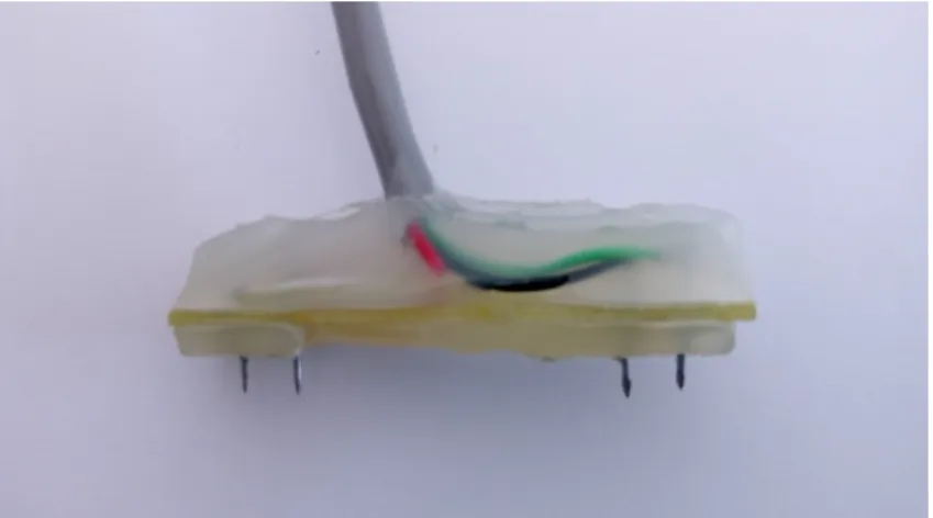
3.2 First generation of electrodes . . . 52
3.3 flexible Electrodes . . . 53
3.4 AD5933 Hand-held bioimpedance measurement system . . . 53
3.5 AD5933 system function blocks diagram . . . 54
3.6 The MFIA impedance analyzer . . . 54
3.7 Structure of a : (a) current excitation system (b) voltage excita-tion system . . . 56
3.8 2-electrode configuration . . . 58
3.9 4-electrodes configuration . . . 58
3.10 4-electrode measurements on bluefin tuna using the MFIA impedance analyzer . . . 59
3.11 2-electrode measurements on bluefin tuna using the MFIA impedance analyzer . . . 60
3.12 Experimentation setup . . . 61
3.13 modified portable BIA device schematic . . . 61
3.14 Data processing algorithm . . . 62
3.16 BIS and EMG electrodes implanted on the gilt-head bream . . 64 3.17 Bioimpedance module of a gilt-head bream during different
swim-ming speeds . . . 65 3.18 Bioimpedance phase of sea bream during different swimming speeds 65 4.1 Constraints summary for each BIS measurement approach . . . 73 4.2 Wideband simultaneous excitation bioimpedance spectroscopy
architecture . . . 77 4.3 Frequency sweep bioimpedance spectroscopy architecture . . . . 77 4.4 Hybrid bioimpedance spectroscopy architecture . . . 79 4.5 Layout of the first generation integrated circuit measurement
sys-tem . . . 81 5.1 Excitation signal generation block . . . 83 5.2 Sine wave construction block . . . 84 5.3 Symmetry of sine-wave signal generated by summing square-wave
signals . . . 87 5.4 Sine-wave signal generated using square-wave signals with various
amplitudes for five signals (M = 5) . . . 88 5.5 Spectrum of sine-wave signal generated using square-wave signals
with various amplitudes for five signals (M = 5) . . . 89 5.6 Sine-wave signal generated using square-wave signals with various
phase shifts for six signals M = 6 with [ϕ1; ϕ2; ϕ3] = [0.2037; 0.4701; 0.9784]rad 90
5.7 Spectrum of sine-wave signal generated using square-wave signals with various phase shifts for six signals M = 6 . . . 91 5.8 THD ratio variations due to varying phase-shift error
Monte-Carlo results for a sine-wave signal generated using square-wave signals with various phase shifts for six signals M = 6 . . . 92 5.9 Comparison of harmonic cancellation techniques . . . 93 5.10 THD of a sine-wave generated using square-wave signals with
approximated amplitudes with fraction with denominator d . . . 94 5.11 SFDR of a sine-wave generated using square-wave signals with
approximated amplitudes with iteration on continued fraction . 95 5.12 THD up to harmonic 11 of a sine-wave generated using
square-wave signals with approximated amplitudes with n iterations on continued fractions . . . 96 5.13 SFDR of a sine-wave generated using square-wave signals with
approximated amplitudes with n iterations on continued fractions 97 5.14 Spectrum of a sine-wave signal affected by amplitude error and/or
jitter on the square-wave signals for sine-wave signal generated using square-wave signals with various amplitudes five signals M = 5 . . . 98 5.15 Total Harmonic distortion ratio of a sine-wave generated using
square-wave signals with various amplitudes deviations and af-fected by various level of jitter . . . 99 5.16 Multitone excitation signal generation block . . . 100 5.17 Bioimpedance magnitude of bluefin tuna using the MFIA impedance
5.18 Square-wave signal in time and frequency domain . . . 104
5.19 Linear feedback shift register based pseudo random generation . 105 5.20 Frequency components of the BMS excitation technique . . . 106
5.21 Frequency components of the BMS excitation technique after fil-tering . . . 107
5.22 Functional diagram of a sigma delta modulator . . . 108
5.23 Frequency components of a classic sigma delta based multitone excitation . . . 110
5.24 Customized loop filter bode diagram . . . 112
5.25 Nyquist diagram of the customized transfer function . . . 113
5.26 Signal transfer function of the proposed excitation . . . 114
5.27 Noise transfer function of the proposed excitation . . . 114
5.28 Frequency components of the proposed excitation . . . 115
5.29 Frequency components of the proposed excitation after filtering 116 6.1 Current driver position in the integrated circuit architecture . . 119
6.2 current source model . . . 121
6.3 Output impedance vs load impedance illustration . . . 121
6.4 Saturation voltage illustration . . . 122
6.5 OTA architectures : (a) Simple OTA (b) Telescopic OTA (c) Miller OTA (d) Symmetrical OTA (e) Folded cascode OTA . . . 123
6.6 Current driver architecture . . . 126
6.7 Current mirror structures: (a) Simple current mirror(b) cascode (c) high swing cascode (d) gain boosted cascode (e) regulated cascode . . . 127
6.8 Improved regulated cascode architecture . . . 128
6.9 Layout of the current drive . . . 130
6.10 Current driver bandwidth . . . 131
6.11 Output impedance of the current driver . . . 131
6.12 Improved regulated cascode (NMOS) stability check . . . 132
6.13 Impedance magnitude of the electrode-tissue-interface of bluefin tuna in a 2 electrodes configuration . . . 133
6.14 Output current vs output voltage characteristic of the high swing cascode, the regulated cascode and the improved regulated cas-code for an input current of 300 µA . . . 134
List of Tables
2.1 Optical parameters of muscle and fat tissue, reproduced from
(Song et al., 2009) . . . 34
2.2 Comparative status on the studied techniques . . . 39
5.1 local SNR’s for the binary multi-frequency excitation . . . 107
5.2 local SNR’s for a classic sigma delta excitation . . . 110
5.3 local SNR’s for the proposed excitation . . . 113
6.1 Comparison of amplifier architectures . . . 124
6.2 Current mirror performances comparison . . . 128
To my family,
Abstract
Biologging consists in attaching data-recording devices to animals to collect data on their environment and infer information about their biology and ecol-ogy. In the last decades, advances in science and engineering, especially in microelectronics, biotechnology, and materials have fueled the development of sophisticated devices, which resulted in smaller species being tagged for longer periods and in different types of environment (terrestrial, aquatic or aerial). This allowed researchers to address a diverse panel of scientific questions re-lated to the behavior and ecology of wild species. Current technology provides a description of the physical environment of animals, but no device allows to describe their physiological aspects over relevant time scales. Therefore, physi-ological parameters tracking is on the rise as it offers extremely valuable infor-mation to answer key questions such as the response of individuals to climate change. The miniaturization of silicon electronics has benefited the development of implantable devices that opened up new opportunities for the long-term mon-itoring of physiological states. In that respect, a promising line of research is to use tissue composition to infer information on physiological states of animals.
The ultimate goal of this research work is to design an integrated measure-ment system capable of assessing biological tissue composition. This research work intends to be an exploratory work aiming to bring to the scientific commu-nity tools that are capable of measuring changes in a wide range of physiological processes. Thus, this measurement system will be adjustable to a large scope of physiological variables and long-term monitoring applications. The research work presented in this thesis is part of the POP-up Satellite Tag for Advanc-ing Research in marine ecology (POPSTAR) project which aims at developAdvanc-ing a new generation of tagging devices for fish. The main goal of the project is to enhance our understanding of fish behavior by linking the analysis of the environment in which the fish travels and lives to its physiological states. the POPSTAR project will be used to validate research hypotheses and narrow down the specifications of the integrated measurement system.
After comparing several tissue composition techniques, bioimpedance spec-troscopy has been chosen as the technique to be used for the design of the integrated measurement system. Since the electrical impedance of the biologi-cal tissue is a function of physiologibiologi-cal processes, variations in the composition of the biological tissue are expected to change its electrical properties, which by extension can be seen as variations of the electrical impedance of the tissue.
After defining bioimpedance spectroscopy as the technique to be used, we have identified the design challenges of such measurement system by using an in-depth literature review of the existing bioimpedance measurement systems as well as the bioimpedance theoretical concepts. Because design challenges are very application dependent, we also took into account the constraints related
to the measurement environment and configuration. Given that the majority of bioimpedance measurements on fishes that can be found in the literature use single frequency, we have conducted several bioimpedance spectroscopy experi-ments on fish species such as bluefin tuna, sea bass, sardine or sea bass in order to assess the missing parameters. Following this methodology we have been able to narrow down as much as possible the integrated circuit specifications, which layed the foundation of the integrated circuit design guidelines.
The integrated circuit design guidelines being defined, we have demonstrated that the constraints ruling the design of the measurement system could be cat-egorized depending on the frequency band of excitation. In order to break the trade-off between short measurement durations and precision, we have decided to use a wideband simultaneous excitation approach for the low and the nominal frequency bands, and a frequency sweep excitation for the high frequency band. A hybrid architecture providing fast measurements while maximizing precision has therefore been proposed. It has been defined for the POPSTAR project as a special research context, and for biological tissue electrical properties explo-ration over a wide frequency range as a general research context.
As the excitation signal generation blocks are critical and their performances affect the whole architecture performances, the second part of this research fo-cuses on the design and optimization of the signal generation part of the archi-tecture which is composed of stimuli generation blocks and a current driver that transforms the voltage stimuli to an excitation current. In the stimuli genera-tion part of the architecture, two stimuli generators have been designed, one for frequency sweep excitation and one for wideband simultaneous excitation. For the frequency sweep excitation, we compared on-chip sine wave construction techniques. This techniques are used to generate sine-wave signals by summing digital signals in order to cancel low-order harmonics. We first compared sev-eral approaches and analytically proven that for a given number of square-wave generators, an approach allows to cancel a higher number of harmonics com-pared to the others. The sine-wave constructor implementation that suits our research context has been discussed. For the wideband simultaneous excitation, several techniques have been compared, and key parameters defining their per-formances presented. A novel multitone generation technique based on a delta sigma modulator has thus been proposed. Finally, a simple implementation of the multitone generator has been presented.
These two signal generation approaches have a common block, which is the current driver of the architecture. The current driver transforms the voltage optimized stimuli to a proportional current signal. A current driver architec-ture optimized for bioimpedance measurements has been proposed. It uses an improved regulated cascode to enhance the output impedance, enabling accu-rate measurements of transfer impedances at low and high frequencies. The current driver uses a common-mode feedback compensation technique capable of accurately setting the output common mode voltage independently of process variations. The proposed architecture therefore offers a high output swing for a given supply voltage. A first prototype chip containing the critical blocks of the architecture has been successfully designed and simulated in a 0.18 µm AMS (Austria MicroSystems) CMOS process operating at 1.8 V power supply.
Résumé
Le biologging consiste à attacher des dispositifs à des animaux dans le but d’enregistrer des données liées à leur état biologique ou à leur environnement. Au cours des dernières décennies, les progrès de la science et de l’ingénierie en microélectronique, en biotechnologie et en science des matériaux, ont aidé au développement de dispositifs évolués, ce qui a permis de marquer des espèces de petite taille pour des périodes de plus en plus longues dans des environnements différents (terrestre, marin ou aérien). Cette évolution a donné aux chercheurs les outils nécessaires pour traiter un large panel de questions scientifiques liées au comportement et à l’écologie d’espèces sauvages.
Le suivi de paramètres physiologiques est de plus en plus utilisé car il per-met d’obtenir des informations extrêmement utiles sur les espèces en question. La miniaturisation de l’électronique intégrée a favorisé le développement de dis-positifs implantables qui ont ouvert de nouvelles opportunités pour le suivi à long terme de l’état physiologique de plusieurs espèces. L’un des indicateurs physiologiques les plus intéressants à suivre est la composition des tissus. Les techniques d’évaluation de la composition des tissus biologiques permettent de mieux comprendre les processus physiologiques et leur impact global sur l’état biologique des sujets expérimentaux.
Les techniques d’évaluation de la composition tissulaire varient en préci-sion mais aussi en types de tissus qu’ils sont capables de mesurer. Historique-ment, ces techniques étaient difficiles à intégrer. Des techniques telles que l’absorptiométrie à rayons X à double énergie (DEXA), la résonance magné-tique nucléaire (RMN) ou la conductivité électrique totale du corps offrent des performances élevées, mais nécessitent des équipements de grande taille qui les rendent impropres à des mesures in vivo. Grâce aux améliorations tech-nologiques continues, de plus en plus de travaux de recherche visent à fournir à la communauté scientifique des outils et des techniques hautement intégrés qui répondent au besoin de suivi continu des paramètres physiologiques.
Le but principal de ce travail de recherche est de concevoir un système de mesure intégré capable d’évaluer la composition des tissus biologiques. Ce tra-vail de recherche se veut être un tratra-vail exploratoire visant à apporter à la com-munauté scientifique des outils capables de mesurer un large éventail de biomar-queurs. Ainsi, ce système de mesure pourra être adapté à une large gamme d’applications de suivi à long terme de variables physiologiques. Néanmoins, les travaux de recherche présentés dans cette thèse font partie du projet POPSTAR, qui vise à développer une génération innovante de systèmes de marquage de poissons. L’objectif principal du projet est d’améliorer notre compréhension du comportement des poissons en analysant non seulement l’environnement dans lequel les poissons se déplacent et vivent, mais aussi les poissons eux-mêmes.
Le projet POPSTAR servira de cas d’étude afin de valider des hypothèses sci-entifiques et d’affiner les spécifications du système de mesure intégré.
Les systèmes de biologging disponibles dans le commerce aujourd’hui pour tagger des poissons utilisent des circuits électroniques pour enregistrer des paramètres environnementaux tels que la lumière, la température ou la pres-sion. Après une durée préétablie, le système est libéré du poisson et émerge à la surface de l’eau grâce à une flottabilité légèrement positive. Les données enreg-istrées sont ensuite transmises à un satellite et récupérées pour être analysées (voir figure 1.1).
Figure 1: Principe de fonctionnement d’un système de marque pour poisson
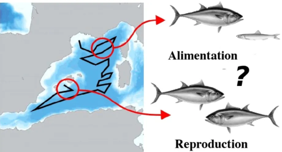
Bien que ces systèmes de marquage aient permis de mieux comprendre l’environnement dans lequel évoluent les poissons, ils ne fournissent pas d’informations sur les poissons eux-mêmes. Par conséquent, les informations scientifiques nécessaires pour conserver et gérer efficacement le poisson font actuellement défaut. Une des solutions possibles pour améliorer notre compréhension des espèces marines consiste à utiliser des capteurs implantables pour surveiller les paramètres physiologiques liés aux principaux processus biologiques tels que l’alimentation et le frai. En effet, de tels processus offrent des informations signi-ficatives sur une foule de paramètres démographiques clés tels que la croissance, la survie ou la reproduction (voir figure 1.2). Par exemple, un paramètre phys-iologique clé est la teneur en graisse musculaire. La variation de ce paramètre dans l’espace et le temps est en effet liée aux processus mentionnés précédem-ment, tels que l’alimentation, qui peut se traduire en une accumulation des
réserves en gras, ou la reproduction pendant laquelle le poisson va puiser dans ses réserves de gras.
Figure 2: Processus biologiques non-qualifiés des poissons dans leur milieu sauvage
Suivre l’état physiologique des poissons dans leur environnement naturel est une première mondiale. Il va donc de soi que nous n’avons pas d’informations précises sur la pertinence d’un paramètre biologique en particulier. Partant de ce constat, les travaux de recherche présentés dans cette thèse visent à concevoir un système de mesure intégré capable d’explorer un large éventail de variables physiologiques. L’objectif est de fournir à la communauté scientifique un outil de suivi continu fournissant des informations sur l’état physiologique des espèces sauvages dans leur environnement naturel.
Après avoir comparé plusieurs techniques d’estimation de la composition tissulaire, la spectroscopie de bioimpédance a été choisie comme technique à utiliser pour la conception du système de mesure intégré. Etant donné que l’impédance électrique du tissu biologique est fonction des processus physi-ologiques, les modifications de la composition du tissu biologique se traduisent en une modification de ses propriétés électriques, ce qui par extension, se traduit en une variation de l’impédance électrique du tissu.
Concevoir un système de mesure intégré capable de mesurer une large gamme de biomarqueurs sur de longues périodes est un défi majeur. En effet, en adap-tant le système pour explorer un large éventail de variables physiologiques, la conception d’un tel système devient rapidement régie par de fortes contraintes telles que la bande passante, la précision, le temps de mesure ou la sureté de fonctionnement. Le choix entre différentes architectures de système potentielles devient une tâche difficile. En outre, certains blocs d’architecture sont critiques et il convient de veiller à optimiser leur conception.
Après avoir défini la spectroscopie de bioimpédance comme la technique à utiliser, nous avons identifié les défis de conception d’un tel système de mesure intégré en utilisant une analyse bibliographique approfondie des systèmes de mesure de bioimpédance existants ainsi que des concepts théoriques de bioim-pédance. Les défis de conception étant dépendant des applications, nous avons également pris en compte les contraintes liées à l’environnement de mesure et à la configuration. Étant donné que la majorité des mesures de bioimpédance sur
les poissons que l’on peut trouver dans la littérature utilisent une mesure mono-fréquence (à 50 kHz, nous avons mené plusieurs expériences de spectroscopie de bioimpédance sur des espèces de poissons telles que le thon rouge, le bar, la sardine ou le bar. En suivant cette méthodologie, nous avons pu affiner autant que possible les spécifications du circuit intégré, ce qui a permis de définir les lignes directrices de conception du circuit de mesure.
Les lignes directrices pour la conception du circuit intégré étant définies, nous avons démontré que les contraintes régissant la conception du système de mesure pouvaient être classées en fonction de la bande de fréquence de l’excitation. Afin de rompre le compromis entre une durée de mesure courte et la précision de mesure, nous avons décidé d’utiliser une approche d’excitation simultanée à large bande pour les bandes de fréquences basses et nominales et une excitation en balayage de fréquences pour la bande haute fréquence. Une architecture hybride permettant des mesures rapides tout en maximisant la précision a donc été proposée. Cette architecture a été défini pour le projet POPSTAR comme contexte de recherche particulier et pour l’exploration des propriétés électriques des tissus biologiques sur une large gamme de fréquences en tant que contexte de recherche général.
Les blocs de génération du signal d’excitation de l’architecture proposée sont critiques car leurs performances affectent les performances de l’ensemble de l’architecture. L’accent a ainsi été mis sur la conception et l’optimisation de ces blocs. De l’optimisation de la création des stimuli à l’aide de techniques de traitement du signal numérique à l’amélioration des performances de la source de courant pour laquelle nous avons proposé une nouvelle architecture analogique. Pour la partie génération de stimuli de l’architecture, deux générateurs de stimuli ont été conçus, le premier sert à injecter dans le tissu biologique un signal sinusoïdal mono-fréquentiel, le deuxième générateur permet l’excitation du tissu biologique avec un signal multi-fréquentiel.
Pour la génération mono-fréquentielle, nous avons comparé les techniques de construction sur puce intégrée d’un signal sinusoïdal. Ces techniques sont util-isées pour générer des signaux sinusoïdaux en sommant des signaux numériques afin d’annuler les harmoniques de faible ordre.
Plusieurs approches sont possibles. Dans cette partie nous avons d’abord comparé les différentes approches possibles. Ensuite, nous avons prouvé de manière analytique que pour un nombre donné de signaux carrés, une approche en particulier permet d’annuler un nombre plus élevé d’harmoniques par rapport aux autres. Finalement, l’implémentation du générateur sinusoïdal qui convient à notre contexte de recherche a été discutée.
Pour l’excitation multi-fréquentielle, les techniques de génération d’un sig-nal composite ont été comparées et les paramètres clés définissant leurs per-formances ont été présentés. Une nouvelle technique de génération d’un signal multi-fréquentiel basée sur un modulateur delta sigma a été proposée. Enfin, une implémentation simple de ce générateur a été présentée.
La source de courant est l’un des blocs les plus critiques de l’architecture. Ce bloc étant commun aux deux approches de mesure, il doit opérer sur une large gamme de fréquence (de 8 Hz à 8 MHz).
Les paramètres clés définissant ses performances ont d’abord été énumérés et discutés. Un de ces paramètres clé d’une source de courant est l’impédance de sortie. L’impédance de sortie détermine non seulement la plage de fréquence de fonctionnement mais également la plage d’impédance de charge pour laquelle la source de courant est capable de maintenir un courant de sortie constant. Un autre paramètre clé est l’amplitude maximale de la tension de sortie, qui détermine la plage d’impédance de charge dans laquelle la source de courant est capable de maintenir ses performances de linéarité. Ce paramètre est d’autant plus critique quand la tension d’alimentation est basse et l’amplitude de courant de sortie élevée (nécessaire pour faciliter l’analyse de la réponse). Partant de ce constat, nous avons cherché à optimiser l’architecture en fonction de ces contraintes majeures.
Différentes architectures de sources de courant pour la spectroscopie de bioimpédance ont donc été comparées, et finalement une nouvelle architecture analogique présentant des performances supérieures par rapport à l’état de l’art a été présentée. Une puce contenant les blocs critiques de l’architecture a été conçue et simulée avec succès avec un process CMOS AMS (Austria MicroSys-tems) 0.18 µm fonctionnant sous une alimentation de 1,8 V.
Chapter 1
Thesis Introduction : Context and
Objectives
Biologging consists in attaching data-recording devices to animals to collect data on their environment and infer information about their biology and ecol-ogy. Biologging found its beginnings in the marine environment. In fact, this technique has first been introduced by Pers Scholander who had the idea of at-taching a capillary depth gauge onto a freshly harpooned whale in the objective of studying the maximum immersion depth of the cetacean (Ropert-Coudert et al.,2010).
In the last decades, advances in science and engineering, especially in mi-croelectronics, biotechnology, and materials have fueled the development of so-phisticated devices, which resulted in smaller and smaller species being tagged for longer periods and in different types of environment (terrestrial, aquatic or aerial), thus allowing researchers to address a diverse panel of scientific questions related to the behavior and ecology of wild species.
In the process of widening our understanding of wild species, physiological parameters tracking is on the rise as it offers extremely valuable informations. The mass adoption of consumer electronics as well as the rise of open source software and hardware in the past decade have permitted the development of cheap and miniaturized sensors. The miniaturization of silicon electronics has benefited the development of implantable devices which opens up new opportu-nities for physiological states long-term monitoring. One of the most interesting physiological indicators is tissue composition. Tissue composition assessment techniques are used to help better comprehend physiological processes and their overall impact on the biological state of the experiments subjects.
The tissue composition assessment techniques vary in precision and in the target tissue of interest. Historically, these techniques were difficult to integrate. Techniques such as Dual-energy X-ray absorptiometry, Nuclear Magnetic Res-onance (NMR) or Total Body Electrical Conductivity, offer high performances but need large size equipments which make them unsuitable for in-vivo mea-surements. Thanks to continuous technology improvements more and more research studies are aiming to provide the scientific community with highly in-tegrated tools and techniques that satisfies the need for continuous monitoring of physiological parameters.
From this perspective, the research work presented in this thesis intents to design an integrated measurement system capable of exploring a wide range of physiological variables. The goal is to provide the scientific community with a
continuous monitoring tool that provides valuable informations related to the physiological state of wild species in their natural environment.
1.1
Research Project Background: case study
The research work presented in this thesis has been equally funded by the LabEx NUMEV and IFREMER. LabEx NUMEV is an Excellence Laboratory created in 2011 in the framework of the French Program of Investments to the Future. NUMEV seeks to harmonize hard and computational sciences approaches with life sciences and pave the way for emerging interdisciplinary groups with an international profile.
IFREMER has a research project named POP-up Satellite Tag for Advanc-ing Research in marine ecology (POPSTAR), which is the result of a close collaboration between several research units from IFREMER and LIRMM. In particular, this PhD project has been funded and yearly evaluated by IFRE-MER and LabEx NUMEV.
POPSTAR aims at developing an innovative generation of electronic tags for large pelagic fish (e.g. tunas and billfishes). The main goal of the project is to improve our understanding of fish behavior through the joint analysis of the evolution of its physical environment and physiological state.
Figure 1.1: Operating principle of electronic tags for large pelagic fish
Electronic tags currently available on the market use electronic circuits to record environmental parameters, typically light, temperature and pressure.
The tag is programmed to be released after a given duration and floats up to the surface thanks to its slightly positive buoyancy. The recorded data are then transmitted through the ARGOS system, and retrieved in order to be analyzed (see figure 1.1).
Although these electronic tags have allowed a better understanding about the environment in which large pelagics evolve, they generally do not provide any information about their physiological state across time scales consistent with their large geographical movements. Thus, key scientific information to conserve and manage fish efficiently is currently lacking. A way to improve our understanding of marine species is to use implantable sensors for monitoring physiological parameters related to key biological processes such as feeding and spawning. Indeed, such processes offer significant insights into a host of key population parameters such as growth, survival, and reproduction. One key physiological parameter is muscle fat content. The variation of this parameter in space and time can indeed be related to the previously mentioned processes as fish feed to accumulate reserves, which are then used during reproduction (see figure 1.2).
Figure 1.2: Unqualified fish biological processes in the wild
POPSTAR is a multi-disciplinary project that brings several improvements to existing tag technology. It intends to decrease the size and cost of currently available tags on the market. It also aims at improving the transmission system efficiency. However, the most outstanding innovation of the POPSTAR project is the in situ collection of data related to biological processes of the fish. For the first time, it will be possible to jointly analyze the physiological state of the fish in their natural environment and their geolocation. The recorded data will provide new scientific insights to the scientific community.
1.2
Research goals and methodology
As previously noted, POPSTAR is a multi-disciplinary project that brings sev-eral improvements to current state of the art in tag technology. Other than the collection of data related to biological processes, it aims to improve the ratio of
the acquired vs lost transmitted data by improving data transmission technol-ogy. The project aims also to improve the mechanics of the tagging system and to develop the interface between the biological tissue and the electronic system. The ultimate goal of the present research work is to design an integrated measurement system capable of assessing biological tissue composition. This thesis focuses on the choice of the tissue composition technique, the integrated circuit design as well as signal processing. The other innovations previously enumerated are out of the scope of this work (see figure 1.3). The POPSTAR project will thus serve as a case study in order to validate research hypotheses and narrow down the specifications of the integrated measurement system. To do so, this research work has also been backed up by experimentations conducted using a hand-held measurement system for in the field operations as well as a high precision tool for in-lab measurements. The experiments specifically targeted bluefin tuna, but they were also conducted on other species that are easier to handle for lab experiments such as sea bream, sardine and sea bass. The large size of bluefin tuna, up to 4m and 650kg, makes it unsuitable for most of lab experiments and warrants the use of more lab-friendly alternative animal models.
Figure 1.3: Scope of the thesis research work
Given that the measurement device is intended to be used to improve our understanding of wild species, the research and development process was not restricted to one particular physiological parameter. Instead, the present work intends to be exploratory in order to provide the scientific community with tools capable of measuring a wide range of physiological processes. Thus, this measurement system will be adjustable to a large scope of physiological vari-ables for long time monitoring applications and the technique chosen for tissue composition analysis must be capable of assessing a wide range of biomark-ers. From this perspective, several tissue composition assessment techniques
were first compared and the most suitable was chosen for the design of the integrated measurement system.
Designing such a system is an outstanding challenge. Indeed, by tailoring the system to explore a large scope of physiological states, the design of such system becomes rapidly ruled by strong constraints such as bandwidth, preci-sion, measurement time and safety. An overview of the design challenges of the integrated measurement system was therefore layed out.
Due to the nature of the constraints, the choice between various potential system architectures becomes a difficult task. To address this issue, the exist-ing integrated circuit architectures have been studied and a novel architecture suitable for the exploration of biological variables has been proposed.
Given that the excitation signal generation blocks are critical and their per-formances affect the whole architecture perper-formances, care must be taken in or-der to optimize their design. therefore, emphasis has been placed on the signal generation part of the architecture. The stimuli creation have been optimized for different generation scenarios and a novel analog current driver architec-ture has been proposed in order to enhance its performances. The presented research aims to provide improved circuit blocks that can be used in Integrated Circuit (IC) architectures for physiological parameters assessment applications specifically, and biomedical/healthcare applications generally.
Taking into consideration the aforementioned aspects, the following research questions need to be answered :
• Research Question 1 : What is the most suitable biological tissue compo-sition assessment technique in the context of our research project ? • Research Question 2 : What are the design challenges of an integrated
measurement system within the context of our research project ?
• Research Question 3 : Taking into consideration these challenges, what is the most suitable integrated circuit architecture for the measurement system ? How can the measurement system adapt itself to different sources of errors ?
As the emphasis has been placed on the blocks of the architecture responsible for the generation of the stimulation signal, the following research questions are related to this part of the architecture:
• Research Question 4 :For the signal generation part of the architecture, what are the metrics defining the quality of the signal ? What is the most optimized stimuli with regards to those metrics ? What is the most efficient circuit implementation of a stimuli generator ?
• Research Question 5 : What are the analog topologies for the current driver of the architecture? What are their limitations in terms of perfor-mance ?
• Research Question 6 : How can an integrated design overcome perfor-mance limitations of the current drivers ?
1.3
Thesis content and outline
This thesis is structured as follows :
Chapter 1 presents the context of the present research work, the research goals and methodology as well as the key research questions that this work is aiming to answer.
Chapter 2 deals with the biological tissue composition assessment tech-niques, it begins by a comparison of several well-adopted techniques. The result is the choice of the technique to be used for this research work. In its second part, this chapter presents a theoretical background for the chosen technique from a single cell electrical properties to the correlation methods used to link the electrical parameter with physiological variables.
Chapter 3 gives an overview of the design challenges of an integrated mea-surement system. The chapter tries to build the floor for the meamea-surement circuit specifications.
Chapter 4 begins by presenting the existing integrated system architectures and discusses their advantages and drawbacks. With the set of constraints derived from chapter 3, this chapter proposes a novel hybrid architecture that is suitable within the context of the exploration of biological variables.
Chapter 5 dives into the signal generation block of the architecture. In this chapter, the stimuli generation part is presented. The key metrics defining the quality of the generated signal are presented, then different signal Generation optimizations are proposed for two scenarios: a frequency sweep generation and a wideband composite signal generation. lastly, simple implementations of this generators are presented and discussed.
Finally, Chapter 6 deals with the current driver of the architecture, which is a critical block. The chapter begins by highlighting the key parameters defining its performances, then different current driver architectures are compared, and finally a novel architecture that presents high performances compared to the state of the art is proposed.
Chapter 2
Biological Tissue Composition
assessment
2.1
Introduction
The analysis of biological tissue composition is of particular interest in the field of biologging. Ranging from the fast and easy to the most complex and precise ones, a large range of measurement techniques are used to better comprehend the physiological processes and their overall impact on the biological state of the studied subjects. Depending on the application, they provide different but insightful informations that could assist efficient decision making. Researchers have debated the ideal way to measure body composition for years, testing, comparing, and refining correlation formulas in order to determine the most appropriate approach (Lukaski, 1987; Wagner and Heyward, 1999; Nelson et al., 1996; Breitenstein and Shaw, 1998; Lee and Gallagher, 2008; Mehta et al.,
2014). However, with the variation in the existing techniques comes fluctua-tion in terms of accuracy, design complexity, portability, width of the scope of measurable biomarkers, . . . .
The main objective of this thesis is to design an integrated measurement system able to analyze biological tissue composition. Therefore, the first steps of the present research were the analysis and comparison of existing techniques in the literature. As depicted in chapter 1, throughout this process the choice made had to be consistent with the case study within the POPSTAR project. As a result, a technique to analyze tissue composition matching the requirements of our research will be selected as the basis on top of which the integrated measurement system will be conceived.
The second section of this chapter provides a comparison of some of the largely used tissue composition assessment techniques. It acts as a justification for the choice of the technique that will be used for the design of the integrated measurement system. The third section of this chapter provides a theoretical background for the chosen technique. The main goal is not to be theoretically exhaustive, rather it is meant to bring a detailed understanding of the key principles that will be used for the design of the integrated measurement system in the next chapters.
2.2
Tissue composition analysis techniques
2.2.1
Introduction
In this part, we present the exploration of several techniques that can be found in the literature for the estimation of biological tissue composition.
Our approach consists in studying the following aspects for each technique: operating principle, advantages and the drawbacks taking into consideration our main constraints which are integration, measurement accuracy and power consumption. In addition, a particular consideration was given to the range of applications possible for each technique and its possible risks and limitations. A preliminary scan of the literature allowed us to quickly eliminate several techniques, mainly because they are difficult to integrate. In this category we found techniques such as Dual-energy X-ray absorptiometry (DEXA), Nuclear magnetic resonance (NMR) or Total Body Electrical Conductivity (TOBEC). These techniques need large-size equipments which make them ultimately un-suitable for live fish deployment and miniaturization. Other techniques such as biochemical analysis, anthropometric measurement based estimations, or hy-drostatic densitometry have obviously been eliminated because they were not suitable for in-vivo measurements.
Finally, we have chosen to limit our comparative study to the exploration of the following techniques for biological tissue composition analysis:
• Micro-wave propagation • Near Infrared Spectroscopy • Ultrasonic waves
• Bioimpedance Spectroscopy
In the following subsections we provide for each of these techniques: the op-erating principle, advantages and drawbacks, followed by a comparative status and a conclusion.
2.2.2
Micro-wave propagation
Operating principle(Kent, 1990) describes a portable device for measuring fat levels in fish. The principle is to put a transmission line in contact with the biological tissue. Since this biological tissue is largely composed of body water, it presents dielectric properties when an electromagnetic field is applied to it. In fact, the complex dielectric permittivity could be analyzed since the real part corresponds to the capacity of the tissue to absorb energy and the complex part corresponds to the dissipated energy. Thus, by putting a transmission line (a planar coaxial conductor) in which an electromagnetic wave is generated in contact with the fish tissue (see figure 2.1 ), the dielectric characteristics of the biological tissue will modify the parameters of the transmission.
Figure 2.1: Design of the portable device propagating mi-crowaves, reproduced from (Kent,1990)
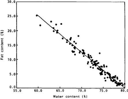
The transmission parameter that is measured is the signal energy attenua-tion, also called the insertion loss. This parameter can be correlated with the amount of water present in the tissue, which itself is correlated with the fat content by regression models already established for different fish species (see figure 2.2).
Figure 2.2: Correlation between total body water and the fat rate for Atlantic herring (Kent,1990)
Advantages, drawbacks and limitations
This technique has the advantage of being simple, fast and non-invasive. One of the drawbacks of this technique is that only the amount of body water is mea-sured which is then correlated with the fat content via regression models, this process may produce inaccuracies. Indeed, a great variability of the measure-ments results can be observed in (Clerjon, 2007). The correlation between the measurements made by this device and the measurements made with a conven-tional method such as biochemical analysis is not very strong (Douirin, 1998). This may be due to several parameters that can distort the measurement results of the device such as the density of cutaneous fat as well as subcutaneous fat, which decreases the propagation of the wave in the muscle.
Because of its strong variability, we chose to not select this technique for the context of our research.
2.2.3
Near Infrared Spectroscopy
Operating principleNear-infrared spectroscopy exploits the principle of absorption and reflection of light by the biological tissue. Indeed, each type of tissue presents a different behavior when subjected to near-infrared radiation (Weijun, 2009). The fat tissue contains less chromophore (the part of the molecule responsible for color) and therefore absorbs less light. These cells have a size of several hundred microns while the near-infrared wavelengths range from 600 nm to 1300 nm, thus we have a stronger reflection. Muscle tissue, on the other hand, behaves in an opposite way, there is more absorption and less reflection. The figure 2.1 presents the results of a study (Song et al., 2009; Lin Ling, 2001) highlighting the optical parameters of the muscular tissues, notably the absorption coefficient noted ua and the reflection coefficient noted us.
Subject u
a(mm
−1)
u
s(mm
−1)
Fat
1
2
0.003
0.005
1.200
1.200
3
0.004
1.200
Muscle
4
0.022
0.450
5
0.026
0.450
6
0.024
0.450
7
0.026
0.500
8
0.020
0.400
9
0.022
0.450
10
0.022
0.500
Table 2.1: Optical parameters of muscle and fat tissue, repro-duced from (Song et al., 2009)
The emitter and receiver electronic circuitry is relatively simple to imple-ment. Indeed, for the emission, one or several components emitting near-infrared light can be used, either Light Emitting Diodes (LED) or Lasers. For reception, photoelectric conversion circuits based on photo-voltaic cells (Weijun,
2009) can be used (See figure 2.3)
+
-C2
R
Amp
R
C1
Figure 2.3: Photo-electric conversion circuit
Advantages, drawbacks and limitations
This technique has the advantage of being simple to implement, accurate and fast. The disadvantage of this method is the relatively large power consumption. Indeed, near-infrared LEDs and lasers commercially available generally have consumptions around 70 mA. In our case study, where measurement campaigns of several months are required, such power consumption can not be accepted. Moreover, near-infrared spectroscopy causes increases in the temperature of the tissue to which it is applied, up to 10° C (Bozkurt, 2004), this might induce discomfort that could change the behavior of the species under study. This risk is even more important in the case of frequent measurements. The increase in temperature is mainly caused by the overheating of the emitter components.
Because of these two main disadvantages, this technique was abandoned.
2.2.4
Ultrasound
Operating principle
Ultrasound operates in the frequency range between 20 kHz and 20 MHz. The operating principle of a measurement consists in propagating ultrasound waves through the biological tissue and then the reflected acoustic wave is analyzed. In fact, the reflected wave parameters will vary depending on the acoustic
impedance of the different components of the biological sample that the wave will pass through during its propagation. For humans, for example, we can find the following acoustic impedance values :
• Air : 0 g cm−1s−1
• Fat : 0.138 g cm−1s−1
• Muscle : 0.170 g cm−1s−1
• Bones : 0.78 g cm−1s−1
In order to measure tissue composition using ultrasound, a probe is needed as well as the electronic circuitry for emission and response signal processing.
Regarding the probe : it is generally composed of the following elements
(Basix principle of medical ultrasonic probes) (See figure 2.4):
Figure 2.4: An ultrasound probe components, reproduced from (Basix principle of medical ultrasonic probes)
• The piezoelectric transducer: It contains piezoelectric crystals. This crys-tals are deforming (expansion and contraction) at the frequency of the electrical voltage that will be applied to them, thus generating an acous-tic wave that propagates in the biological tissue. The higher the acousacous-tic wave frequency is, the better the resolution, the lesser wave penetration depth we have. However, we observe the use of the 5 MHz frequency in most biomedical applications. Conversely, if a vibration is applied to the transducer, this element will generate an electrical voltage proportional to the applied vibration.
• The backing material: This element limits excessive vibrations of the transducer.
• The matching layer: This element is used to adapt the acoustic impedance of the biological tissue by reducing the mechanical friction.
• The acoustic lens: It is used in order to focus the ultrasound waves on the tissue region to be analyzed.
Regarding the ultrasound wave generation, level shifters (charge pumps or boost-converters) are used to create high voltages (typically around 40 V) re-quired to deform the piezoelectric element.
In order to analyze the response signal, several techniques are used:
• A signal detection block with a timer, in order to measure the delay of the response signal.
• A peak detection block followed by an integrator in order to measure the energy of the response signal.
• High speed Analog-to-Digital Converters (ADC) may be used to analyze the response signal in the frequency domain.
This blocks may be used separately or combined depending on the con-straints of the application.
Advantages, drawbacks and limitations
Biological tissue composition analysis using ultrasound has the advantage of being precise and fast, it also allows with a single measurement to collect infor-mation on fat, muscle and bone content of the analyzed tissue.
The main disadvantage of this technique is the use of level-shifters neces-sary to deform the piezoelectric element. These level-shifters require external components (inductances / capacitors), which is cumbersome in the case of an implanted device. The need to use a high voltages is also problematic since it results in a significant power consumption, this can be reduced by exciting the tissue with shorter time frame waveforms (Johansson J, 2006), unfortunately this can only be done at the cost of the accuracy of the measurement.
2.2.5
Bioimpedance Spectroscopy
Operating PrincipleBioimpedance Spectroscopy consists in estimating the composition of a given biological tissue by measuring its complex electrical impedance over several frequencies. Using 2, 3 or 4 electrodes, a controlled current or voltage is used to stimulate the tissue to be analyzed. Conversely, a voltage or a current is then measured, thus making it possible to recover the electrical impedance (see figure 2.5).
Bioimpedance is a function of physiological processes, thus offering interest-ing opportunities for monitorinterest-ing a large field of bio-markers. In the literature several studies demonstrating its wide range of applications can be found, from the estimation of body composition (Smith, Johnson, and Nagy, 2009), to can-cerous tissue detection (Aberg et al.,2004), or blood characterization (Dai and Adler,2009).
Each biomolecule has a different response for a given frequency. Scwhan (Schwan,1957) identified 3 frequency regions: dispersion α (Hz - KHz), disper-sion β (KHz - MHz) and disperdisper-sion γ (MHz - GHz). Each disperdisper-sion is ruled by different physiological processes. To be able to make a rigorous electrical
Figure 2.5: Bioimpedance analysis operating principle
characterization of the biological tissue, it is necessary to analyze its complex impedance over a wide frequency range.
Advantages, drawbacks and limitations
Bioimpedance spectroscopy is a relatively simple technique to implement. The ability to monitor multiple biological indicators with a single measurement is a great advantage. In addition, bioimpedance measurements have already been carried out on several fish species (Cox and Hartman,2005; Duncan et al.,2007; Willis and Hobday,2008) in the objective of assessing their tissue composition. This research studies have shown more or less good correlations between elec-trical parameter at 50 kHz and body composition parameters such as the fat content or water content.
It is also interesting to note that bioimpedance analysis is a widely used technique in biomedical/healthcare applications. Studies (Aberg et al., 2004; Dai and Adler,2009) have shown that the technique has a great potential as a non-invasive characterization and monitoring tool of the human biological tissue properties.
The main disadvantage of bioimpedance spectroscopy is its sensitivity to several parameters such as temperature or muscle activity.
2.2.6
Comparative status
Table 2.2 shows a status of advantages and drawbacks of the techniques studied above
The microwave propagation and near infrared spectroscopy having been eliminated for the reasons detailed in the preceding subsections, the choice of the techniques to be used in the context of our application was restricted to ultrasound and bioimpedance spectroscopy.
Technique Microwave propagation
NIR
Spec-troscopy
Ultrasound Bioimpedance Spec-troscopy
Advantages Simple to imple-ment, fast, non invasive
Simple, fast,
ac-curate Accurate,Measures fat/muscle/bone content, fast
Fast, low cost, low power consumption, wide range of biomark-ers could be monitored, measurements already performed on several fish species
Drawbacks Wide variability of measurements, Measures only water content
Relatively impor-tant power con-sumption, Risk of tissue damaging on the long term
Use of cumber-some external components, relatively im-portant power consumption Measurement sensitiv-ity to temperature and muscle activity.
Table 2.2: Comparative status on the studied techniques
For its low complexity, its ability to achieve low power consumption and especially for the wide range of biological indicators it allows to monitor, we chose bioimpedance spectroscopy as the technique to be used for our application.
2.3
Bioimpedance spectroscopy : From electrical
properties of tissue to its electrical impedance
2.3.1
Introduction
Bioelectrical methods deal with the interactions of endogenic or exogenic elec-tricity and biological tissue. While Bioelecelec-tricity is about studying the ability of biological tissue to generate electricity, as it is the case for the heart for example (electrocardiography). Bioimpedance, on the other hand, deals with ability of the biological tissue to oppose (impede) an applied electric current flow. From this perspective, Bioimpedance describes the passive electrical properties of tis-sue (muscle, fat, blood or bones, . . .), as changes in the bioimpedance can reflect changes in the biomaterial.
To be able to measure such a bioimpedance we can use electrodes, a con-trolled current or voltage is generated to stimulate the tissue to be analyzed and, conversely, a voltage or a current is then measured. When studying bioimpedance, it is important to decompose the study into circuit and bio-chemical issues. In fact, charge carriers flowing into wires are electrons while the charge carriers in biological tissue are ions. The electrodes interface is re-sponsible for the conversion of the charge carriers from electrons to ions and from ions to electrons.
2.3.2
Cell electrical properties
The cell is the basic structural unit of living tissue. The understanding of its structure and components is essential to better comprehend the properties of biological tissue. Cells are of uneven size with various functions (muscle cells, nerve cells, bone cells, . . .). Therefore, for an accurate analysis, the cell model must take into account the various nature of cell types. However, in order to showcase the basic principles of cell electrical modeling, we consider a simple case of a volume containing cells in interstitial fluids (see figure 2.6). We can observe that the cells are contained in an Extracellular Fluid (ECF). The intracellular space is composed of the cell nucleus, and the Intracellular Fluid (ICF) which contains components such as the cytoplasm and organelles. The cell membrane separates the intracellular space from the extracellular space.
The ICF, ECF and cell membrane account for cell and tissue electrical prop-erties, which can be divided to conductance properties and dielectric properties (Grimnes and Martinsen,2014).
The intracellular and extracellular fluids contain ions. Due to this free ions (mostly Na+ and K+), ECF and ICF are considered as electrolytes, which
means that they have the ability to conduct electric current in the presence of an external electrical field. Therefore, the biological tissue can be considered as an ionic conductor.
The cell membrane is predominately constituted of polar lipids. These lipids are responsible of the capacitive nature of cells and by extent of biological tissue. The cell membrane is a layer that separates ICF and ECF and forms two sharp boundaries against them. The cell membrane consists of phospholipids
Extracellular fluid Intracellular fluid
Nucleus
Cell membrane
Figure 2.6: Cell components
that form a Bilayer Lipid Membrane (BLM). The structure of the phospholipid molecules consists of two hydrophobic fatty acid tails and a hydrophilic head. Each mono-layer has its hydrophobic part oriented inward and its hydrophilic part outward facing the intracellular or extracellular fluids (see figure 2.7), which makes the membrane a bad conductor for ions.
Figure 2.7: Bilayer lipid membrane
This structure forms a continuous double layer known as the lipid bilayer. The conductivity of the lipid bi-layer is very poor and is mainly considered as a dielectric.
2.3.3
Cell electrical model
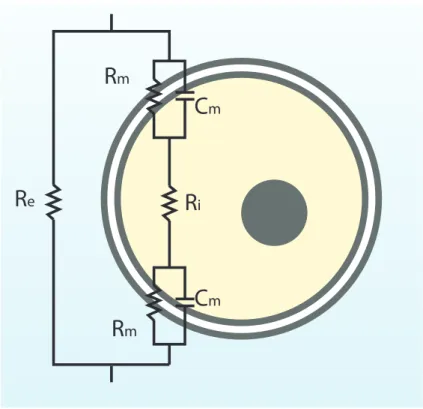
The conductance and dielectric properties of biological tissue can be modeled as an equivalent electrical circuit. Obviously, the complexity of biological processes makes it difficult to accurately model the cell properties using simple electrical components. However, for illustrative purposes we will consider a simple elec-trical model which is the Fricke model (Fricke,1924; Fricke and Morse,1925).
The Fricke model is one of the earliest equivalent electrical models to be introduced in the literature. It is illustrated in figure 2.8. The extracellular and intracellular fluid are associated with resistive components, while the cell membrane is associated with capacitive and resistive components. The ionic currents can flow between the extracellular space of the cell and its intracellular space through existing ionic channels across the bilayer lipid membrane.
C
mC
mR
mR
mR
eR
iFigure 2.8: Fricke’s Electrical Equivalent Model
The cell equivalent circuit is then represented by the ECF Re resistor, the
ICF Riresistor, and the cell membrane is represented by a resistor Rmin parallel
with a capacitor Cm. Since the conductivity of the cell membrane is poor, the
cell electrical model could be simplified by representing the cell membrane only by the capacitor Cm.
The total impedance can be written as : Zcell = Re//(Ri+ 1 jC0 mω ) (2.1) With C0 m = Cm
2 and j the imaginary unit
Zcell = Re// 1 + jRiCm0 ω jC0 mω (2.2) Zcell = Re(1 + jRiCm0 ω) 1 + jC0 mω(Re+ Ri) (2.3) It is interesting to observe the frequency dependency introduced by the cell membrane capacitance. Indeed, at low frequencies the cell membrane acts as
an insulator blocking the current from penetrating into the intracellular space ( see figure 2.9 ).
Figure 2.9: Low frequency current path
Therefore, at low frequencies the biological tissue impedance is equivalent to the resistance of the extracellular fluids. This can be easily checked by using equation 2.3 :
ω → 0 =⇒ Zcell = Re (2.4)
On the other hand, at high frequencies, the impedance of the cell membrane decreases, and the current flows through the extracellular space as well as the intracellular space ( see figure 2.10 ).