Université de Montréal
Manipulation of the Ubiquitin-Proteasome System by HIV-1: Role of the
Accessory Protein Vpr
par
Jean-Philippe Belzile
Département de Microbiologie et Immunologie
Faculté de Médecine
Thèse présentée à la Faculté des Études Supérieures
en vue de l’obtention du grade de Philosophiae Doctor (Ph.D.)
en Microbiologie et Immunologie
Février, 2010
Université de Montréal Faculté des Études Supérieures
Cette thèse intitulée:
Manipulation of the Ubiquitin-Proteasome System by HIV-1: Role of the
Accessory Protein Vpr
présentée par: Jean-Philippe Belzile
a été évaluée par un jury composé des personnes suivantes
Dr Jacques Thibodeau, Ph.D. President-rapporteur Dr Éric A. Cohen, Ph.D. Directeur de recherche Dr Gerardo Ferbeyre, Ph.D. Membre du jury Dr Carlos de Noronha, Sc.D. Examinateur externe Dr Jacques Archambault, Ph.D. Représentant du doyen
SOMMAIRE
Le virus de l’immunodéficience humaine de type 1 (VIH-1), l’agent étiologique du SIDA, est un rétrovirus complexe arborant plusieurs protéines accessoires : Nef, Vif, Vpr, et Vpu. Celles-ci sont impliquées dans la modulation de la réplication virale, dans l’évasion immunitaire et dans la progression de la pathogenèse du SIDA. Dans ce contexte, il a été démontré que la protéine virale R (Vpr) induit un arrêt de cycle cellulaire en phase G2. Le mécanisme par lequel Vpr exerce cette fonction est l’activation, ATR (Ataxia telangiectasia and Rad3 related)-dépendante, du point de contrôle de dommage à l’ADN, mais les facteurs et mécanismes moléculaires directement impliqués dans cette activité demeurent inconnus.
Afin d’identifier de nouveaux facteurs cellulaires interagissant avec Vpr, nous avons utilisé une purification d’affinité en tandem (TAP) pour isoler des complexes protéiques natifs contenant Vpr. Nous avons découvert que Vpr s’associait avec CRL4A(VprBP), un complexe cellulaire d’E3 ubiquitine ligase, comprenant les protéines Cullin 4A, DDB1 (DNA damage-binding protein 1) et VprBP (Vpr-binding protein). Nos études ont mis en évidence que le recrutement de la E3 ligase par Vpr était nécessaire mais non suffisant pour l’induction de l’arrêt de cycle cellulaire en G2, suggérant ainsi que des événements additionnels seraient impliqués dans ce processus. À cet égard, nous apportons des preuves directes que Vpr détourne les fonctions de CRL4A(VprBP) pour induire la polyubiquitination de type K48 et la dégradation protéosomale de protéines cellulaires encore inconnues. Ces événements d’ubiquitination induits par Vpr ont été démontrés comme étant nécessaire à l’activation d’ATR. Finalement, nous montrons que Vpr forme des foyers ancrés à la chromatine co-localisant avec VprBP ainsi qu’avec des facteurs impliqués dans la réparation de l’ADN. La formation de ces foyers représente un événement essentiel et précoce dans l’induction de l’arrêt de cycle cellulaire en G2. Enfin, nous démontrons que Vpr est
capable de recruter CRL4A(VprBP) au niveau de la chromatine et nous apportons des preuves indiquant que le substrat inconnu ciblé par Vpr est une protéine associée à la chromatine.
Globalement, nos résultats révèlent certains des ménanismes par lesquels Vpr induit des perturbations du cycle cellulaire. En outre, cette étude contribue à notre compréhension de la modulation du système ubiquitine-protéasome par le VIH-1 et son implication fonctionnelle dans la manipulation de l’environnement cellulaire de l’hôte.
Mots clés: Virus, protéines accessoires, ATR, Point de contrôle de domage à l’ADN, cycle cellulaire, ubiquitination, ubiquitine ligase.
ABSTRACT
Human immunodeficiency virus 1 (HIV-1), the etiologic agent of AIDS, is a complex retrovirus with several accessory proteins. HIV-1 accessory proteins Nef, Vif, Vpr, and Vpu have been implicated in the modulation of viral replication, enhancement of viral fitness, immune evasion, and progression of AIDS pathogenesis. In that regard, viral protein R (Vpr) induces a cell cycle arrest in the G2 phase by activating the canonical ATR (Ataxia telangiectasia and Rad3 related)-mediated DNA damage checkpoint, but cellular factors and mechanisms directly engaged in this process remain unknown.
To identify novel Vpr-interacting cellular factors, we used tandem affinity purification (TAP) to isolate native Vpr-containing complexes. We found that Vpr hijacks a cellular E3 ubiquitin ligase complex, CRL4A(VprBP), composed of Cullin 4A, DDB1 (DNA damage-binding protein 1) and VprBP (Vpr-binding protein). Moreover, we observed that recruitment of the E3 ligase by Vpr was necessary but not sufficient for the induction of G2 cell cycle arrest, suggesting that additional events are involved. In this context, we provide direct evidence that Vpr usurps the function of CRL4A(VprBP) to induce the K48-linked polyubiquitination and proteasomal degradation of as-yet-unknown cellular proteins. These ubiquitination events mediated by Vpr were necessary for the activation of ATR. Moreover, we show that Vpr forms chromatin-associated foci that co-localize with VprBP and DNA repair factors. Our data indicate that formation of these foci represent a critical early event in the induction of G2 arrest. Finally, we show that Vpr is able to recruit CRL4A(VprBP) on chromatin and we provide evidence that the unknown substrate targeted by Vpr is a chromatin-associated protein.
Overall, our results reveal some of the mechanisms by which Vpr induces cell cycle perturbations. Furthermore, this study contributes to our understanding of the modulation of the ubiquitin-proteasome system by HIV-1 and its functional implication in the manipulation of the host cellular environment.
Keywords: Virus, accessory proteins, ATR, DNA damage checkpoint, cell cycle, ubiquitination, ubiquitin ligase.
TABLE OF CONTENTS
SOMMAIRE... iii
ABSTRACT... v
TABLE OF CONTENTS... vii
LIST OF FIGURES ... x
ABBREVIATIONS ... xi
ACKNOWLEDGEMENTS... xv
INTRODUCTION... 1
1. HUMAN IMMUNODEFICIENCY VIRUS TYPE 1 (HIV-1)... 1
1.1. HIV-1 and the retrovirus family... 2
1.2. Genetic organization ... 2
1.3. Overview of the HIV-1 replication cycle... 4
1.4. Roles of HIV-1 accessory proteins in pathogenesis and immune evasion... 8
1.4.1. VIF ... 9
1.4.2. VPU... 11
1.4.3. NEF ... 13
1.4.4. VPR ... 17
2. THE UBIQUITIN-PROTEASOME SYSTEM ... 23
2.1. Principles and mechanisms of ubiquitination ... 23
2.2. Functional implications of ubiquitin chain topologies... 27
2.3. Cullin-RING ubiquitin ligases (CRLs) ... 31
2.3.1. Structure and composition ... 31
2.3.2. Regulation ... 34
3. CELL CYCLE REGULATION... 37
3.1. Regulation of cell cycle progression... 37
3.2. Mitogenic restriction point... 41
3.3. DNA damage checkpoint ... 44
3.4. Mitotic checkpoint ... 50
CHAPTER 1: HIV-1 VPR-MEDIATED G2 ARREST INVOLVES THE
DDB1-CUL4VPRBP E3 UBIQUITIN LIGASE ... 54
ABSTRACT... 56
INTRODUCTION ... 56
RESULTS ... 58
DISCUSSION ... 64
MATERIALS AND METHODS... 68
ACKNOWLEDGMENTS ... 74
REFERENCES... 75
FIGURES ... 83
CHAPTER 2: HIV-1 VPR INDUCES THE K48-LINKED POLYUBIQUITINATION AND PROTEASOMAL DEGRADATION OF TARGET CELLULAR PROTEINS TO ACTIVATE ATR AND PROMOTE G2 ARREST ... 95
ABSTRACT... 97
INTRODUCTION ... 97
RESULTS ... 101
DISCUSSION ... 107
MATERIALS AND METHODS... 112
ACKNOWLEDGMENTS ... 115
REFERENCES... 115
FIGURES ... 124
SUPPLEMENTAL MATERIAL ... 138
CHAPTER 3: FORMATION OF CHROMATIN-ASSOCIATED NUCLEAR FOCI CONTAINING HIV-1 VPR AND VprBP IS CRITICAL FOR THE INDUCTION OF G2 CELL CYCLE ARREST... 144
ABSTRACT... 146
INTRODUCTION ... 146
RESULTS ... 150
DISCUSSION ... 156
ACKNOWLEDGMENTS ... 164
REFERENCES... 164
FIGURES ... 171
SUPPLEMENTAL MATERIAL ... 189
DISCUSSION ... 195
1. HIV-1 VPR HIJACKS A CELLULAR CULLIN-RING E3 UBIQUITIN LIGASE ... 195
2. IDENTIFICATION OF VPR’S G2 ARREST SUBSTRATE ... 198
3. ROLE OF G2/M ARREST DURING INFECTION... 200
4. IMPLICATION OF CRL4A(VprBP) IN OTHER FUNCTIONS OF VPR ... 201
5. ARCHITECTURE AND COMPOSITION OF THE E3 LIGASE COMPLEX RECRUITED BY VPR... 203
6. INTERACTIONS BETWEEN VPR AND OTHER COMPONENTS OF UPS .. 205
7. DEVELOPMENT OF DRUGS TARGETING THE VPR-VprBP BINDING INTERFACE... 207
CONCLUSION... 208
LIST OF FIGURES
Figure 1. HIV-1 genomic organization. ... 3
Figure 2. HIV-1 replication cycle. ... 5
Figure 3. Conservation of Vpr throughout primate lentiviral evolution. ... 18
Figure 4. NMR structure of HIV-1 Vpr. ... 19
Figure 5. The ubiquitin conjugation cycle. ... 24
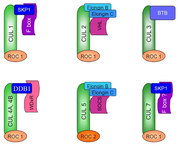
Figure 6. Composition and architecture of Cullin-RING E3 ubiquitin ligases... 32
Figure 7. Regulation of cell cycle progression by CDKs and APC/C ... 39
Figure 8. The DNA damage checkpoint. ... 45
ABBREVIATIONS
53BP1 Tumor suppressor protein 53-binding protein 1
9-1-1 Rad9-Hus1-Rad1 complex
ABRA1 Abraxas
ANT Adenine nucleotide translocator
AP-1 Adaptor protein 1
AP-2 Adaptor protein 2
APC/C Anaphase-promoting complex/cyclosome
APOBEC3F Apolipoprotein B mRNA-editing enzyme catalytic polypeptide-like 3F
APOBEC3G Apolipoprotein B mRNA-editing enzyme catalytic polypeptide-like 3G
ATM Ataxia telangiectasia mutated
ATR Ataxia telangiectasia and Rad3 related
ATRIP ATR-interacting protein
β-COP Coatomer protein complex subunit beta β-TrCP Beta-Transducin repeat-containing protein
BARD1 BRCA1-associated RING domain 1
BER Base excision repair
BRCA1 Breast cancer 1, early onset
BTB Broad complex, tramtrack, bric-a-brac
Bub1 Budding uninhibited by benzimidazoles homolog 1 Bub3 Budding uninhibited by benzimidazoles homolog 3
BubR1 Bub1 homolog beta
CA Capsid
CAND1 Cullin-associated and neddylation-dissociated
CCR5 C-C chemokine receptor type 5
Cdc25 Cell division cycle 25
CDK1 Cyclin-dependent kinase 1 CDK2 Cyclin-dependent kinase 2 CDK4 Cyclin-dependent kinase 4 CDK6 Cyclin-dependent kinase 6 Cdt1 Cdc10-dependent transcript 1 Cdt2 Cdc10-dependent transcript 2 Chk1 Checkpoint kinase 1 Chk2 Checkpoint kinase 2
CKI Cyclin-dependent kinase inhibitor
CRL Cullin-RING E3 ubiquitin ligase
CRL1 Cullin 1-RING E3 ubiquitin ligase CRL2 Cullin 2-RING E3 ubiquitin ligase CRL3 Cullin 3-RING E3 ubiquitin ligase CRL4A Cullin 4A-RING E3 ubiquitin ligase CRL4B Cullin 4B-RING E3 ubiquitin ligase CRL5 Cullin 5-RING E3 ubiquitin ligase
CRL7 Cullin 7-RING E3 ubiquitin ligase
CSN COP9 signalosome
CSN6 COP9 signalosome subunit 6
CUL4A Cullin 4A
CXCR4 Chemokine (CXC motif) receptor 4
DC-SIGN Dendritic Cell-Specific Intercellular adhesion molecule-3-Grabbing Non-integrin
DC Dendritic cells
DDA1 DET1 and DDB1 associated 1
DDB1 DNA Damage-Binding protein 1
DDB2 DNA Damage-Binding protein 2
DET1 De-etiolated homolog 1
DNA-PKcs DNA-dependent protein kinase catalytic subunit
DSB DNA double-strand break
DUB Deubiquitinase
EGFR Epidermal growth factor receptor
Emi1 Early mitotic inhibitor 1
Env Envelope
ERAD Endoplasmic reticulum-associated degradation ERK Extracellular signal regulated kinase
ESCRT-I Endosomal sorting complex I required for transport
ESCRT-III Endosomal sorting complex III required for transport
FA Fanconi anemia
Gag Group-specific antigen
γ-H2AX Phosphorylated H2AX
gp41 Glycoprotein 41
gp120 Glycoprotein 120
gp160 Glycoprotein 160
GR Glucocorticoid receptor
H2AX Histone 2A, variant X
HBV Hepatitis B virus
HBx Hepatitis B virus protein X
HCMV Human cytomegalovirus
HECT Homologous to the E6-AP carboxyl terminus
hHR23A Human homolog A of RAD23
HIV-1 Human immunodeficiency virus type 1 HIV-2 Human immunodeficiency virus type 2
HLA Human leukocyte antigen
IκBα Inhibitor of NF-κB alpha
IKK IκB kinase
KSHV Kaposi’s sarcoma-associated hespesvirus LEDGF/p75 Lens epithelium-derived growth factor, p75
LTR Long terminal repeat
MA Matrix
MAD1 Mitotic arrest deficient 1
MAD2 Mitotic arrest deficient 2
MAPK Mitogen-activated protein kinase
MCM Mini-chromosome maintenance complex
MDC1 Mediator of DNA damage checkpoint 1
MHC-I Major histocompatibility complex class I MHC-II Major histocompatibility complex class II
Mps1 Monopolar spindle 1
MRN Mre11-Rad50-NBS1
MVB Multivesicular body
NBS1 Nijmegen breakage syndrome 1
NC Nucleocapsid
NDC80 Ndc80 homolog, kinetochore complex component
Nef Negative factor
NEDD8 Neuronal precursor expressed, developmentaly down-regulated 8
NEMO NF-κB essential modulator
NER Nucleotide excision repair
NES Nuclear export signal
NF-κB Nuclear factor kappa-light-chain-enhancer of activated B cells
NHEJ Non-homologous end joining
HR Homologous recombination
NK Natural killer cell
NKG2D Natural killer group 2, member D
NLS Nuclear localization signal
P-TEFb Positive transcription elongation factor b
p300 E1A-binding protein 300
PARC Parkin-like cytoplasmic protein
PBMCs Peripheral blood mononuclear cells
PIC Pre-integration complex
PIKK Phosphoinositide-3-kinase-related protein kinase PCNA Proliferating cell nuclear antigen
PLK1 Polo-like kinase 1
Pol Polymerase
PP Protein phosphatase
Pre-RC Pre-replicative complex
R Repeated sequence
RAP80 Receptor-associated protein 80
Rb Retinoblastoma tumor suppressor protein Rev Regulator of virion gene expression
RFC Replication factor C
RING Really interesting new gene
RNF8 RING finger protein 8
ROC1 Regulator 1 of Cullins
ROC2 Regulator 2 of Cullins
ROS Reactive oxygen species
RPA Replication protein A
RRE Rev-responsive element
SCF Skp1/Cullin 1/F-Box complex
SIV Simian immunodeficiency virus
SIVagm SIV in African green monkeys
SIVcpz SIV in chimpanzees
SIVmac SIV in rhesus macaques
SIVsm SIV in sooty mangabeys
Skp1 S-phase kinase-associated protein 1 Skp2 S-phase kinase-associated protein 2 SOCS Suppressor of cytokine signaling
SP1 Transcription factor SP1
STAT2 Signal transducer and activator of transcription 2
SV5 Simian virus 5
SV5 V Simian virus 5 protein V
SUMO Small ubiquitin-related modifier
TAP Tandem affinity purification
Tat Transcriptional transactivator
TLS Translesion synthesis
TNF Tumor necrosis factor
TopBP1 Topoisomerase 2 binding protein 1
TS Template switching
U3 Unique to 3’end
U5 Unique to 5’end
Ub Ubiquitin
UBD Ubiquitin-binding domain
UNG2 Uracil-DNA glycosylase 2
UPS Ubiquitin-proteasome system
VCP Valosin-containing protein
Vif Viral infectivity factor
Vpr Viral protein R
VprBP Vpr-binding protein
Vpu Viral protein U
Vpx Viral protein X
ACKNOWLEDGEMENTS
I would like to thank my research director, Dr Éric Cohen, for his supervision and advices throughout these years. Research is rarely easy and at time can be particularly difficult. Without your support and the constructive discussion we had, I would not have been able to satisfactorily complete my studies. Under your supervision, my Ph.D. has been a very stimulating and enriching experience.
I would also like to thank Nicole Rougeau who has helped me in my research work almost since the beginning of my Ph.D. studies. The conditions we worked in were not always ideal but at the end we were able to succeed in our projects. My thanks also go to Dr Levon Abrahamyan, Mathieu Dubé, Dr Francine Gérard, Johanne Mercier, Jonathan Richard, and Dr Yong Xiao for their invaluable technical assistance and for the great scientific discussions we had. I am grateful also to the other members of the laboratory for their friendship and support.
Most and foremost, I would like to thank my wife for her ever-present support throughout my graduate studies and my daughter for lightening up my days when skies were gloomy. I could never have made it without you. I would also like to thank my parents for their constant encouragements.
Finally, this thesis would not have been realizable without financial support from my research director, from the Canadian Institute of Health Research (CIHR), from the Institut de Recherches Cliniques de Montréal (IRCM), from the department of microbiology and immunology and the Faculté des Études Supérieures (FES) of Université de Montréal.
INTRODUCTION
1. HUMAN IMMUNODEFICIENCY VIRUS TYPE 1 (HIV-1)
Human immunodeficiency virus type 1 (HIV-1) is the causative agent of the global epidemics of AIDS. In 2008, 33.4 million individuals were HIV-1-positive. An estimated 2.7 millions new HIV-1 infections occurred and 2 million people died of AIDS- related diseases. Due to the beneficial effects of the introduction of anti-retroviral therapy in low-income countries, the estimated number of new HIV-1 infection cases was 30% lower than at the peak of the epidemics in 1996 and the number of AIDS-related death was 10% lower than at the peak of mortality in 2004 [1].
HIV-1 is a highly heterogeneous virus and, based on genetic similarities, is subdivided into 4 groups: M, O, N, and P. Group M is responsible for the present epidemic and, due to founder effects, can be further subdivided into clades or subtypes (A to K). Clade B viruses are the most prevalent in Western countries whereas C is the most prominent globally. Because the epidemic originated in Africa, it has the most heterogeneous viral distribution and inter-clade or inter-group recombinants are commonly observed. Groups N, O, and P only represent a minority of cases and are typically restricted to some regions of Africa [2-4]. HIV is a zoonosis and each group is thought to have originated from a single independent cross-species transmission from a primate, the natural reservoir, to a human. Group M and N likely originated from chimpanzees (Pan troglodytes troglodytes) in South Cameroon, group O from either chimpanzees or gorillas (Gorilla gorilla gorilla) [5-8], and Group P from gorillas [2]. There exists a second human immunodeficiency virus called HIV-2. HIV-2 is characterized by a lower pathogenicity and is principally restricted to Western Africa. It probably arose from at least 8 independent cross-species transmissions from sooty mangabeys (Cercocebus atys) to humans [5,9]. Recent molecular clocking analyses situate the origins of the HIV-1 epidemic to the early 20th century in Belgian Congo (now Democratic Republic of Congo). Trans-species transmissions between chimpanzees and humans might have occurred before this period but social conditions
and population densities resulting from the establishment of colonial cities such as Leopoldville (now Kinshasa) probably generated the optimal conditions for the global spread of the sexually transmitted disease [10,11].
1.1. HIV-1 and the retrovirus family
HIV-1 belongs to the retrovirus family (Retroviridae), which comprises enveloped viruses with linear, non-segmented, positive, single-stranded RNA genomes. The hallmarks of this family of viruses are that they require reverse transcription of the viral genome into linear double-stranded DNA and subsequently stable integration of their genome into the host DNA. Retroviruses are ubiquitous and are present in all classes of vertebrates. All retroviruses possess three major genes: gag (group-specific antigen), pol (polymerase), and env (envelope). Particles vary in size from 80 to 120 nm in diameter and are covered by envelope glycoprotein spikes [12,13]. The genus lentivirus, of which HIV-1 is the prototypic example, comprises complex retroviruses that encode additional unique auxiliary proteins. Other defining characteristics of the lentiviral genus include a curved hexagonal viral core called a fullerene cone and a biphasic mode of gene expression [14,15]. All lentiviruses can infect macrophages and primate lentiviruses exhibit a strong tropism for CD4-expressing cells, including macrophages and T helper lymphocytes. In contrast to several other retroviruses, lentiviruses do not directly induce oncogenesis. Common manifestations of lentiviral diseases include long incubation time, persistent chronic viral replication, encephalopathy, and suppression of specific haematopoietic or immune cell types [16].
1.2. Genetic organization
As mentioned above, HIV-1 like all retroviruses harbours the gag, pol, and env genes (Figure 1, p.3). The HIV-1 gag gene encodes the viral structural proteins matrix (MA, p12), capsid (CA, p24), and nucleocapsid (NC, p7) expressed as part of a myristilated precursor polyprotein (p55). Individual components are released following the proteolytic processing of the polyprotein by the viral protease. The gag precursor protein also possesses p1, p2, and p6 domains which are not thought to play functions as individual proteins but rather contribute to the regulation or functions of the precursor polyprotein. The pol gene produces a precursor polyprotein fused to the Gag
Figure 1. HIV-1 genomic organization.
The HIV-1 genome is flanked by two long-terminal repeats (LTR) and takes advantage of its three reading frames. HIV-1 possesses the conserved retroviral genes gag, pol, and env. The HIV-1 genome also contains six additional genes that encode two regulatory proteins (Tat and Rev) and four accessory proteins (Nef, Vif, Vpr, and Vpu).
polyprotein via a ribosomal frameshift. Proteolytic processing of Gag-pol yields the viral enzymatic proteins protease (Pr), reverse transcriptase (RT), and integrase (IN). The env gene encodes the two envelope subunits, gp120 and gp41, which are first expressed as a single precursor protein (gp160) and later cleaved by a cellular furin-like protease. The HIV-1 genome also contains six additional genes that encode the two regulatory proteins Tat (transcriptional transactivator) and Rev (regulator of virion gene expression) and the four accessory proteins: Nef (negative factor), Vif (viral infectivity factor), Vpr (viral protein R), and Vpu (viral protein U). HIV-2 (human immunodeficiency virus type 2) and some simian immunodeficiency virus (SIV) isolates of the HIV-2/SIVsm (sooty mangabey) lineage possess a fifth accessory gene called Vpx (viral protein X), but do not encode for a Vpu protein. Tat and Rev are involved in HIV gene expression and proper splicing and export of the different mRNA species whereas the accessory proteins modulate host immune responses and facilitate viral replication in specific cell types. Except for Vpu and Env, which are expressed from the same mRNA, and Pol, which is expressed following a ribosomal frameshift, all the other HIV proteins are expressed from their own unique singly or multiply spliced mRNA. HIV genes are enclosed between two identical copies of the long terminal repeat (LTR). The LTR is subdivided in three regions: U3 (unique to 3’end), R (repeated sequence), and U5 (unique to 5’end). The transcription start site is located at the junction of U3 and R whereas the poly(A) signal is at the boundary of R and U5. Finally, the U3 region contains most of the transcriptional regulatory elements [13].
1.3. Overview of the HIV-1 replication cycle
The HIV-1 infection cycle (Figure 2, p.5) starts with the docking of the trimeric envelope glycoproteins (containing the gp41 and gp120 subunits) to its cognate cellular receptor CD4 [17,18]. The dendritic cell surface molecule DC-SIGN (Dendritic Cell-Specific Intercellular adhesion molecule-3-Grabbing Non-integrin) can also be used as an attachment factor for HIV-1. Although it can sometimes lead to productive infection of dentritic cells, it is generally exploited by HIV-1 as vehicle for dissemination of the virus within the host and as a mean to facilitate cell-to-cell transmission [19]. Binding of gp120 to CD4 induces a conformational change in gp120 that exposes its V3 loop
Figure 2. HIV-1 replication cycle.
The HIV-1 replication cycle can be subdivided in two phases: early and late. The early phase includes docking of the viral envelop to its primary receptor CD4 and a co-receptor, inducing fusion and released of the viral core into the cytoplasm. Entry is followed by reverse transcription of the viral genomic RNA into double-stranded DNA and ultimately by the stable integration of the viral genome into the host genomic DNA. The late phase of the infection consists of the expression of the various viral proteins and of the viral genomic RNA, resulting in the assembly and budding of progeny virions. Please refer to the main text for a detailed description of each step.
[20,21] enabling an interaction with its primary co-receptor CCR5 (C-C chemokine receptor type 5) [22]. A switch to CXCR4 (chemokine (CXC motif) receptor 4) in co-receptor usage happens late in infection in approximately 50% of infected individuals and is associated with rapid disease progression [23]. This second interaction event induces a series of conformation changes, this time in gp41, which induces the fusion of the viral membrane with the cellular plasma membrane following the insertion of the N-terminal fusion peptide [24-26]. It was long thought that HIV-1 was entering cells through fusion at the plasma membrane. However, recent evidence shows that although fusion might be initiated at the plasma membrane, it is completed after the virus has engaged the classical endocytic route [27,28]. Membrane fusion effectively releases the viral core into the cytoplasm of the cell [24,25]. There follows a series of poorly understood steps called uncoating whereby capsid proteins are shed from the viral core [29]. Once the RNA-capsid complex attains a certain level of maturation, reverse transcription is initiated [29,30]. It is a complex mechanism that involves multiple priming and initiation steps. The end product of reverse transcription is a double-stranded linear proviral DNA with a short overhanging structure called DNA flap on the positive strand [31]. As reverse transcription proceeds, the viral core continues its maturation and is transported along the microtubule networks toward the nuclear membrane. Once it reaches the nuclear membrane it is called the pre-integration complex and is now competent for nuclear import and integration into the host genome [29,30]. It is actively imported into the nucleoplasm via a nuclear pore and with the help of its karyophilic elements Vpr, MA, integrase, and the DNA flap [32]. As the provirus reaches the nucleoplasm it is tethered to chromatin via an interaction between the viral integrase and the stress-induced transcription factor LEDGF/p75 (Lens epithelium-derived growth factor, p75) [33-35]. Multimers of the viral integrase then trims two nucleotide at each end of the proviral DNA and catalyzes the nucleophilic attack of these ends onto the host genomic DNA [36]. The integration reaction leaves DNA overhangs that are subsequently repaired by the cellular DNA repair machinery. Which repair and sensor proteins are involved in this process remains highly controversial and is probably cell-type specific [37-40]. Once repair is complete, the late phase of the infection starts.
The dominant factor that promotes expression from the HIV-1 LTR is the viral regulatory protein Tat. It trans-activates the LTR by recruiting the positive transcription elongation factor b (P-TEFb) and histone acetyl transferase complexes and by activating NF-κB (nuclear factor kappa-light-chain-enhancer of activated B cells). Overall, Tat increases LTR activity by several hundred folds [41-43]. Cis-acting elements in the viral RNA leads to the generation of unspliced, partially spliced or fully spliced RNA that are going to produce the different viral proteins [44]. Since nuclear export of RNA is tightly coupled with splicing, HIV has developed its own mechanism to promote efficient nuclear export of unspliced or partially spliced RNA. Multimers of the viral regulatory protein Rev binds to the rev-responsive element (RRE) and mediate nuclear export of RNA via an interaction with the nuclear export factor exportin 1. This mode of export is energy-dependent and requires Ran GTPase [45,46]. The primary sites of virion assembly are at the plasma membrane and appear to be concentrated in some membrane subdomains such as lipid rafts [47,48], tetraspanin-rich domains [49,50] and regions of cell-to-cell contact [51]. One of the defining features of viral assembly is the interaction of the MA portion of the myristoylated Gag precursor protein with membranes containing the lipid PI(4,5)P2. Gag-Gag and Gag-lipid interactions probably cooperate to stabilize Gag assembly at the plasma membrane [52-54]. Gag-Pol is similarly incorporated into assembling virions [55,56]. Encapsidation of the viral genomic RNA requires an interaction between its packaging signal and the NC domain of Gag [57-59]. The envelope precursor protein gp160 is translated in the endoplasmic reticulum and forms trimers via disulfide bonds. It is then transported through the Golgi secretory pathway where it is heavily glycosylated and cleaved into its individual subunits gp120 and gp41 [60]. The incorporation of envelope glycoproteins into virions is still not well understood but probably involves viral as well as cellular proteins [61]. Budding of virions is mediated by the direct recruitment of the cellular ESCRT-I (endosomal sorting complex 1 required for transport) and ESCRT-III (endosomal sorting complex 3 required for transport) complexes. This is achieved via interactions between late domains in the p6 region of Gag and the ESCRT protein TSG101 (Tumour susceptibility gene 101) and AIP1/Alix (ASK-interacting protein 1). The ESCRT
machinery then induces membrane curvature and finally release of virions [54,62,63]. The final step of the viral cycle is characterized by the maturation of the spherical viral core into a conical core. This process involves a major conformational rearrangement mediated by the viral protease. The protease sequentially cleaves the individual components of the Gag and Gag-Pol precursor proteins resulting in the realignment of capsid proteins around the RNA/protein complex [64,65].
1.4. Roles of HIV-1 accessory proteins in pathogenesis and immune evasion
The hallmarks of HIV-1 infection have long been considered as the progressive infection and destruction of the pool of CD4+ lymphocytes, thus inducing profound immune dysfunction and ultimately immunodeficiency [66,67]. However, recent observations in simian models have considerably changed our understanding of AIDS as well as of the immunobiological paradigms involved in progression towards this condition. Indeed, extensive studies of non-pathogenic infections in sooty mangabeys have revealed that despite high levels of viral replication and dramatic CD4+ T-cell depletion, these rarely develop AIDS [68]. One central phenotype that distinguishes pathogenic versus non-pathogenic infections is sustained immune activation. In this context, disease progression positively correlates with markers of T cells activation and is associated with widespread apoptosis in B and T cells as well as increased levels of pro-apoptotic and immunosuppressive tumour necrosis factor (TNF), TRAIL (TNF-related apoptosis-inducing ligand) and FAS ligand (FASL, CD154). Multiple factors and events likely contribute to the establishment of chronic immune activation early during HIV-1 infection. These include direct viral infection of immune cells, release of pro-inflammatory cytokines, perturbation of mucosal immunity, translocation of microbes across a damaged intestinal epithelium, and an aberrant balance between pro-inflammatory TH17 (T helper 17) and immunosuppressive TReg cells (regulatory T cells) [69-72]. The ability of the virus to establish a persistent infection and avoid immune eradication is therefore a prime contributor to the exhaustion of the immune system. The main culprits responsible for ongoing viral replication, immune evasion, and immunomodulatory adverse effects are likely to be the HIV-1 accessory proteins:
Vif, Vpu, Nef, and Vpr. They are referred to as ‘accessory’ because they generally only have a marginal role in viral replication in vitro but are essential for viral replication and pathogenesis in the host [73].
1.4.1. VIF
HIV-1 viral infectivity factor (Vif) is a 23-kDa acid cytoplasmic protein that is expressed at high levels late in the infection [74,75]. Vif is also incorporated at low levels in budding virions [76,77]. The most significant function of Vif was recently uncovered at the molecular level but had been evident for some years. Vif was known to be essential for the replication of HIV-1 in peripheral blood lymphocytes, macrophages, and some cell lines characterized as ‘non-permissive’ cells [78-80]. Vif was capable of increasing viral infectivity of virions produced from ‘non-permissive’ cells but had no effect on virions produced in ‘permissive’ cells [81-84]. Heterokaryon experiments between permissive and non-permissive cells suggested that Vif was able to counteract a cellular factor restricting viral infectivity [85,86].
This cellular ‘restriction factor’ was later identified as APOBEC3G (apolipoprotein B mRNA-editing enzyme catalytic polypeptide-like 3G) [87]. APOBEC3G is a ssDNA cytidine deaminase of the APOBEC family which also includes AID. In the absence of Vif, ABOBEC3G is incorporated into progeny virions and impairs the efficiency and accuracy of reverse transcription [88]. The mechanistic nature of this inhibition has been however controversial. Some investigators found that APOBEC3G could restrict HIV-1 replication independently of its deaminase activity [89-93], whereas others, using stable expression systems mimicking physiological conditions, observed that the enzymatic function was critical for the restriction [94,95]. APOBEC3F, another protein of the same family, was also found to restrict HIV-1 replication and to be inactivated by Vif [96-99]. Both enzymes preferentially target CC dinucleotides although not exclusively and induce G-to-A hypermutations in the plus-strand of the proviral DNA [96,97,100-102]. In absence of Vif, they are packaged
inside virions and associate with the viral core [103,104]. The mechanism of encapsidation appears to rely mainly on the packaging of viral genomic RNA [103,104], although other components including NC and the cellular 7SL RNA might also be required [105]. Vif from HIV-1 and SIVagm (SIV African green monkey) directly interacts with APOBEC3G in a highly species-specific manner. On the other hand, SIVmac (SIV macaque) Vif can inhibit APOBEC3G from humans, African green monkeys, and rhesus macaques [106] . Surprisingly, a single amino acid residue at position 128 in human APOBEC3G was responsible for the species specificity of the interaction with Vif. Conversion of this residue to match the agm sequence (D128K) was sufficient to exchange the sensitivity to HIV-1 and SIVagm Vif [107-110]. Vif targets APOBEC3G and 3F to a Cullin 5 RING (really interesting new gene)–E3 ubiquitin ligase complex (CRL5) through a direct interaction with some of its components [111-114]. A SLQ(Y/F)LA motif similar to a conserved motif in the BC box of the suppressors of cytokine signalling (SOCS) proteins was found to mediate association of Vif to elongin C, an adaptor of CRL5 ligases [113]. Vif also directly associate with Cullin 5 via its HCCH zinc-binding domain [112,113]. Vif would target APOBEC3G and 3F to the CRL5 complex and induce their polyubiquitination and resulting in their proteasomal degradation [106,111,113,115]. Evidence for APOBEC3G polyubiquitination have however only been obtained in vitro [116,117]. Mutations of all the possible lysine acceptor sites yielded inconsistent results with some investigators finding that it prevented APOBEC3G degradation [118] whereas others observed no effect [119]. Some have proposed that Vif itself could be ubiquitinated and could act as the degradation signal [119]. Additionally, increasing evidence showed that Vif can also inhibit APOBEC3G in a degradation-independent manner. Expression of Vif led to reduced viral incorporation of a degradation-insensitive mutant of APOBEC3G [120]. Vif (S144A) induces degradation of APOBEC3G but is unable to prevent the restricting activity of APOBEC3G on progeny virions [113]. Therefore, Vif has the ability to inactivate APOBEC3G and 3F using degradation-dependent as well as degradation-independent mechanisms.
Besides the inactivation of APOBEC3G and 3F, Vif performs other functions in the viral replication cycle. Vif was shown to induce cell cycle perturbation in the G2 phase in infected cells [121,122]. In contrast to Vpr, which arrests cells in G2, Vif would only cause delays in cell cycle progression [123]. Recruitment of the CRL5 ubiquitin ligase by Vif was found to be essential for this activity but the presence of APOBEC3 family members was dispensable [123]. It is therefore thought that Vif would target a yet-unknown cellular factor for ubiquitination and degradation in order to induce this cell cycle delay [123]. The role of this activity of Vif and its potential interplay with Vpr-induced G2 arrest is not understood. Interestingly, a recent report shows that Vif might modulate Vpr-induced cell cycle arrest by inducing the degradation of Vpr [124]. Other poorly understood functions of Vif include contribution to viral assembly [125,126] and protection of viral cores during uncoating [127].
1.4.2. VPU
Vpu is a 16-kDa class I transmembrane protein present in HIV-1 and some SIV lineages (chimpanzee, greater spot-nosed monkey, mustached monkey, mona monkey, Dent's mona monkey, and gorilla). The protein is amphipathic and composed of an N-terminal hydrophobic domain and C-N-terminal hydrophilic cytoplasmic domain. Vpu is expressed late during infection from an Env-Vpu bicistronic RNA and is inserted in membranes at the level of the endoplasmic reticulum [73,128,129]. The C-terminal domain of Vpu contains two cysteine residues (S52 and S56) that are phosphorylated by casein kinase II [130]. Vpu performs two main biological functions during infection: down-regulation of neo-synthesized CD4 and enhancement of viral release by counter-acting the cellular restriction factor Tetherin.
Down-modulation by HIV-1 of its own primary receptor, CD4, is of capital importance for replication and pathogenesis, as evidenced by the fact that the virus devotes three proteins (Vpu, Nef, and the envelop precursor gp160) to this process [131]. Nef is expressed early during infection and rapidly removes cell-surface CD4
molecules by enhancing their endocytosis and lysosomal degradation (see section 1.4.3). On the other hand, gp160 and Vpu are expressed late in the infection and interfere with transport of neo-synthesized CD4 by respectively sequestering it in the endoplasmic reticulum and inducing its proteasomal degradation [132]. The exact beneficial role of CD4 down-modulation has remained debated but might include enhancement of virion release and infectivity [133-138], diminution of super-infection [139-141], and interference with T-cell activation [142]. Degradation of CD4 by Vpu involves the recruitment of the cellular Cullin 1-based E3 ubiquitin ligase complex SCF (Skp1/Cullin 1/F-box) via a direct interaction with the substrate receptor β-TrCP (β-transducin repeat-containing protein) [143]. Recruitment of this complex requires phosphorylation at S52 and S56 on Vpu [143]. Vpu was found to act as a bridge between this complex and neo-synthesized CD4, inducing its ubiquitination and proteasomal degradation [143-145]. Additional membrane dislocation events reminescent of ERAD (endoplasmic reticulum associated degradation) might also be required for CD4 degradation [146,147].
Vpu had been known for several years to enhance viral particle release in a cell type-dependent manner [148-151]. Heterokaryon experiments between Vpu-sensitive and insensitive cells have shown that Vpu was counteracting a putative cellular restriction factor [152]. This factor was later discovered to be the interferon-inducible cell-surface protein BST-2/CD317, renamed Tetherin because of its ability to trap budding viruses onto the plasma membrane and to subsequently induce their endocytosis [153-156]. Tetherin activity is not specific to HIV-1 but can also restrict release of other retroviruses or even of non-related enveloped viruses including KSHV (Karposi’s sarcoma-associated herpesvirus) and Ebola [157-160]. Owing to its unusual topology, which includes both transmembrane and GPI (glycosylphosphatidylinositol) anchor domains [161], Tetherin is incorporated into nascent viral buds and effectively retain them by acting as a bridge to the plasma membrane [162]. The mechanism by which Vpu is able to counteract Tetherin has been the subject of an intense debate. Some investigators found that Vpu could enhance the endocytosis of cell-surface Tetherin, targeting it for lysosomal degradation [163,164] whereas others found that Vpu, via recruitment of SCFβ-TrCP, could induce the proteasomal degradation of
neo-synthesized Tetherin at the level of the endoplasmic reticulum [165,166] or target Tetherin for lysosomal degradation from the trans-Golgi network or early endosomes [167]. The results of this last study are supported by the observation that localization of Vpu to the trans-Golgi network correlates with its anti-Tetherin activity [168]. In contrast, other studies did not find that cell-surface down-modulation or intracellular degradation of Tetherin could fully account for the antagonizing effect of Vpu [169]. Moreover, the effect of Vpu was found to be specific to human Tetherin and could not relieve the restriction imposed by simian or rodent Tetherins [166,170-172]. Interestingly, Vpu from SIVcpz (SIV chimpanzee) does not antagonize Tetherin. Rather, Nef performs this activity [129]. In HIV-2 or in most other SIV isolates, Nef [170,173] or the envelope glycoproteins [174,175] are responsible for inhibiting Tetherin, suggesting that intense selective pressure during interspecies transmission would have driven different evolutional solutions [129]. Initially, another cellular protein called CAML (calcium-modulating cyclophilin ligand) had also been proposed to act as an additional Vpu-sensitive viral release restriction or perhaps as a cofactor for Tetherin [176], but it has now become apparent that it is probably not the case [177].
Besides its two main biological functions, Vpu has also been shown to down-modulate cell-surface expression of MHC-I (major histocompatibility complex class I) [178] and mature MHC-II (major histocompatibility complex class II) molecules [179]. However, the mechanisms and functional implications of these events still remain poorly understood. Moreover, expression of Vpu was shown to induce apoptosis by sequestering β-TrCP and thus inhibiting its normal function in cell cycle regulation [180]. It remains unknown whether Vpu-induced apoptosis in infected cells would contribute to HIV-1 pathogenesis and what would be the interplay with other pro-apoptotic viral factors (i.e. Vpr, Tat, and gp41).
1.4.3. NEF
The erroneously named negative factor (Nef) is a 27-kDa accessory proteins present in all lentiviral lineages and expressed very early in the viral replication cycle.
Myristoylation of its N-terminus is essential for its association to the cytoplasmic side of membranes and is critical for all of Nef functions [181]. A striking array of biological and immunomodulatory activities have been attributed to Nef including down-modulation of CD4, MHC class I and MHC class II as well as enhancement of viral infectivity and replication [142]. The importance of these Nef functions for viral immune evasion and pathogenesis has been illustrated by the observed prolonged survival of humans [182,183] or rhesus macaques [184] infected with viral strains lacking fully functional Nef proteins. Nef is however not absolutely required for progression to AIDS but instead appears to accelerate it given that individuals [185] or macaques [186] infected with Nef-defective viruses eventually develop immunodeficiency in absence of reversion to wild type Nef.
As mentioned above, down-modulation of cell-surface CD4 molecules by Nef complements the activity of Vpu and gp160 on neo-synthesized CD4 [131]. Nef is able to accelerate the endocytosis of CD4 by directly interacting with the cytoplasmic tail of the receptor [187,188] and by rerouting a number of intracellular trafficking factors [189]. CD4 cell-surface down-modulation requires the recruitment of AP-2 (adaptor protein 2) [190-193] by dileucine and diacidic motifs present on the Nef C-terminal flexible domain and targets CD4 for endocytosis via clathrin-coated vesicles [194,195]. Nef subsequently directs CD4 to the multivesicular body (MVB) pathway leading to its degradation in the lysosomes in a process that requires ESCRT complexes but was surprisingly independent of CD4 and Nef ubiquitination [196]. In contrast, another study showed that ubiquitination of Nef on lysine 144 was necessary for CD4 down-modulation [197]. Finally, other investigators reported that targeting to lysosomes involved the direct recruitment of β-COP (coatomer protein complex subunit beta) by Nef [198]. It is unclear at present whether the MVB and β-COP pathways might somehow be functionally linked.
Nef has also been implicated in the selective down-modulation of the MHC-I molecules HLA-A and HLA-B without affecting HLA-C and HLA-E [199]. This selective preservation of HLA-C and HLA-E would permit immune evasion from
cytotoxic T-lymphocytes but would protect infected cells against subsets of natural killer (NK) cells [199,200]. In contrast to CD4, Nef does not primarily target cell-surface MHC-I molecules but rather reroute them from the trans-Golgi network to lysosomes [189]. Nef accomplishes this by interacting directly with the cytoplasmic tail of MHC-I and by recruiting the clathrin adaptor protein 1 (AP-1) [201,202]. Recruitment of β-COP by Nef would then be necessary to target MHC-I to lysosomes [198]. Additionally, Nef marginally accelerates the endocytosis of MHC-I from the cell surface in an AP-1-dependent but clathrin-independent pathway and targets it to lysosomes [203]. This latter mechanism is thus distinct from cell-surface CD4 internalization and requires the GTPase ARF6 (ADP-ribosylation factor 6) [204].
HIV-1 Nef was also shown to down-regulate cell-surface MHC-II molecules, while increasing cell-surface levels of the invariant chain (Ii). Both mechanisms would lead to defective antigen presentation in macrophages, dentritic cells as well as activated helper T-lymphocytes [205]. Additionally, HIV Nef can down-regulate the co-stimulatory molecule CD28 and chemokine receptors including CXCR4 from the cell surface via its interaction with AP-2 (adaptor protein 2)[206,207]. Various other cell-surface molecules have also been shown to be down-modulated by Nef including NKG2D (natural-killer group 2, member D) ligands [208] and NKp44L (natural killer protein 44 ligand) [209], effectively promoting viral immune evasion from cytotoxic effector cells. Importantly, down-modulation of CD28, CD4 and MHC-I by Nef are genetically separable, suggesting that these molecules are selected independently rather than targeted by an overall increase in endocytosis [207]. Conversely and despite down-modulation of these cell-surface receptors, expression of HIV-1 Nef has been associated with an increased T-cell activation following its recruitment to the immunological synapse [210-212]. Another recently elucidated function of Nef is its effect on isotype-class switching in B-lymphocytes. Nef induces the formation of intercellular long-range actin-propelled conduits between infected macrophages and B-lymphocytes in systemic as well as gastrointestinal lymphoid follicles. Nef can penetrate B-cells by travelling along these structures in both clathrin-dependent and clathrin-independent vesicles [213]. Nef inhibits immunoglobulin class-switch DNA recombination by up-regulating
IκBα (inhibitor of NF-κB alpha) and SOCS proteins, thus blocking CD154 and cytokine signalling and inhibiting AID expression [214]. It remains to be seen if Nef-mediated formation of long-range intercellular conduits could have other implications for viral replication and pathogenesis.
Significant phenotypic and structural differences exist between the various alleles of Nef. Notably, most SIV Nef alleles encode longer Nef species of approximately 35 kDa that display only low levels of amino acid identity (30%) compared to those of the HIV-1 allele [142]. The down-regulation of CD4, CD28, and MHC-I is generally well conserved between HIV-1 Nef and several SIV alleles [215]. One striking difference however is the efficient down-modulation of CD3-TCR by most SIV and HIV-2 Nef alleles but not by those of pathogenic HIV-1 or SIV of the chimpanzee lineage [215]. Some investigators have recently proposed that down-modulation of CD3-TCR by SIV would lead to reduced CD4+ T-cell activation, proliferation and apoptosis, allowing the host to maintain functional immune responses [142].
In addition to its immunomodulatory role, Nef additionally increases viral infectivity at an early step of infection [216-218] by a poorly understood mechanism, which requires its expression in virus-producing cells [219] but does not necessitate its incorporation in viral particles [220,221]. Increased virion cholesterol content [222], increased viral entry [223], reduced susceptibility of viral cores to proteasomal degradation [224] as well as facilitated transport of the viral genome through the cortical actin network [225] have all been proposed to account for this effect of Nef. Recently, an interaction between Nef and the cellular protein dynamin-2 has also been implicated in this function [226]. Finally, Nef can increase viral production by at least two distinct mechanisms. HIV-1 Nef is transcribed very early from pre-integrated proviral DNA [227] in a selective process mediated by Vpr [228] and modulates the transcriptional activity of resting T-cells [227]. The second mechanism involves the enhancement of Tat-mediated transactivation of the LTR [229].
1.4.4. VPR
Viral protein R (Vpr) is a small protein of 96 amino acids (in HIV-1) that is well conserved among the different HIV and SIV lineages (Figure 3, p.18) [230,231]. The mRNA encoding for Vpr is expressed late during the course of infection [232]. Vpr is however present in the early phase of the infection as a PIC (pre-integration complex)-associated proteins [233-238] since it is encapsidated in virions [239,240] via a direct interaction with the p6 domain of the Gag precursor protein [241-244] and is a component of the viral core [245,246]. It is also released in the extracellular medium of in vitro infected cells [247] and can be found in the plasma and cerebrospinal fluid of infected patients [248,249]. Interestingly, Vpr, in its soluble form, has the ability to transduce non-infected cells [250-252]. Vpr performs several roles in viral replication and pathogenesis including the induction of G2 arrest and apoptosis, the promotion of viral replication, the up-regulation of LTR activity and immunomodulation.
Vpr has a simple secondary structure composed of disordered N- and C-termini and of three central alpha helices [253-256]. The tertiary structure of a full-length synthetic Vpr in 30% acetonitrile has recently been resolved by nuclear magnetic resonance (Figure 4, p.19). In these conditions, Vpr forms a compact hydrophobic core centred on the third helix (residues 56-77) with stabilizing hydrophobic intramolecular interactions with the first (residues 17-33) and second helices (residues 38-50) [253]. This closed tertiary structure of Vpr is thought to be important for its functions [253,257]. The determinants responsible for viral incorporation have been mapped to the first helix [242,258] but may required additional structural elements in the second helix [259]. Moreover, Vpr possesses two non-classical nuclear localization signals (NLS), in the first and third helices [258,260-264]. The third helix also contains a nuclear export signal (NES) and is involved in the nucleocytoplasmic shuttling of the protein [260]. A hydrophobic leucine/isoleucine-rich region in the third helix is also thought to act as a leucine-zipper, mediating interactions with several cellular proteins including Sp1 transcription factor, E1A-binding protein 300 (p300), GR
Figure 3. Conservation of Vpr throughout primate lentiviral evolution.
The amino acid sequences of HIV-1 Vpr (laboratory strains NL4-3 and HxBru) were aligned with Vpr sequences from HIV-2 (strains ROD and GH1), SIVsm (isolate PBJ14), and SIVmac (isolate 239) using the ClustalW software. Amino acids highlighted in red display perfect identity whereas amino acids written in red show conservation of charges.
Figure 4. NMR structure of HIV-1 Vpr.
Two different views of the NMR structure of Vpr are shown. The first alpha-helix (residues 17-33 in blue), the second alpha-helix (residues 38-50 in orange) and the third alpha-helix (residues 56-77 in red) are displayed in ribbon form. Flexible disordered structures, including the N- and C-termini, are shown in green. The side chains of residues (T19, L20, L23, L26, H33, F34, L39, L42, I46, W54, I60, L64, L68, H71, and F72) stabilizing the hydrophobic core are depicted. The position of the phospho-residue S79 is also indicated.
(glucocorticoid receptor), and VprBP (Vpr-binding protein) [265-269]. The flexible C-terminus of Vpr is enriched in arginine residues and is critical for its biological functions [262,270-273]. Finally, the role of the flexible N-terminus is poorly understood [257]. Several groups have also reported that Vpr can form dimers as well as higher-order multimers in vitro as well as in vivo [274-278]. The exact regions of Vpr responsible are not known but appear to involve all three alpha helices, implying that formation of the hydrophobic core would be essential [257,274-278]. The physiological relevance of this oligomerization is however poorly understood but might be important for its interaction with Gag and incorporation into budding particles [276].
In 1995, several groups reported that Vpr, when expressed alone or in the context of infection, arrested cells in the G2/M phase of the cell cycle [279-282]. This activity of Vpr is conserved among all five primate lentiviral lineages [283,284] and can be recapitulated with soluble Vpr [251,252]. Abnormal accumulation of cells in G2 has also been observed in infected patients, suggesting that Vpr-induced G2 arrest would play an important role in replication and pathogenesis [285]. The most likely mechanism by which Vpr would perturb cell cycle is that it would induce the canonical DNA damage checkpoint by activating ATR (ataxia telangiectasia and Rad3 related) but not its homolog ATM (Ataxia telangiectasia mutated) [285-287]. Activation of ATR by Vpr leads to phosphorylation of several effector molecules including H2AX (histone 2A, variant X), RPA (replication protein a), and Chk1 (checkpoint kinase 1) and is accompanied by the formation of DNA repair foci containing γ-H2AX (phosphorylated H2AX), 53BP1 (tumour suppressor protein 53-binding protein 1), BRCA1 (breast cancer 1, early onset), RPA as well as the 9-1-1 (Rad9-Hus1-Rad1), and Rad17-RFC (Rad17-replication factor C) complexes [285,286,288-290]. The downstream result of this series of events is the up-regulation of Wee1 following its phosphorylation and the 14-3-3-dependent inhibition of inactive phosphorylated Cdc25 (cell division cycle 25), resulting in the stable phosphorylation of CDK1 (cyclin-dependent kinase 1, also known as cdc2 in yeast) to prevent entry into mitosis [279,280,291,292]. Several mechanisms have been proposed to explain how Vpr would induce checkpoint activation. These include direct inactivation of Cdc25 by Vpr [293] or direct modulation of the activity of
the cellular factors 14-3-3 [294], Wee1 [295], CSN6VIP/mov34 (COP9 signalosome subunit 6) [296], hHR23A (human homolog A of RAD23) [297,298], and SAP145 (spliceosome-associated protein 145) [299]. However, in most cases, interaction between Vpr and these cellular proteins did not correlate with the induction of G2 arrest [266,300,301]. These interactions might potentiate checkpoint function by affecting downstream effectors but are unlikely to be responsible for ATR activation [302]. Finally, Vpr was shown to induce the formation of transient nuclear membrane deformations or herniations by a yet-unknown mechanism [303]. Although it is conceivable that perturbation or the laminar network might result in DNA damages or DNA replication stress recognized by ATR, the link between these two possible causative events has not been formally established. Therefore, the proximal causes of the activation of the ATR checkpoint by Vpr remain poorly understood.
Early on, the role of Vpr in facilitating viral replication was established by the observation that it weakly increased replication in transformed CD4+ T-lymphocytes [304,305]. This effect of Vpr was linked to its ability to transactivate the HIV LTR [304]. Infection of rhesus macaques with Vpr-defective mutant viruses reduced viral replication and delayed disease progression [306,307]. Moreover, Lang and colleagues reported that Vpr mutant viruses quickly reverted to wild type in a majority of cases [306]. Mutations affecting Vpr functions have also been identified in individuals naturally controlling the infection (long-term non-progressors) [308-313], thus emphasizing the relevance of this accessory protein for viral replication and pathogenesis in vivo. It is generally accepted that Vpr, particularly at low multiplicity of infection, has a weak (2 to 4 folds) stimulating effect on replication in transformed T-cell lines, primary CD4+ lymphocytes, PBMCs (peripheral blood mononuclear T-cells), and human lymphoid tissues (HLT) [304,305,314-316]. In contrast, other investigators did not observe a significant effect of Vpr on viral replication in quiescent or activated primary T-cells nor in cell lines [273,317,318]. Vpr, either as a virion-associated protein [236,270,314,317] or as a soluble factor [319], has also been implicated in the nuclear import of the pre-integration complex in non-dividing cells such as macrophages producing a significant increase in viral replication in these cell types. Finally,
extracellular Vpr can re-activate viral gene expression in latently infected cell lines and PBMCs [319]
As mention above, one of the first functions attributed to Vpr is its ability to transactivate the HIV-1 LTR and a variety of other viral and cellular promoters [304]. Although several mechanisms, including interaction with SP1, TFIIB (Transcription factor IIB), p300, and GR have been proposed [320], Vpr-induced modulation of transcription is likely a direct consequence of the induction of G2 arrest since the phenotype can be recapitulated by artificially arresting cells in G2 [321]. Vpr was also shown to induce apoptosis of infected cells [302]. The underlying molecular mechanism still remains controversial with some investigators finding that it is a direct consequence of prolong G2 arrest [288,322,323] whereas others observing that Vpr can induce apoptosis independently of G2 arrest [278,324,325]. The latter scenario was correlated with the ability of Vpr to bind ANT (adenine nucleotide translocator) and perturb the mitochondrial membrane potential [326,327]. Several investigators described an interaction between Vpr and the cellular DNA repair enzyme UNG2 (uracil-DNA glycosylase 2) [328]. While this interaction does not correlate with the induction of G2 arrest [329], its potential functional implications has remained controversial. Some investigators found that Vpr could mediate the incorporation of UNG2 in viral particles leading to an increased fidelity of reverse transcription [330,331] whereas others found that Vpr would instead induce down-modulation of UNG2 by proteasomal degradation [332,333] or by transcriptional repression [334]. Down-modulation of UNG2 was found to be associated with an up-regulation in LTR activity [333] or with an inhibition of APOBEC3G activity [332]. On the other hand, other investigators did not see any effect of Vpr on the expression and viral incorporation of UNG2 [335]. Finally, expression of Vpr was shown to perturb many immune functions in infected cells but also in bystander non-infected cells. These immune functions include T-cell activation, DC (dendritic cell) maturation, and NK cell function [336-339]. Recently, we and other investigators demonstrated that Vpr-induced G2 arrest Vpr-induced the up-regulation of cell-surface NKG2D ligands, leading to increased NK cell cytolytic activity [340,341]. However, the functional consequences
of all these immunomodulatory functions of Vpr still need to be addressed in a physiologically relevant animal model.
2. THE UBIQUITIN-PROTEASOME SYSTEM
Vpu, Vif, and as will be exposed later in this thesis, Vpr, all perform their roles by hijacking Cullin-RING E3 ubiquitin ligases, which are components of the host ubiquitin-proteasome system, in order to inactivate cellular factors. In contrast, Nef appears to bypass the requirement for ubiquitination by directly linking its cellular target molecules to trafficking adaptors. A thorough understanding of the activities and regulatory mechanisms of ubiquitin-conjugating enzymes (ubiquitin ligases) is therefore warranted to fully appreciate the molecular processes involved in accessory protein functions.
2.1. Principles and mechanisms of ubiquitination
Ubiquitination (also known as ubiquitylation) is characterized by the covalent post-translational conjugation of a small protein called ubiquitin (Ub) to target proteins. Ubiquitin is 76-amino acid long and is highly conserved among all eukaryotes. Ubiquitin is expressed from four different genes: Ubc, Ubb, Uba52, and Uba80. These genes encode polyubiquitin precursors with C-terminal extensions or single ubiquitin molecules fused to ribosomal proteins. These must be proteolytically processed by several classes of ubiquitin-specific proteases known as deubiquitinases (DUBs) in order to release functional ubiquitin monomers [342-344]. Ubiquitin adopts a common structure known as a ubiquitin beta-grasp or fold, exposing its carboxy-terminal tail to form covalent linkages with target proteins [345].
In order to be conjugated to target proteins (Fig 5, p.24), ubiquitin must first be ‘activated’ by the enzymes Uba1 (ubiquitin-activating enzyme 1) or Uba6 (ubiquitin-
Figure 5. The ubiquitin conjugation cycle.
An E1 ubiquitin-activating enzyme ‘activates’ ubiquitin by forming a high-energy thioester bond on its catalytic cysteine. Ubiquitin is then transferred to the active site of an E2 conjugating enzyme. The E2 interacts with an E3 RING ligase and transfer the ubiquitin to an acceptor amino group on the E3-bound substrate protein. Multiple rounds of ubiquitination (polyubiquitination) target the substrate for proteasomal degradation. Finally, ubiquitin is recycled by the proteasome and can be used again for additional cycles of conjugation.
activating enzyme 6) commonly referred to as activating enzymes or E1. This is accomplished by the ATP-dependent attachment of the carboxyl group at the extreme C-terminus of ubiquitin to the sulfhydryl group of the active-site cysteine residue, forming a high-energy thioester conjugate [346,347]. The E1-conjugated ubiquitin is then transferred to the active-site cysteine residue in the ubiquitin-conjugating domain of an E2 conjugating enzyme. The human genome encodes at least 38 E2s. Once ubiquitin is transferred, the E1 is ejected from the E1-E2 complex, allowing the E2 to specifically interact with one of the estimated 600-1000 E3 ubiquitin ligases [346,348,349]. E3 ubiquitin ligases direct the specificity of ubiquitination by directly interacting with target substrate proteins or via multiple cofactors. They are subdivided in three main classes: HECT (Homologous to the E6-AP Carboxyl Terminus), RING, and U-Box. HECT ligases possess a conserved cysteine residue that first forms a thioester intermediate with ubiquitin and then catalyses the isopeptidic bond formation between an acceptor group and ubiquitin. HECT ligases represent a minor fraction with only 28 identified members to date. Notable examples include NEDD4, NEDD4L and HUWE1 [350]. RING ligases are characterized by their zinc-binding RING finger globular domain and are structurally and functionally related to the zinc-free U-Box ligases. RING-containing proteins are the most prevalent and constitute more than 95% of the E3 ubiquitin ligases. In contrast to HECT E3s, which directly catalyses transfer of ubiquitin to the target protein, RING and U-Box E3s merely serve as adaptors, connecting a catalytically active E2 with a target cellular protein. Although RING E3 ligases sometimes function as independent units, they are usually part of multiprotein complexes in which the E2- and substrate-interacting domain are on different subunits, as exemplified by APC/C (anaphase-promoting complex/cyclosome) and Cullin-RING ligases (CRLs), [351-353]. Mechanistically, ubiquitin transfer involves the E2- or E3- catalyzed deprotonation of the acceptor group, usually an amino group, and its nucleophilic attack on the ubiquitin-cysteine thioester conjugate, resulting in an isopeptidic (i.e. amide) bond between the C-terminal carboxy group of ubiquitin and the amino acceptor group [354]. The acceptor group is usually the ε-amino group of the lysine side-chain. However, more and more examples of ubiquitin transfer at the free N-terminal amino group of proteins are uncovered [355,356]. Moreover, a few reports
have documented ubiquitin transfer to cysteine, threonine, and serine residues. Because of the unstable nature of the thioester or ester bonds formed between ubiquitin and non-lysine residues, it is unclear at present whether such modifications are widespread or if they are only rare occurrences [357-361].
Not only can E3 ubiquitin ligases promote the transfer of a single ubiquitin (monoubiquitination) to a lysine residue on the target protein, but they can also induce the formation of an isopeptidic linkage on any of the seven lysine residues of a ubiquitin already conjugated to a target protein, forming different types of ubiquitin chains (K6, K11, K27, K29, K33, K48, and K63) [362,363]. The functional consequences of this diversity in ubiquitin modifications will be discussed in the next section. The molecular processes underlying the processivity and specificity of polyubiquitin chain synthesis (polyubiquitination) have been the subjects of considerable work and several models have already been proposed to explain the differential behaviours of E3 ligase complexes. 1) Rapid E2-RING E3 assembly and disassembly through high affinity electrostatic interactions can lead to efficient elongation of the ubiquitin chain. Specialized domains in the E2 might interact with the elongating ubiquitin chain and help to orient the attacking lysine residue for optimal transfer. 2) The E2 dissociates from the RING domain allowing re-loading of ubiquitin but remains in association with other domains of the E3 ligase complex. This topology would permit rapid association and dissociation of the E2 at the RING interface. 3) A first E2 initiates chain elongation by adding one or a few ubiquitin moieties. The substrate or the E3 is then bound by a chain-elongating E2 (sometimes called E4) to complete the formation of the polyubiquitin chain. 4) The ubiquitin chain is pre-formed on the E2 active site and transferred en bloc to the substrate. These models are not mutually exclusive and probably explain the different modes of action of E2-E3 pairs [348,364-373]. As for the selection of correct linkages, the intrinsic properties of some E2s might be responsible for the specificity. For instance, whether or not cdc34 and UBE2S (ubiquitin-conjugating enzyme E2S) are in complex with an E3, they specifically catalyze K48- and K11 polyubiquitination, respectively [366,367,374]. It is thought that the preference for a specific linkage is probably the result of the interaction between the E2s
and the acceptor ubiquitin, orienting it to expose solely a specific lysine to its active site. Other E2s including UBC13 (ubiquitin-conjugating enzyme 13) form a complex with the pseudo-E2 MMS2. In these cases, MMS2 binds to the acceptor ubiquitin to provide linkage specificity [375,376]. On the other hand, E2s like UBCH5 do not show any linkage specificity when bound to their cognate E3s and can induce the assembly of homotypic chains on all seven lysine residues as well as mixed-linkage chains [377,378]. Various combinations of E2s with the same E3 can therefore induce the formation of a variety of linkages. The E3 ligase BRCA1, when in complex with the E2s UBCH6 and UBE2E2, induces monoubiquitination of its substrate, whereas when it is in complex with MMS2-UBC13 and UBE2K, catalyses the formation of K63- and K48-linkages respectively [379]. In the case of HECT ligases, the E3 but not the E2 appears to be fully responsible for chain elongation processivity and specificity [380,381].
2.2. Functional implications of ubiquitin chain topologies
In contrast to phosphorylation, which primarily acts as a binary switch, ubiquitination constitutes a versatile multimodal class of post-translational modifications. The different types of ubiquitination can be subdivided in five main categories: monoubiquitin, homotypic polyubiquitin chains, mixed chains, head-to-tail linear chains, and heterologous chains.
Monoubiquitination or the conjugation of a single ubiquitin moiety to an acceptor lysine residue is one the most abundant types of ubiquitination. It is involved in multiple processes including protein trafficking, signalling, transcription, and DNA repair [382]. Although monoubiquitination has been implicated in receptor endocytosis, its exact role and its intricate interplays with other types of ubiquitin modifications has remained controversial [383,384]. On one hand, directly fusing a polyubiquitination-defective ubiquitin moiety to the N-terminal of receptor tyrosine kinases such as epidermal growth factor receptor (EGFR), demonstrated that monoubiquitination was