Letters to the Editor
|400| haematologica/the hematology journal | 2005; 90(3)
Hematopoiesis
Differential expression of vascular endothelial
growth factor and its receptors in hematopoietic
and fatty bone marrow: evidence that neuropilin-1
is produced by fat cells
Vascular endothelial growth factor (VEGF), its receptors (VEGFR-1, VEGFR-2) and neuropillin-1 (NRP-1) are expressed at variable levels in bone marrow. NRP-1expression is higher in fatty bone marrow than in hematopoietic marrow. Adipo-cytes are responsible for NRP-1 expression sug-gesting that they may play a role in hematopoiesis by producing NRP-1 or that NRP-1 may regulate adipocyte activity.
haematologica 2003; 90:400-401
(http://www.haematologica.org/journal/2005/03/400.html)
Recent evidence suggests that vascular endothelial growth factor (VEGF) is involved in hematopoiesis and in the pathogenesis of hematopoietic malignancies.1
Neuro-pilin-1 (NRP1) was recently recognized as an isoform-spe-cific receptor for VEGF1652 acting as coreceptor for the
VEGFR-2. Our primary goal was to evaluate the expression of NRP-1in human hematopoietic marrow and to identify cells responsible for its expression in vivo. Key partners of NRP-1 such as VEGF, VEGFR-1 and VEGFR-2 were also studied. Samples from iliac bone (rich in hematopoietic marrow), and from femurs (mostly composed of fatty cells) were from 18 human bone marrow core samples obtained from autopsies performed at the Department of Pathology and from 8 human core biopsies obtained from patients undergoing hip surgery at the Department of Orthopedic Surgery. The total RNA was purified by centrifugation on a cesium chloride cushion3and from cell suspensions using
the High Pure RNA Isolation kit (Roche Diagnostics, Mannheim, Germany). Reverse transcription-polymerase chain reaction (RT-PCR) was performed under non-com-petitive conditions in the presence of a synthetic RNA used as internal standard in order to make the procedure quan-titative.4To evaluate a possible correlation between the
hematopoietic activity and the mRNA level of the VEGF isoforms and their receptors, total RNA was isolated from femoral fatty bone marrow and from iliac crest hematopoi-etic bone marrow collected from the same donor and RT-PCR was performed as above. The hematopoietic activity was evaluated by calculating the relative percentages of the tissue hematopoietic cells versus adipose cells.5In all
tested specimens, the main isoforms found were VEGF165 and VEGF121 expressed at variable levels. This pattern of expression seems to be specific to bone marrow and is in agreement with the data reporting the RT-PCR analysis of the VEGF121 and VEGF165 in a series of human hematopoietic cell lines.6 A significant expression of the
189 isoform was observed in femoral bone marrow where-as it wwhere-as barely detectable in iliac bone marrow (Figure 1A). In most of samples of femoral bone marrow, the total VEGF mRNA was higher than in iliac crest bone marrow:
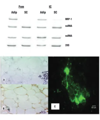
Figure 1. A: Representative examples of the electrophoretic pat-terns of RT-PCR products of A: VEGF, B: VEGFR-1, C: VEGFR-2 and D: NRP-1 mRNA measured in duplicate using total RNA isolated from the femoral (fem) and the iliac crest (IC) bone marrow of the same donor. The total RNA in the sample solution was evaluated by measuring the 28S rRNA. A synthetic RNA (ssRNA) was added in each tube reaction to monitor the yield of both the reverse tran-scription and the amplification steps (only for 28S rRNA, VEGF and NRP-1). 189 165 ssRNA 121 ssRNA 28s VEGFR-1 ssRNA 28S VEGFR-2 ssRNA 28S NRP-1 ssRNA 28S A B D C Fem IC Fem IC Fem IC Fem IC
Figure 2. A. Representative examples of electrophoretic patterns of RT-PCR products of NRP-1 mRNA using total RNA isolated from adipocytes (adip) and sedimented cells (SC) from femoral (Fem) and iliac crest (IC) bone marrow. B. NRP-1 immunoreactivity on bone marrow sections (a and b) and adipocytes smears (c). NRP-1 immunoperoxidase staining in femoral bone marrow adipocytes (b). (a) are negative controls for the specific signal using normal goat immunoglobulins. NRP-1 immunoreactivity is revealed by immunofluorescence in isolated adipocytes smears (c).
A B C NRP-1 ssRNA ssRNA 28S Adip SC Adip SC Fem IC
haematologica/the hematology journal | 2005; 90(3) | 401| Letters to the Editor
this could be related to the predominance of adipocytes in the femoral samples. Indeed, it has been shown that VEGF mRNA is upregulated during the conversion of 3T3 preadipocytes to adipocytes.7The VEGFR-1, VEGFR-2 and
NRP-1 were measured in the same series of samples. VEGFR-1 mRNA levels were quite variable from case to case. VEGFR-1 mRNA was either absent in iliac crest and femoral bone marrow or expressed at the same level in both tissues or expressed only in femoral bone marrow or was expressed at higher level in femoral bone marrow than in iliac crest bone marrow (Figure 1B). Values for VEGFR-2 were available in five samples only: there was no significant difference between femoral and iliac crest mar-row (Figure 1C).
VEGFR-2 is essential for the development of hematopoi-etic stem cells during early embryonic development, it may be redundant in adult bone marrows. Since activation of VEGFR-1 is fully sufficient to rescue hematopoietic stem cell survival in vitro and hematopoietic repopulation in vivo,8
the presence of VEGFR-2 may be related to the mainte-nance of bone marrow vasculature. NRP-1 was expressed at higher level in femoral bone marrow than in iliac crest in each donor (Figure 1D) and it seems to be inversely cor-related with the hematopoietic activity. The cellular origin of NRP-1 was assessed on isolated cell populations by a floatation/sedimentation procedure. By contrast to sedi-mented cells (hematopoietic and stromal cells), high levels of NRP-1 mRNA were detected in the adipocytic popula-tion (Figure 2A). This was confirmed by in situ hybridiza-tion (not shown) and at the protein level by immunohisto-chemistry (Figure 2B). This is the first report demonstrat-ing neuropilin-1 expression in bone marrow in vivo. The capacity of adipocytes to produce NRP-1, previously sus-pected to play an interactive role with hematopoietic cells9
suggests that adipocytes may contribute to the regulation of hematopoiesis and/or that NRP-1 may be a novel regu-lator of adipocyte activity in the bone marrow, possibly as a receptor for VEGF. Although this study does not provide a definitive link between NRP-1, adipocyte function and hematopoiesis, such a relationship may exist and deserves further studies.
Zakia Belaid,* Frederique Hubint,* Chantal Humblet,* Jacques Boniver,° Betty Nusgens,#Marie-Paule Defresne*
*Laboratoire d’Histologie-Cytologie; °Laboratoire d’Anatomie et Cytologie Pathologiques;
#Laboratoire de Biologie des Tissus Conjonctifs, Tour de Pathologie
B23, Universitè de Liège, Sart Tilman, Liège, Belgium Acknowledgments: we thank Colige CA, Lambert CA and Munaut C for expert technical assistance. We also thank Prof. P. Gillet and Dr. A. Rodriguez for providing bone marrow biopsies. We also thank Prof. Kolodkine from John Hopkins University, Baltimore USA, for providing us the anti-neuropilin-1 antibody. Funding: this work was supported by the “Belgian Federation Against Cancer”, non-profit organization and the National Foundation for Scientific Research, Belgium. Zakia Belaid and Fredèrique Hubin are Tèlèvie Fellows granted from the National Foundation for Scientific Research.
Correspondence: Zakia Belaidz, Laboratoire d’Histologie-Cytologie, Tour de Pathologie B23, Universitè de Liège, Sart Tilman, B-4000 Liège, Belgium. Phone: international +32.43662403.
Fax: international +32.43662919.
E-mail: belaid@ulg.ac.be or defresne@ulg.ac.be
References
1. Gerber HP, Ferrara N. The role of VEGF in normal and neo-plastic hematopoiesis. J Mol Med 2003;81:20-31.
2. Soker S, Takashima S, Miao HQ, Neufeld G, Klagsbrun M.
Neuropilin-1 is expressed by endothelial and tumor cells as an isoform-specific receptor for vascular endothelial growth fac-tor. Cell 1998; 92:735-45.
3. Chirgwin JM, Przybyla AE, MacDonald RJ, Rutter WJ. Isolation of biologically active ribonucleic acid from sources enriched in ribonuclease. Biochemistry 1979;18:5294-9. 4. Lambert CA, Colige CA, Munaut C, Lapière CM, Nusguens
BV. Distinct pathways in the over-expression of matrix metal-loprotreinases in human fibroblasts by relaxation of mechani-cal tension. Matrix Biology 2001;20:397-408.
5. Kerndrup G, Pallesen G, Melsen F, Mosekilde L. Histomorpho-metrical determination of bone marrow cellularity in iliac crest biopsies. Scand J Haematol 1980;24:110-4.
6. Bellamy WT, Richter L, Frutiger Y, Grogan TM. Expression of vascular endothelial growth factor and its receptors in hematopoietic malignancies. Cancer Res 1999;59:728-33. 7. Ziegler BL, Valtieri M, Porada GA, De Maria R, Muller R,
Masella B, et al. KDR receptor: a key marker defining hematopoietic stem cells. Science 1999;285:1553-8.
8. Emoto M, Anno T, Sato Y, Tanabe K, Okuya S, Tanizawa Y, et al. Troglitazone treatment increases plasma vascular endothe-lial growth factor in diabetic patients and its mRNA in 3T3-L1 adipocytes. Diabetes 2001;12:1166-70.
9. Tordjman R, Ortega N, Coulombel L, Plouet J, Romeo PH, Lemarchandel V. Neuropilin-1 is expressed on bone marrow stromal cells: a novel interaction with hematopoietic cells? Blood 1999;94:2301-9.
Red Cell Disorders
Decreased plasma endothelin-1 levels in children
with sickle cell disease treated with hydroxyurea
Plasma endothelin-1 (ET-1) is elevated in patients with sickle cell disease (SCD). Hydroxy-urea (HU) is the only drug with demonstrated clin-ical efficacy in SCD. Here we show that treatment with HU results in a decreased concentration of circulating ET-1 which is not correlated with the HU-induced increase in HbF level. Blunting of the ET-1 vasoconstrictive stimulus could contribute to the beneficial effects of HU.haematologica 2003; 90:401-403
(http://www.haematologica.org/journal/2005/03/401.html)
Sickle cell disease (SCD) is characterized by unpre-dictable painful crises resulting from vaso-occlusion by rigid, sickled red blood cells (RBC). Still, factor(s) initiat-ing vasoocclusive crises (VOC) remain largely unknown. In SCD, the vascular endothelium is chronically activated and expresses various adhesion molecules on its surface. Exacerbation of this activation, in particular within an inflammatory context, is believed to be (one of) the major triggering factor(s) of VOC by promoting the abnormal adhesion of RBC and other circulating cells to the endothelium.1The concentration of endothelin-1 is
ele-vated in the plasma of SCD patients, especially during bouts of acute chest syndrome (ACS) and other compli-cations of VOC.2Given that it is a powerful
vasoconstric-tor and pro-inflammavasoconstric-tory agonist, ET-1 might be also play an important role in VOC.
Hydroxyurea (HU) significantly reduces the incidence of VOC and ACS, as well as global morbidity and mortality.3,4
Its initial intended use was to induce fetal hemoglobin (HbF). However, the increment in HbF levels is not con-stant and it appears that effects of HU are the results of multi-targeted actions.5 We recently demonstrated that
HU down-regulates ET-1 gene expression by endothelial cells in culture both in basal conditions and after stimula-tion with pro-inflammatory cytokines.6 The aim of the