Down-Regulation of Vascular Endothelial Growth Factor by Tissue
Inhibitor of Metalloproteinase-2: Effect on in Vivo Mammary Tumor
Growth and Angiogenesis
1Amin Hajitou, Nor-Eddine Sounni, Laetitia Devy, Christine Grignet-Debrus, Jean-Marc Lewalle, Hong Li, Christophe F. Deroanne, He Lu, Alain Colige, Betty V. Nusgens, Francis Frankenne, Anne Maron, Patrice Yeh, Michel Perricaudet, Yawen Chang, Claudine Soria, Claire-Michelle Calberg-Bacq, Jean-Michel Foidart, and Agnès Noël
Laboratories of Tumor and Development Biology [A. H., N-E. S., L. D., C. G-D., J-M. L., F. F., J-M. F., A. N.], Connective Tissues Biology [C. F. D., A. C.], and Fundamental Virology [C-M. C-B.], University of Liège, Sart-Tilman 4000 Liege, Belgium; Centre National de la Recherche Scientifique-Aventis UMR 1582, Institute Gustave Roussy, Villejuif France [A. M., P. Y., M. P.]; Institut National de la Santé et de la Recherche Médicale 353, St Louis Hospital, Paris; France [H. Li, H. Lu, C. S.]; and Aventis, Hayward, California 94545 [Y. C.]
Abstract
The tissue inhibitor of metalloproteinases-2 (TIMP-2) has at least two independent functions, i.e., regulation of matrix metalloproteinases and growth promoting activity. We investigated the effects of TIMP-2
over-expression, induced by retroviral mediated gene transfer, on the in vivo development of mammary tumors in syngeneic mice inoculated with EF43.fgf-4 cells. The EF43.fgf-4 cells established by stably infecting the normal mouse mammary EF43 cells with a retroviral expression vector for the fgf-4 oncogene, are highly tumorigenic and overproduce vascular endothelial growth factor (VEGF). Despite a promotion of the in vitro growth rate of EF43.fgf-4 cells overexpressing timp-2, the in vivo tumor growth was delayed. At day 17 post-cell injection, the volume of tumor derived from TIMP-2-overexpressing cells was reduced by 80% as compared with that obtained with control cells. Overexpression of TIMP-2 was associated with a down-regulation of VEGF expression in vitro and in vivo, a reduction of vessel size, density, and blood supply in the induced tumors. In addition, TIMP-2 completely inhibited the angiogenic activity of EF43.fgf-4 cell-conditioned medium in vitro using a rat aortic ring model. Our findings suggest that overexpression of TIMP-2 delays growth and
angiogenesis of mammary carcinoma in vivo and that down-regulation of VEGF expression may play an important role in this TIMP-2-mediated antitumoral and antiangiogenic effects. Finally the in vivo delivery of TIMP-2, as assessed by i.v. injection of recombinant adenoviruses vectors, significantly reduced the growth of the EF43.fgf-4-induced tumors. This effect of TIMP-2 was shown to be equally comparable with that of angiostatin, a known potent inhibitor of angiogenesis.
Abbreviations : ECM, extracellular matrix; MMP, matrix metalloproteinase; TIMP, tissue inhibitor of
metalloproteinase; VEGF, vascular endothelial growth factor; rTIMP, recombinant TIMP; RT-PCR, reverse transcription-PCR; pfu, plaque-forming unit(s)
INTRODUCTION
Angiogenesis is a pivotal step during cancer progression because it promotes both primary tumor growth and metastasis dissemination (1). It is a multistep process characterized by (a) the activation of vascular endothelial cells by angiogenic factors; (b) migration of endothelial cells toward the source of chemotactic stimuli; (c) degradation of the ECM32 to allow endothelial cell invasion into surrounding tissue; and (d) differentiation of
endothelial cells into microvessels and stabilization (2, 3). The matrix breakdown is mediated by ECM-degrading enzymes including MMPs secreted, at least by endothelial cells, in response to angiogenic stimuli. MMPS form a large family of structurally related zinc endopeptidases (4, 5) that are collectively able to degrade
1 Supported by grants from Communauté Française de Belgique (Actions de Recherches Concertées), the Commission of European
Communities, Fonds de la Recherche Scientifique Médicale, DGTRE of the "Region Wallone" (Belgium), Fonds National de la Recherche Scientifique (FNRS, Brussels), Fédération Belge Contre le Cancer, Center Anticancéreux près l'Université de Liège, CGER-Assurances, Fondation Léon Frédéricq (University of Liège), Fonds d'Investissements de la Recherche Scientifique (CHU, Liège), General RE-Luxembourg, and Roche Diagnostics GmbH (Penzberg, Germany) L. D. is the recipient of a grant from FNRS-Télévie, A. N. is a Research Senior Associate, C. F. D. is a Postdoctoral Research Associate, and A. C. is a Research Associate, all from FNRS.
a wide variety of ECM proteins. MMP activity is tightly regulated by proenzyme activation and by the action of specific physiological inhibitors, the tissue inhibitors of metalloproteinases or TIMPs. The TIMP family comprises at least four distinct members, TIMP-1, TIMP-2, TIMP-3, and TIMP-4 (reviewed in Refs. 6,7). TIMP-2 is a Mr 21,000 nonglycosylated protein that forms a 1:1 complex with the latent and activated form of
MMP-2 (8). It is also known to interact with the activated form of different MMPs including MMP-9 (9). Recent studies have shown that MMP-deficient mice exhibit delayed or reduced angiogenic responses during embryonic development or in response to tumor xenograft (10, 11). More direct evidence of the MMP involvement in the angiogenic process is that MMP inhibitors, both synthetic and physiological, inhibit angiogenesis in vitro and in vivo (2, 12). TIMP-2 blocks neoangiogenesis in collagen or fibrin matrix (13, 14). Furthermore, a number of reports have demonstrated a TIMP-2-mediated inhibition of tumor growth and invasion in murine models, when administrated exogenously, as well as overexpressed through timp-2 cDNA transfection of different tumor cell lines (15-18). However, high tissue concentration in TIMP-2 are associated with poor prognosis in breast carcinomas (19). This paradoxical clinical observation can be partly explained by the multiple functions of TIMP-2. Indeed, TIMP-2 not only exhibits a broad specificity of inhibition against MMPs but also modulates cell properties such as proliferation and apoptosis (7, 20). Also, although TIMP-2 inhibits MMP-2 activity at high concentrations, it also promotes its activation at low concentrations and in association with MT-MMPs (21, 22).
We have previously demonstrated that the expression of the fgf-4 oncogene in normal murine mammary EF43 cells yielded a cell population that induced in vivo rapidly developing and highly vascularized tumors (23, 24). The angiogenic potential of these cells was attributed to their capacity to produce VEGF, as demonstrated in
vitro (25). In the present study, we induced the overexpression of TIMP-2 by introducing timp-2 cDNA into
these murine EF43.fgf-4 cells by retroviral mediated gene transfer. This led to a delay of in vivo tumor growth and angiogenesis. This antiangiogenic effect of TIMP-2 observed both in vitro and in vivo was associated with a down-regulation of VEGF expression by tumor cells. All together, these results emphasize the potency of TIMP-2 as an antiangiogenic agent and provide a new possible mechanism of TIMP-TIMP-2 action, through the down-regulation of VEGF expression.
MATERIALS AND METHODS
Cell Cultures and TIMP-2 Analysis
The normal mouse mammary EF43 cell line and its tumorigenic derivatives, the EF43.fgf-4 cells (23), were grown at 37°C in 5% CO2 in DMEM supplemented with 10% fetal bovine serum (Life Technologies, Inc.),
penicillin (100 units/ml), and streptomycin (100 µg/ml).
To measure their proliferation rate in vitro, cells (2 x 10⁴) were seeded in quadruplicate in 24-well plates in
complete medium for 48 h and were then cultured in serum-depleted medium for 7 days. DNA content was evaluated daily by fluorimetry (26).
To prepare conditioned medium, cells were cultured to confluence, washed twice with serum-free medium and covered, with 2 or 5 ml of fresh serum-free medium in 3.3- or 10-cm diameter dishes, respectively. Conditioned medium was collected 24 h later and clarified by centrifugation or filtration through a 0.45 µm filter. The results were corrected for cellular DNA content measured by fluorimetry in each culture dish. TIMP-2 amounts in the conditioned media were measured by ELISA as described previously (15, 19, 22).
When indicated, human rTIMP-2, purified as described previously (15, 27), was added to the culture medium every 2 days.
Retroviral Vector Construction and Production
The 2-carrying retroviral plasmid was constructed as described previously (15). It contains the human timp-2 sequence cDNA under the control of viral 5'-LTR sequence, and a selection gene (puro conferring resistance to puromycin) driven by an internal SV40 early promoter. Production of retroviruses with titers of 1 to 5 x 10⁵
cfu/ml and cell transduction were performed as described previously (23). The 4.C and
EF43.fgf-4.timp-2 cells were populations of cells resistant to puromycin (1 µg/ml) and containing the control empty vector
Analyses of VEGF Expression
Northern Blotting.
Total cellular RNA was purified as described previously (28), with slight modifications as described elsewhere (22). RNA samples (15 µg) were run on 1% agarose gels containing 10% formaldehyde and transferred onto nylon membranes (Hybond-N; Amersham, Aylesbury, United Kingdom). The probe used was the murine VEGF164 cDNA generously provided by P. D'Amore (Laboratory for Surgical Research, Children's Hospital,
Boston, MA). Hybridization was performed according to the procedure described previously (25).
RT-PCR.
Total RNA was extracted from cell layers or tumors by RNA Instapure treatment (Eurogentec, Liège, Belgium). RT-PCR was performed on 10 ng of total RNA using a Perkin-Elmer kit (Foster City, CA) and following the manufacturer's instructions. RT was carried out with 5'-CTC ACC GCC TCG GCT TGT CAC A-3' as primer during 15 min at 70°C PCR products were generated with the same primer as reverse primer and with 5'-CCT GGT GGA CAT CTT CCA GGA GTA-3' as forward primer. PCR conditions were 95°C for 2 min, followed by 29 cycles consisting of 94°C for 20 s, 66°C for 20 s, 72°C for 30 s, and a final elongation step of 72°C for 2 min. The amplification products of the RNAs coding for VEGF188, VEGF164, and VEGF, 2o, were 479, 407, and 265
bp sizes, respectively. To control the efficiency of the RT-PCR, we designed a synthetic RNA (CTR1) that could be reverse transcribed and amplified with the same primers and giving rise to a 311-bp fragment. A total of 8000 copies of this internal control were added to each sample. VEGF expression was normalized to that of the 28S. PCR conditions for 28S were 95°C for 2 min, 17 cycles consisting of 94°C for 15 s, 68°C for 20 s, 72°C for 10 s, and a final elongation step of 72°C for 2 min. A synthetic RNA (CTR2) was also used to control the efficiency of RT-PCR for 28S. The amplification products were electrophoresed on a polyacrylamide gel, stained with Gelstar (Sanver Tech, Antwerpen, Belgium), scanned with FluorSImager, and analyzed using multianalyst software (Bio-Rad, Belgium).
Ratios VEGF:CTR1 and 28S:CTR2 were determined. VEGF expression was expressed as the ratio of VEGF transcripts/28S transcripts.
Western Blot Analysis in Tumors.
Tumors were frozen in liquid nitrogen, powdered, extracted with lysis buffer (0.1 M Tris-HCl/0.4% Triton X-100), and centrifuged at 12,000 rpm. Samples containing 15 µg of protein were electrophoresed on a 15% SDS-polyacrylamide gel. Western blotting was performed as described previously (25), using a 1:500 dilution of a monoclonal antibody against human VEGF165 and mouse VEGF164 (V-4758; Sigma) and, as secondary antibody, peroxidase-conjugated goat antimouse IgG (Dako; P0447) diluted 1:1000. Peroxidase was revealed by the enhanced chemiluminescence assay (Amersham Corp).
Tumorigenicity and Antitumoral Effect of Adenoviral Vectors
The cells were trypsinized, counted, centrifuged, and resuspended in serum-free medium. EF43.fgf-4.timp-2 and EF43.fgf-4.C control cells (5 x 10⁴, 105, 5 x 10⁵, and 106) were injected s.c. into syngeneic 4-week-old female
BALB/c mice. Tumor growth was monitored twice a week by caliper measurement of 2 diameters, and expressed as mean tumoral volume ± SD. In each assay, the number of mice per group (n) was 10.
To evaluate the antitumoral effect of a TIMP-2 treatment on the EF43.fgf-4 tumor growth, we used adenoviral vectors (Ad.timp-2) that carry a cytomeg-alovirus-driven cDNA for human TIMP-2. To obtain this adenovirus, the TIMP-2 cDNA was inserted in pC05 shuttle vector at the downstream of cytomegalovirus early gene promoter (pCMV). The SV40 late gene poly-adenylation signal sequence was inserted after the stop codon of cDNA. The recombinant adenovirus was obtained in 293 cells by homologous recombination between shuttle vector containing TIMP-2 expression cassette and E1/E3-deficient human type 5 AdRSV.βgal DNA (29). The viruses (5 x 10⁹ pfu) were injected into the tail vein of mice (n = 7) bearing a 20-mm3 preestablished tumor.
Control mice with the same tumor size (n = 7) were similarly injected with AdCO1 vectors that carried the empty expression cassette only. Vectors carrying the β-galactosidase gene, Ad.β-gal (30) were also injected into mice to confirm the adenoviral vector expression. At the end of the experiment, the livers from all of the animals were positive for β-galactosidase staining. Adenoviral vectors, AdK3 (30) carrying a cDNA for the NH2-terminal
fragment from human plasminogen, including the first three kringle domains (angiostatin K3), (5 x 10⁹ pfu) were
Evaluation of Tumor Vascularization
The extent of tumoral vascularization was assessed by immunostaining using a rabbit polyclonal serum raised against laminin (diluted 1:100; Ref 31) or a rat monoclonal antibody raised against PECAM (PharMinogen, SanDiego, CA; diluted 1:20). Frozen sections (6-µm) of tumor tissues were fixed in paraformaldehyde 4% for 5 min and then were incubated with the primary antibodies for 60 min at room temperature. After three washes with PBS, the sections were incubated with fluorescein-conjugated swine antirabbit (Dako; dilution 1:50) for 30 min. Preparations were mounted with Eukitt (Labonord, Villeneuve, France) and viewed with an Olympus microscope.
Vessel density was evaluated under light microscopy on sections of whole tumors. Ten sections were made in each tumor. Image analysis and quantification of stained vessel sections were performed on a computer using the Microimage 3.0.1.0. software from Olympus (Bio-Rad, Belgium). Results were expressed as mean (± SD) vessel number per mm2.
In Vitro Angiogenesis: the Aorta Ring Assay
Rat aortic expiant cultures were prepared as described previously (32). For the preparation of culture wells, 30 ml of 1.5% agarose solution (type VII, cell culture-tested; Sigma, St. Quentin Fallavier, France) were poured into 100-mm-diameter Petri dishes (Corning Costar Corporation, Cambridge, MA) and allowed to gel. Agarose rings were obtained by punching two concentric circles in the agarose with punchers of 10- and 17-mm diameter and were transferred to additional 100-mm diameter Petri dishes (bacteriological polystyrene; Falcon; Becton Dickinson, Lincoln Park, NJ). The bottom of each agarose well was coated with 200 µl of rat tail tendon (type I) collagen (1.5 mg/ml; Collagen R; Serva, Heidelberg, Germany) that was allowed to gel at 37°C One aortic ring obtained by sectioning the rat aorta at 1-mm intervals (32), was carefully positioned in each well, which was then completely filled with collagen solution. Aorta rings were maintained at 37°C in MCDB131 (Life Technologies LTD., Paisley, Scotland) supplemented with 25 mM NaHCO3, 100 units/ml penicillin, and 100 µg/ml
streptomycin. Cultures were examined every 2 days by phase-contrast microscopy. When indicated, human recombinant VEGF165 (20 ng/ml; Sigma) was added to the medium. The identity of endothelial cells was verified
by Dil-Ac-LDL incorporation in the aorta ring before embedding into collagen gel (33).
RESULTS
In Vitro Development of Cells Overproducing TIMP-2.
The highly tumorigenic EF43.fgf-4 cells were infected with retroviral vectors carrying the timp-2 cDNA or the control vectors carrying only the puro selection gene. Stable transformants derived from timp-2 cDNA transduction (EF 43.fgf-4.timp-2 cells), and cell populations derived from control retroviral vector transduction (EF43.fgf-4.C cells) were obtained by puromycin selection.
The serum-free media conditioned by these different cell populations were analyzed for the presence of TIMP-2 by ELISA. Endogenous TIMP-2 production was weak in control EF43.fgf-4.C cells (0.13 ± 0.02 ng/µg DNA), whereas it was increased in EF43.fgf-4.timp-2 cells (0.98 ± 0.09 ng/µg DNA). The FGF-4 production was not modulated by the increased TIMP-2 secretion as determined by Western blot (data not shown).
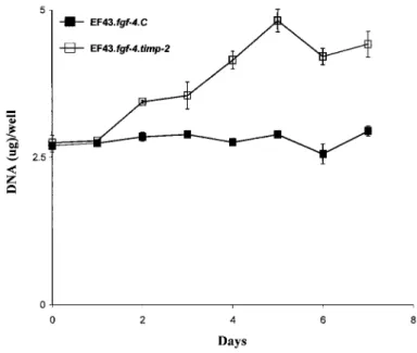
Because it was reported that TIMP-2 can stimulate the growth of some cell types (7, 34), we compared the in
vitro growth rate of EF43.fgf-4.timp-2 cells and EF43.fgf-4.C cells in serum-free medium. EF43.fgf-4.C cells did
not multiply in these serum-free conditions, whereas overexpression of timp-2 significantly promoted the proliferation of EF43.fgf-4 cells (Fig. 1).
In Vivo Development of Tumoral Cells Overproducing TIMP-2.
We previously reported that the EF43.fgf-4 cells were highly tumorigenic when injected s.c. into syngeneic BALB/c mice, leading to tumor onset within 1 week (23, 24). Here, we show that injection of both EF43.fgf-4.C and EF43.fgf-4.timp-2 cells led to tumor formation, but the growth of tumors induced by EF43.fgf-4.timp-2 cells was strikingly delayed (Fig. 2). At day 17 postimplantation, the volume of these tumors was reduced by 80% as compared with the ones induced in EF43.fgf-4.C control tumors. Fig. 2 illustrates an experiment based on the injection of 5 x 10⁵ cells. Similar delays in tumor development were observed after inoculation of 5 x 10⁴, 105, or
Fig. 1: In vitro cell growth rate. The EF43fgf-4.C and EF43.fgf-4.timp-2 cells (2 X 10⁴) were cultured in
serum-free medium as described in "Materials and Methods”. Cell proliferation was expressed as mean ± SD DNA amount/well of quadruplicate cultures
Fig. 2: Tumorigenicity. Tumors were induced by s.c. injection of 5 X 10⁵ EF43.fgf-4.C or EF43.fgf-4. timp-2
cells in BALB/c syngeneic mice. Symbols, mean tumor volumes (cm3) ± SD.
When observed macroscopically directly after resection, the EF43.fgf-4.C tumors appeared more vascularized than their EF43.fgf-4.timp-2 counterparts. Large-size blood vessels were closely associated with the edge of tumor derived from EF43.fgf-4.C cells. In contrast, EF43.fgf-4.timp-2 tumors displayed only a weakly developed peripheral neovascularization. In addition, during tumor growth, hemorrhagic areas appeared regularly inside the 4.C tumors. Such vascular ectasias and blood extravasation were always absent from the
EF43.fgf-4.timp-2 tumors, at any stage of development (Fig. 3).
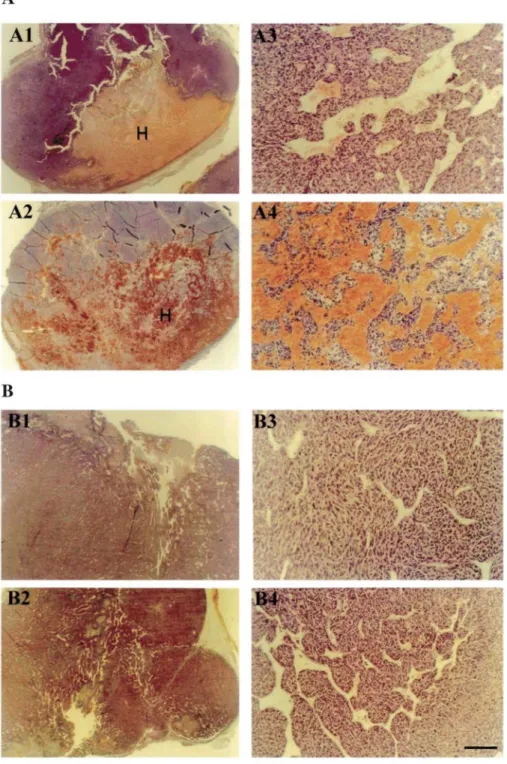
Tumors grown from EF43.fgf-4.C cells appeared as dense adenocarcinoma (Fig. 3, A1 and A2) with a clearer zone formed by large vascular cavities. At higher magnification, a well-organized network of anastomosing vessels was seen in these tumors (Fig. 3. A3). At a more advanced stage, we observed an angiomatoid pattern characterized by cellular necrosis and the presence of large vessels with a dilated lumen, as well as large vascular cystic cavities filled with blood (Fig. 3. A4). The large size vessels were always limited by a subendothelial basement membrane containing laminin (Fig. 4C). On the other hand, tumors derived from EF43.fgf-4.timp-2 cells essentially appeared at all stages as dense adenocarcinoma (Fig. 3, B1 and B2), but well- defined vessels were observed with a narrow lumen or even collapsed, probably caused by the pressure of the dense tumoral mass (Fig. 3, B3 and B4). These tumors were devoid of vascular cavities, and the amount of blood was greatly
reduced as compared with control EF43.fgf-4.C tumors.
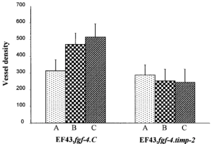
Vascular Density in Tumors Overproducing TIMP-2.
Tumor vascularization was thus measured on tumor sections immunostained with an antilaminin antibody (Fig. 4). Tumors were compared when they had reached the same volume of 0.4 cm3, 3.2 cm3, and 6.25 cm3. Such
volumes were reached 10, 20, and 24 days after implantation of the EF43.fgf-4.C, respectively, and 17, 40, and 48 days after implantation of EF43.fgf-4.timp-2 cells, respectively (Fig. 2). Tumors formed by EF43fgf-4.C cells harbored numerous long and anastomosing vessels (Fig. 4A). Shorter vessels were observed in tumors derived from timp-2-overexpressing cells (Fig. 4B). Such reduction of tumor angiogenesis in EF43.fgf-4.timp-2 tumors was confirmed by quantitative analysis. Indeed, a significant reduction of vessel density within the
EF43.fgf-4.timp-2 tumors was observed as compared with the EF43.fgf-4.C control tumors (Fig. 5). Although the vessel
density increased progressively during the growth of EF43.fgf-4.C tumors, it remained constant during the evolution of EF43.fgf-4.timp-2 tumors (Fig. 5).
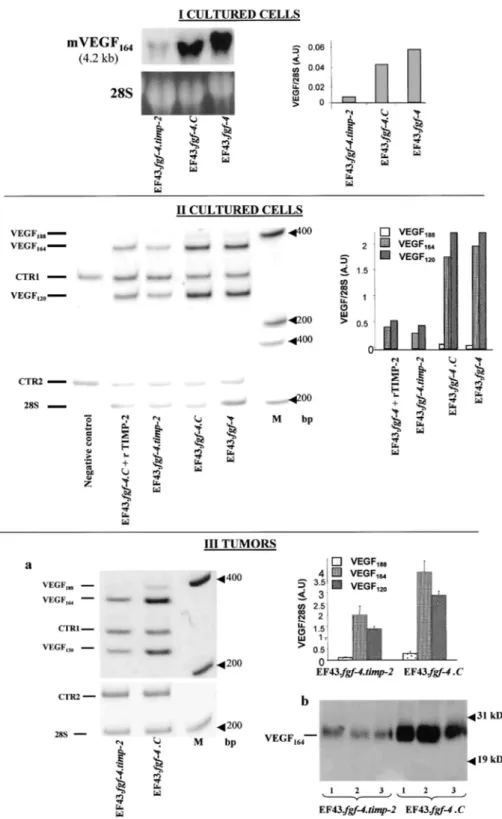
Effect of TIMP-2 on VEGF Expression.
We previously reported that EF43.fgf-4 cells induce in vitro angiogenesis through VEGF overexpression (25). We, therefore, evaluated the VEGF expression in cells that overexpressed TIMP-2 or not. Interestingly, Northern blot analysis of total RNA showed a 7.3-fold decrease in VEGF mRNA level in EF43.fgf-4.timp-2 cells as compared with EF43.fgf-4.C cells (Fig. 6,I). By RT-PCR, overexpression of TIMP-2 in EF43.fgf-4 cells was shown to be associated with more than 5-fold down-regulation of VEGF188, VEGF164, and VEGF120 mRNA
isoforms (Fig. 6, II). Moreover, the addition of exogenous rTIMP-2 (100 ng/ml) to EF43.fgf-4.C cell cultures, resulted in a similar down-regulation of VEGF mRNA isoforms expression (Fig. 6, II). In accordance with these
in vitro data, a 2- to 3-fold reduction of VEGF mRNA isoforms expression was detected by RT-PCR in
EF43.fgf-4.timp-2 tumors, as compared with tumors obtained from EF43.fgf-4.C cells (Fig. 6, III, a). This modulation of VEGF expression was confirmed by measuring the VEGF protein by Western blot analysis (Fig. 6, III, b).
Effect of TIMP-2 on in Vitro Angiogenesis.
Media conditioned by EF43.fgf-4.C or EF43.fgf-4.timp-2 cells were assayed for their ability to modulate in vitro angiogenesis. Rings of rat aorta were embedded in collagen gels and maintained in culture, in the presence of medium conditioned by EF43.fgf-4.C or EF43.fgf-4.timp-2 cells. Microvessels outgrew from rat aorta cultured in the presence of EF43.fgf-4.C cell-conditioned medium. In such conditions, the generation of micro vessels was similar to that observed in the presence of 20 ng/ml human recombinant VEGF165 (Fig. 7). Interestingly, no
angiogenic response was observed with medium conditioned by EF43.fgf-4.timp-2 cells. A similar effect was obtained by adding exogenous rTIMP-2 (100 ng/ml) to the conditioned medium of EF43.fgf-4 cells.
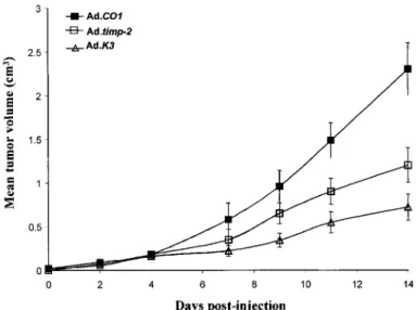
In Vivo Antitumoral Effect of Adenovirus-mediated Gene Transfer of TIMP-2 or Angiostatin.
To assess potential therapeutic action of in vivo TIMP-2 delivery, we used a defective adenovirus (Ad.timp-2) expressing human TIMP-2 under the control of the cytomegalovirus promoter. A single i.v. administration of
Ad.timp-2 (5 X 10⁹ pfu) to mice bearing a 20-mm3-preestablished EF43.fgf-4 tumor resulted in a significant
impairment of tumor growth (Fig. 8). Fourteen days after virus injection, the tumoral volume was reduced by 50%. In contrast, a similar injection of control adenoviruses (AdCO1) did not affect tumor growth.
The antitumoral effect of TIMP-2 on EF43.fgf-4 tumor growth was compared with the effect of a known antiangiogenic factor, angiostatin, identified as the Mr 38,000 NH2-terminal fragment of plasminogen. A single i.v. injection of AdK3 (5 X 10⁹ pfu) viruses also exerted an antitumoral effect that was slightly more pronounced
Fig. 3: H&E staining of EF43.fgf-4.C and EF43.fgf-4.timp-2 tumors. Representative tumors obtained after
injection of 5 x 10⁵ EF43.fgf-4.C cells (A) and EF43.fgf-4.timp-2 cells (B). Sections were performed on tumors
with a defined volume of 3.2 cm3 and 6.2 cm3, which correspond to 20 (A1, A3) and 24 days (A2, A4) after
implantation of the 4 C cells, and to 40 (B1, B3), and 48 days (B2, B4) after implantation of EF43.fgf-4.timp-2 cells, respectively. Each tumor type is presented at low (A1,A2, Bl, B2) and higher (A3, A4, B3, B4) magnifications. Bar, 800 µm (A1, A2, B1, B2) and 80 µm (A3, A4, B3, B4). H, hemorrhagic area
Fig. 4: Immunostaining of vessels in tumors. Frozen sections of EF43.fgf-4. C (A) or EF43.fgf-4.timp-2 (B)
tumors with a volume of 3 cm3 were stained with antilaminin antibody. C, a section from EF43.fgf-4.C tumor,
showing a large vascular cavity limited by a subendothelial basement membrane. Laminin labeling was always codistributed with endothelial cells recognized by the antimouse PECAM antibody (data not shown). White arrows, vessels. Bars, 20 µm
DISCUSSION
Numerous preclinical studies have demonstrated that synthetic MMP inhibitors inhibit the growth of a variety of tumors in murine xenograft models (reviewed in Refs. 35, 36). On the basis of its inhibitory function against MMPs, it is anticipated that TIMP-2 blocks tumor growth and dissemination. This concept is supported by several reports on experimental models showing a TIMP-2-mediated reduction of tumor growth, invasion, and metastasis (15, 16, 37). Although TIMP-2 overexpression in EF43.fgf-4 cells sustained their proliferation in
vitro, their in vivo growth was considerably reduced and led to 80% tumor growth reduction. TIMP-2
overexpression in tumor cells resulted in: (a) a reduced blood supply as visualized at both microscopic and macroscopic levels; (b) the absence of large dilated vessels and vascular cavities filled with blood on tumor sections; (c) a decrease in vessel density evaluated by immunostaining; and (d) the absence of vascular ectasias. These data demonstrate in vivo that TIMP-2 is a potent antiangiogenic agent. The angiogenesis inhibition by TIMP-2 was confirmed in vitro, in a rat aorta ring model, by using either medium conditioned by TIMP-2-overexpressing cells or rTIMP-2. This in vitro model (32, 38) using intact vascular expiants reproduces more
accurately the environment in which angiogenesis takes place than cultures of isolated endothelial cells. The inhibition of angiogenesis by TIMP-2 is consistent with the recent implication of MT1-MMP (14), MMP-9 (10), and MMP-2 (11) in neoangiogenesis.
TIMP-2 may inhibit tumor growth and angiogenesis by several mechanisms. We provide evidence for a new mechanism by which TIMP-2 exerts its antitumoral effect: the down-regulation of VEGF expression. This growth factor originally discovered as a permeability factor (VPF; Ref. 39) exhibits both potent endothelial mitogenic and vascular permeability-inducing activity, as well as vessel dilation (3). We previously demonstrated that EF43.fgf-4 cells induced, through the secretion of VEGF, an angiogenic phenotype of
endothelial cells in an in vitro assay (25). The regulation of VEGF expression by TIMP-2 was here demonstrated
in vitro and in vivo, by Northern blot, RT-PCR, and Western blot analysis. Both endogenous and rTIMP-2
reduced VEGF expression of EF43.fgf-4 cells. The three different mRNA isoforms of VEGF that were expressed at different levels by EF43.fgf-4 cells were all affected by 2 expression. The mechanisms by which TIMP-2 modulates VEGF expression remain to be elucidated.
All together, these data suggest that TIMP-2 exerts its antiangiogenic effect through different mechanisms including MMP inhibition and down-regulation of VEGF expression. In the aortic ring assay, the antiangiogenic role of TIMP-2 is probably mainly related to its antiproteolytic function independently of its capacity to regulate VEGF expression. Indeed, inhibition of microvessel outgrowth was observed when exogenous rTIMP-2 was added to the EF43.fgf.4 cell supernatant, which contains VEGF. Furthermore, the addition of exogenous rVEGF to the medium conditioned by EF43.fgf-4.timp-2 cells did not bypass the inhibition of angiogenesis (data not shown). As a protease inhibitor, TIMP-2 may control the degradation of the ECM and, thereby, may prevent endothelial cell migration and/or, control the activation of growth factors or limit their release from the matrix (40). In this context, TIMP-2 could control the MMP-9 effect on the release of VEGF sequestrated in the ECM (41). Alternatively, TIMP-2 may operate by a different mechanism, independently of its antiproteolytic activity. For instance, TIMP-2 may affect directly endothelial cell functions by inhibiting their proliferation or migration (42).
Finally, the antitumoral effect of TIMP-2 was compared with that of angiostatin, which is known for its efficiency on suppressing primary tumor growth (30). Delivery of TIMP-2 or angiostatin was achieved by systemic administration of recombinant defective adenoviruses. In both cases, this resulted in a reduced tumor growth demonstrating that TIMP-2 and angiostatin can have anticancer actions.
Altogether, our data emphasize the importance of TIMP-2 as an anticancer agent. We demonstrate that TIMP-2 reduces tumor growth and angiogenesis and provide a new mechanism for this antitumoral action by reducing dramatically VEGF expression. These data have direct implication in anticancer therapies. They potentially extend the use of TIMP-2 in cancer beyond the control of invasion and metastasis.
Fig. 5: Vessel density. Tumor vascularization was quantified in EF43.fgf-4.C and EF43.fgf-4.timp-2 tumors with
defined volumes of 0.4 cm3 (A), 3.2 cm3 (B), and 6.2 cm3 (C). Frozen sections were immunostained with an
antilaminin antibody, and tumor vascularization was estimated by image analysis. Results are expressed as mean vessel density ± SD (number/mm2) as described in "Materials and Methods."
Fig. 6: Analysis of VEGF expression. I, Northern blot analysis of in vitro cultured cells. Samples, fractionated
by gel electrophoresis, were transferred to nylon membrane, verified for gel loading (28S), and hybridized with a probe for mouse VEGF (4.2 kb). The histogram corresponds to the quantification of VEGF mRNA by
densitometry. A. U, arbitrary unit. II, quantitative RT-PCR analysis of in vitro cultured cells. EF43.fgf-4.C control cells were cultured in the absence or in the presence of rTIMP-2 (EF43.fgf-4.C cells + rTlMP-2). Negative control, no template; M, molecular marker (Life Technologies, Inc.). 28S was used as a control for RNA expression CTR1 and CTR2 correspond to synthetic internal control RNA for VEGF and 28S, respectively. The histogram corresponds to the densitometric quantification of VEGF mRNA isoforms. III,a, quantitative RT-PCR analysis on extracts from tumors with a volume of 3 cm3. The histogram corresponds to the densitometric
quantification of VEGF mRNA isoforms. III,b, Western blot analysis of tumor extracts. Three different representative tumors (1, 2, 3) derived from EF43.fgf-4. timp-2 or EF43.fgf-4. C cells were used.
Fig. 7: Photomicrographs of rat aorta in collagen gel cultures. Aortic expiants were cultured in the presence of
media conditioned by EF43.fgf-4.C (A) or EF43.fgf-4.timp-2 cells (B). C, the conditioned medium of EF43.fgf-4 cells was supplemented with rTIMP-2 (100 ng/ml). As positive control, human rVEGF165 (20ng/ml) was added to
the MCDB131 control medium in D. Arrows, microvessels. Bar, 250 µm.
Fig. 8: In vivo efficiency of adenovirus-mediated gene delivery. Ad.timp-2 or Adk3 (angiostatin) in the amount of
5 x 10⁹ pfu was injected i.v. into mice with tumors (20 mm3) derived from EF43.fgf-4.C cells. The control AdCO1
vectors were similarly injected into control mice. Each experimental animal group contained seven mice, and the tumor growth was expressed by the tumor mean volume (cm3) ± SD
REFERENCES
1. Folkman, J. Angiogenesis in cancer, vascular, rheumatoid, and other disease. Nat Med., 1: 27-31, 1995.
2. Stetler-Stevenson, W. G. Matrix metalloproteinases in angiogenesis: a moving target for therapeutic intervention. J. Clin. Investig., 103: 1237-1241, 1999
3. Carmeliet, P. Mechanisms of angiogenesis and arteriogenesis. Nat. Med., 6: 389-395, 2000.
4. Noël, A., Gilles, C., Bajou, K., Devy, L., Kebers, F., Lewalle, J-M., Maquoi, E., Munaut, C., Remacle, A., and Foidart J-M. Emerging roles for proteinases in cancer Invasion Metastasis, 17: 221-239, 1997.
5. Nagase. H. Matrix metalloproteinases. J. Biol. Chem., 274: 491-494, 1999
6. Blavier, L., Henriet, P., Imren, S., and Declerck, Y. A. Tissue inhibitors of matrix metalloproteinases. Ann. NY Acad. Sci., 878: 108-119, 1999
7. Gomez, D. E., Alonso, D. F., Yoshiji, H., and Thorgeirsson, U. P. Tissue inhibitors of metalloproteinases: structure, regulation, and biological functions. Eur. J. Cell Biol., 74: 111-122, 1997.
8. Goldberg, G. I, Marmer, B. L., Grant, G. A., Eisen, A. Z., Wilhelm, S., and He, C. S Human 72-kilodalton type IV collagenase forms a complex with a tissue inhibitor of metalloproteases designated TΓMP-2. Proc. Natl. Acad. Sci. USA, 86: 8207-8211, 1989.
9. Nagase, H., Meng, Q., Malinovskii, V., Huang, W., Chung, L., Bode, W., Maskos, K., and Brew, K. Engineering of selective TIMPs. Ann. NY Acad. Sci., 878: 1-11, 1999.
10. Vu, T. H., Shipley, J. M., Bergers, J., Berger, J. E., Helms, J. A., Hanahan, D., Shapiro, S. D., Senior, R. M., and Werb, Z. MMP-9/gelatinase B is a key regulator of growth plate angiogenesis and apoptosis of hypertrophic chondrocytes. Cell, 93: 411-422, 1998. 11. Itoh, T., Tanioka, M., Yoshida, H., Yoshioka, T., Nishimoto, H., and Itohara, S Reduced angiogenesis and tumor progression in gelatinase A-deficient mice. Cancer Res., 58: 1048-1051, 1998.
12. Zhu, W. H., Guo, X., Villaschi, S., and Nicosia, R. F. Regulation of vascular growth and regression by matrix metalloproteinases in the rat aorta model of angiogenesis Lab. Investig., 80: 545-555, 2000.
13. Schnaper, H. W., Grant, D. S., Stetler-Stevenson, W. G., Fridman, R., D'Orazi, G., Murphy, A., N, Hoythya, M., Fuerst, T. R., French, D. L. et al. Type IV colla-genase(s) and TIMPs modulate endothelial cell morphogenesis in vitro. J. Cell Physiol., 156: 235-246, 1993. 14. Hiraoka, N, Allen, E., Apel, I. J., Gyetko, M. R., and Weiss, S. J. Matrix metalloproteinases regulate neovascularization by acting as pericellular fibrinolysins. Cell, 95: 365-377, 1998.
15. Noël, A., Hajitou, A., L'hoir, C, Maquoi, E., Baramova, E., Lewalle, J-M., Remacle, A., Kebers, F., Brown, P., Calberg-Bacq, C-M., and Foidart, J-M. Inhibition of stromal matrix metalloproteases: effects on breast-tumor promotion by fibroblasts Int. J. Cancer, 76: 267-273, 1998.
16. Noël, A., Boulay, A., Kebers, F., Kannan, R., Hajitou, A., Calberg-Bacq, C-M., Basset, P., Rio, M. C, and Foidart, J-M. Demonstration in vivo that stromelysin-3 functions through its proteolytic activity. Oncogene, 19: 1605-1612, 2000
17. DeClerck, Y. A., Perez, N, Shimada, H., Boone, T. C, Langley, K. E., and Taylor, S. M. Inhibition of invasion and metastasis in cells transfected with an inhibitor of metalloproteinases. Cancer Res., 52: 701-708, 1992
18. Montgomery, A. M. P., Mueller, B. M., Reisfeld, R. A., Taylor, S. M., and DeClerck, Y. A. Effect of tissue inhibitor of the matrix metalloproteinases-2 expression on the growth and spontaneous metastasis of a human melanoma cell line. Cancer Res., 54: 5467-5473, 1994.
19. Remacle, A., McCarthy, K., Noel, A., Maguire, T., McDermott, E., O'Higgins, N, Foidart, J. M., and Duffy, M. J. High levels of TIMP-2 correlate with adverse prognosis in breast cancer. Int. J. Cancer, 89: 118-121, 2000.
20. Guedez, L., Lim, M. S., and Stetler-Stevenson, W. G. The role of metalloproteinases and their inhibitors in hematological disorders. Crit. Rev. Oncog., 7: 205-225, 1996.
21. Nagase, H. Activation mechanisms of matrix metalloproteinases. Biol. Chem., 378: 151-160, 1997.
22. Maquoi, E., Frankenne, F., Baramova, E., Munaut, C, Sounni, N. E., Remacle, A., Noël, A., Murphy, G., and Foidart, J-M. Membrane type 1 matrix metalloproteinase-associated degradation of tissue inhibitor of metalloproteinase 2 in human tumor cell lines. J. Biol. Chem., 275: 11368-11378, 2000.
23. Hajitou, A., and Calberg-Bacq, C-M. Fibroblast growth factor 3 is tumorigenic for mouse mammary cells orthotopic ally implanted in nude mice. Int. J. Cancer, 63: 702-709, 1995.
24. Hajitou, A., Baramova, E. N, Bajou, K., Noë, V., Bruyneek, E., Mareel, M., Collette, J., Foidart, J-M., and Calberg-Bacq, C-M. FGF-3 and FGF-4 elicit distinct oncogenic properties in mouse mammary myoepithelial cells. Oncogene, 17: 2059-2071, 1998.
25. Deroanne, C. F., Hajitou, A., Calberg-Bacq, C-M., Nusgens, B. V., and Lapiere, C. M. Angiogenesis by fibroblast growth factor 4 is mediated through an autocrine up-regulation of vascular endothelial growth factor expression. Cancer Res., 57: 5590-5597, 1997. 26. Labarca, C, and Paigen, K. A simple, rapid, and sensitive DNA assay procedure. Anal. Biochem., 102: 344-352, 1980.
27. Declerck, Y. A., Yean, T. D., Chan, D., Shimada, H., and Langley, K. E. Inhibition of tumor invasion of smooth muscle cell layers by recombinant human metalloproteinase inhibitor. Cancer Res., 51: 2151-2157, 1991.
28. Chirgwin, J. M., Przybyla, A. E., Mac Donald, R. J., and Rutter, W. J. Isolation of biologically active ribonucleic acid from sources enriched in ribonuclease. Biochemistry, 18: 5294-5299, 1979.
29. Stratford-Perricaudet, L. D., Makeh, I, Perricaudet, M., and Brian, P. Widespread long-term gene transfer to mouse skeletal muscles and heart. J. Clin. Investig., 90: 626-630, 1992.
30. Griscelli, F., Hong, L., Griscelli-Bennaceur, A., Soria, J., Opolon, P., Soria, C, Perricaudet, M., Yeh, P., and Lu, H. Angiostatin gene transfer: inhibition of tumor growth in vivo by blockage of endothelial cell proliferation associated with a mitosis arrest. Proc. Natl. Acad. Sci. USA, 95: 6367-6372, 1998.
31. Foidart, J-M., Bere, E. W., Yaar, M., Rennard, S. I, Gullino, M., Martin, G. R., and Katz, S. I. Distribution and immunoelectron microscopic localization of laminin, a noncollagenous basement membrane glycoprotein. Lab. Investig., 42: 336-342, 1980.
32. Nicosia, R. F., and Ottinetti, A. Growth of microvessels in serum-free matrix culture of rat aorta. A quantitative assay of angiogenesis in vitro. Lab. Investig., 63: 115-122, 1990.
33. Voyta, J. C, Via, D. P., Butterflied, C. E., and Zetter, B. R. Identification and isolation of endothelial cells based on their increased uptake of acetylated-low density lipoprotein. J. Cell Biol., 99: 2034-2040, 1984.
34. Corcoran, M. L., and Stetler-Stevenson, W. G. Tissue inhibitor of metalloproteinase-2 stimulates fibroblast proliferation via a c AMP-dependent mechanism. J. Biol. Chem., 270: 13453-13459, 1995.
35. Brown, P. D., and Giavazzi, R. Matrix metalloproteinase inhibition: a review of antitumor activity. Ann. Oncol., 6: 967-974, 1995. 36. Talbot, D. C, and Brown, P. D. Experimental and clinical studies on the use of matrix metalloproteinase inhibitors for the treatment of cancer. Eur. J. Cancer, 32A: 2528-2533, 1996.
37. Henriet, P., Blavier, L., and Declerck, Y. A. Tissue inhibitors of metalloproteinases (TΓMP) m invasion and proliferation. APMIS, 107: 111-119, 1999.
38. Nicosia, R. F., Nicosia, S. V., and Smith, M. Vascular endothelial growth factor, platelet-derived growth factor, and insulin-like growth factor-1 promote rat aortic angiogenesis in vitro. Am. J. Pathol., 145: 1023-1029, 1994.
39. Dvorak, H. F., Orenstem, N. S., Carvalho, A. C, Churchill, W. H., Dvorak, A. M., Gaslli, S. J., Feder, J., Bitzer, A. M., Rypysc, J., and Giovinco, P. Induction of a fibrin-gel investment: an early event in line 10 hepatocarcinoma growth mediated by tumor-secreted products. J. Immunol., 122: 166-174, 1979.
40. Rifkin, D. B., Mazzien, R., Munger, J. S., Noguera, I, and Sung, J. Proteolytic control of growth factor availability. APMIS, 107: 80-85, 1999.
41. Bergers, G., Brekken, R., McMahon, G., Vu, T. H., Itoh, T., Tamaki, K., Tanzawa, K., Thorpe, P., Itohara, S., Werb, Z., and Hanahan, D. Matrix metalloproteinase-9 triggers the angiogenic switch during carcinogenesis. Nat. Cell Biol., 2: 737-744, 2000.
42. Murphy, A. N, Unsworth, E. J., and Stetler-Stevenson, W. G. Tissue inhibitor of metalloproteinases-2 inhibits bFGF-induced human microvascular endothelial cell proliferation. J. Cell. Physiol., 157: 351-358, 1993.