The precursor of the Streptomyces R61 DD-peptidase containing a
C-terminal extension is inactive
Laurence Fanuela, Benoît Graniera, Jean-Marc Wilkina, Catherine Bellefroid-Bourguignona, Bernard Jorisa, Jeremy Knowlesb, Elisabeth Komivesb, Jozef Van Beeumenc, Jean-Marie Ghuysena, Jean-Marie Frèrea
aLaboratoire d'Enzymologie et Centre d'Ingénierie des Protéines, Université de Liège, Institut de Chimie, B6, B-4000 Sart-Tilman, Belgium b
Department of Chemistry, Harvard University, 12 Oxford Street, Cambridge, MA 02138, USA
cVakgroep Biochemie, Fysiologie en Microbiologie, Rijksuniversiteit-Gent, K.L. Ledeganckstraat 35, 9000 Gent, Belgium
Abstract
The Streptomyces R61 DD-peptidase gene encodes a 26-residue C-terminal extension which is not found in the mature protein. When the gene was expressed in Escherichia coli, the extension was not cleaved and the
precursor protein was not enzymatically active. It also reacted with penicillins significantly more slowly than the mature protein.The introduction of a 'stop' codon after that corresponding to the C-terminal residue of the mature protein resulted in the production of an active protein in the periplasm of E. coli.
Keywords : DD-peptidase ; Post-translational processing ; Protein maturation ; Heterologous expression ;
Streptomyces
Abbreviations : LMM, low molecular mass ; HMM, high molecular mass ; Ac2-L-Lys-D-Ala-D-Ala, Nα,Nε
-diacetyl-L-lysyl-D-alanyl-D-alanine.
Enzymes : DD-peptidase from Streptomyces R61 = EC 3.4.16.4. Lysozyme = EC 3.2.1.17.
1. Introduction
The Streptomyces R61 DD-peptidase, a soluble, secreted protein, has been widely used as a model penicillin-sensitive enzyme [1]. The corresponding gene has been cloned and expressed in Streptomyces lividans TK24 [2,3]. These studies have shown that the gene encoded a C-terminal extension which was not found in the mature, active protein.
The possible influence of this C-terminal peptide on the activity of the enzyme has now been studied. To solve difficulties due to the time-consuming transformation procedures and to the relatively long generation time of
Streptomyces [1], the gene was first successfully expressed in Escherichia coli. 2. Materials and methods
2.1. Enzymes, chemicals and antisera
The enzymes, the recombinant DNA techniques, the antisera and the antibiotics were as described by Bourguignon-Bellefroid et al. [4], The various β-lactams were kindly given by the respective companies. Oligonucleotides were obtained from Eurogentec, Liège, Belgium and purified as before [4], [35S-thio]dATP (1000 Ci/mmol) and [l4C]benzylpenicillin (50 µCi/µmol) were from NEN (Boston, MA, USA) and Amersham International, UK, respectively.
2.2. Plasmids
For expression of the S. R61 DD-peptidase in the periplasm of E. coli, the nucleotide sequence coding for the protein devoid of its own signal peptide and obtained from plasmid pDML114 [2] was positioned behind the
TEM β-lactamase promoter and signal peptide sequences derived from plasmid pTG2 [5], thus yielding plasmid pBK8 (Fig. 1A). The details on the strategy and the restriction maps of the intermediate constructs are available from the authors on request. Fig. 1 shows the junction between the TEM signal peptide and the N-terminus of the mature DD-peptidase. Three d-nucleotides encoding a His residue were added during the construction process. The shortened gene coding for the DD-peptidase devoid of its C-terminal extension was obtained by site-directed mutagenesis in phage M13 [6]. The SphI-PstI fragment of pBK8 was sub-cloned and the GCG codon of Ala-350 was replaced by TGA, with the help of the synthetic complementary nucleotide
5'CGCAGCTTCTCAGGTCGTCGGCT-TGCCGC3' (where the modified bases are underlined) and of an oligonucleotide-directed in vitro mutagenesis kit (Amersham Int.). The complete sequence of the SphI-PstI fragment was verified and the ApaI-PstI fragment was recovered from the phage and recloned into the corresponding sites of pBK8 yielding ρDML41 (Fig. 1B). The presence of the mutation in plasmid pDML41 could easily be demonstrated by the appearance of an additional DdeI restriction site.
Note that the previously published sequence [2] of the R61 DD-peptidase gene (sddpepg) has recently been modified (EMBL data bank; PRO access number X05109) and that an additional error has been found during this work upon resequencing the DNA encoding the C-terminal extension. The corrected sequence has been submitted to Eur. J. Biochem.
Fig. 1. Restriction maps of plasmids pBK8 (A) containing the R61 peptidase gene with its intact C-terminal
portion and pDML41 (B) where a 'stop' codon has been inserted at the beginning of this extension. The insert shows the sequence of the junction between the TEM signal peptide and the mature S. R61 peptidase N-terminal sequence in detail. The CAC triplet encoding the His residue was added during the construction. DdeI* = additional DdeI site.
2.3. Production and extraction
The proteins were produced in E. coli TG1, grown at 37°C in Terrific Broth liquid medium (Glycerol 5 ml, Bacto-tryptone 12 g, Yeast Extract 24 g, K2HPO4 12.5 g and KH2PO4 2.3 g per litre of water), supplemented with 25 mg/l of tetracycline (Sigma, St. Louis, MO, USA). Two techniques were used to isolate the content of the periplasmic space.
(i) The cell pellet was suspended in an equal volume of 10 mM Tris-HCl buffer, pH 8.0, containing 0.2 mM EDTA and 1% phenethyl-alcohol. The suspension was submitted to three cycles of freezing (-20°C) and thawing (37°C) and centrifuged at 39,000 × g for 15 min. The resulting supernatant contained the R61 protein.
(ii) The cell pellet was suspended in 7-fold its volume of 30 mM Tris-HCl buffer, pH 8.0, containing 27% (w/v) of sucrose. EDTA and lysozyme were added to final concentrations of 5 mM and 0.1 mg/ml, respectively. After 15 min, spheroplasting was usually complete as indicated by the decrease of the A600 nm value after dilution (1:50) in water. The suspension was then supplemented with CaCl2 (final concentration: 20 mM) and centrifuged at 39,000 × g for 15 min. The supernatant contained the S. R61 protein.
2.4. Protein purification and chemistry procedures
2.4.1. Precursor. After spheroplasting at 0°C, the Iysozyme extract corresponding to 3.8 litres of culture was
dialysed against 10 litres of 10 mM Tris-HCl buffer, pH 8.0, containing 50 µM EDTA, clarified by centrifugation and concentrated to 6 ml.
An inactive but immunopositive protein of Mr 40,000 was detected by SDS-PAGE and purified by
chromatography on Q-Sepharose fast flow (1.6 × 12 cm) with elution by a linear NaCl gradient, and
chromatofocusing on a Mono-P 16-20 column (Pharmacia, Uppsala, Sweden) in the pH range of 6-4 with the help of a 1:10 dilution of polybuffer 74, pH 4.0.
Isoelectric focusing indicated the presence of a pure protein (>95%) exhibiting an isoelectric pH value of 4.6 and a Mr value of about 40,000 (40,273 as measured by electrospray mass spectrometry and corresponding well to
the theoretical value of 40,283 for Ala-l-Asp-375 with the additional N-terminal His).
2.4.2. Active protein(s) from plasmid pBK8. A larger culture was utilized (15 litres) and, to obtain complete
spheroplasting, it was necessary to perform the lysozyme treatment at 20°C and during 2 h. Under these conditions, the major immunoreactive protein exhibited a Mr value of about 37,500 and much more enzymatic activity was present in the lysozyme supernatant (about 700 units/litre vs. 90 units/litre above. The specific activity of the pure, mature enzyme is 86 units/mg of protein).
By spheroplasting a 20 ml aliquot of the culture at 0°C, it was, however, verified that the incompletely processed form was again the major protein present in the intact cells. The active enzyme was purified by successive chromatographies on DEAE-cellulose, Sephadex G-100 [7] and Q-Sepharose fast flow, and by
chromatofocusing as above.
Three distinct, equally active proteins were obtained in similar proportions (see below).
2.4.3. Active protein from plasmid pDML41. The cells from a second large culture (15 litres) were Iysozyme
treated at 0°C during 15 min and the enzyme purified from the supernatant as above. One major, immunoreactive protein was obtained, whose Mr value was 37,543 as determined by electrospray mass spectrometry. This is in
good agreement with the theoretical value of 37,530 for Ala-l-Thr-349 with the additional N-terminal His.
2.4.4. Chemistry procedures. N-Terminal sequences were determined as before [8] on a 477A pulsed liquid
sequenator. Approximate and accurate Mr values were obtained by SDS-PAGE and electrospray mass
spectrometry (120A Applied Biosystems Analyser, Foster City, CA, USA), respectively.
2.5. DD-peptidase assay and kinetic measurements
The enzyme activity was measured using Nα,Nε-diacetyl-L-lysyl-D-alanyl-D-alanine (Ac2-L-Lys-D-Ala-D-Ala, UCB-Bioproducts, Braine-l'Alleud, Belgium) as described by Frère et al. [9]. The thiolester substrate, C6H5-CO-NH-CH2-CO-S-CH2-COOH (carboxymethylbenzoylaminothioacetate) was synthesised as described by Adam et al. [10]. The rate of enzyme acylation by penicillins and cephalosporins was determined by monitoring the
time-dependent decrease of the enzyme fluorescence [11 ].
Rabbit anti-(R61 DD-peptidase) antisera were used to visualise the R61 protein after SDS-PAGE and transfer onto nitrocellulose membranes. Revelation was done with the help of the BioRad Immuno-Blot Alkaline Phosphatase Assay System.
3. Results and discussion
3.1. Production of the precursor and mature proteins
Attempts to express the S. R61 DD-peptidase with its own signal peptide in E. coli failed (results not shown). By contrast, using plasmid pBK8, synthesis and export of the same enzyme into the periplasm of E. coli was readily observed. However, when the mild Iysozyme procedure was utilized to isolate the periplasmic fraction, the major protein (>90%) detected by immunoblotting was larger than expected (Mr ≈ 40,000), a result which indicated that the C-terminal peptide was not or incorrectly cleaved (Fig. 2). A similar problem of incorrect processing of a
Streptomyces protein in the periplasm of E. coli has been mentioned by other authors [12,13]. Moreover, the
peptidase activity was very low, corresponding to less than 1 mg of active enzyme per litre of culture. By contrast, when the cells were extracted by the freezing and thawing procedure or when the lysozyme treatment was performed at 20°C and during 2 h, this value increased to 10 mg of active enzyme per litre and the Mr of the major immunoreactive protein was about 37,500 (Fig. 2), similar to that of the mature enzyme purified before from the culture supernatants of S. R61 or S. lividans TK24 [3]. Note that attempts to purify the 40.000 Mr precursor from a freezing/thawing supernatant failed, since 'spontaneous' processing occurred during the various purification steps. Incubation of a lysozyme supernatant with a cytoplasmic extract of E. coli TG1 resulted in the appearance of a lower Mr form, concomitant with an increase of the DD-peptidase activity. It was thus tentatively concluded that an inactive, incompletely matured protein was first produced in the periplasm of E. coli. It could however be further processed by a (probably) cytoplasmic protease liberated by the rather harsh freezing and thawing procedure. Accordingly, the purified 40,000 Mr inactive protein remained stable when incubated at 37°C in buffer, but yielded an inactive and smaller protein after incubation with an E. coli cytoplasmic extract. This was confirmed by the results obtained with plasmid pDML 41 where a stop (TGA) codon had been introduced just after the Thr-349 codon, in replacement of the GCG triplet which encodes the first residue (Ala) of the C-terminal extension. Indeed, in this case, SDS-PAGE analysis of the lysozyme supernatant only revealed one immunoreactive protein exhibiting the Mr value of the mature protein (Fig. 2). A production of about 270 units per litre was obtained and a comparison with the intensity of the 37,500-Mr band as revealed by the Coomassie blue staining of the SDS-gel and quantified by densitometry demonstrated that at least 80% of the total 37,500-Mr protein represented active DD-peptidase. These results suggested that the C-terminal extension was not necessary for a proper folding of the protein.
3.2. Properties of purified proteins
After purification to protein homogeneity, the larger protein (HMM; Mr ≈ 40,000) exhibited a very poor
enzymatic activity on the thiolester and no detectable activity on the peptide substrate (Table 1). Similarly, the rate of acylation by cephalosporin C was strongly decreased when compared to that observed with the mature protein but this reaction was accompanied by a quenching of the protein fluorescence , a behaviour similar to that of the mature enzyme [11]. This suggested the absence of major structural differences.
After processing of the HMM protein by the cytoplas-mic contaminant, three distinct, active proteins were isolated (LMM; Mr ≈ 37,500; S, M, I). They exhibited different isoelectric pH values and could be separated by ion exchange and chromatofocusing. Their enzymatic and penicillin-binding properties were very similar if not identical (Table 1) and their mobilities during SDS-PAGE were not significantly different.
From the periplasmic extract of the cells containing plasmid pDML41, one major protein was purified to
homogeneity (R61-STOP). Its N-terminal sequence was HADLPAPDDT, exactly corresponding to that expected (Fig. 1). The enzymatic properties were identical to those of the proteins purified from the S. R61 or S. TK24 supernatants.
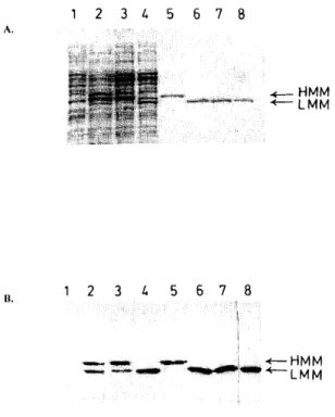
Fig. 2. Coomassie staining of the gel (A) and immunoblot (B) after SDS-PAGE of the following samples: (1)
Supernatant of lysozyme-treated E. coli TG1 cells; (2) supernatant after the freezing/thawing procedure of E. coli TG1 cells harbouring plasmid pBK8; (3) supernatant of lysozyme-treated E. coli TG1 cells harbouring plasmid pBK8. In the immunoblot, the ratio of HMM-to-LMM is clearly much larger than in 2. (4) Supernatant of lysozyme-treated E. coli TG1 cells containing plasmid pDML41; (5) purified HMM immunoreactive but inactive protein (from pBK8 and 'short' lysozyme treatment); (6) purified LMM, enzymatically active protein (from pBK8 and 'long' lysozyme treatment); (7) purified LMM, enzymatically active protein (from pDML41); (8) standard S. R61 DD-peptidase produced by Slreptomyces lividans TK24.
Table 1 Properties of the different DD-peptidase forms at 37° C
Carboxymethyl benzoylaminothioacetate
Cephalosporin C Carbenicillin Ac2-L-Lys-D-Ala-D-Ala pI kcat (s-1) Km (µM) kcat/Km (M-1-s-1) k2/K (M-1-s-1) Q (%) k2/K (M-1-s-1) Q (%) kcat/Km (M-1-s-1)
HMM 4.6 0.90 ± 0.06 130.0 ± 5.5 7000 ± 164 25 ± 0.5 38 ± 1.3 ND ND ND LMM 'S' 4.7 3.59 ± 0.40 53 ± 3 65,200 ± 2800 1900 ± 100 58.6 ± 0.1 637 ± 80 15 ±2 ND LMM 'M' 4.4 3.63 ± 0.04 54 ± 1 67,000 ± 1600 1700 ± 200 51 ± 5.0 850 ± 130 19.3 ± 0.2 ND LMM 'I' 4.15 3.64 ± 0.84 55 ± 26 71,000 ± 8600 1700 ± 100 58.4 ± 0.2 670 ± 50 19.6 ± 0.5 ND R61-stop 4.15 3.84 ±0.31 65 ± 3 59,300 ± 7700 ND ND 625 ± 280 ND 3500 ± 300 S. TK24 4.2 5 50 100,000 1500 50 ± 6 830 ± 80 15 ± 5 3300 ± 300 4.5
HMM = high Mr precursor; LMM 'S', 'M' and 'I' = active forms obtained after processing by E. coli cytoplasmic protease(s); R-61 STOP =
active form encoded by plasmid pDML41 ; S. TK24 = active form produced by S. lividans TK24; k2/K = second-order rate constant for
acylation [10]; Q = % of fluorescence quenching upon acylation; ND = not determined.
4. Conclusions
Cleavage of the C-terminal extension is necessary to obtain an active enzyme but its role at the level of the precursor remains obscure. This peptide did not appear to be involved in a specific interaction with the
cytoplasmic membrane since the intact precursor did not remain membrane-bound and that the protein encoded by the truncated gene appeared to be normally exported. A possible function as an intramolecular chaperone [14] also appeared to be rather unlikely since its absence in the protein encoded by the plasmid pDML41 did not result in the formation of improperly folded protein. Only effects on the rate of the folding process or on a specific secretion mechanism in Streptomyces remain possible. It did not seem that the presence of this
the precursor on the thiolester substrate and the fluorescence properties similar to those of the wild-type protein. This contrasts with the results obtained for the Actinomadura R39 DD-peptidase [15]. In this case, an incomplete cleavage of the signal peptide yielded a completely inactive protein, a probable consequence of the high
hydrophobicity of the uncleaved segment. The C-terminal extension of the R61 peptidase has no such properties. A curious result was the efficient processing by an E. coli cytoplasmic extract, a rather non-specific reaction which yielded at least 3 polypeptides. These could only be separated on the basis of their charges suggesting additional minor proteolysis or deamidation reactions [16].
Despite the large difference in G/C content between the two genomes, the S. R61 DD-peptidase gene was reasonably well expressed in E. coli. However, to obtain export of the protein to the periplasm, it was necessary to utilize a specific E. coli promoter and signal peptide. The overall yields appeared to be different with pBK8 and pDML41, but this was observed under non-optimised culture conditions.
In consequence, this study confirms that heterologous expression of Streptomyces genes in E. coli is indeed possible but difficulties might arise at the level of post-translational modifications and export phenomena.
Acknowledgements
This work was supported in part by the Belgian Government in the frame of the Pôle d'Attraction
Interuniversitaires (PAI no. 19), an Action Concertée with the Belgian Government (convention 89/94-130), the Fonds de la Recherche Scientifique Médicale (contrat no. 3.4537.88), and a convention tripartite between the Région Wallone, SmithKline Beecham UK, and the University of Liège. L.F. and B.J. are respectively Aspirant and Chercheur Qualifié of the Fonds National de la Recherche Scientifique (F.N.R.S., Brussels, Belgium).
References
[1] Frère, J.M., Nguyen-Distèche, M., Coyette, J. and Joris, B. (1992) in: The Chemistry of β-Lactams (Page, M.I. ed.) pp. 148-197, Blackie Academic and Professional, UK.
[2] Duez, C., Piron-Fraipont, C., Joris, B., Dusart, J., Urdea, M., Martial, J., Frère, J.M. and Ghuysen, J.M. (1987) Eur. J. Biochem. 162, 509-518.
[3] Joris, B., Jacques, P., Frère, J.M., Ghuysen, J.M., Van Beeumen, J. (1987) Eur. J. Biochem. 162, 519-524.
[4] Bourguignon-Bellefroid, C., Joris, B., Van Beumen, J., Ghuysen, J.M. and Frère, J.M. (1992) Biochem. J. 283, 123-128. [5] Kadonaga, J., Gautier, A., Straus, D., Charles, A., Edge, M. andKnowles, J. (1984) J. Biol. Chem. 259, 2149-2154. [6] Taylor, J.W., Ott, J. and Eckstein, F. (1985) Nucleic Acids Res. 13, 8765-8784.
[7] Fossati, P., Saint-Ghislain, M., Sicard, P., Frère, J.M., Dusart, J., Klein, D. and Ghuysen, J.M. (1978) Biotechnol. Bioeng. 20, 577- 587. [8] Joris, B., De Meester, F, Galleni, M., Reckinger, G., Coyette, J. and Frère, J.M. (1985) Biochem. J. 228, 241-248.
[9] Frère, J.M., Leyh-Bouille, M., Ghuysen, J.M., Nieto, M. and Perkins, H. (1976) Methods Enzymol. 45B, 610-636. [10] Adam, M., Damblon, C., Plaitin, B., Christiaens, L. and Frère, J.M. (1991) Biochem. J. 270, 525-529.
[11] Frère, J.M, Ghuysen, J.M and Iwatsubo, M. (1975) Eur. J. Biochem. 57, 343-351. [12] Robbins, P., Wirth, D. and Hering, C. (1981) J. Biol. Chem. 256, 10640-10644.
[13] Srivastava, R., Ali, S.S. and Srivastava, B.S. (1991) FEMS Micro- biol. Lett. 78, 201-206. [14] Shinde, U. and Inouye, M. (1993) Trends Biochem. Sci. 18, 442- 446.
[15] Granier, B., Duez, C., Lepage, S., Englebert, S., Dusart, J., Dideberg, O., Van Beeumen, J., Frère, J.M. and Ghuysen J.M. (1992) Biochem. J. 282, 781-788.