HAL Id: tel-01681314
https://pastel.archives-ouvertes.fr/tel-01681314v2
Submitted on 12 Jan 2018HAL is a multi-disciplinary open access archive for the deposit and dissemination of sci-entific research documents, whether they are pub-lished or not. The documents may come from teaching and research institutions in France or abroad, or from public or private research centers.
L’archive ouverte pluridisciplinaire HAL, est destinée au dépôt et à la diffusion de documents scientifiques de niveau recherche, publiés ou non, émanant des établissements d’enseignement et de recherche français ou étrangers, des laboratoires publics ou privés.
the era of high-throughput RNA sequencing.
Elsa Bernard
To cite this version:
Elsa Bernard. Deciphering splicing with sparse regression techniques in the era of high-throughput RNA sequencing.. Bioinformatics [q-bio.QM]. Université Paris sciences et lettres, 2016. English. �NNT : 2016PSLEM063�. �tel-01681314v2�
THÈSE DE DOCTORAT
de l’Université de recherche Paris Sciences et Lettres
PSL Research University
Préparée à MINES ParisTech
Deciphering splicing with sparse regression techniques
in the era of high-throughput RNA sequencing
Etude de l’épissage grâce à des techniques de
régression parcimonieuse dans l’ère du séquençqge
haut débit de l’ARN
COMPOSITION DU JURY :
M. Franck PICARD
LBBE, Président
M. Daniel GAUTHERET
Université Paris-Sud, Rapporteur
M. Wolfgang HUBER
EMBL, Rapporteur
M. Didier AUBOEUF
ENS Lyon, Examinateur
M. Claude HOUDAYER
Institut Curie, Examinateur
M. Jean-Philippe VERT
MINES ParisTech, Examinateur
Soutenue par Elsa BERNARD
le 21 septembre 2016
Dirigée par Jean-Philippe VERT
Ecole doctorale
n°
432
ECOLE DOCTORALE SCIENCES DES METIERS DE L’INGENIEUR
The number of protein-coding genes in a human, a nematode and a fruit fly are roughly equal. The paradoxical miscorrelation between the number of genes in an organism’s genome and its phenotypic complexity finds an explanation in the alternative nature of splicing in higher organisms.
Alternative splicing largely increases the functional diversity of proteins encoded by a limited number of genes. It is known to be involved in cell fate decision and embryonic development, but also appears to be dysregulated in inherited and acquired human genetic disorders, in particular in cancers.
High-throughput RNA sequencing technologies allow us to measure and question splicing at an unprecedented resolution. However, while the cost of sequencing RNA decreases and throughput increases, many computational challenges arise from the discrete and local nature of the data. In particular, the task of inferring alternative transcripts requires a non-trivial deconvolution procedure.
In this thesis, we contribute to deciphering alternative transcript expressions and alternative splicing events from high-throughput RNA sequencing data.
We propose new methods to accurately and efficiently detect and quantify alternative tran-scripts. Our methodological contributions largely rely on sparse regression techniques and takes advantage of network flow optimization techniques. Besides, we investigate means to query splicing abnormalities for clinical diagnosis purposes. We suggest an experimental protocol that can be easily implemented in routine clinical practice, and present new statistical models and algorithms to quantify splicing events and measure how abnormal these events might be in patient data compared to wild-type situations.
Le nombre de g`enes codant pour des prot´eines chez l’homme, le vers rond et la mouche des fruits est du mˆeme ordre de grandeur. Cette absence de correspondance entre le nombre de g`enes d’un eucaryote et sa complexit´e ph´enotypique s’explique en partie par le caract`ere alternatif de l’´epissage.
L’´epissage alternatif augmente consid´erablement le r´epertoire fonctionnel de prot´eines cod´ees par un nombre limit´e de g`enes. Ce m´ecanisme, tr`es actif lors du d´eveloppement embryon-naire, participe au devenir cellulaire. De nombreux troubles g´en´etiques, h´erit´es ou acquis (en particulier certains cancers), se caract´erisent par une alt´eration de son fonctionnement.
Les technologies de s´equen¸cage `a haut d´ebit de l’ARN donnent acc`es `a une information plus riche sur le m´ecanisme de l’´epissage. Cependant, si la lecture `a haut d´ebit des s´equences d’ARN est plus rapide et moins coˆuteuse, les donn´ees qui en sont issues sont complexes et n´ecessitent le d´eveloppement d’outils algorithmiques pour leur interpr´etation. En particulier, la reconstruction des transcrits alternatifs requiert une ´etape de d´econvolution non triviale.
Dans ce contexte, cette th`ese participe `a l’´etude des ´ev´enements d’´epissage et des transcrits alternatifs `a partir de donn´ees de s´equen¸cage `a haut d´ebit de l’ARN.
Nous proposons de nouvelles m´ethodes pour reconstruire et quantifier les transcrits alternatifs de fa¸con plus efficace et pr´ecise. Nos contributions m´ethodologiques impliquent des techniques de r´egression parcimonieuse, bas´ees sur l’optimisation convexe et sur des algorithmes de flots. Nous ´etudions ´egalement une proc´edure pour d´etecter des anomalies d’´epissage dans un con-texte de diagnostic clinique. Nous sugg´erons un protocole exp´erimental facilement op´erant et d´eveloppons de nouveaux mod`eles statistiques et algorithmes pour quantifier des ´ev´enements d’´epissage et mesurer leur degr´e d’anormalit´e chez le patient.
Contents vii
List of Figures x
List of Tables xii
Abbreviations xiii
1 Preambule 1
2 Splicing: from molecular mechanisms to personalized therapies 4
2.1 Molecular mechanisms resulting in the expression of transcript isoforms . . . 5
2.1.1 A bit of history: pre-mRNA splicing . . . 5
2.1.2 Alternative splicing and alternative transcription . . . 8
2.1.3 What makes splicing alternative? . . . 10
2.2 Some aspects of the functional importance of alternative transcript expression . . 11
2.2.1 A word of evolution . . . 11
2.2.2 Alternative splicing regulation during development and cell fate decision . 12 2.2.3 Coupling of alternative splicing with nonsense-mediated decay . . . 13
2.3 Splicing dysregulation in human diseases . . . 14
2.3.1 Mutated regulatory sequences . . . 14
2.3.2 Trans-acting factors . . . 15
2.3.3 A focus on cancer . . . 16
2.4 Emerging therapies targeting splicing defects . . . 16
2.4.1 Cancer-specific isoforms as biomarkers . . . 16
2.4.2 Splice modulating therapies . . . 17
2.4.3 Antisense oligonucleotides: the example of Duchenne muscular dystrophy 17 3 Questioning splicing: from data to algorithms 19 3.1 Measuring splicing with data evolving in time . . . 20
3.1.1 Heritage of Sanger sequencing. . . 20
3.1.2 Successes and limitations of microarray splicing profiling. . . 22
3.1.3 High-throughput sequencing of the RNA as the new gold standard . . . . 23
3.2 Computational challenges associated with RNA-seq reads . . . 29
3.2.1 Mapping RNA-seq reads . . . 29
3.2.2 Modeling RNA-seq reads . . . 32
3.2.3 The isoform deconvolution problem. . . 37 vii
3.3 Genome-guided transcript estimation . . . 39
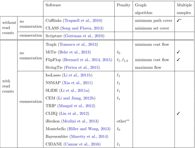
3.3.1 Inferring transcripts with various techniques. . . 39
3.3.2 `1-norm penalization . . . 44
3.3.3 Network flow optimization. . . 48
4 Efficient transcript isoform identification and quantification from RNA-seq data with network flows 52 4.1 Background and related works . . . 53
4.2 Proposed approach . . . 54
4.2.1 Statistical model . . . 55
4.2.2 Isoform detection by sparse estimation . . . 57
4.2.3 Isoform detection as a path selection problem . . . 58
4.2.4 Optimization with network flows . . . 60
4.2.5 Flow decomposition . . . 63
4.2.6 Model selection . . . 63
4.3 Experimental validation . . . 64
4.3.1 Simulated human RNA-seq data . . . 64
4.3.2 Real RNA-Seq data . . . 70
4.4 Conclusion . . . 70
5 A convex formulation for joint transcript isoform estimation from multiple RNA-seq samples 72 5.1 Background and related works . . . 73
5.2 Proposed approach . . . 74
5.2.1 Multi-dimensional splicing graph . . . 74
5.2.2 Joint sparse estimation . . . 75
5.2.3 Candidate isoforms . . . 76
5.2.4 Model selection . . . 77
5.3 Experimental validation . . . 77
5.3.1 Influence of coverage and sample number . . . 78
5.3.2 Influence of hyper-parameters with realistic simulations . . . 82
5.3.3 Experiments with real data . . . 83
5.3.4 Illustrative examples . . . 84
5.4 Conclusion . . . 87
6 A time- and cost-effective clinical diagnosis tool to quantify abnormal splicing from targeted single-gene RNA-seq 88 6.1 Background . . . 89
6.1.1 Molecular diagnosis context . . . 89
6.1.2 Targeted single-gene RNA-seq. . . 89
6.2 Results and discussion . . . 90
6.2.1 A pipeline to query splicing abnormalities . . . 90
6.2.2 BRCA1 pilot study . . . 91
6.2.3 Data normalization. . . 92
6.2.4 Quantifying splicing events on controls . . . 97
6.2.5 Detecting abnormal events as deviation from control distributions . . . . 98
6.2.6 Deciphering complex splicing events with full-length transcript prediction 100 6.3 Conclusion . . . 103
6.4 Methods . . . 103
6.4.1 RNA isolation and sequencing. . . 103
6.4.2 Bioinformatics pre-processing . . . 104 6.4.3 Data normalization. . . 104 6.4.4 Transcript prediction. . . 105 7 Discussion 111 A Supplementary figures 115 B Supplementary tables 118 C Software 120 Bibliography 121
List of Figures
2.1 Typical structure of a multi-exon eukaryotic gene . . . 6
2.2 The two steps of the pre-mRNA splicing reaction . . . 7
2.3 Main modes of alternative splicing . . . 9
2.4 Cis-acting sequences regulating alternative splicing . . . 10
2.5 Coupling of alternative splicing and nonsense-mediated decay . . . 14
2.6 Use of antisense oligonucleotides to modulate pre-mRNA splicing . . . 18
3.1 Illustration of the Sanger sequencing technique . . . 21
3.2 Illustration of a splicing microarray experiment . . . 23
3.3 A typical RNA-seq experiment . . . 25
3.4 RNA-seq reads aligned on a reference genome . . . 31
3.5 RNA-seq coverage density . . . 31
3.6 Comparison of Binomial and Poisson distribution . . . 35
3.7 Benefits of using read count levels to assemble transcripts . . . 40
3.8 Sparsity induction by the `1-norm . . . 47
3.9 Pyramidal shape of the `1-ball . . . 47
4.1 Computation of the effective length. . . 56
4.2 Construction of the DAG generalizing the splicing graph . . . 59
4.3 Flow interpretation of isoforms. . . 61
4.4 Precision and recall on simulated reads . . . 66
4.6 Average CPU times in milliseconds . . . 68
4.7 Precision and recall on simulated reads with FluxSimulator . . . 69
4.8 Precision and recall on human embryonic stem cells data. . . 71
5.1 Multi-dimensional splicing graph . . . 75
5.2 Human simulations with increasing coverage and number of samples . . . 79
5.3 Human simulations with various read lengths . . . 81
5.4 Simulation using both paired or single-end reads at comparable coverage . . . 81
5.5 Fscore results on the Flux Simulator simulations . . . 82
5.6 Fscore results on the modENCODE data . . . 84
5.7 Running time on the D.melanogaster RNA-seq data . . . 85
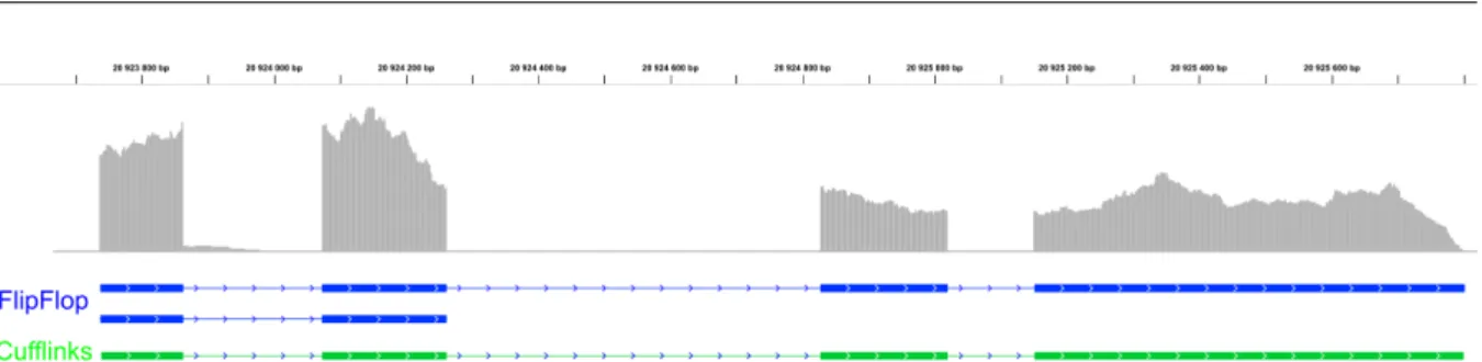
5.8 Transcriptome predictions of gene CG15717 . . . 86
6.1 BRCA1 amplicon design . . . 91
6.2 5’ read count on the set of BRCA1 exons . . . 94
6.3 Distribution of Spearman correlation across the set of controls. . . 95
6.4 Scaling factors . . . 95
6.5 Effect of data normalization on a control sample . . . 96 x
6.6 Effect of data normalization on a patient sample . . . 96
6.7 Percentage of splicing of different regions over the controls . . . 98
6.8 Detection and quantification of abnormal splicing events on a patient sample . . 99
6.9 Effect of puromycin on the quantification of splicing abnormalities . . . 100
6.10 Visualization of the set of inferred transcripts on a patient sample . . . 101
6.11 Transcripts inferred on the ENIGMA cell line . . . 102
6.12 Illustration of the loess-based normalization procedure on a control sample. . . . 106
6.13 Schematic design with 2 amplicons . . . 107
A.1 MiTie results on a first set of human simulations . . . 115
A.2 MiTie results on a second set of human simulations . . . 116
List of Tables
3.1 Overview of genome-guided transcript estimation softwares . . . 45
5.1 Statistical testing on human simulation results . . . 80
6.1 Summary of samples analyzed in the BRCA1 pilot study . . . 92
B.1 Details on the optimized pre-processing parameters . . . 118
B.2 Details on the optimized prediction parameters . . . 118
B.3 Description of the D.melanogaster RNA-seq data . . . 119
B.4 Primer pairs defining each amplicon on the BRCA1 study . . . 119
DNA DeoxyriboNucleic Acid
RNA RiboNucleic Acid
A Adenine
T Thymine
G Guanine
C Cytosine
bp base pair
UTR UnTranslated Region
ESE Exonic Splicing Enhancer ESS Exonic Splicing Silencer ISE Intronic Splicing Enhancer ISS Intronic Splicing Silencer
ESC Embryonic Stem Cell
NMD Nonsense Mediated Decay
ASO AntiSense Oligonucleotide
EMT Ephytelial Mesenchymal Transition EST Expressed Sequence Tag
PCR Polymerase Chain Reaction NGS Next Generation Sequencing RNA-seq RNA-sequencing
DAG Directed Acyclic Graph
VUS Variant of Unknown Significance
Preambule
Through alternative splicing of precursor messenger RNAs, eukaryote genes produce multiple transcript isoforms that may lead to proteins with distinct or even opposite functions.
Alternative splicing not only greatly increases the repertoire of proteins that can be encoded by a genome, it is also a fundamental regulatory mechanism of gene expression at the crossroad between transcription and translation. Alternative splicing is deeply involved in cell fate decision and tissue differentiation.
The importance of alternative splicing is underscored by the fact that splicing defects are respon-sible for many human diseases such as retinitis pigmentosa or Duchenne muscular dystrophy, and that splicing aberrations are believed to contribute to tumor progression in several cancers.
Detecting transcript isoforms in different cell types or samples is therefore crucial to understand the cells’ regulatory programs and to identify splicing variants responsible for diseases. Fur-thermore, fully characterizing the transcripts expressed in tumor samples will contribute to our understanding of cancer mechanisms, provide new diagnostic and prognostic biomarkers and reveal possible drug targets, improving personalized patient treatment.
Recent technological advances decreased the cost of RNA sequencing while increasing the throughput. This allows the profiling of numerous RNA landscapes from various species, tissues and conditions and to get closer to RNA profiling in routine clinical practice.
High-throughput RNA sequencing is accelerating our understanding of alternative splicing reg-ulation and dysregreg-ulation and gives a better insight into fascinating questions such as (i) how
much alternative splicing contributes to cell fate decision, (ii) to what extent alternative splic-ing events are functionally relevant, or (iii) whether there are splicsplic-ing aberrations that drive tumorigenesis.
However, while an accurate reconstruction and quantification of transcript isoforms is a crucial step to answer the above questions and for downstream analysis such as differential analysis of transcript abundances, the task is not trivial due to the nature of RNA sequencing data. Indeed, recovering the structure of the transcripts and estimating their abundances from this data need an accurate deconvolution procedure. Furthermore, their discrete nature requires an appropriate statistical modeling, and their high dimensionality asks for the development of efficient algorithmic tools.
The contributions of this thesis lie in the fields of transcriptome assembly and alternative splicing events quantification from high-throughput RNA sequencing. We propose new methods to reconstruct transcript isoforms from one or several RNA sequencing samples, and we investigate means to query splicing abnormalities in a clinical diagnosis context.
Organization and contributions of the thesis
We detail below the organization of the thesis, and highlight, when appropriate, our contribu-tions to the fields of transcriptome assembly and alternative splicing events quantification.
• Chapter2 is an introductory chapter that briefly reviews the alternative splicing process, mentions some of its functional properties and discusses its implication in human diseases as well as emerging therapies tailored to correct splicing abnormalities.
• Chapter3is an other introductory chapter that surveys sequencing and profiling protocols developed since the 90’s and which give access to alternative splicing, with a focus on modern high-throughput RNA sequencing that emerged a decade ago. We also describe the computational challenges associated with RNA sequencing data, and review to the best of our knowledge the state-of-the-art methods to assemble and quantify transcript isoforms. We finally introduce the notions of `1-norm penalization and network flow optimization that we intensively use in the following chapters.
• Chapter4 describes a new method to reconstruct and quantify transcript isoforms from RNA sequencing data. The main novelty of our approach is to translate a computation-ally hard sparse regression problem formulated with a `1-penalized maximum likelihood estimation into a network flow optimization problem that can be solved very efficiently.
• Chapter5extends the sparse regression setting of the previous chapter to the joint analysis of several RNA sequencing samples. We formulate a convex problem that allows us to share information across samples when inferring transcript isoforms, hence increasing the power of the statistical inference and resulting performances.
• Chapter 6 describes a clinical diagnosis tool to detect and quantify alternative splicing events as well as full-length transcripts from targeted RNA sequencing experiments where the sequencing efforts are concentrated on a subset of the transcriptome. Our method focuses on revealing splicing abnormalities by measuring discrepancies between patient estimates and wild-type distributions derived from control samples. We apply our method-ology on RNA sequencing data from patients characterized by mutations in a breast cancer susceptibility gene, and experimentally validate some of our results.
• Chapter7concludes the thesis by summarizing the main results and giving some prospects on how to extend the proposed methodologies to other emerging RNA sequencing protocols and on how the techniques we developed during the thesis could be used to answer other molecular biology questions.
Splicing: from molecular
mechanisms to personalized
therapies
“The discovery of split genes has been of fundamental importance for today’s basic research in biology, as well as for more medically oriented research concerning the development of cancer and other diseases”
“the genetic message, which gives rise to a particular product, is not definitely established at the stage when the RNA is first synthesized. Instead, it is the splicing pattern that determines the nature of the final product”
Nobel Prize Press Release, 1993.
Ce chapitre introductif fournit aux lecteurs les cl´es pour comprendre comment les eucaryotes peuvent exprimer plusieurs ARN messagers `a partir d’un unique g`ene. Les notions d’´epissage, d’´epissage alternatif et de transcription alternative sont donc introduites. Les aspects fonction-nels de l’´epissage sont ´egalement discut´es, son rˆole adaptatif et son implication dans le devenir cellulaire. Enfin, la d´er´egulation de l’´epissage dans plusieurs maladies g´en´etiques comme le can-cer et l’´emergence de th´erapies ciblant les dysfonctionnements de l’´epissage sont mentionn´ees.
In this introductory chapter, we start by explaining how eukaryotes can express several mes-senger RNAs (mRNAs) from the same gene, that is we introduce the concepts of splicing, alternative splicing and alternative transcription. We then discuss some functional aspects of alternative splicing as a fundamental gene expression regulatory mechanism that shows adapta-tive significance and is deeply involved in cell fate decision. We finally illustrate how alternaadapta-tive splicing can be dysregulated in human diseases and in particular in cancer, before discussing certain emerging therapies tailored to target splicing abnormalites.
2.1
Molecular mechanisms resulting in the expression of
tran-script isoforms
In this section we describe the molecular mechanisms behind splicing and resulting in the ex-pression of several transcript isoforms from the same locus. We do not claim that the following explanations would satisfy the curiosity of a molecular biologist, but we hope they can benefit non-specialists by introducing some key concepts. In particular, we do not detail the different proteins known to be involved in the splicing machinery and their mechanisms of action, but we rather give a schematic view of their effects and refer to the literature for more detailed explanations of molecular mechanisms.
2.1.1 A bit of history: pre-mRNA splicing
The gene expression field made an important step forward in the late 80’s when the split nature of most eukaryotic genes was discovered. In 1977, several groups working with adenoviruses that infect and replicate in mammalian cells obtained surprising results: RNA molecules from infected cells containing sequences from non-contiguous sites in the viral genome (Berget et al.,
1977;Chow et al.,1977). What they termed “mosaic RNA” at the time was the result of the excision of what came to be called intragenic sequences (introns) from precursor mRNA. This process of removing or “splicing out” introns is now known as precursor mRNA splicing (pre-mRNA splicing or splicing in short form). However, the concept of pre-(pre-mRNA is nowadays thought to be a virtual entity due to the co-transcriptional nature of splicing (Merkhofer et al.,
2014).
Formally, an intron is defined as a gene segment that is present in the primary (or precursor) transcript but absent from the mature RNA as a consequence of splicing. The term intron refers to both the DNA sequence within a gene and the corresponding sequence in the unprocessed RNA transcript. On the contrary, an exon denotes a gene segment that is or can be present in mature RNA. Most human genes contain multiple exons, and the average length of exons (50 − 250bp1) is much shorter than that of introns (frequently thousands of bp). Figure 2.1
illustrates the split nature of eukaryotic genes: figure 2.1(a) shows the exons and introns of a gene as well as the untranslated regions (UTRs), the initiation codon and the termination codon at the 5’ and 3’ ends of the first and last exons. It also depicts a promoter region
1
translation initiation codon (ATG) transcription initiation
exon 1 intron 1 exon 2 intron 2 exon 3
translation termination codon (TAA, TAG, TGA)
{
3’ UTR
{
5’ UTR
polyA addition signal
{
promoter region polyA addition site(a) Double stranded DNA gene.
exon 1 intron 1 exon 2 intron 2 exon 3
5’ cap AAAA
(b) Single stranded pre-mRNA.
exon 1 exon 2 exon 3 AAAA
5’ cap
(c) Mature RNA.
Figure 2.1: Typical structure of a multi-exon eukaryotic gene (a) and its associated pre-mRNA resulting from transcription, 5’ capping and polyA addition (b) and mature pre-mRNA resulting from splicing (c).
that contributes to define the transcription inition site and a polyadenylation (polyA) addition sequence signal that contributes to define the polyA addition site. The polyA addition site delineates the transcription termination site. Figure 2.1(b) shows the pre-mRNA that results from transcription, 5’ capping (i.e. the addition of a methylated guanine at the 5’ end of the pre-mRNA) and polyA addition. Finally figure2.1(c) corresponds to the mature mRNA resulting from pre-mRNA splicing.
How splicing happens?
The biochemical mechanism by which splicing occurs is fairly well understood (Clancy,2008). Introns are removed from primary transcripts by cleavage at conserved sequences called splice sites. These sites are found at the 5’ end (donor site) and 3’ end (acceptor site) of introns. The splice donor site includes an almost invariant sequence GU within a larger and less highly conserved region while the splice acceptor site terminates the intron with an almost invariant AG sequence. These consensus sequences are known to be critical, as changing one of the conserved nucleotides often results in the inhibition of splicing (Cartegni et al.,2002). Another important sequence occurs at what is called the branch point, characterized by an A residue, and located anywhere from 18 to 40 nucleotides upstream from the 3’ end of an intron.
exon 1 AG GU intron A AG G exon 2 3’ splice site branch point 5’ splice site exon 2 G exon 1 AG A AG AG A exon 1 AGG exon 2 G U G U
+
step 1 step 2Figure 2.2: The two steps of the pre-mRNA splicing reaction.
Figure2.2schematically illustrates the two steps of the splicing chemical reaction: the A residue from the branch point interacts with the 5’ splice site to form a so-called intronic lariat before ligation of the two exons and liberation of the intron. Splicing is carried out in the nucleus of eukaryote cells by the spliceosome, a megaparticle in which ribonucleoprotein particles (the so-called small nuclear ribonucleoprotein particles or snRNPs) and a large number of auxiliary proteins (denoted as splicing factors) cooperate to accurately recognize the splice sites and catalyse the two steps of the splicing reaction. A multitude of RNA-RNA, RNA-protein and protein-protein interactions allows for the precise excision of each intron and appropriate joining of the exons.
We refer toHastings and Krainer (2001) and Black(2003) for more details about the splicing biochemistry.
2.1.2 Alternative splicing and alternative transcription
How come there are ⇠ 120000 mRNA molecules mapped out in the human cells while the human genome contains only ⇠ 25000 protein-coding genes? The solution lies in the alternative nature of splicing in eukaryotes.
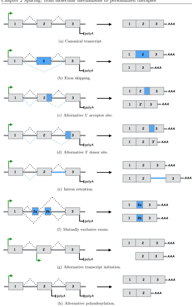
Alternative splicing is the mechanism through which multiple mature mRNA transcripts (or mRNA isoforms) are expressed from a single gene. The ability of cells to exhibit variations of mature mRNA from the same pre-mRNA adds a layer of complexity to the central dogma DNA ! RNA ! protein of molecular biology. It is accomplished by excluding one or more exons (exon skipping), by moving exon/intron boundaries (acceptor or donor splice site shift) or by retention of introns. The main modes of alternative splicing are illustrated in figures
2.3(b),2.3(c),2.3(d),2.3(e), 2.3(f). This widespread mechanism is estimated to affect ⇠ 90% of mammalian protein-coding genes (Wang et al.,2008a) and is now considered a fundamental regulatory process at the crossroad between transcription and translation. Some functional aspects of alternative splicing are discussed in section 2.2.
Perhaps the most striking example of alternative splicing comes from Drosophila melanogaster. Its Dscam gene, which codes for a cell surface protein involved in neuronal connectivity, has 24 exons, with 12 alternative versions of exon 4, 48 versions of exon 6, 33 versions of exon 9 and 2 versions of exon 17. Each version of a particular exon is used to the exclusion of all the others. Thus the combinatorial use of alternative exons can potentially generate 38016 different protein isoforms (Schmucker et al.,2000). The Dscam gene exemplifies both the extreme expansion in coding capacity that alternative splicing provides and the tight regulation of alternative splicing that must be in place to somehow enforce mutual exclusion of the different versions of the exons.
In addition to the alternative splicing mechanisms mentioned above and illustrated in figure2.3
(exon skipping, alternative acceptor or donor splice sites and intron retention), the exon com-position of RNA transcripts can also vary by the differential selection of 5’ end transcription initiation and 3’ end termination sites – also known as multiple promoter or multiple polyA usage (Kornblihtt,2005). Figures2.3(g)and 2.3(h)illlustrate as well these two distinct mecha-nisms which are not splicing events stricto sensu but similarly participate to creating a variety of RNA transcripts from a single locus.
Identifying the different transcript isoforms produced by a single gene, that is the different combinations of exons included in the expressed mRNA, is the main scope of chapters4 and5.
1 2 3 1 2 3 AAA
polyA
(a) Canonical transcript.
1 2 3 1 2 3 AAA polyA 1 3 AAA (b) Exon skipping. AAA AAA 1 2 3 polyA 1 2 3 1 2’ 3
(c) Alternative 5’ acceptor site.
AAA 1 2 3 polyA 1 2 3 AAA 1 2 3’
(d) Alternative 3’ donor site.
1 2 3 1 2 3 AAA polyA AAA 3 1 2
(e) Intron retention.
1 2a 3 1 3 AAA polyA 2b 2a 1 2b 3 AAA
(f) Mutually exclusive exons.
1 2 3
2 3 AAA
polyA
1 2 3 AAA
(g) Alternative transcript initiation.
AAA 1 1 2 3 2 polyA 1 2 3 AAA polyA (h) Alternative polyadenylation.
Figure 2.3: Main modes of alternative splicing ((b) to (f)), alternative transcription initiation site (g) and alternative polyadenylation site (h).
ISS ESS ISS
ESE ISE
Figure 2.4: Cis-acting sequences regulating alternative splicing. ESE: exonic splicing en-hancer, ISE: intronic splicing enen-hancer, ESS: exonic splicing silencer, ISS: intronic splicing enhancer. Enhancers can activate adjacent splice sites whereas silencers can repress splice sites. The competing influences of the different enhancers and silencers determine the inclusion or skipping of the exon. Figure is inspired fromMatlin et al.(2005).
2.1.3 What makes splicing alternative?
The decision as to which exon is removed and which exon is included involves RNA sequence elements and protein regulators.
First of all, splice sites can be strong or weak depending on how far their sequences diverge from the consensus sequences, which determine their affinity for splicing factors. The relative position and use of weak and strong sites give rise to the different alternative splicing modes described in figure2.3. Unsurprisingly, it has been shown that alternative exons possess weaker splice sites than constitutive exons (Sorek et al.,2004).
Second, the degree to which weak sites are used is regulated by both cis-regulatory sequences and trans-acting factors. Depending on the position and function of the cis-regulatory elements, they are divided into four categories: exonic splicing enhancers (ESEs), exonic splicing silencers (ESSs), intronic splicing enhancers (ISEs) and intronic splicing silencers (ISSs). Trans-acting factors include proteins and ribonucleoproteins that bind to the splicing enhancers and silencers. Figure2.4shows how these enhancers and silencers act combinatorially to regulate the alterna-tive use of splice sites. Of note, a machine learning algorithm has been developed that is capable of automatically extracting combinations of cis-elements that are accurately predictive of brain, muscle, digestive and embryo versus adult specific alternative splicing patterns (Barash et al.,
Finally, alternative splicing is also believed to be regulated by the secondary structure of the pre-mRNA transcript and by interactions with the transcription and chromatin machiner-ies (Schwartz and Ast,2010;Luco et al.,2011).
For accurate reviews of alternative splicing mechanisms and regulation we suggestMatlin et al.
(2005),Chen and Manley (2009) and Kornblihtt et al. (2013).
In line with what has been presented above, chapter 6 focuses on detecting splicing defects on transcripts expressed from alleles harboring mutations in their cis-regulatory splicing enhancers or silencers.
2.2
Some aspects of the functional importance of alternative
transcript expression
2.2.1 A word of evolution
Alternative splicing is believed to occur in all metazoan organisms, but is more prevalent in ver-tebrates. The number of protein-coding genes in vertebrates is not radically different from the number in invertebrates (for example the number of human genes is roughly equal the the num-ber of nematode genes and barely four times the numnum-ber of genes in budding yeast), suggesting a link between alternative splicing prevalence and phenotypic complexity (Nilsen and Graveley,
2010). Kim et al.(2007) studied in depth the different levels of splicing among eukaryotes and proposed alternative splicing as a possible solution to the paradoxical miscorrelation between the number of genes in an organism’s genome and its phenotypic complexity.
The split organization of eukaryotic genes into exons and introns and the existence of pre-mRNA splicing process is believed to confer at least two evolutionary advantages. The first –relatively obvious– advantage is that alternative splicing allows a single gene to produce several mRNA variants, greatly expanding the coding capacity of eukaryotic genomes (Keren et al., 2010). The second advantage lies at a phylogenic level, as intronic recombination events (such events leave the exons intact) allow protein-coding exons to be placed together to form new genes. Recombined mRNAs have high chance of encoding novel functional polypeptides that combine functional domaines previously tested by natural selection. This mutational process is known as exon shuffling (Ast,2004). Moreover it has been proposed that alternative splicing represents a major source of species-specific differences: for exempleBarbosa-Morais et al.(2012) recently
showed that there is a decline in alternative splicing frequency in vertebrates as the evolutionary distance from primates increases.
However, the prevalence of alternative splicing raises questions about its biological significance. What fraction of multiple mRNA isoforms expressed from each of ⇠ 20000 alternatively spliced human genes has a functional impact? It has been proposed that many alternative splicing events do not have functional significance but rather represent stochastic noise in the splicing process (Melamud and Moult, 2009; Skandalis et al., 2010). In any case, the adaptive role of alternative splicing remains elusive, in part because few variant transcripts have been charac-terized functionally, making it difficult to assess the contribution of alternative splicing to the generation of phenotypic complexity and to study the evolution of splicing patterns (Mudge et al.,2011).
2.2.2 Alternative splicing regulation during development and cell fate
deci-sion
The Drosophila sex determination pathway provides a simple and central example of how a choice between different splicing patterns contributes to cell fate decision and tissue specificities. Indeed, sex determination in flies is a binary decision based on alternative splicing (Salz,2011): splicing of the sex-lethal (Sxl ) gene in females gives rise to a functional protein product, while in male alternative splicing leads to the inclusion of a stop codon so that the functional protein in not produced. Remarkably the Sxl gene is a splicing factor that regulates as well the splicing of its target genes also involved in the sex determination pathway. Interestingly, related insects such as the housefly do not splice the Sxl pre-mRNA in a specific manner while the sex-determination cascade of the honeybee is different in almost all its components although relying on alternative splicing as well. This shows as previously discussed the evolutionary plasticity provided by alternative splicing (Nilsen and Graveley,2010).
In addition to alterations by sex, metazoan organisms regulate the splicing of thousands of other transcripts depending on cell type, developmental state or external stimulus. High-throughput studies have shown that 50% or more of alternative splicing isoforms are differently expressed among tissues, indicating that most alternative splicing is subject to tissue-specific regula-tion (Yeo et al.,2004;Wang et al.,2008a).
Large-scale profiling studies have also revealed sets of alternative splicing events associated with changes in cell differentiation and development (Blencowe,2006). In particular, alternative splic-ing has been identified to contribute to the differentiation of embryonic stem cells (ESCs) into distinct lineages. Wu et al.(2010) provided evidence that isoform complexity is more extensive in ESCs and becomes restricted and more specialized as ESCs differentiate, whileGabut et al.
(2011) showed that an ESC-specific alternative splicing switch stimulates the expression of key pluripotency genes.
For a detailed review of the functional consequences of developmentally regulated alternative splicing we refer toKalsotra and Cooper (2011).
2.2.3 Coupling of alternative splicing with nonsense-mediated decay
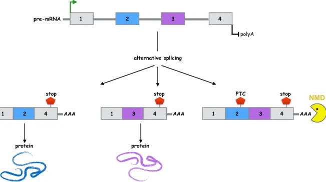
Most human genes exhibit alternative splicing, but not all alternatively spliced transcripts pro-duce functional proteins. Some alternative splicing events in humans result in mRNA isoforms harboring a premature termination codon (PTC), i.e. a stop codon located upstream from the last exon. A single-nucleotide mistake during the pre-mRNA splicing process often results in a frameshift and consequent PTC appearance. These transcripts characterized by a PTC are pre-dicted to be degraded by the nonsense-mediated mRNA decay (NMD) pathway (Lareau et al.,
2007). Figure 2.5illustrates the NMD degradation process.
NMD is then considered as an mRNA quality-control mechanism by degrading transcripts en-coding truncated proteins with no or undesired functions. However, while it prevents the accu-mulation of potentially harmful polypeptides, NMD is also believed to regulate the expression of 10 − 20% of normal transcripts. Briefly, it is the coupling of alternative splicing and NMD that allows the downregulation of specific transcripts: alternative splicing events that occur in exons located in the 3’ UTR and that generate a PTC activate NMD even though the degraded transcript would have encoded a full-length protein. This regulation phenomenon is believed to restrict the expression of several stress-related mRNA under non-stress conditions ( Lykke-Andersen and Jensen,2015).
We will encounter the NMD pathway again in chapter6when we pay attention to its inhibition in a clinical diagnosis setting in order to reveal the expression of aberrant transcripts from mutated BRCA1 alleles.
NMD 1 2 3 1 2 4 AAA polyA 4 1 3 4 AAA 1 2 3 4 AAA alternative splicing
stop stop PTC stop
pre-mRNA
protein protein
Figure 2.5: Coupling of alternative splicing and nonsense-mediated decay. PTC: premature stop codon, NMD: nonsense-mediated decay. In the depicted example, exons 2 and 3 are mutually exclusive, so that the simultaneous inclusion of both exons generate a PTC that activate NMD. Figure is inspired fromLareau et al. (2007).
2.3
Splicing dysregulation in human diseases
The link between alternative splicing and disease is well established (Scotti and Swanson,2016a), and many different human diseases can be caused by errors in RNA splicing or its regulation. We briefly discuss here the link between splicing and human diseases via the alteration of both cis- or trans-acting factors, with a particular focus on cancer. In addition, we emphasize that the identification of abnormal splicing as a primary mechanism of diseases raises the possibility of therapeutic approaches targeting splicing.
2.3.1 Mutated regulatory sequences
Mutations in regulatory sequences that affect alternative splicing are a widespread cause of hu-man hereditary diseases and cancers. These mutations can disrupt existing splicing enhancers or silencers or create new ones, thereby perturbing the use of alternative or constitutive exons. A single nucleotide mutation that does not change the encoded amino acid of a protein (silent mutation) can disrupt for instance a crucial splicing enhancer and be a disease-causing muta-tion (Wang and Cooper, 2007). Examples of human disease genes known to be targeted by synonymous and non-synonymous mutations often altering splicing regulatory elements include
the BRCA1 (breast cancer 1) gene involved in heriditary breast cancer, the SMN1 (survival of motor neuron 1) gene involved in spinal muscular atrophy and the DMD gene involved in Duchenne muscular dystrophy or the MAPT (microtubule-associated protein tau) gene involved in Alzheimer’s disease (Cartegni et al.,2002).
It has been estimated that as many as 50% of disease mutations in exons may impact on splic-ing (Lopez-Bigas et al.,2005). This strongly suggests that, in a clinical diagnosis perspective, genetic variants that are linked with a disease phenotype need to be evaluated for disruption of the correct splicing patterns. For example, it is important to know that a mutation results in a loss of expression due to aberrant splicing and NMD-mediated degradation, rather than the expression of a wild-type level of a protein containing a missense mutation. Knowing that the primary effect of an exonic mutation is a splicing defect, rather than a protein-coding mutation, is crucial in order to understand the detailed pathogenic mechanism of a disease.
In chapter 6, we underline the importance of introducing routine transcript analysis in order to properly assess possible mechanisms accounting for human diseases, and propose a new methodology to implement such routine mRNA screenings.
2.3.2 Trans-acting factors
Mutations in genes encoding trans-acting factors that regulate alternative splicing can also cause diseases. Unlike the cis-acting mutations that only affect the compromised gene, this second type of mutation can affect large sets of genes. Mutations in different constituents of the spliceosome are involved in several diseases, such as retinal degenerative disorders and cancers. As an example, the familial form of retinitis pigmentosa –the most common form of blindness– is characterized by mutations in genes required for the proper assembly and function of a core component of the spliceosome (Wang and Cooper,2007).
As we discuss in the next section, cancers are associated with splicing changes, such as switches of the expression level of the predominant transcript isoforms of developmental genes. Most of these cancer-associated splicing changes are not associated with nucleotide changes in the affected genes, implying an alteration of trans-acting factors (Srebrow and Kornblihtt,2006). To illustrate this, recent large scale studies have uncovered recurrent somatic mutations in splicing factor genes linked to poor prognosis in myelodysplastic syndromes and chronic lymphocytic leukemia (Papaemmanuil et al.,2011;Malcovati et al.,2014;Yoshida and Ogawa,2014).
2.3.3 A focus on cancer
Cancer is a heterogeneous and complex disease, and the role of alternative transcription ( Davu-luri et al., 2008) and alternative splicing (David and Manley, 2010) has been known to be implicated in cancer for long. As previously described, a combination of factors influences alter-native splicing events in a cell-type and developmental-specific manner. The transcript isoforms produced by the cells are tightly regulated during normal development, but often dysregulated in tumors. In short, cancer cells use the flexibility brought by alternative splicing to express specific isoforms that confer survival advantages and drug resistance (Pal et al.,2012).
A striking phenomenon illustrating how alternative splicing is intrinsically linked to tumor’s de-velopment is the existence of cancer-specific transcript isoforms. In particular, specific isoforms known to be involved in epithelial-mesenchymal transition (EMT) during embryonic develop-ment are reactivated in cancer cells, leading to enhance invasion and metastasis and associated with poor prognosis (Shapiro et al., 2011; Biamonti et al., 2012). These development-specific isoforms are important candidates in understanding the pathogenesis and progression of can-cer (Pal et al.,2012).
Another common phenomenon in tumors related to the regulation of alternative splicing is the switch of the predominant transcript isoforms expressed in cancer cells compared to normal cells, while the protein isoforms produced often have opposite functions. As an example, transcripts from a large number of genes involved in apoptosis are alternatively spliced, resulting in isoforms with opposite roles in promoting or preventing cell death (Schwerk and Schulze-Osthoff,2005).
David and Manley (2010) provides a series of examples of such genes implicated in apoptosis that produce two isoforms with antagonist functions such that the pro-apoptotic form is over-expressed in several cancers.
2.4
Emerging therapies targeting splicing defects
2.4.1 Cancer-specific isoforms as biomarkers
Splicing abnormalities are commonly reported in various cancers (Wang and Cooper, 2007). Therefore, alternative spliced variants are potential biomarkers for the cancer diagnosis or prognosis and may be good targets for cancer therapies based on specific splicing correction treatments (Pal et al.,2012).
Zhang et al. (2013) recently reported that cancer cells could be more accurately discriminated from non-oncogenic cells using transcript isoform expression rather than solely gene expression, highlighting the importance of providing cancer signatures at the isoform level. By comparing matched tumor and normal tissues of hundreds of samples across several cancer types, other re-cent studies (Dvinge and Bradley,2015;Danan-Gotthold et al.,2015;Tsai et al.,2015;Sebestyen et al., 2015) reported recurrent splicing alterations both across cancers and specific to cancer types. Splicing markers include cassette exons or intron retentions as well as switches in the predominant transcript isoforms.
2.4.2 Splice modulating therapies
Splice modulating therapies (Douglas and Wood,2011;Scotti and Swanson,2016b) are emerging as an opportunity to correct splicing defects and potentially treat numerous genetic disorders, including cancer. These emerging therapies are of two main types: some modulating the spliceo-some’s activity, others targeting specific transcript isoforms or aberrant regulatory sequences of the pre-mRNA.
The first category corresponds to small molecules (bacterial fermentation products) that show antitumoral activity by modulating the functions of the spliceosome (Bonnal et al.,2012). The second category corresponds to nucleic acid-based tools that target mRNA or pre-mRNA to correct or attenuate splicing defects (Spitali and Aartsma-Rus,2012). Among these tools, RNA interference (RNAi) can target disease-specific transcript isoforms and inhibit their expres-sion, while antisense oligonucleotides (AONs) can interact with splicing regulatory elements to specifically manipulate pre-mRNA splicing. AONs are short oligonucleotides synthesized to be complementary to a particular RNA sequence. By designing AONs that hybridize with specific splice sites or with enhancer or silencer elements, the splicing mechanism of the targeted pre-mRNA can be drastically manipulated. Figure 2.6 sketches the AON mode of action. AONs show particular promise in the therapeutic area as illustrated in the next section.
2.4.3 Antisense oligonucleotides: the example of Duchenne muscular
dystro-phy
As described above and in figure2.6, splicing can be modulated with antisense oligonucleotides, offering prospects of personalized medicine tailored to specific mutations.
3 ESE 1 2 1 2 3 3 ESE 1 2 1 3 AON ESE ESE A B pre-mRNA pre-mRNA mRNA mRNA
Figure 2.6: Use of antisense oligonucleotides to modulate pre-mRNA splicing. ESE: exonic splicing enhancer, AON: antisense oligonucleotide. In that specific example, an ESE located within the second exon activates the use of the exon’s splice site (A). When the ESE interacts with AONs such that it becomes inaccessible to the splicing machinery, splicing is shifted toward exon 3 so that exon 2 is skipped (B).
A successful AON strategy has been developed for treating Duchenne muscular dystrophy (DMD). DMD is a progressive muscular disease that roughly affects 1 over 3500 newborn males. DMD mutations are often multi-exon deletions that cause frameshift at exon 51. The reading frame can however be restored by skipping of exon 51, leading to the production of internally deleted DMD proteins that retain partial function. This can be achieve in vivo by the binding of AONs to an exon 51 splicing enhancer that shift splicing to exon 52 (Scotti and Swanson,
2016b). Notably, AON strategies are currently under evaluation in DMD patients in clinical trials.
Questioning splicing: from data to
algorithms
“knowledge of sequences could contribute much to our understanding of living matter”
Frederick Sanger.
“the way we do RNA-seq now ... is you take the transcriptome, you blow it up into pieces and then you try to figure out how they all go back together again. If you think about it, its kind of a crazy way to do things”
Michael Snyder.
Ce chapitre recense les techniques experimentales existantes pour d´etecter les ´ev´enements d’´epissage et les transcrits alternatifs. Les m´ethodes de s´equen¸cage `a haut-d´ebit de l’ARN sont finement d´etaill´ees ainsi que les d´efits pos´es par l’analyse algorithmique des donn´ees. Les notions de p´enalisation par la norme `1 et d’optimisation de flots, deux concepts cl´es dans le domaine de l’assemblage du transcriptome, sont introduites.
In this chapter, we review some sequencing or profiling techniques that can be used to detect and quantify alternative splicing events and transcript isoforms. We focus on describing high-throughput RNA sequencing technologies as well as the computational challenges associated with the data and the variety of methods that exist to assemble and quantify transcripts. We end the chapter by introducing the notions of `1-norm penalization and network flow optimization as two key concepts used in the field of transcriptome assembly.
3.1
Measuring splicing with data evolving in time
3.1.1 Heritage of Sanger sequencing
Sanger sequencing
Nucleic acid sequencing denotes a method for determining the exact order of nucleotides present in a given DNA or RNA molecule. A major foray into DNA sequencing was the Human Genome Project (ConsortiumInternational, 2004). It was completed in 2003 after a $3 billion and 13-year-long endeavor using techniques that relied on Sanger sequencing.
The Sanger sequencing technology, named after its inventor Frederick Sanger, was developed in 1977 (Sanger et al.,1977). It can be defined as a “chain-termination” enzymatic sequencing method. It uses the combination of a polymerase enzyme and fluorescently labeled terminator nucleotides to decipher a DNA nucleotidic sequence. More precisely, single stranded DNA is replicated by a polymerase in the presence of chemically altered versions of the A, C, G, and T bases among regular nucleotides. The altered bases stop the replication process when they are incorporated into the growing strand of DNA, resulting in varying lengths of short DNA. In addition, in the optimized version1 of Sanger sequencing (Smith et al., 1986), each of the four
altered base is incorporated with a different fluorescent dye. The DNA strands are then ordered by size (using capillary electrophoresis), and by reading the end letters (using laser excitation and spectral emission analysis) from the shortest to the longest piece, the whole sequence of the original DNA is revealed. Figure 3.1illustrates the Sanger sequencing technique.
The key strength of Sanger sequencing is that it remains the most available technology nowadays and that it is very accurate in reading the nucleotidic bases. However, the requirement for electrophoretic separation of DNA fragments limits the number of samples that can be run in parallel and is the primary bottleneck for throughput.
Expressed sequence tag
The combination of reverse transcription of RNA to complementary DNA (cDNA) and Sanger sequencing was the first mean to generate abundant information on the transcriptome. This procedure, fully developed in the 90’s initially as part of the human genome project (Adams
1
in its first version the Sanger protocol divides a DNA sample into four separate sequencing reactions each one containing only one of the terminator nucleotide A,C,G or T.
3’…GATCAGCTTCAAGTC… 5’ DNA template primer 5’…CTAGT …CTAGTC …CTAGTCG …CTAGTCGA …CTAGTCGAA …CTAGTCGAAG …CTAGTCGAAGT …CTAGTCGAAGTT …CTAGTCGAAGTTC …CTAGTCGAAGTTCA …CTAGTCGAAGTTCAG capillary electrophoresis tube laser detector larger fragments smaller fragments dye terminator nucleotides sequence output C G A A G T T C A G reaction cycle
Figure 3.1: Illustration of the Sanger sequencing technique. Figure is inspired from https://www.abmgood.com/marketing/knowledge_base/next_generation_sequencing_ introduction.php.
et al.,1991), produces the so-called expressed sequence tags (ESTs). Formally an EST is a short sub-sequence of a cDNA sequence. A RNA population is reverse transcribed to double-stranded cDNA using a specialized enzyme, the reverse transcriptase. The resultant cDNA is cloned2 to make libraries representing a snapshot of the transcriptome of the original tissue. The cDNA clones are sequenced randomly in a single-pass run from either their 5’ or 3’ end, producing 100 to 800bp long ESTs. More than 70 million ESTs are available in public databases, such as GenBank (Benson et al.,2005).
Alignment of EST data to sequenced genomes afforded initial glimpses into the extend of al-ternative splicing and other forms of transcript processing complexity (Nagaraj et al., 2007). Analysis of 3’ end EST data for instance gave significant insights into the use of polyA sites in human tissues. Gautheret et al.(1998) identified previously unreported polyA sites in human mRNAs and Yan and Marr (2005) demonstrated that at least 49% of human polyadenylated transcription units show alternative polyA sites. In addition to the study of polyA sites with EST data, Modrek et al.(2001) performed a genome-wide appreciation of alternative splicing. They
2
molecular cloning corresponds to the process of amplification of DNA molecules via its replication in bacteria. Note that modern sequencing technologies rather use in vitro amplification with the polymerase chain reaction (PCR).
estimated that ⇠ 40% of human protein coding genes are alternatively spliced. As explained in section3.1.3, this number has increased significantly with the emergence of high-throughput RNA sequencing techniques, reaching an estimate of ⇠ 90% (Pan et al.,2008).
While EST libraries have first provided genome-wide evidence of alternative splicing and al-ternative transcription sites, allowing the design of specific probes for microarray profiling (see section 3.1.2), it remains relatively low-throughput and generally not quantitative. Moreover, since ESTs are generated from the 5’ and 3’ ends of cDNA clones, detection of mRNA process-ing events is biased towards the ends of the transcripts. In comparison, section3.1.3 describes how high-throughput RNA sequencing allows for the efficient detection and quantification of a diverse range of RNA processing events.
3.1.2 Successes and limitations of microarray splicing profiling
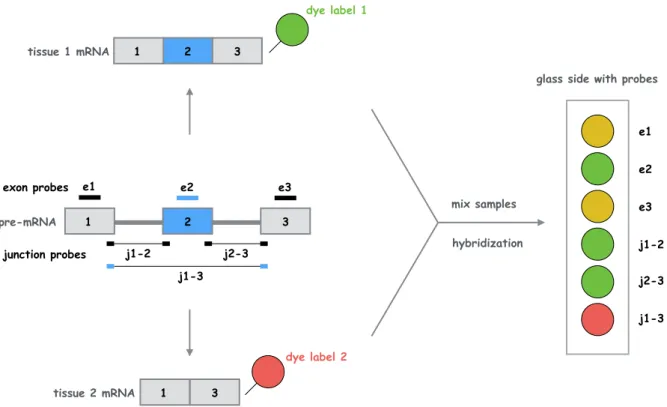
Microarray technologies have played a predominant role in shaping our understanding of tran-scriptome complexity and regulation (Blencowe, 2006). Microarray approaches rely on the hy-bridization of fluorescently labeled target RNA sequences to anchored oligonucleotides of known composition, often called probes, previously attached to a glass-slide. The abundance of target RNA is then inferred using laser fluorescence that measures the extent of hybridization on the probes. The development of custom microarrays with probe sets designed to detect individual exons and splice junction sequences overcame many of the obstacles encountered when analyz-ing EST data, in particular throughput and quantification aspects (Pan et al., 2004). Splicing microarrays can indeed be designed to hybridize to isoform-specific mRNA regions, which al-lows for the detection and quantification of distinct spliced isoforms. The concept of splicing microarrays is illustrated in figure 3.2.
Splicing microarray successes include the discovery of new alternative splicing events and the detection of cell- and tissue-specific alternative splicing events. For example, Johnson et al.
(2003) used arrays with probes for all adjacent exon-exon junctions in 10000 human genes and hybridized these with samples from 52 human tissues, revealing tissue-specific clustering of alternative splicing events.
The major drawbacks of splicing microarray are two folds: the limited dynamic range of signal detection and the reliance upon an existing genomic sequence. Indeed, array measurements are
1 2 3 1 3 e1 e2 2 1 3 e3 exon probes j1-2 j2-3 junction probes j1-3 pre-mRNA tissue 1 mRNA tissue 2 mRNA dye label 1 dye label 2 mix samples hybridization e1 e2 e3 j1-2 j2-3 j1-3
glass side with probes
Figure 3.2: Illustration of a splicing microarray experiment. Probes are complementary to individual exons or exon-exon junctions. Dye-labeled mRNAs hybridize with the corresponding probes, which allows the comparison of expression levels between the two samples. Figure is inspired fromMatlin et al.(2005).
limited by a strong background noise level and by saturation of high fluorescent signals. It also requires prior knowledge of target RNA sequences to design the probes.
The next section explains how modern high-throughput RNA sequencing does not require transcript-specific probes and measures a large dynamic range of expression levels.
3.1.3 High-throughput sequencing of the RNA as the new gold standard
Demand for cheaper and faster sequencing methods has increased greatly after the first hu-man genome sequence was completed in 2003. This dehu-mand has driven the emergence of fast, cost-effective, accurate and high-throughput sequencing technologies. The so-called “next-generation” sequencing (NGS) technologies enable to sequence an entire human genome in less than one day by sequencing massive amount of DNA in parallel. High-throughput RNA se-quencing or “RNA-seq” is an experimental protocol that uses NGS technologies to sequence RNA molecules within a biological sample.
In comparison to EST sequencing by Sanger technology, which is low-throughput and only de-tects the more abundant transcripts, RNA-seq can target lowly express transcripts and can
sequence millions of cDNA sequences in a single reaction. In contrast to other high-throughput technologies, such as hybridization-based microarrays, RNA-seq achieves base-pair level res-olution, offers a much higher dynamic range of expression levels and does not require prior knowledge of the sequences to be profiled.
We explain below the principles of RNA-seq, that is the use of NGS technologies to sequence and latter quantify RNA molecules after their conversion to cDNA by reverse transcription. We refer to Goodwin et al. (2016) for a detailed description of the different existing NGS technologies and to Wang et al.(2009) for a focus on the RNA-seq protocol.
RNA-seq technology
Next-generation sequencing, also referred as “deep sequencing”, “high-throughput sequenc-ing”, “massively-parallel sequencing” or “shotgun sequencsequenc-ing”, has revolutionized genomics, epigenomics and transcriptomics by allowing massively parallel sequencing at a relatively low cost (Koboldt et al., 2013). The key strength of NGS technologies is to perform real-time identification of millions of nucleotidic sequences in parallel. This differs greatly from Sanger sequencing technology where complementary strands of target cDNA first have to be separated by size before being revealed.
Various NGS platforms exist and use different chemistry or different ways to iteratively read the target nucleotides. Mardis (2011) and van Dijk et al. (2014) provide a comparison of the dif-ferent NGS platforms. However, all technologies monitor the sequential addition of nucleotides to immobilized and spatially arrayed DNA templates. We choose here to focus on the strat-egy developed by the Illumina platform (Bentley et al., 2008), which is the most widely used NGS technology worldwide. Illumina sequencing uses a “sequencing by synthesis” approach (described below) combined with fluorescence image analysis. Note that other platforms use a “sequencing by ligation” technique (McKernan et al.,2009) or identify the growing nucleotidic strands with analysis of electric rather then fluorescent signals (Rothberg et al.,2011).
The different steps of RNA-seq, additionally illustrated in figure 3.3, are the following:
1. Mature RNA selection and reverse transcription. As ribosomal RNA (rRNA) constitutes the predominant fraction of the transcriptome, it needs to be removed to avoid wasting sequencing efforts on a few superabundant molecules. rRNA for which the sequence is
AAA
AAA
AAA AAA
AAA
AAA
polyA selection reverse transcription
input RNA input mRNA input cDNA
(a) Selection and reverse transcription
fragmentation input cDNA size selection adaptor ligation sequencing library (b) Library preparation amplification bridge P CR emul sion PCR (c) Amplification 3’ template 5’ primer C C C G G G A C T terminator cap fluorescent dye
fluorescent emission cleavage
!
(d) Sequencing by synthesis
Figure 3.3: A typical RNA-seq experiment. Figure is inspired fromhttps://www.abmgood. com/marketing/knowledge_base/next_generation_sequencing_introduction.php.
known can be directly subtracted from the transcript pool, or alternatively mRNA har-boring a polyA tail can be enriched by capture with oligo-dT3. Selected RNA molecules
are converted to cDNA by a reverse transcriptase.
2. Library preparation. Starting material must be converted into a library of sequencing reaction templates which require fragmentation, size selection and adapter ligation. Given that most NGS technologies cannot sequence fragments longer than 1000 bases (often only a hundred bases), cDNA molecules need to be sheared into pieces so that all nucleotides of the molecules are sequenced. Fragmentation can be enzymatic or performed via hydrolysis or physical methods such as acoustic shearing or sonication. Note also that in some protocols fragmentation can be done at the RNA level before reverse transcription. Adapter ligation adds synthetic oligonucleotides of a known sequence onto the ends of the cDNA fragments, which serve as primers for downstream amplification and/or sequencing reactions. In strand-specific RNA-seq protocols (Levin et al.,2010), different primers are attached to the 5’ and 3’ ends of the RNA molecules, which further allows overlapping transcripts expressed from opposite strands of the genome to be distinguished.
3. Template generation and amplification. One of the key steps of NGS is to immobilize and separate the DNA fragments from a population (typically on a flow cell or on microbeads), allowing the downstream sequencing reaction to operate in parallel on millions of spatially distinct DNA templates.
Additionally, a template amplification step is required for most sequencing platforms in order to obtain sufficient signal for base calling. Amplification strategies are based on a polymerase chain reaction (PCR) step (emulsion PCR onto microbeads or bridge ampli-fication to form clusters on a flow cell).
Note that amplification-free protocols are emerging as promising technologies. SMRT (single molecule real-time) platforms (Eid et al.,2009) are indeed based on single-molecule
template sequencing hence bypassing the need for fragmentation and amplification. Amplification-based and single-molecule sequencing technologies have been respectively referred to as
“second-generation” and “third-generation” sequencing.
4. Sequencing and base calling. The sequencing by synthesis strategy implemented by Illu-mina uses the cDNA library fragments as templates of which new DNA fragments are synthesized by a polymerase enzyme.
Similarly to Sanger sequencing it employs fluorescently-labeled terminator nucleotides. However, the key innovation compared to Sanger sequencing is the use of reversible ter-minators. Hence during each reaction cycle a single nucleotide is added to the growing DNA strand and the fluorescent dye is imaged to identify the base. The terminator is then enzymatically cleaved so that it allows incorporation of the next nucleotide.
Sequencing typically occurs solely at the ends of the cDNA fragments. The sequenced ends are called reads. Sequencing only one end of the fragments produces the so-called “single-end reads” whereas sequencing both the 5’ and 3’ “single-ends produces “paired-“single-end reads”. An Illumina platform typically produces reads of ⇠ 100bp.
The millions of reads produced by RNA-seq further need to be pre-processed and analyzed in order to answer relevant questions such as i) what are the levels of expression of the mRNA transcripts in a biological sample? ii) are some transcripts differentially expressed between different conditions or iii) are there any alternative splicing events specific to a given tissue?
In that context, section3.2focuses on the analysis of RNA-seq reads, in particular in the aim of identifying and quantifying the different transcript isoforms present in a given sample. Chapters
4and5provide new computational methods to infer the transcript isoforms from RNA-seq data.
Opportunities raised by RNA-seq
A new appreciation of the complexity of the transcriptome has emerged with the use of RNA-seq data (Blencowe et al., 2009). The biological applications that RNA-seq makes it possible to target are very diverse (Ozsolak and Milos,2011), ranging from the profiling of mRNA and non-coding RNA expression to the study of alternative splicing, alternative polyadenylation or transcription initiation sites as well as the study of small RNA, antisense transcripts or the detection of fusion genes.
In particular, datasets generated with the RNA-seq technology have facilitated the identification of thousands of regulated alternative splicing events in various biological contexts. Pioneer works that gave new insights into the complexity of alternative splicing include Pan et al.
(2008); Wang et al. (2008b) and Mortazavi et al. (2008) By analysing mRNA-seq data across different human tissues, bothPan et al.(2008) and Wang et al.(2008b) estimated that ⇠ 95% of human multi-exon genes undergo alternative splicing. Wang et al.(2008b) identified “switch-like” splicing events where exons exhibit dramatically different inclusion levels between different